bFGF promotes adipocyte differentiation in human mesenchymal stem cells
derived from embryonic stem cells
Xinghui Song
1, Yanwei Li
1, Xiao Chen
2, Guoli Yin
3, Qiong Huang
1, Yingying Chen
2,
Guowei Xu
2and Linlin Wang
21
Core Facilities, Zhejiang University School of Medicine, Hangzhou, China.
2
Department of Basic Medicine Sciences, School of Medicine, Zhejiang University, Hangzhou, China.
3
Department of Basic Medicine, Zhejiang Medical College, Hangzhou, China.
Abstract
In this work we describe the establishment of mesenchymal stem cells (MSCs) derived from embryonic stem cells (ESCs) and the role of bFGF in adipocyte differentiation. The totipotency of ESCs and MSCs was assessed by immunofluorescence staining and RT-PCR of totipotency factors. MSCs were successfully used to induce osteo-blasts, chondrocytes and adipocytes. MSCs that differentiated into adipocytes were stimulated with and without bFGF. The OD/DNA (optical density/content of total DNA) and expression levels of the specific adipocyte genes PPARg2 (peroxisome proliferator activated receptorg2) and C/EBPs were higher in bFGF cells. Embryonic bodies had a higher adipocyte level compared with cells cultured in plates. These findings indicate that bFGF promotes adipocyte differentiation. MSCs may be useful cells for seeding in tissue engineering and have enormous therapeutic potential for adipose tissue engineering.
Key words:embryonic stem cells; mesenchymal stem cells; fibroblast growth factor; adipocyte differentiation. Received: August 23, 2013; Accepted: October 14, 2013.
Introduction
Embryonic stem cells (ESCs) are pluripotent stem cells (Thomsonet al., 1998) and have the ability to remain undifferentiated and proliferate indefinitelyin vitro, while maintaining the potential to differentiate into cells of all three embryonic germ layers (Thomsonet al., 1995). The capacity for virtually unlimited self-renewal and differenti-ation in ESCs has opened up the prospect of widespread ap-plications for these cells in biomedical research, including cell differentiation, developmental studies, elucidation of gene function, drug screening and cell therapy (Bishopet al., 2002).
Although ESCs have a strong potential to differenti-ate, the diretion of differentiation is difficult to control. This problem has been resolved to some extent with the de-velopment of mesenchymal stem cells (MSCs). MSCs are multipotent stromal cells that have a high capacity for self-renewal while maintaining multipotency. MSCs there-fore have enormous therapeutic potential for tissue repair. MSCs have the capacity to differentiate into multiple cell types (Nardi and da Silva Meirelles, 2006), including os-teoblasts (Brighton and Hunt, 1991), chondrocytes
(Brigh-ton and Hunt, 1997) and adipocytes. In theory, this capacity can be used to generate any cell, tissue or even organ (Rippon and Bishop, 2004), a characteristic that could be useful in the treatment of human disease. However, there are still problems related to the use of MSCs, such as poor homology, inefficient proliferation and differentiation, and long culture times (Gondaet al., 2008; Rodehefferet al., 2008; Schipperet al., 2008; De Rosaet al., 2009; Lundet al., 2009). In addition, MSCs may cause serious trans-location osteogenesis (Chenet al., 2009) during the treat-ment of diseases.
The difficulties associated with the surgical treatment of trauma, burns, tumor resection, congenital malforma-tions and other causes of soft tissue defects are well-known. With increased development of adipose tissue engineering, new ideas have been developed in relation to the treatment of soft tissue defects and expansion. Adipose-derived stem cells are ideal seed cells for adipose tissue engineering (Rodehefferet al., 2008). Adipocytes differentiate primar-ily from MSCs through the embryonic body (EB) with the help of induction factors (Daniet al., 1997). But long in-duction times and a low differentiation efficiency are still major problems. Basic fibroblast growth factor (bFGF) is one of the most effective induction factors in cellular differ-entiation and promotes adipocyte differdiffer-entiation through activation of Ras-MAPin vivo(Hutleyet al., 2004; Newell
Send correspondence to Linlin Wang. Department of Basic Medi-cine Sciences, School of MediMedi-cine, Zhejiang University, 866 Yuhangtang Road, Hangzhou, Zhejiang Province, 310058, China. E-mail: [email protected].
et al., 2006). Whether there is a similar effect on ESC dif-ferentiation remains to be determined.
In this study, human ESCs were obtained and used to generate MSCs. The totipotency of these cells was as-sessed. MSCs were induced to differentiate into osteo-blasts, chondrocytes and adipocytes. bFGF was used to induce adipocytes from MSCs in a variety of conditions. Finally, the OD/DNA (optical density/content of total DNA) values and the expression level of specific adipocyte genes were measured. Our findings indicate that bFGF can promote adipocyte differentiation from MSCs and that the latter cells could be of enormous therapeutic potential for adipose tissue engineering.
Materials and Methods
Cell culture
An undifferentiated NIH-registered human ESCS H9 cell line (a gift from the National University of Singapore) was cultured on mitotically inactivated mouse embryonic fibroblasts. Briefly, the head and extremities were removed from 12.5-13.5d C57B6 mouse fetuses and fibrocytes were generated from the remains after digestion with 0.25% pancreatin. The cells were passaged when they reached 80% confluence and then cryopreserved in liquid nitrogen. Feeder layer cells were obtained by treatment with 1% mitomycin C for 30 min. When differentiation occurred at the clone center, mechanical isolation was used to passage undifferentiated ESCs to a new feeder layer within a 5-7-day period. The medium used was Dulbecco’s modified Eagle’s medium (DMEM, Gibco), supplemented with 20% serum (SR, Gibco), 1 mM glutamine (Gibco), 1% nones-sential amino acids (Gibco) and 0.1%b-mercaptoethanol. The cells were cultured at 37 °C in a 5% CO2atmosphere.
ESC identification
ESCs were cultured with and without feeder layer medium. The activity of totipotency factors Oct-4, SSEA-3 Tra-1-60 and Tra-1-81 was assessed by immunofluores-cence staining (Wang and Matise, 2013), while the expres-sion of totipotency factors C-MYC, KLF-4, OCT-4 and SOX-2 was assessed by RT-PCR. The totipotency of ESCs
in vivowas determined by analyzing teratomas in severe combined immunodeficiency disease (SCID) mice (Shihet al., 2007).
MSCs induction and identification
To induce MSCs, the ESCs in the feeder layer were transferred to a feeder-free system (Xuet al., 2005). After 3-4 days, the cells were digested with dispase, separated and seeded at very low density (10 cells/cm2) to allow the formation of MSCs (Chen et al., 2009). The ability of MSCs to differentiate was assessed as described elsewhere (Pittengeret al., 1999; Ouyanget al., 2006; Biet al., 2007). MSCs were induced to osteoblasts, chondrocytes and
adipocytes through treatment with osteoblast inducer (b -glycerol phosphate, dexamethasone, ascorbic acid), chon-drocyte inducer (TGF-b1) and adipocyte inducer (1-methyl-3-isobutylxanthine, dexamethasone, insulin, indo-methacin). The differentiated cells were identified by stain-ing with alkaline phosphatase, Safranin O and Oil Red O.
Adipocyte induction in plate cultures
MSCs were cultured until 80% confluence, after which they were dissociated with pancreatin and cultured in 24-well plates at a density of 5x10⁵cells/mL. After incu-bation for 24 h to allow attachment, the cells were divided into bFGF-treated (inducer supplemented with 4 ng bFGF/mL, Gibco) and non-bFGF-treated (inducer without bFGF) cells (16 wells/group). One, three, five and seven days after induction four wells for each group were washed three times with phosphate-buffered saline (PBS) and then fixed in 4% paraformaldehyde. After removal of this solu-tion, the cells were stained with Oil Red O for 30 min and then washed three times with isopropanol. Finally, the cells were suspended in PBS and examined by light microscopy. In addition, OD was measured after the Oil Red O dye solubilized in isopropanol. RNA was extracted from the cells and the specific adipose genes PPARr2 and C/EBPs were detected by RT-PCR. GDPH was used as an internal housekeeping gene (control).
Adipocyte induction in EB
EBs were formedin vitro. Briefly, medium was re-moved from MSCs and then the cells were washed three times with PBS. After treated with pancreatin for 3 min, the cells were dispersed and the overlying solution was re-moved by centrifugation. The underlying cells were sus-pended in medium and cultured in hanging upside down plates (2x10⁵ cells/mL, 20mL/EB) at 37 °C. Three days later, the EBs were transferred to new plates and cultured in suspension. A similar experiment in which adipocytes were induced in EBs and then cultured in plates was also done. The EBs were transferred to 24-well plates (five EBs/well) and induced. The EBs were removed on the first, third and seventh days after induction: two EBs were stained with Oil Red O and the remaining three were used to detect specific adipose genes.
Results
Morphology and identification of ESCs
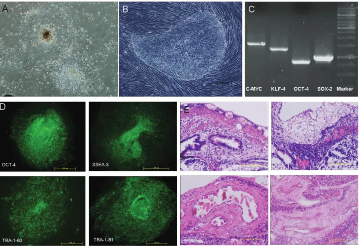
ESCs were cultured with and without feeder layer medium. In both of these conditions, the cells exhibited typical ESCs morphology,i.e., a flat, close, smooth edge and a high nuclear cytoplasmic ratio. However, cells grown in feeder layer medium were thicker and piled up, whereas cells grown without feeder layer medium were flatter and more expanded (Figure 1A,B).
Immunofluorescence staining and RT-PCR of several totipotency factors were used to identify and characterize the ESCs. RT-PCR showed that C-MYC, KLF-4, OCT-4 and SOX-2 were expressed in ESCs (Figure 1C). In addi-tion, OCT-4, SSEA-3, TRA-1-60 and TRA-1-80 were de-tected by immunofluorescence staining (Figure 1D). These results indicated that these cells were totipotent. The injec-tion of ESC subcutaneously into SCID mice resulted in teratoma formation after eight weeks, as shown by hemoto-xylin and eosin staining of tissue sections, thus confirming the totipotency of the cells. As shown in Figure 1E, the teratoma consisted of three germ layers: digestive tract epi-thelial layer derived from entoderm, adipose tissue layer derived from mesoderm and neuroectodermal tissue layer derived from ectoderm.
Morphology and identification of MSCs
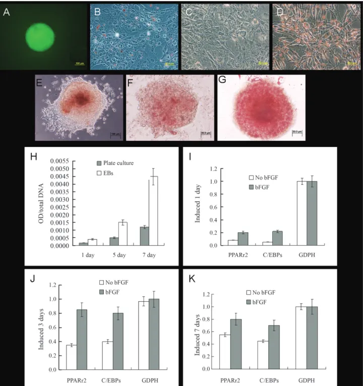
MSCs were derived from ESCs. With continuous passage, the number of heterologous cells decreased while the number of homologous cells gradually increased. After three passages, the cells began to exhibit MSC morphol-ogy. ESCs-derived fibroblast colonies were finally formed after at least 13 passages (Figure 2A-C). The ability of
MSCs to differentiate into different cell types was assessed by exposing these cells to various inducers. Three types of cells were induced and identified by staining. Two weeks after induction, osteoblasts were identified by the presence of black mineral calcium deposits (Figure 2D) and four weeks after induction adipocytes showed red (lipid) drop-lets and chondrocytes were stained watermelon red (Figu-re 2E,F).
Adipocyte induction in plate cultures
The cells were removed 1, 3, 5 and 7 days after induc-tion, respectively. There was no obvious adipocyte forma-tion in non-bFGF-treated cells, as judged by staining with Oil Red O (Figure 3A-C). In contrast, in bFGF-treated cells, adipocytes appeared after three days and reached 60% confluence after seven days (Figure 3D-F). The level of staining was quantified by measuring the optical density (OD) of Oil Red O in adipocytes and the OD/DNA values of the cells were compared. As shown in Figure 3I, the OD/DNA values of bFGF-treated cells were three times higher than those of the non-bFGF-treated cells. RT-PCR was used to detect the expression of specific adipose genes in these cells. The expression levels of PPARr2 and
C/EBPs in bFGF-treated cells were significantly higher than in the control group and increased with the number of passages (Figure 3J-M). These results indicated that the adipogenic rates in bFGF-treated cells were higher than in non-bFGF-treated cells and suggested that bFGF may play an important role in adipocyte differentiation.
Adipocyte induction in EBs
EBs were formed by MSCs at a density of 2 x 10⁵ cells/mL (Figure 4A). The ability of EBs to differentiate was assessed by comparing cell induction in plate cultures and EBs. The cells were detected 1, 3 and 7 days after in-duction. Adipocytes began to grow after one day and three days in EBs and plate cultures, respectively (Figure 4B-G); this growth increased significantly with the number of pas-sages. In addition, the OD/DNA values, as well as the ex-pression levels of PPARr2 and C/EBPs, were significantly higher in EBs than in plate cultures (Figure 4I-K). These re-sults showed that adipocyte induction was easier and greater in EBs.
Discussion
ESCs are pluripotent stem cells with a capacity for in-definite proliferation and a strong potential for differentia-tion. However, the direction of ESC differentiation is diffi-cult to control. MSCs derived from ESCs are multipotent stromal cells with a high capacity for self-renewal and maintenance of multipotency. MSCs can differentiate into
various cell types and are considered to be of major thera-peutic potential for tissue repair. Recently, MSC-derived adipocytes have provided a new approach for treating soft tissue defects and expansion. In this context, bFGF is an important inducer of adipocyte differentiation.
In this study, ESCs were cultured on mouse embry-onic fibroblasts. These cells can be cultured, passaged and cryopreserved, and retain their proliferative capacity after continuous passage. Several totipotency molecular markers with an important role in maintaining the capacity for self-renewal (Eigeset al., 2001; Richardset al., 2002) oc-cur in these cells, including stage-specific embryonic anti-gens (SSEA-1, SSEA-3, SSEA-4), transcriptional regulation antigens (TRA-1-60, TRA-1-81), homologous protein transcription factor (Nanog) and transcription fac-tors OCT3\4 and SOX-2. As shown here, three germ layer teratomas were also generated. These results indicate these cells are totipotent and that the method for establishing them is feasible. MSCs were generated from ESCs. With continuous passage, the cells began to form fibroblast colo-nies. MSCs can proliferate indefinitely while retaining their potential for differentiation, as shown by the identification of totipotency factors. Indeed, MSCs were totipotent and could differentiate into osteoblasts, chondrocytes and adipocytes after stimulation with a specific inducer. MSCs can therefore be regarded as seed cells for tissue engineer-ing. The relatively controllable differentiation may provide a new approach for adipose tissue engineering.
130 Songet al.
FGFs are heparin-binding protein mitogens that stim-ulate the division of most cell types in culture. During em-bryonic development, FGFs have diverse roles in regulat-ing cell proliferation, migration and differentiation. In adult organisms, FGFs are homeostatic factors and have impor-tant functions in tissue repair and response to injury (Ornitz and Itoh, 2001). FGFs regulate a broad spectrum of biologi-cal functions by signaling through FGF receptors.
Knock-down of FGF1 expression by small-interfering RNA re-duces FGF-1-stimulated signaling events, proliferation and priming (Widberget al., 2009). The FGF signal pathways include the RAS/MAP kinase, PI3 kinase/AKT and PLCg
pathways, of which the RAS/MAP kinase pathway is the most important (Yunet al., 2010).
Various studies have shown that bFGF promotes pocyte differentiation. For example, FGF-1 promotes
adi-132 Songet al.
pogenesis of primary human preadipocytes (phPA) through ERK1/2. ERK1/2 activation is necessary for human adipo-genesis in the absence of mitotic clonal expansion. FGF-1 induces robust phosphorylation of ERK1/2 in early differ-entiation and the inhibition of ERK1/2 activity signifi-cantly reduces phPA differentiation (Newellet al., 2006); FGF-2 significantly enhances the differentiation of adipo-cytes from human adipose-derived stem cells. In cells treated with FGF-2 before adipocyte induction, the mRNA expression of peroxisome proliferator-activated receptor
g2, a key transcription factor in adipogenesis, is unregu-lated (Kakudoet al., 2007). As shown here, bFGF plays a similar role in ESC differentiation. In bFGF-treated cells, induced adipocytes began to appear after three days and reached 60% confluence after seven days; in contrast, no adipocytes formed in non-bFGF-treated cells. The high OD/DNA values and expression levels of specific adipo-cyte genes seen here clearly showed that bFGF improved the efficiency and speed of adipocyte differentiation.
Different cellular morphologies may influence the differentiation of MSCs. EBs are formed from cell aggre-gates in non-adherent spheroids (Höpflet al., 2004). The molecular and cellular morphogenic signaling and events in EBs recapitulate numerous aspects of embryonic devel-opment and, as shown here, result in differentiation of the cells into three embryonic germ layers (endoderm, meso-derm and ectomeso-derm), in a manner similar to that associated with the gastrulation of an epiblast-stage embryoin vivo
(Itskovitz-Eldor et al., 2000). In this study, EBs were also induced and compared with cells grown in plate cultures. Adipocytes were easier to induce in EBs than in plated cells. This phenomenon provides additional evidence for bFGF function, but also highlights the high differentiation capacity of EBs.
In conclusion, human ESCs and MSCs were success-fully generated and shown to be totipotent. bFGF signifi-cantly enhanced the differentiation of adipocytes from MSCs, and adipocyte induction was greater in EBs than from MSCs. These results indicate that bFGF promotes adipocyte differentiation and that MSCs may be of enor-mous therapeutic potential for adipose tissue engineering.
Acknowledgments
This study was supported by Zhejiang provincial Nat-ural Science Foundation of China (grant nos. Y2100095, Y2091197), National Natural Science Foundation of China (81272067), Zhejiang Provincial Education Department Fund (Y200908872), Zhejiang Institute of Higher Educa-tion Research Projects Key Project (Z201213), Zhejiang University, independent research projects (2013QNA7025), Experimental Technology Department of Zhejiang University laboratory research projects (188100-560101\033).
References
Bi Y, Ehirchiou D, Kilts TM, Inkson CA, Embree MC, Sonoyama W, Li L, Leet AI, Seo B-M and Zhang L (2007) Identifica-tion of tendon stem/progenitor cells and the role of the extracellular matrix in their niche. Nat Med 13:1219-1227. Bishop AE, Buttery LD and Polak JM (2002) Embryonic stem
cells. J Pathol 197:424-429.
Brighton CT and Hunt RM (1991) Early histological and ultra-structural changes in medullary fracture callus. J Bone Joint Surg Am 73:832-847.
Brighton CT and Hunt RM (1997) Early histologic and ultra-structural changes in microvessels of periosteal callus. J Orthop Trauma 11:244-253.
Chen X, Song XH, Yin Z, Zou XH, Wang LL, Hu H, Cao T, Zheng M and Ouyang HW (2009) Stepwise differentiation of human embryonic stem cells promotes tendon regenera-tion by secreting fetal tendon matrix and differentiaregenera-tion fac-tors. Stem Cells 27:1276-1287.
Dani C, Smith A, Dessolin S, Leroy P, Staccini L, Villageois P, Darimont C and Ailhaud G (1997) Differentiation of embry-onic stem cells into adipocytesin vitro. J Cell Sci 110:1279-1285.
De Rosa A, De Francesco F, Tirino V, Ferraro GA, Desiderio V, Paino F, Pirozzi G, D’Andrea F and Papaccio G (2009) A new method for cryopreserving adipose-derived stem cells: An attractive and suitable large-scale and long-term cell banking technology. Tissue Eng Part C Methods 15:659-667.
Eiges R, Schuldiner M, Drukker M, Yanuka O, Itskovitz-Eldor J and Benvenisty N (2001) Establishment of human embry-onic stem cell-transfected clones carrying a marker for un-differentiated cells. Curr Biol 11:514-518.
Gonda K, Shigeura T, Sato T, Matsumoto D, Suga H, Inoue K, Aoi N, Kato H, Sato K and Murase S (2008) Preserved proliferative capacity and multipotency of human adipose-derived stem cells after long-term cryopreservation. Plast Reconstr Surg 121:401-410.
Höpfl G, Gassmann M and Desbaillets I (2004) Differentiating embryonic stem cells into embryoid bodies. Methods Mol Biol 254:79-98.
Hutley L, Shurety W, Newell F, McGeary R, Pelton N, Grant J, Herington A, Cameron D, Whitehead J and Prins J (2004) Fibroblast growth factor 1, a key regulator of human adipo-genesis. Diabetes 53:3097-3106.
Itskovitz-Eldor J, Schuldiner M, Karsenti D, Eden A, Yanuka O, Amit M, Soreq H and Benvenisty N (2000) Differentiation of human embryonic stem cells into embryoid bodies com-prising the three embryonic germ layers. Mol Med 6:88-95. Kakudo N, Shimotsuma A and Kusumoto K (2007) Fibroblast
growth factor-2 stimulates adipogenic differentiation of hu-man adipose-derived stem cells. Biochem Biophys Res Commun 359:239-244.
Lund P, Pilgaard L, Duroux M, Fink T and Zachar V (2009) Effect of growth media and serum replacements on the prolifera-tion and differentiaprolifera-tion of adipose-derived stem cells. Cyto-therapy 11:189-197.
Nardi NB and da Silva Meirelles L (2006) Mesenchymal stem cells: Isolation, in vitro expansion and characterization. Stem Cells 174:249-282.
func-tional effects of fibroblast growth factor-1 on human preadipocyte differentiation. FASEB J 20:2615-2617. Ornitz DM and Itoh N (2001) Fibroblast growth factors. Genome
Biol 2:3005.3001-3005.3012.
Ouyang HW, Cao T, Zou XH, Heng BC, Wang LL, Song XH and Huang HF (2006) Mesenchymal stem cell sheets revitalize nonviable dense grafts: Implications for repair of large-bone and tendon defects. Transplantation 82:170-174.
Pittenger MF, Mackay AM, Beck SC, Jaiswal RK, Douglas R, Mosca JD, Moorman MA, Simonetti DW, Craig S and Marshak DR (1999) Multilineage potential of adult human mesenchymal stem cells. Science 284:143-147.
Richards M, Fong C-Y, Chan W-K, Wong P-C and Bongso A (2002) Human feeders support prolonged undifferentiated growth of human inner cell masses and embryonic stem cells. Nat Biotechnol 20:933-936.
Rippon H and Bishop A (2004) Embryonic stem cells. Cell Prolif 37:23-34.
Rodeheffer MS, Birsoy K and Friedman JM (2008) Identification of white adipocyte progenitor cellsin vivo. Cell 135:240-249. Schipper BM, Marra KG, Zhang W, Donnenberg AD and Rubin
JP (2008) Regional anatomic and age effects on cell function of human adipose-derived stem cells. Ann Plast Surg 60:538-544.
Shih C-C, Forman SJ, Chu P and Slovak M (2007) Human embry-onic stem cells are prone to generate primitive, undifferenti-ated tumors in engrafted human fetal tissues in severe com-bined immunodeficient mice. Stem Cells Dev 16:893-902.
Thomson JA, Kalishman J, Golos TG, Durning M, Harris CP, Becker RA and Hearn JP (1995) Isolation of a primate em-bryonic stem cell line. Proc Natl Acad Sci USA 92:7844-7848.
Thomson JA, Itskovitz-Eldor J, Shapiro SS, Waknitz MA, Swier-giel JJ, Marshall VS and Jones JM (1998) Embryonic stem cell lines derived from human blastocysts. Science 282:1145-1147.
Wang H and Matise MP (2013) Immunofluorescence staining with frozen mouse or chick embryonic tissue sections. Me-thods Mol Biol 1018:175-188.
Widberg CH, Newell FS, Bachmann AW, Ramnoruth SN, Spelta MC, Whitehead JP, Hutley LJ and Prins JB (2009) Fibro-blast growth factor receptor 1 is a key regulator of early adipogenic events in human preadipocytes. Am J Physiol Endocrinol Metab 296:E121-E131.
Xu R-H, Peck RM, Li DS, Feng X, Ludwig T and Thomson JA (2005) Basic FGF and suppression of BMP signaling sustain undifferentiated proliferation of human ES cells. Nat Me-thods 2:185-190.
Yun Y-R, Won JE, Jeon E, Lee S, Kang W, Jo H, Jang J-H, Shin US and Kim H-W (2010) Fibroblast growth factors: Biol-ogy, function, and application for tissue regeneration. J Tis-sue Eng Regen Med 1:1-18.
Associate Editor: Alysson Muotri
All the content of the journal, except where otherwise noted, is licensed under a Creative Commons License CC BY-NC.