UNIVERSIDADE DE LISBOA
FACULDADE DE FARMÁCIA
EFFECT OF CELASTROL IN LEUKOCYTES FROM
RHEUMATOID ARTHRITIS PATIENTS
S
USANA
M
ARIA DA
S
ILVA
O
LIVEIRA
Dissertação orientada por:
Doutora Ana Rita Cascão Rodrigues
João Eurico Fonseca Lab, Instituto de Medicina Molecular João Lobo Antunes
Professor Doutor João Manuel Braz Gonçalves
Faculdade de Farmácia da Universidade de Lisboa
M
ESTRADO EM
C
IÊNCIAS
B
IOFARMACÊUTICAS
i
A
BSTRACT
Rheumatoid arthritis (RA) is an autoimmune inflammatory disease characterized by synovial inflammation and joint deformity. The pathogenesis of RA is mediated by several immune cells (such as monocytes, granulocytes, T and B lymphocytes) that infiltrate the synovial membrane and secrete a complex network of pro-inflammatory cytokines, chemokines and other inflammatory mediators, which perpetuate the inflammatory process associated with bone resorption and damage. RA remains an incurable, progressive and debiliting disease for the great majority of patients despite profound evolution of the therapeutics options over the last decades. Indeed, the new biotechnological treatments for RA are administered parenterally, have safety issues and constitute a significant economic burden to national health services. Thus, the development of therapeutic strategies able to control both synovial inflammation and bone erosion, with a high rate of disease remission, low incidence of side effects and low production costs is still an unmet medical need in RA.
Celastrol, a bioactive component of the Chinese herb Tripterygium wilfordii, has shown significant anti-inflammatory properties in vitro and in vivo, mainly attributed to the regulation of cytokine and chemokine production, the inhibition of cell invasion and proliferation, osteoclast modulation and suppression of bone resorption. Our group has previously characterized the efficacy and safety of celastrol in an animal model of arthritis, showing a reduction of synovial leukocyte infiltration.
The main goal of this work was to analyze the effect of celastrol with or without LPS stimulation on primary human leukocytes (such as monocytes, granulocytes, T and B cells and their subsets) activation, maturation, survival and apoptosis using peripheral blood samples collected from chronic RA patients (with more than 1 year of disease duration) in comparison with healthy controls, using Flow Cytometry and performingalarmarBlue® cell vitality assay.
For that, we first proceeded to the optimization of the experimental conditions (assessing the optimal compounds concentrations and incubation time to be tested), where peripheral leukocytes from healthy controls (n=4) were incubated at a range of celastrol (0,001 µM, 0,01 µM, 0,05 µM, 0,1 µM, 0,3 µM, 0,5 µM and 1 µM) and LPS (2 μg/mL, 5 μg/mL and 10 μg/mL) concentrations, and under a varying incubation times (0h, 4h, 16h and 24h). After incubation, cell viability was assessed using alarmarBlue® by spectrophotometry assay, and an immunophenotyping characterization of monocytes,
ii
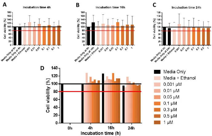
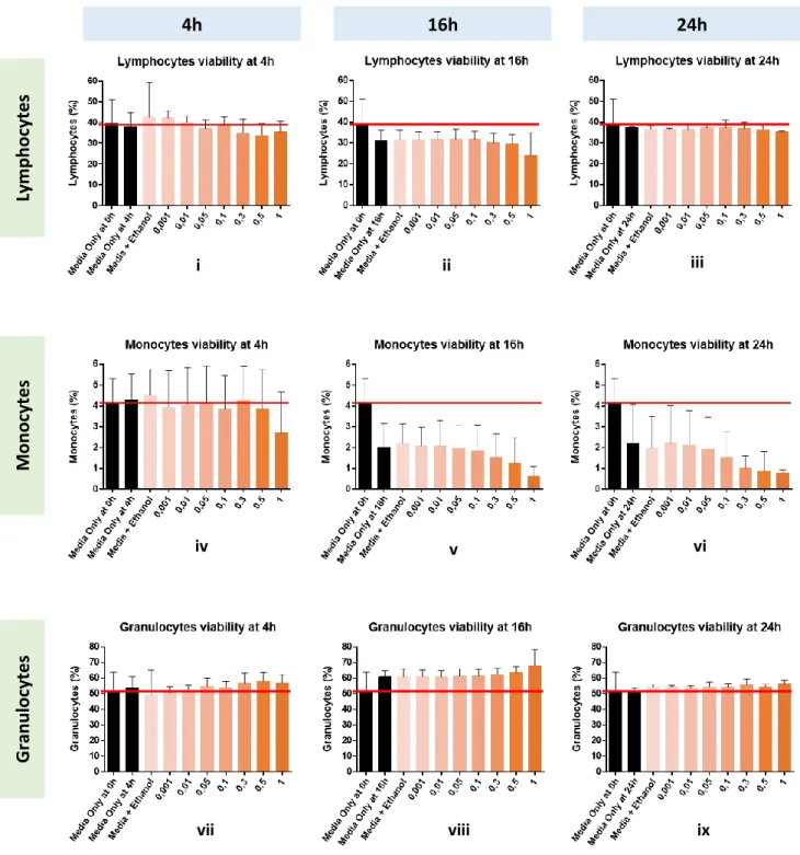
granulocytes, B and T cells was performed by Flow Cytometry. We found that incubation for 4h with celastrol 0,3 µM and LPS 10 μg/mL was the condition that showed less lymphocyte’s, monocyte’s and granulocyte’s cell death, appearing to be the bestcondition to be tested in the following experiments. Next, we studied the effect of celastrol in peripheral leukocytes of the recruited chronic RA patients (n=5) and healthy controls (n=10), using the pre-optimized experimental conditions.
Our results have shown that celastrol had no significant effect on CD14+ monocytes, CD66b+ granulocytes, CD19+ B cell and CD3+ T cell levels, both in healthy controls and in chronic RA patients.
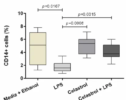
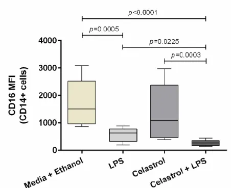
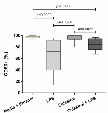
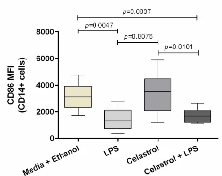
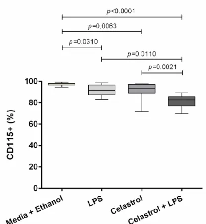
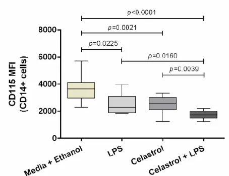
Accordingly, the overall data about monocytes have shown that celastrol was able to diminish (after LPS stimulation) CD16 and CD115 frequency and expression, and restore HLA-DR expression and CD86 frequency and expression to basal levels in monocytes (on total CD14+ cells) from healthy controls. In addition, in chronic RA patients, celastrol was able to reduce CD115 frequency and decrease (after LPS stimulation) CD86 frequency on monocytes (on total CD14+ cells).
Regarding granulocytes, our results have shown that celastrol was able to diminish CD62L expression and restore CD11b expression and CXCR2 frequency to basal levels in granulocytes (on total CD66b+ cells) from healthy controls. In chronic RA patients, celastrol was able to restore CD11b expression to basal levels in granulocytes (on total CD66b+ cells).
Our data regarding T cells have shown that celastrol was able to increase central memory Th2-like cells CCR6-) and effector memory Th2-like cells (CXCR3-CCR6-) frequency and restore naïve activated T helper cells (CD45RO-HLA-DR+) frequency to basal levels from healthy controls. In chronic RA patients, celastrol was able to increase memory activated T helper cells (CD45RO+HLA-DR+) frequency. Moreover, our results have shown that celastrol was able to reduce (after LPS stimulation) CXCR3 expression to basal levels in T cells (on total CD3+ cells and on T helper cells (CD3+CD4+)) from healthy controls.
Concerning B cells, our findings have shown that celastrol was able to diminish RANKL and CD95 frequency on transitional B cells (IgD+CD38++) and CD95 expression on naïve B cells (IgD+CD27-) and transitional B cells (IgD+CD38++) from healthy controls. In addition, celastrol was able to restore RANKL frequency to basal levels in B cells (on total CD19+ B cells, naïve (IgD+CD27-), pre-switch memory (IgD+CD27+), post-switch memory (IgD-CD27+) B cells)) from healthy controls. Also, in healthy controls,
iii
celastrol was able to restore CD95 frequency and expression and FcgRIIB expression to basal levels in B cells (on plasmablasts (IgD-CD38++); restore CD95 frequency and CD21 expression to basal levels on naïve B cells (IgD+CD27-) and CD21 expression to basal levels in B cells (on total CD19+ B cells); and restore (after LPS stimulation) HLA-DR frequency to basal levels in B cells (on transitional B cells (IgD+CD38++).
Celastrol seems to have a stronger effect on innate immune system cells, reducing their overall activation, differentiation and migration potential.
Accordingly, celastrol is a promising candidate for further testing in the clinic for RA therapy. Furthermore, the results suggest that celastrol might also be beneficial for the treatment of a few other autoimmune diseases besides arthritis.
iv
R
ESUMO
A artrite reumatóide (AR) é uma doença progressiva autoimune caracterizada por inflamação crónica sinovial e deformação irreversível das articulações, que pode levar à destruição da cartilagem e erosão óssea, causando incapacidade funcional e redução da esperança média de vida do doente, se não for devidamente e precocemente tratada. Inicialmente, manifesta-se através de inchaço, dor e rigidez das pequenas articulações das mãos, alastrando-se progressivamente para as articulações maiores dos joelhos e tornozelos. Deste modo, o diagnóstico e tratamento precoce e adequado é essencial para prevenir a progressão da doença e a destruição óssea e articular a ela associada.
O processo inflamatório da AR é caracterizado pelo recrutamento de células imunitárias (leucócitos) da circulação sanguínea periférica para o líquido e tecido sinovial. Nesta patologia estão envolvidas células imunitárias tanto do sistema imunitário inato (macrófagos e neutrófilos) como do adaptativo (linfócitos B e T), que infiltram a membrana sinovial (que reveste as articulações) e secretam citocinas pro-inflamatórias – particularmente as interleucinas (IL)-1β, IL-6 e IL-17 e o factor de necrose tumoral (TNF) – , quimiocinas e outros mediadores inflamatórios, tornando-a hiperplásica. Assim, o tecido sinovial torna-se, maioritariamente, repleto de macrófagos e linfócitos (células B e T), enquanto que o líquido sinovial é concentrado com neutrófilos.
As interações entre células apresentadoras de antigénios (APCs), células T e células B activam a resposta imunitária, levando à produção de autoanticorpos e excessiva activação de células T. Subsequentemente, estas células T activadas vão continuamente activar monócitos, macrófagos e fibroblastos sinoviais, induzindo a produção de citocinas pro-inflamatórias e quimiocinas que perpetuam o processo inflamatório, bem como mediadores de actividade óssea e enzimas com capacidade de reabsorver e destruir cartilagem e osso. Monócitos/macrófagos (precursores de osteoclastos) são induzidos a expressar RANK, que após se ligar ao seu ligando, RANKL, secretado pelos osteoblastos, estimula a sua diferenciação em osteoclastos maduros, responsáveis pela reabsorção e erosão óssea na AR.
A AR é uma doença de etiologia desconhecida e que afecta cerca de 0,5% a 1% da população mundial, sendo mais frequente nas mulheres (≈75%). Apesar da profunda evolução das opções terapêuticas ao longo das últimas décadas, a AR continua a ser uma doença incurável, progressiva e debilitante para a grande maioria dos doentes. O objectivo do tratamento na AR é a remissão, que é alcançada apenas em 20% - 30% dos doentes, se
v
precocemente e adequadamente tratados, no entanto, a maioria experiencia efeitos colaterais graves. Os primeiros tratamentos administrados aos doentes com AR são glucocorticóides, úteis no alívio dos sintomas clínicos e na diminuição da inflamação. Contudo, por terem efeitos secundários adversos em doses elevadas, são substituídos, numa segunda fase por drogas antirreumáticas modificadoras da doença (csDMARDs) sintécticas convencionais, que incluem o metrotexato (MTX), o fármaco mais usado no tratamento da AR e eficaz no controlo da actividade e progressão da doença. Ainda assim, uma grande percentagem de doentes é não-respondedora ou deixa de responder ao tratamento, pelo que a introdução de terapias biológicas (bDMARDs), como agentes anti- TNF, IL-1β, IL6, células B e T, têem surgido como uma alternativa aos convencionais DMARDs. Mais recentemente, têem também sido desenvolvidos agentes inibidores que são pequenas moléculas (tsDMARDs), como o tofacitinib, que apesar de inovadoras, infelizmente são terapias excessivamente caras, limitando o acesso aos serviços de saúde. Para além disso, muitas destas terapias são apenas eficazes no controlo da inflamação ou no do dano ósseo, pelo que para alguns doentes nenhuma destas terapias é eficaz. Assim, o desenvolvimento de estratégias terapêuticas capazes de controlar tanto a inflamação sinovial como a erosão óssea, com uma elevada taxa de remissão da doença, baixa incidência de efeitos colaterais e baixos custos de produção ainda é uma necessidade médica não atendida na AR.
O celastrol, um componente bioactivo extraído da planta chinesa Tripterygium wilfordii, que tem sido muito usado na medicina Chinesa para tratar inflamação e doenças auto-imunes incluindo a AR, foi previamente identificado pelo nosso grupo como um potencial candidato terapêutico para a AR, por inibir simultaneamente a produção das citocinas pro-inflamatórias TNF e IL-1β, directamente relacionadas com a inflamação e a degradação óssea na AR. Vários estudos têem apontado significantes propriedades anti-inflamatórias do celastrol in vitro e in vivo, maioritariamente atribuídas à regulação da produção de citocinas e quimiocinas, à inibição da invasão/infiltração e proliferação celular, modulação de osteoclastos e supressão da reabsorção óssea. Particularmente, o nosso grupo tem determinado a eficácia e segurança do celastrol num modelo animal de artrite, mostrando uma redução da infiltração leucocitária sinovial.
Assim, o principal objectivo deste trabalho foi avaliar o efeito do celastrol, com ou sem estimulação de LPS, na activação, maturação, sobrevivência e apoptose de leucócitos humanos primários (monócitos, granulócitos, células B e T e suas subpopulações), utilizando amostras de sangue periférico recolhidas de doentes com AR crónica (com mais de 1 ano de duração de doença) em comparação com controlos saudáveis, recorrendo à técnica de Citometria de Fluxo
vi
e ao ensaio de viabilidade alamarBlue®.
Para isso, primeiramente procedemos à optimização das condições experimentais (com vista à determinação das concentrações ideais de compostos e tempo de incubação a testar), onde leucócitos periféricos de controlos saudáveis (n=4) foram incubados numa gama de concentrações de celastrol (0,001 µM, 0,01 µM, 0,05 µM, 0,1 µM, 0,3 µM, 0,5 µM e 1 µM) e LPS (2 μg/mL, 5 μg/mL e 10 μg/mL), e sob tempos de incubação variáveis (0h, 4h, 16h e 24h). Após cada incubação, a viabilidade celular foi determinada usando o reagente de alamarBlue® através de um ensaio de espectrofotometria, e a caracterização imunofenotípica dos monócitos, granulócitos, células B e T, foi realizada por Citometria de Fluxo. De entre as condições testadas, verificou-se que a incubação de 4h com 0,3 µM de celastrol e 10 µg/mL de LPS foi a condição que apresentou menos morte celular das populações de linfócitos, monócitos e granulócitos, surgindo como a condição ideal a ser testada nas experiências seguintes. Em seguida, procedemos ao recrutamento de doentes com AR crónica (n=5) e controlos saudáveis (n=10) e estudámos o efeito do celastrol em leucócitos periféricos, testando as condições experimentais pré-optimizadas.
O nosso trabalho demonstrou que o celastrol não teve efeito significativo nos níveis de células CD14+ (monócitos), CD66b+ (granulócitos), CD19+ (células B) e CD3+ (células T), tanto nos indíviduos saudáveis como nos doentes com AR crónica.
Em relação aos monócitos, o celastrol mostrou ser capaz de diminuir (após estimulação com LPS) a frequência e a expressão de CD16 e CD115, e restaurar a expressão de HLA-DR e a frequência e expressão de CD86 para níveis basais nas células CD14+ totais de controlos saudáveis. Além disso, nos doentes com AR crónica, celastrol foi capaz de reduzir a frequência de CD115 e diminuir (após estimulação com LPS) a frequência de CD86 nas células CD14+ totais.
No que diz respeito aos granulócitos, o celastrol foi capaz de reduzir a expressão de CD62L e restaurar a expressão de CD11b e a frequência de CXCR2 para níveis basais nas células CD66b+ totais de controlos saudáveis. Nos doentes com AR crónica, o celastrol foi capaz de restaurar a expressão de CD11b para níveis basais nas células CD66b+ totais.
Quanto às células T, o celastrol foi capaz de aumentar a frequência de células central memory Th2-like (CXCR3-CCR6-) e effector memory Th2-like (CXCR3-CCR6-) e restaurar a frequência das naïve activated T helper (CD45RO-HLA-DR+) para níveis basais de controlos saudáveis. Nos doentes com AR crónica, o celastrol foi capaz de aumentar a frequência de células memory activated T helper (CD45RO+HLA-DR+). Para além disso, o celastrol foi capaz de reduzir (após estimulação com LPS) a expressão de CXCR3 para
vii
níveis basais nas células CD3+ totais e nas T helper (CD3+CD4+) de controlos saudáveis. No que concerne às células B, o celastrol foi capaz de diminuir a frequência de RANKL e CD95 nas células transitional (IgD+CD38++) e a expressão de CD95 nas naïve (IgD+CD27-) e transitional (IgD+CD38++) de controlos saudáveis. Além disso, o celastrol foi capaz de restaurar a frequência de RANKL para níveis basais nas células CD19+ totais, nas naïve (IgD+CD27-), pre-switch memory (IgD+CD27+) e post-switch memory (IgD-CD27+) de controlos saudáveis. Nos doentes com AR crónica, o celastrol foi capaz de restaurar a frequência e expressão de CD95 e a expressão de FcgRIIB para níveis basais nos plasmablasts (IgD-CD38++); restaurar a frequência de CD95 e a expressão de CD21 para níveis basais nas células naïve (IgD+CD27-) e a expressão de CD21 para níveis basais nas células CD19+ totais; e restaurar (após estimulação com LPS) a frequência de HLA-DR para níveis basais nas células transitional (IgD+CD38++).
Celastrol parece ter um efeito mais forte sobre as células do sistema imune inato, reduzindo a sua activação geral, diferenciação e potencial de migração. Desta forma, celastrol é um candidato promissor para futuros ensaios clínicos em doentes com AR. Além disso, estes resultados sugerem que o celastrol também pode ser benéfico para o tratamento de algumas outras doenças autoimunes além da artrite.
viii
A
CKNOWLEDGEMENTS
Aos meus queridos pais e maninho e à fofinha da avó Mina <3
A eles por serem a minha almofada e por fazerem de tudo para me verem bem! Obrigada família, vocês fazem-me um bem danado. Se o mundo acabasse amanhã,
posso dizer que enquanto vivi, vivi feliz e mais feliz por vos ter! Amo-vos!
Às RItas, que me orientaram. À Vânia, ao Ângelo e ao Rui que sempre me ajudaram. Ao Parceiro de sempre, que partilha da dor de ouvir pronunciar certas palavras proibidas (aka Citómetro e MAC). À mais recente aquisição do lab, a Dory alentejana, que vai pelo mesmo caminho. Às Xuxus e às Muchachas, que me acompanham nesta nossa amizade tão pura e tão louca desde sempre. À Talia, por seres a amiga que me aparece com sushi para o jantar nos dias mais off de sempre. Dispensas agradecimentos, porque todo e qualquer agradecimento não chega para agradecer verdadeiramente o quanto significas para mim. Ao meu melhor ‘amigo’, que apesar de tudo, nunca o esquecerei. À “família” do Starbucks, que me tem acolhido tão bem e aturado quase diariamente nestes longos últimos meses de escrita!
ix
Um especial e carinhoso agradecimento vai para a verdadeira Dory (também conhecida como Rita Moura), que sem ela e as suas Dorices, esta caminhada teria sido muito mais difícil, senão mesmo impossível. Graças a ela, “Do not stressate” tornou-se o melhor lema que aprendi em Ciência e que nunca encontrei em nenhum paper! Obrigada Rita Moura por tudo isso, e por todos os “Olá! Tudo bem?” no início de cada frase!
x
A
BBREVIATIONS
7-AAD 7-Amino-actinomycin D
ACPA Anti-citrullinated protein antibody
ACR American College of Rheumatology
AIA Adjuvant-induced arthritis
APCs Antigen-presenting cells
BAFF-R B cell activating factor -receptor
bDMARDs Biologic disease-modifying anti-rheumatic drugs
CCR Chemokine (C-C motif) receptor
CD Cluster of differentiation
c-Fms Colony-stimulating factor-1 receptor
CIA Collagen-induced arthritis
COX-2 Cyclooxygenase-2
CRP C-reactive protein
csDMARDs Conventional synthetic DMARDs
CXCR Chemokine (C-X-C motif) receptor
DAS28 Disease activity score of 28 joints
DFZ Deflazacort
EDTA Ethylenediamine tetraacetic acid
ELISA Enzyme-linked immunosorbent assay
ESR Erythrocyte sedimentation rate
EULAR European League Against Rheumatism
FasR Fas receptor
FBS Fetal bovine serum
FcgRIIB Fc gamma-receptor IIB
FVD Fixable Viability Dye
FLSs Fibroblast-like synoviocytes
Foxp3 Forkhead box protein 3
GCs Glucocorticoids
HC Healthy control
HIF-1α Hypoxia-inducible factor-1α
HLA Human leukocyte antigen
IFN-γ Interferon-gamma
Ig Immunoglobulin
IL Interleukin
iNOS Inducible nitric oxide synthase
JAK Janus kinase
LPS Lipopolysaccharide
MCP-1 Monocyte chemotactic protein-1
M-CSF Macrophage-colony stimulating factor
xi
MHC Major histocompatibility complex
MMPs Matrix metalloproteinases
MTX Methotrexate
NF-kB Nuclear factor kappa-light-chain-enhancer of activate B cells
NSAIDs Nonsteroidal anti-inflammatory drugs
PDN Prednisone
PBS Phosphate Buffered Saline
RA Rheumatoid arthritis
RANK Receptor activator of nuclear factor-κB
RANKL RANK ligand
RANTES Regulated upon activation, normally T cell expressed and secreted
RBC Red Blood Cell
RF Rheumatoid factor
RORgT Retinoic acid receptor-related orphan receptor gamma T
ROS Reactive oxygen species
RT Room temperature
SLZ Salazopyrin
TCRs T-cell receptors
Tc T cytotoxic cells
Th T helper cells
Th1 T helper 1 cells or IL-1-producing T cells
Th2 T helper 2 cells or IL-2-producing T cells
Th17 T helper 17 cells or IL-17-producing T cells
TNF Tumor necrosis factor
tsDMARDs Targeted synthetic disease-modifying anti-rheumatic drugs
Treg Regulatory T cells
VAS Visual Analogue Scale
xii
T
ABLE OF CONTENTS
ABSTRACT ... i RESUMO ... iv ACKNOWLEDGEMENTS ... viii ABBREVIATIONS ... xTABLE OF CONTENTS ... xii
FIGURE INDEX ... xv
TABLE INDEX ... xvii
CHAPTER I - INTRODUCTION ... 1
1. Rheumatoid Arthritis (RA) ... 1
1.1. Definition ... 1 1.2. Etiology ... 2 1.3. Pathophysiology ... 3 1.4. Treatment options ... 8 2. Celastrol... 10 2.1. Celastrol ... 10 2.2. Celastrol in arthritis ... 11 CHAPTER II - AIMS ... 14
CHAPTER III - MATERIALS AND METHODS ... 15
1. Compounds ... 15
2. Human samples ... 15
3. Isolation of leukocytes from blood ... 16
4. In vitro test of celastrol ... 16
4.1. Optimization of experimental conditions ... 16
4.2. Optimized in vitro test of celastrol ... 17
5. Flow cytometry ... 17
5.1. Antibodies ... 17
5.2. Leukocyte staining - Preparation of samples to Flow cytometry ... 18
5.2.1. Cell surface staining protocol ... 18
5.2.2. Intracellular staining protocol ... 19
6. AlamarBlue® cell viability assay ... 19
xiii
8. Statisitical analysis ... 21
CHAPTER IV - RESULTS ... 21
1. In vitro test of celastrol ... 21
1.1. Optimization of experimental conditions ... 21
1.1.1. Incubation time optimization of celastrol ... 21
AlamarBlue® viability assay... 22
Flow cytometry ... 23
1.1.2. Concentration optimization of celastrol ... 24
7-AAD Apoptosis Staining and Flow cytometry ... 24
1.1.3. Concentration optimization of LPS ... 25
AlamarBlue® cell viability assay ... 25
Flow cytometry ... 26
7-AAD Apoptosis Staining and Flow cytometry ... 27
2. Human samples ... 28
2.1. Characterization of patients cohort ... 28
3. Flow cytometry ... 29
3.1. Healthy controls ... 29
3.1.1. Monocytes ... 29
Classification of monocyte subpopulations and the effect of celastrol ... 29
Monocyte markers and the effect of celastrol ... 30
3.1.2. Granulocytes ... 38
Classification of granulocyte population and the effect of celastrol ... 38
Granulocyte markers and the effect of celastrol ... 38
3.1.3. T cells ... 42
Classification of T cell subpopulations and the effect of celastrol ... 42
T cell markers and the effect of celastrol ... 44
3.1.4. B cells ... 47
Classification of B cell subpopulations and the effect of celastrol ... 47
B cell markers and the effect of celastrol ... 48
3.2. Chronic RA patients ... 52
3.2.1. Monocytes ... 52
Effect of celastrol on monocyte population ... 52
Effect of celastrol on monocyte markers ... 52
3.2.2. Granulocytes ... 55
Effect of celastrol on granulocyte population ... 55
xiv
3.2.3. T cells ... 57
Effect of celastrol on T cell subpopulations ... 57
Effect of celastrol on T cell markers ... 58
3.2.4. B cells ... 60
Effect of celastrol on B cell subpopulations ... 60
Effect of celastrol on B cell markers ... 60
3.3. Chronic RA patients vs Healthy controls ... 62
3.3.1. Monocytes ... 62
Effect of celastrol on monocyte population ... 62
Effect of celastrol on monocyte markers ... 63
3.3.2. Granulocytes ... 66
Effect of celastrol on granulocyte population ... 66
Effect of celastrol on granulocyte markers ... 67
3.3.3. T cells ... 72
Effect of celastrol on T cell subpopulations ... 72
Effect of celastrol on T cell markers ... 74
3.3.4. B cells ... 79
Effect of celastrol on B cell subpopulations ... 79
Effect of celastrol on B cell markers ... 80
CHAPTER V - DISCUSSION AND CONCLUSIONS ... 91
REFERENCES ... 102
APPENDIX ... 118
I. Multicolor flow cytometry panel ... 118
II. Celastrol effect on Monocytes ... 120
III.Celastrol effect on T cells ... 126
IV. Celastrol effect on B cells ... 131
xv
F
IGURE INDEX
Figure 1. Representation illustrating and comparing healthy and arthritic articular joint ... 3
Figure 2. Schematic representation of the inflammatory microenvironment in inflamed joints and events occurring in Rheumatoid Arthritis ... 4
Figure 3. RANK/RANKL signaling pathway representation and osteoclastogenesis ... 6
Figure 4. Incubation time optimization of celastrol in in vitro. Cell viability analysis by alamarBlue® assay ... 12
Figure 5. Incubation time optimization of celastrol in in vitro. Cell viability and subpopulation percentage analysis by Flow Cytometry ... 13
Figure 6. Concentration optimization of celastrol in in vitro under incubation time of 4h. Analysis of subpopulations cell death by 7-AAD Apoptosis Staining and Flow Cytometry ... 14
Figure 7. Concentration optimization of LPS in in vitro under incubation time of 4h. Cell viability analysis by alamarBlue® assay... 15
Figure 8. Concentration optimization of LPS in in vitro under incubation time of 4h. Cell viability and subpopulation percentage analysis by Flow Cytometry ... 16
Figure 9. Concentration optimization of LPS in in vitro under incubation time of 4h. Analysis of subpopulations cell death by 7-AAD Apoptosis Staining and Flow Cytometry ... 27
Figure 10. Frequency of total CD14+ cells from healthy controls ... 30
Figure 11. Frequency of CD16+ cells on total CD14+ cells from healthy controls ... 31
Figure 12. Expression of CD16 (MFI) cell marker on total CD14+ cells from healthy controls ... 31
Figure 13. Frequency of CD86+ cells on total CD14+ cells from healthy controls ... 33
Figure 14. Expression of CD86 (MFI) cell marker on total CD14+ cells from healthy controls ... 34
Figure 15. Expression of HLA-DR (MFI) cell marker on total CD14+ cells from healthy controls . 35 Figure 16. Frequency of CD115+ cells on total CD14+ cells from healthy controls... 36
Figure 17. Expression of CD115 (MFI) cell marker on total CD14+ cells from healthy controls .... 37
Figure 18. Expression of CD11b (MFI) cell marker on CD66b+ cells from healthy controls ... 39
Figure 19. Expression of CD62L (MFI) cell marker on CD66b+ cells from healthy controls ... 40
Figure 20. Frequencies of CXCR2+ cells on total CD66b+ cells from healthy controls ... 41
Figure 21. Expression of CXCR3 (MFI) cell marker on total CD3+ T cells from healthy controls .. 46
Figure 22. Expression of CD21 (MFI) cell marker on total CD19+ B cells from healthy controls ... 49
Figure 23. Frequency of RANKL+ cells on total CD19+ B cells from healthy controls ... 50
Figure 24. Frequency of CD86+ cells on total CD14+ cells from chronic RA patients ... 53
Figure 25. Frequency of CD115+ cells on total CD14+ cells from chronic RA patients ... 54
Figure 26. Expression of CD11b (MFI) cell marker on CD66b+ cells from chronic RA patients .... 55
Figure 27. Frequency of CD14+ cells in chronic RA patients vs healthy controls ... 62
xvi Figure 29. Expression of CD86 (MFI) cell marker on CD14+ cells in chronic RA patients vs healthy
controls ... 64 Figure 30. Expression of HLA-DR (MFI) cell marker on CD14+ cells in chronic RA patients vs
healthy controls ... 65 Figure 31. Frequency of total CD66b+ cells in chronic RA patients vs healthy controls ... 66 Figure 32. Frequency of CD15+ cells on total CD66b+ cells in chronic RA patients vs healthy
controls ... 67 Figure 33. Frequency of CD16high cells on total CD66b+ cells in chronic RA patients vs healthy
controls ... 68 Figure 34. Frequency of CD16low cells on total CD66b+ cells in chronic RA patients vs healthy
controls ... 69 Figure 35. Expression of CD62L (MFI) cell marker on CD66b+ cells on chronic RA patients vs
healthy controls ... 70 Figure 36. Frequency of CXCR2+ cells on total CD66b+ cells in chronic RA vs healthy controls .. 71 Figure 37. Expression of CXCR2 (MFI) cell marker on CD66b+ cells on chronic RA patients vs
healthy controls ... 71 Figure 38. Expression of HLA-DR (MFI) cell marker on total CD3+ T cells in chronic RA patients
vs healthy controls ... 75 Figure 39. Expression of CXCR3 (MFI) cell marker on total CD3+ T cells in chronic RA patients vs
healthy controls ... 78 Figure 40. Frequency of total CD19+ B cells in chronic RA patients vs healthy controls ... 79 Figure 41. Frequency of BAFF-R+ cells on total CD19+ B cells in chronic RA patients vs healthy
controls ... 81 Figure 42. Expression of BAFF-R (MFI) cell marker on total CD19+ B cells in chronic RA patients
vs healthy controls ... 82 Figure 43. Frequency of CD21+ cells on total CD19+ B cells in chronic RA patients vs healthy
controls ... 84 Figure 44. Frequency of CD21lowCD38low+ cells on total CD19+ B cells in chronic RA patients vs healthy controls ... 86 Figure 45. Frequency of HLA-DR+ cells on total CD19+ B cells in chronic RA patients vs healthy
controls ... 87 Figure 46. Expression of RANKL (MFI) cell marker on total CD19+ B cells in chronic RA patients
xvii
T
ABLE INDEX
1
C
HAPTER I
–
1.
RHEUMATOID ARTHRITIS (RA)
1.1.
D
EFINITION
Rheumatoid arthritis (RA) is a progressive immune-mediated inflammatory disease characterized by chronic synovial inflammation, bone erosion and cartilage destruction in the joints. If not properly treated, RA leads to deformities, functional disability and reduced life expectancy (Wolfe FE and Hawley DJ, 1998; Pincus TE et al., 1994).
The most common symptoms of RA include symmetrical inflammation of small joints (hands and feet) and latter of larger joints (shoulders, elbows, hips, knees and ankles), accompanied by swelling, pain, morning stiffness and fatigue. This disease is often associated with other comorbidities, such as cardiovascular, pulmonary, psychological and skeletal disorders(Kourilovitch M et al., 2014; Michaud K et al., 2007).
The RA incidence is higher in individuals between 30 to 50 years of age, affecting more frequently women (≈75%), and is a relatively frequent disease with an overall world prevalence of 0,5% - 1% (Alamanos Y et al., 2005). In Portugal, RA affects about 0,7% of the population (Branco JC et al., 2016), which represents a significant impact on health systems (Laires PA et al., 2016).
Currently, RA diagnosis is performed according to the 2010 American College of Rheumatology/European League Against Rheumatism (2010 ACR/EULAR) criteria which focus on some patient parameters including: the clinical history of the patient; the joint involvement (the number of swollen or tender joint); the presence of autoantibodies in the serum, such as rheumatoid factor (RF) and/or anti-citrullinated protein antibody (ACPA); the raised levels of inflammatory markers in the blood such as C-reactive protein (CRP) and erythrocyte sedimentation rate (ESR); the radiologic analyses and symptoms duration (Aletaha D et al., 2010). It is important to diagnose RA in the earliest possible phase of the disease course, since a prompt diagnosis and an accurate early therapeutic strategy are essential to prevent disease progression and joint erosions (Emery P et al., 1997).
2
1.2.
E
TIOLOGY
RA is a disease of unclear etiology, however, various factors including genetic and environmental factors are thought to promote RA development (Davidson A et al., 2001).
RA develops in a genetically susceptible host and some of the alleles associated with RA have been described. Human Leukocyte Antigen (HLA) emerge as the most important genetic factor for the susceptibility of this disease (Huizinga TW et al., 2005). HLA gene complex encodes the major histocompatibility complex (MHC) proteins, which are cell-surface proteins responsible for the immune regulation, by presenting foreign molecules (antigens) to T cells, leading to T cell activation.
HLA-DRB1 risk alleles are the most significant genetic susceptibility locus in RA, and genetic studies have shown that RA in the Caucasian population is strongly linked to the HLA-DRB1*01, HLA-DRB1*04 and HLA-DRB1*10 alleles (Raychaudhuri S et al., 2012). Moreover, regarding the environmental risk factors, several studies have reported that smoking is the most important risk factor for RA in individuals with HLA-DRB1 susceptibility alleles (Symmons DP et al., 1997), promoting the development of autoantibodies, RFs or ACPAs (Morgan AW et al., 2009; Klareskog L et al., 2006), which are predictor factors for a severe form of RA. The female gender, older age, lifestyle, diet, obesity and infectious agents amongst others, are also associated with an increased risk for developing RA (Gerlag DM et al., 2016; Alamanos Y et al., 2005).
3
1.3.
P
ATHOPHYSIOLOGY
RA is characterized by leukocyte recruitment from the peripheral blood circulation into the synovial fluid and synovial tissue. This pathology involves a complex network of immune cells: macrophages and neutrophils from the innate immune system, and lymphocytes (T and B cells) from the adaptive immune system. These cells infiltrate the synovial membrane and secrete pro-inflammatory cytokines, particularly interleukins (IL)-1β, IL-6 and IL-17 and tumor necrosis factor (TNF), as well as the excess of other inflammatory mediators, including chemokines and soluble adhesion molecules (Mclnnes IB et al., 2007). Before these events take place, neutrophils migrate to the synovial fluid, where they phagocyte immune complexes and release powerful proteases (Nathan C, 2006). The RA synovial tissue is highly infiltrated with macrophages and lymphocytes (T and B cells) while the synovial fluid has large numbers of neutrophils(Harris ED., 1994) (Figure 1).
Figure 1 – Representation illustrating and comparing healthy (Normal joint) and arthritic (RA joint) articular
4
Aside from the synovial inflammatory cellular infiltration, the synovial tissue lining, composed of a thin layer of synoviocytes (macrophage-like and fibroblast-like synoviocytes (FLSs)), undergoes hyperplasic and expands, leading to a persistent inflammation associated with articular cartilage (Muller-Ladner U et al., 1996) and bone damage (Tolboom TCA et al., 2005).
Figure 2 – Schematic representation of the inflammatory microenvironment in inflamed joints and events occurring
in Rheumatoid Arthritis. The interactions among antigen-presenting cells (APCs), T cells and B cells would activate immune response, leading to the production of autoantibodies and excessive T cell activation. Subsequently, these T cells would continuously activate monocytes, macrophages and synovial fibroblasts, with up-regulated inflammatory cytokines, chemokines and matrix metalloproteinases (MMPs). Macrophages might differentiate into osteoclasts, which would resorb and destroy bone. Blys – B lymphocyte stimulator. C’ – complement. CP – citrullinated peptide. CR – complement receptor. FcR – receptor for the Fc portion of IgG. IC – immune complex. IFN – interferon. IFN1 – type 1 interferons. IL – interleukin. RF – rheumatoid factor. TACI – transmembrane activator and calcium-modulator and cyclophilin ligand interactor. TCR – T-cell receptor. Th1 – T-helper 1 cell. TLR – Toll-like receptor. Treg – regulatory T cell (Smolen JS et al., 2016).
5
The synovial hyperplasia phenomenon is named of the synovitis and is not only caused by the homing of these cells into the synovia, but also by the perpetuation of the inflammatory process due to the production of cytokines that maintain inflammatory cells activated (Harris ED et al., 1990) (Figure 2).
RA was classically considered mainly as a T-cell driven disease. Invading T cells are activated when it occurs the T-cell receptors (TCRs) interaction and signaling via costimulatory molecules, including Cluster of Differentiation (CD) 28 (on the T cells) binding to CD80/CD86 (on the APCs), which are essential for the initial T cell activation, leading to an upregulation of CD40L (Frauwirth KA et al., 2002; Janeway CAJ et al., 1994; Lenschow DJ et al., 1996). Then, T cells activated will lead to the activation of synovial monocytes, macrophages and synovial fibroblasts, through the production of interferon-gamma (IFN-γ) (Lundy SK et al., 2007; Kinne RW et al., 2007). CD40L interaction with CD40 on the APCs, also leads to activation of synovial monocytes/macrophages, fibroblast-like synoviocytes (FLS), and B cells, and may play a critical role in repeated activation of memory T cells in the synovium and, thus, maintenance of inflammatory reactions(Howland KC et al., 2000).
The subsequent overproduction of IL-1β (Ruscitti P et al., 2015; Wei S et al., 2005; Jules J et al., 2012), IL-6 (Axmann R et al., 2009; Yoshida Y et al., 2014), IL-17 (Lubberts E et al., 2005; Jovanovic DV et al., 1998; Yago T et al., 2009) and TNF (Azuma Y et al., 2000; Kanazawa K et al., 2005; Zhang YH et al., 2001) will induce monocytes/macrophages (osteoclast precursors) to express the receptor activator of nuclear factor (NF)-kB (RANK), which after binding to its ligand (RANKL) secreted by osteoblasts, stimulate their differentiation into mature osteoclasts that are responsible for bone resorption (Figure 3) (Pettit AR et al., 2001; Redlich K et al., 2002; Boyce BF et al., 2008). Differentiation of osteoclasts from their mononuclear precursors also requires macrophage-colony stimulating factor (M-CSF), engaging its receptor CD115 (c-Fms) on the surface of monocyte/macrophage precursors. Both RANKL and M-CSF are abundant in the synovium, providing the prerequisite milieu for osteoclastogenesis (osteoclast formation and maturation process) (Firestein G et al., 1988; Gravallese EM et al., 2000; Shigeyama Y et al., 2000).
6
Physiological bone resorption is typically followed by formation through osteoblasts, permitting the essential replacement of old skeletal tissue with new. This relationship between osteoclast and osteoblast function is disrupted in RA, which is characterized by limited bone formation despite high resorptive activity. Thus, in the RA pathogenesis there is an imbalance between pro- and anti-inflammatory mediators and of bone resorption and formation due to the predominance of the pro-inflammatory signals and bone resorption.
In RA the mechanisms of neutrophil activation, recruitment and apoptosis are altered. Circulating neutrophils from RA patients seem to have decreased levels of spontaneous apoptosis (Weinmann P et al., 2007). Herein, there is an impairment of neutrophil clearance and the ingestion of these cellular by macrophages induces production of pro-inflammatory cytokines, thus amplifying the inflammatory scenario(Sweeney SE, Firestein GS, 2004).
Activated neutrophils specifically play an important role in the onset and perpetuation of RA, not only as pro-inflammatory cytokine (such as IL-1β and TNF) and chemokines-producing cells, but also for being responsible for the release of high amounts of reactive oxygen species (ROS) and destructive enzymes, such as metalloproteinases (MMPs), contributing to inflammation and joint erosions (Nathan C et al., 2006; Scapini P et al., 2000; Cascao R et al., 2010).
B cells contribute to RA pathogenesis not only through antigen presentation, but also through the production of antibodies, autoantibodies and cytokines (Smolen JS et al., 2007). B cells, once differentiated as plasma cells, are able to produce and release autoantibodies such as Rheumatoid factor (RF) and Anti-citrullinated protein antibodies (ACPA), which can occur even before the clinically onset of disease (Edwards CJ et al., 2006).
These autoantibodies can form larger immune complexes which can activate
Figure 3 – RANK/RANKL signaling pathway representation and osteoclastogenesis. Osteoblasts in bone produce
receptor activator of nuclear factor kappa-B ligand (RANKL). RANKL activates its receptor, RANK, which is expressed on the surface of pre-fusion osteoclasts, generating activated osteoclasts (Josse RG, 2009).
7
inflammatory cells (macrophages), and further stimulate the production and release of several pro-inflammatory cytokines (including TNF, IL-1β and IL-6) in synovial tissue (Kinne RW et al., 2007), that exacerbate the inflammatory process as previously described (Smolen JS et al., 2007). Interestingly, it has been evidenced that a decreased B cell count, corresponding to lower memory B cell numbers, in the periphery, is associated with arthritis development (Lubbers J et al., 2015).
Recent evidences have indicated clear involvement of Th17 and regulatory T (Treg) cells in the RA pathogenesis (Alzabin S et al., 2011; Wehrens EJ et al., 2013), and a relatively small contribution of Th1 in the global context of this disease (Boissier MC et al., 1995). Of note, Th2 cells seem to only have a modest contribution for RA pathogenesis as can be appreciated by the low expression level of their cytokines in the synovial tissue (Woods JM et al., 1997; Morita Y et al., 1998).
Previous studies have implicated IL-17-producing T cells (Th17) as important effectors in RA pathogenesis due to the overexpression of IL-17 in the synovial fluid, which is associated with an aggravation of inflammation and joint damage (Lubberts E et al., 2005; Chabaud M et al., 1999). Concretely, this cytokine is involved in overexpression of other cytokines, cartilage-destructive enzymes and also expression of bone destruction-related mediators, such as RANKL (Mclnnes IB et al., 2007; Choy EH et al., 2001). In fact, the percentage of Th17 cells is increased in RA synovial fluid in comparison with RA or normal peripheral blood (Shahrara S et al., 2008). Previous reports have demonstrated that IL-6 and IL-1β secreted by self-reactive T cell-stimulated APCs, are able to induce the differentiation of naïve self-reactive T cells into Th17 cells (Hirota K et al., 2007; Chung Y et al., 2009; Kryczek I et al., 2007), via ‘retinoic acid receptor-related orphan receptor gamma T’
(RORgT). In fact, cytokines that support Th17 differentiation, suppress Treg cells polarization, and consequently shift T cell homeostasis towards inflammation.
Regulatory T (Treg) cells have been detected in the blood and in synovium of RA patients with active disease and particularly in synovial fluid, but they seem to have impaired regulatory function (Ehrenstein MR et al., 2004). Contrarily to Th1, Th2 and Th17 cells, Treg cells are characterized by low proliferative capacity upon triggering the T cell receptor (TCR) and by their ability to suppress CD4+ and CD8+ T-cell immune responses (Suri-Payer E et al., 1998). Thus, to prevent overshooting cell activation, a means of T-cell control is achieved by Treg T-cells, a population of CD4+CD25+ T-cells expressing the transcription factor ‘forkhead box protein 3’ (Foxp3) that effectively suppresses T-cell
8
activation. However, several studies have pointed out that Treg cells are deficient in RA (Ehrenstein MR et al., 2004) suggesting that in fact a breakdown of Treg-mediated peripheral tolerance may have occurred (Suri-Payer E et al., 1998; van Amelsfort JM et al., 2004; Moradi B et al., 2014).
Accordingly, an imbalance between the T regulatory cells (Treg) and the T helper cells (Th17) in RA patients has been reported and associated with the disease development (Niu Q et al., 2012).
1.4.
T
REATMENT OPTIONS
The treatment goal in RA is remission (van Tuyl LH et al., 2009; van Tuyl LH et al, 2010) which is achieved in 20%-30% of patients if early and optimally treated (Lindqvist E et al., 2002).
In the last decades, there was a great improvement in RA therapeutic approaches, however despite all available treatment options, RA remains an incurable, progressive and debilitating disease (Burmester GR et al., 2017). Glucocorticoids (GCs), conventional and targeted synthetic disease modifying anti-rheumatic drugs (DMARDs) and biologic DMARDs, represent the most commonly used therapeutic strategies in the treatment of RA (van Vollenhoven RF et al., 2009; Yang M, et al., 2017).
GCs such as prednisolone and dexamethasone are suppressors of the inflammatory response and, consequently of the pain and swelling (Gaffo A et al., 2006), reducing synovitis in the short-term and decreasing joint damage in the long-term (Kirwan JR et al., 2007). GCs are widely used in treating active RA but side effects such as immunosuppression, osteoporosis, hyperglycaemia and hypertension associated with the dosage cannot be ignored during long-term therapy (Polido-Pereira J et al., 2011; van Vollenhoven RF et al., 2009). Currently, GCs are used in an initial phase of the disease and are useful to control symptoms immediately after diagnosis, until slower-action DMARDs can start to have an effect (Smolen JS et al., 2017).
Conventional synthetic DMARDs (csDMARDs) such as methotrexate (MTX), hydroxychloroquine, sulfasalazine and leflunomide are the most used drugs to efficiently treat RA patients (Donahue KE et al., 2008). csDMARDs can slowly (1 up to 6 months) relieve joint damage and control the disease progression, reducing synovitis and systemic
9
inflammation and improving function (Gaffo A et al., 2006; Cronstein B et al., 2005). MTX is the most commonly used csDMARD agent, and it is effective on standard clinical measures of disease activity (Strand V et al., 1999) cost-effective and comparatively well tolerated. csDMARDs have been shown to provide a more favourable outcome in patients. However, approximately 30% of the patients are either non-responsive to csDMARDs or lose response over time or experience adverse events following treatment (Cash JM et al., 1994; Nagashima M et al., 2006; Keystone EC et al., 1999; Rau R, 2010).
Biologic DMARD agents (bDMARDs) have been developed to target a specific molecule and/or pathway and are used when arthritis is uncontrolled or toxic effects arise with csDMARDs. Among them, inhibition of TNF, IL-1β, IL-6 receptor, T-cell costimulation blockade, and B-cell depletion have appeared as an alternative to csDMARDs (Nam JL et al., 2017). Evidences suggest that biologic agents are highly effective, although 30% of patients still show low efficacy. In addition, various problems are associated with current biological therapies, since they must be administered parenterally, have adverse events and high costs, as they are considerably more expensive than standard treatment options (Koenders MI et al., 2015; van Vollenhoven RF et al., 2009).
Lastly, targeted synthetic DMARDs (tsDMARDs) which are also developed to target a specific molecule/pathway, but unlike the biologics these are small molecules. Tofacitinib and baricitinib are two examples of small-molecule Janus kinase (JAK) inhibitors (Smolen JS et al., 2016; Nam JL et al., 2017). tsDMARDs are recommended only after failure to meet the treatment goal with MTX (Smolen JS et al., 2017) or more than one bDMARD.
Unfortunately, the most recent and innovate therapies are highly expensive limiting the access to the standard of care. Moreover, many of these therapies are only effective in controlling inflammation or protecting bone damage (Fonseca JE et al., 2009; Joosten LA et al., 1999), which is the major burden in RA patients. Consequently, the development of less expensive small molecules for the treatment of RA which are safe and effective in the control of both inflammation and bone damage, is still a medical unmet need.
10
2.
CELASTROL
2.1.
C
ELASTROL
The growing need for safe therapies led to the discovery of a Chinese herb, the Tripterygium wilfordii, which has been used for centuries in the chinese medicine to treat inflammation and autoimmune related diseases including RA (Sassa H et al., 1990; Allison AC et al., 2001; Kim DH et al., 2009; Jaquet V et al., 2011; Venkatesha SH et al., 2011; Brinker AM et al., 2007; Lipsky PE and Tao XL, 1997; Qiu D and Kao PN, 2003; Canter PH et al., 2006). Extracts from Tripterygium wilfordii have already been used in RA patients (Tao X et al., 2002; Goldbach-Mansky R et al., 2009; Jiao J et al., 2012; Cibere J et al., 2003; Lv QW et al., 2015; Wu YJ et al., 2001; He WZ et al., 2014), reducing disease activity as effectively as csDMARDs (Liu Y et al., 2013; Wang HL et al., 2016). However, despite its potencial clinical usefulness, contradictory data regarding efficacy and safety has been published (Canter PH et al., 2006; Cameron M et al., 2011; Jiang Q et al., 2009; Qian SZ et al., 1987; Matlin SA et al., 1993; Yuan YY et al., 1995). Of note, herbal extracts exhibit a high variability in the concentration/quality of their bioactive constituents and the products used for extraction methods might cause tolerability problems, thus, the use of a purified component isolated from Tripterygium wilfordii that possesses the disease-modulating attribute of the natural plant extract may circumvent these limitations (Venkatesha SH et al., 2011).
Celastrol, is a bioactive of Tripterygium wilfordii that has already shown to possess potent anti-inflammatory, anti-tumoral and neuroprotective properties in in vitro and in animals models of cancer (Yu X et al., 2015; Li H et al., 2010; Zhao F et al., 2010; Zhou GS et al., 2011a; Abbas S et al., 2007; Lee JH et al., 2006; Davenport A et al., 2010), inflammatory (Ding Q et al., 2013; Kim DY et al., 2009; Kim DH et al., 2009; Shaker ME et al., 2014; Pinna GF et al., 2004) and neurodegenerative diseases (Paris D et al., 2010; Choi BS et al., 2014). These significant therapeutic properties of celastrol support the growing interest on treating patients with celastrol.
11
2.2.
C
ELASTROL IN ARTHRITIS
The anti-inflammatory properties of celastrol can be mainly attributed to the regulation of cytokine and chemokine production (Kim DH et al., 2009; Lee JH et al., 2006; Venkatesha SH et al., 2011; Venkatesha SH et al., 2012; Lee JY et al., 2015; Cascão R et al., 2012; Cascão R et al., 2015), the modulation of inflammatory cell functions (Li G, Liu D et al, 2013; Li G et al., 2012; Li G et al., 2013; Yu Y et al., 2015; Astry B et al., 2015; Cascão R et al., 2015), osteoclast modulation and bone damage control (Idris AI et al., 2010; Nanjundaiah SM et al., 2012; Gan K et al., 2015; Cascão R et al., 2015), mostly due to its ability to downregulate the NF-kB pathway.
Using an adjuvant-induced rat model of arthritis (AIA), several studies have reported the anti-inflammatory and bone protective effects of celastrol via the inhibition of pro-inflammatory cytokines (such as TNF, IL-1β (Venkatesha SH et al., 2012; Cascão R et al., 2012; Li H et al., 2008; Astry B et al., 2015), IL-17, IL-6 and IL-8 (Nanjundaiah SM et al., 2012; Venkatesha SH et al., 2011)) and the levels of chemokines (such as the ‘regulated upon activation, normally T cell expressed and secreted’ (RANTES) and monocyte chemotactic protein 1 (MCP-1)) that mediate cellular infiltration into the joints (Venkatesha SH et al., 2012). Celastrol also reduces the number of Th17 cells and conversely, significantly increases Treg cells in the synovial tissue, promoting a restore of Th17/Treg balance (Astry B et al., 2015). Moreover, a significant inhibitory effect on antibody response by reducing serum levels of ACPA antibodies of the IgG class has also been observed in the same rat model upon celastrol adiminstration (Venkatesha SH et al., 2011). Using the same model of arthritis (AIA) celastrol has also shown a significant inhibitory effect on inflammatory arthritis and on the reduction of bone and cartilage damage by suppressing mediators of osteoclastic bone remodeling, RANKL expression and osteoclast numbers (Nanjundaiah SM et al., 2012). Also, celastrol can protect chondrocytes by downregulating the expression of metalloproteinases, inducible nitric oxide synthase (iNOS) and cyclooxygenase-2 (COX-2) protein (Ding Q et al., 2013).
Other studies using celastrol have also reported beneficial effects in various models of inflammation, in addition to AIA, such as in the collagen-induced arthritis (CIA) model, where it has also been observed that celastrol suppresses arthritis and bone damage as it is able directly inhibit osteoclast differentiation/formation (osteoclastogenesis) and function and consequently prevent bone destruction (Gan K et al., 2015; Nanjundaiah SM et al.,
12
2012). It has been shown in the joints of CIA mice and in RAW264.7 macrophagic murine cell line, that celastrol inhibits the formation of the osteoclasts cells and reduces the expression of osteoclastic genes and transcriptional factors beyond the bone-resorbing activity (Gan K et al., 2015).
It has also been described in cells isolated from RA patients and cultured in vitro, that celastrol can normalize synovial features inhibiting human RA fibroblast-like synoviocytes (FLSs) proliferation (Xu Z et al., 2013) and their migration and invasion (Li G et al., 2012; Li G et al, 2013; Li G, Liu D et al, 2013), possibly through suppression of Fas receptor (FasR) (also known as CD95; involved in cell apoptosis) (Xu Z et al., 2013), MMP-9 expression (responsible for bone and cartilage matrix degradation) (Li G et al., 2012; Li G et al, 2013), CXC chemokine receptor 4 ( CXCR4) (involved in invasion processes) and hypoxia-inducible factor-1α (HIF-1α) expression (regulator in the cellular response to hypoxic conditions) (Li G, Liu D et al, 2013), respectively. Additionaly, it has been shown that celastrol specifically impairs the development of B cells in peripheral blood (Kusy S et al., 2012).
Based on previous data from our group showing that IL-1β plays an important role since the early phase of RA (Cascão R, Polido-Pereira J et al., 2012; Cascão R et al., 2010) and that pathways regulating this cytokine, together with TNF, can constitute promising combined therapeutic targets for RA, we have performed an in vitro drug screening (Figueiredo et al., ?) for compounds that simultaneously inhibit IL-1β and TNF production (Cascão R et al., 2012) and have identified celastrol as a promising therapeutic candidate.
Using the AIA rat model we have also demonstrated that this compound has significant anti-inflammatory and anti-proliferative properties through suppressing joint inflammation (ankle swelling, joint inflammatory cell infiltration and proliferation) (Cascão R et al., 2012) as well as reducing synovial infiltration of CD3+ T cells, CD19+ B cells, CD163+ macrophages, and most importantly, CD68+ macrophages (a biomarker of drug efficacy in RA) (Cascão R et al., 2015). Importantly, we have also revealed that celastrol has bone protective effects in the AIA rat model, as it is able to decrease the number of osteoclasts and osteoblasts present in arthritic joints and reduce local bone erosions, systemic bone loss and microarchitecture degradation (Cascão R et al., 2017).
Recently our own data showed in vivo in the same animal model, that the doses of 2,5 and 5µg/g/day are effective and non-toxic in the treatment of arthritis. However, lower concentrations immediately lose efficacy and higher concentrations show signs of toxicity
13
determine the maximum safe dose and pharmacokinetics in healthy rats, and predict the first-in-human dose in future experiments.
Altogether, these data showing that celastrol can effectively control the arthritis progression due to suppress not only the inflammation mediators but also to prevent cartilage damage and bone resorption, suggest that this compound constitute a promising candidate to the therapeutic targets of arthritis and subsequently, a new and suitable treatment option for RA disease.
14
C
HAPTER II –
Celastrol has shown inhibitory effects in inflammation as well as in cartilage and bone damage in vivo and in vitro. Thus, this compound can constitute a promising candidate in the treatment of RA. Based on previous data about celastrol effects and considering the unmet medical need in RA therapeutic options, we hypothesize that celastrol is able to restore bone and inflammatory markers to basal levels and counteract joint infiltrating immune cells in RA patients. Therefore, we find convenient and appropriate to investigate the effect of celastrol on several leukocyte cell populations such as monocytes, granulocytes and lymphocytes as well as their subsets, on peripheral blood samples collected from RA patients.
The goal of this project is to analyze the effect of celastrol with or without LPS
stimulation on primary human leukocytes (such as monocytes, granulocytes, T cells and B cells and their subsets) activation, maturation, survival and apoptosis using peripheral blood samples collected from RA patients in comparison with healthy controls. In order
to accomplish this, after celastrol incubation, cell viability is assessed using alarmarBlue® by spectrophotometry assay, and an immunophenotyping characterization of monocytes, granulocytes, T and B cells is performed by Flow Cytometry.
The present project is innovative because it enables a very complete understanding underlying the effect of celastrol on primary immune cells.
15
C
HAPTER III –
1.
COMPOUNDS
Celastrol was purchased from Sigma (Missouri, USA), dissolved in ethanol 100% as solvent (absolute ethanol) (Qi X et al., 2014) and stored as aliquots (stock solution of 10 mg/mL) at –20oC until used.
Purified lipopolysaccharide (LPS) from Escherichia coli was purchased from Sigma (Missouri, USA), dissolved in Phosphate Buffered Saline (PBS) 1x and stored as aliquots (1000 μg/mL) at -20oC until used.
2.
HUMAN SAMPLES
Peripheral blood samples were collected from RA patients (n=5) (Rheumatology Department, Hospital de Santa Maria, Lisbon) with active disease (disease score > 3.2) and healthy individuals (n=10) used as controls (Table 1). Patients cohort included RA patients who fulfilled the 2010 ACR/EULAR criteria for RA(Aletaha D et al., 2010) with more than 1 year of disease duration, RF and/or ACPA positivity (seropositive) and treated with methotrexate (MTX) ± prednisone (PDN) ≤ 7,5 mg/day ± NSAIDs.
The disease activity score using 28 joint counts (DAS28) were applied to all patients. This study was approved by the local ethics committee (Comissão de Ética do Hospital de Santa Maria, Lisbon, Portugal) and all patients and healthy donors signed an informed consent form. Patient care was conducted in accordance with standard clinical practice and the study was performed in accordance with the Declaration of Helsinki as amended in Fortaleza, Brazil (2013).
Blood was collected for tubes containing Sodium Heparin to proceed to the isolation of leukocytes. Additionally, blood were also collected for a vacutainer blood collection tube containing anticoagulant EDTA (BD Vacutainer®), to perform white blood cell (WBC) count using a pocH-100iV Diff ™ Automated Hematology Analyzer (Sysmex).
16
3.
ISOLATION OF LEUKOCYTES FROM BLOOD
Blood was resuspended and incubated in an erythrocyte lysis buffer (Red Blood Cell (RBC) lysis buffer) at 1:10 dilution, for 10 min at room temperature (RT). After centrifugation at 300g, for 15 min at RT, the supernatant was discarded and the white blood cells (WBC) fraction (pellet) was collected. Cells were resuspended in RPMI 1640 ( ThermoFisher Scientific) supplemented with 10% (v/v) Fetal Bovine Serum (FBS), 1% (v/v) Penicillin-Streptomycin (Pen-Strep), 1% (v/v) Sodium Pyruvate, 1% (v/v) L-Glutamine, 1% (v/v) MEM Non-essential aminoacids, 1% (v/v) HEPES Buffer and 0,05 mM 2-mercaptoethanol (ThermoFisher Scientific). Viable peripheral WBCs were count using Trypan Blue dye (BioWhittaker™) on a Neubauer Chamber (hemocytometer).
4.
I
N VITRO TEST OF CELASTROL
4.1.
O
PTIMIZATION OF EXPERIMENTAL CONDITIONS
Cells were seeded into 24-well plates at a density of 1x10⁶ cells per well, incubated with a range of celastrol concentrations (0,001 µM, 0,01 µM, 0,05 µM, 0,1 µM, 0,3 µM, 0,5 µM and 1 µM) and stimulated with LPS (2 μg/mL, 5 μg/mL and 10 μg/mL), for different incubation times (0h, 4h, 16h and 24h) in a 5% CO2 enriched atmosphere at 37oC and
protected from light exposure. Four experimental conditions were studied: Media + Ethanol 100% (solvent); Media + Ethanol 100% + LPS; Media + Ethanol 100% + Celastrol and Media + Ethanol 100% + Celastrol + LPS. The condition Media + Ethanol 100% was used as a control condition. Accordingly, one condition studied either at 0h incubation time as in the incubation time to be tested, in particular, Media (Media Only) was used as control condition. Cell viability was assessed using alamarBlue® cell viability test by spectrophotometry and 7-Amino-actinomycin D (7-AAD) Apoptosis Staining by Flow Cytometry, to subsequently determine the best compounds concentrations and incubation time to use. Triplicates of each experimental condition were used.
The selection of the range of concentrations to be tested were based in in vitro experiments and in in vivo pharmacokinetics tests of celastrol reported by Zhang J et al.
17
4.2.
O
PTIMIZED IN VITRO TEST OF CELASTROL
Cells were seeded into 24-well plates at a density of 1x10⁶ cells per well, incubated with 0,3 µM of celastrol and stimulated with 10 μg/mL LPS for 4h in a 5% CO2 enriched
atmosphere at 37oC and protected from light exposure. Four experimental conditions were used: Ethanol 100% (solvent) + Media; Celastrol 0,3 µM + Media; LPS 10 μg/mL + Media and Celastrol 0,3 µM + LPS 10 μg/mL + Media. Duplicates of each condition were used to evaluate cell viability using alamarBlue® cell viability test by spectrophotometry assay. The immunophenotyping characterization of WBC subpopulations, including monocytes, granulocytes, T and B lymphocytes from each condition was performed by Flow Cytometry.
5.
F
LOW CYTOMETRY
5.1.
A
NTIBODIES
Antibody titration was performed using the concentration indicated in the respective data sheet (1:20 ratio) and two concentrations below (1:40 and 1:100 ratio) directly stained on 100 μL peripheral blood cells of healthy controls, in order to define the concentration that better stain and present well defined cell subsets with the brightest signal.
Immunophenotyping characterization of monocytes, granulocytes, T and B cells and their subsets was performed in primary human leukocytes isolated from peripheral blood samples of healthy controls and chronic RA patients after incubation.
For B cell analysis, combinations of CD19 PerCP-Cy5.5, IgD PE-Cy7, anti-CD27 eFluor450, anti-CD38 APC-eFluor 780, anti-BAFF-R PE, anti-FcgRIIB (CD32) APC, anti-HLA-DR APC, anti-CD21 PE, anti-CD95 APC and anti-RANKL PE were used.
T cells were studied with combinations of CD3 APC, CD4 PerCP-Cy5.5, anti-CD8 PE, anti-CD45RO APC-eFluor 780, anti-CD40L PE-Cy7, anti-HLA-DR eFluor 450, anti-CD25 PE-Cy7, anti-Foxp3 eFluor 450, anti-RORgT PE, anti-CCR6 PE, anti-CXCR3 PE-Cy7 and anti-CCR7 Pacific Blue.
For monocyte analysis, combinations of CD14 PerCP-Cy5.5, CD3 APC, CD86 PE, HLA-DR eFluor 450, CD16 APC-Cy7, CD115 PE-Cy7 and
anti-18
RANK PE were used.
For granulocyte analysis, combinations of anti-CD66b PE-Cy7, anti-CD11b PerCP-Cy5.5, anti-CD16 APC-Cy7, anti-CXCR2 PE, anti-CD62-L APC and anti-CD15 eFluor 450 were used.
Antibodies were purchase from eBioscience (USA), BD Pharmigen (USA), R&D Systems (United Kingdom), BioLegend (San Diego), Santa Cruz Biotechnology.
5.2.
L
EUKOCYTE STAINING – PREPARATION OF SAMPLES TO FLOW
CYTOMETRY
After incubation with celastrol and LPS, cells were carefully ressuspended and transferred from the 24-well plates into to the cytometer tubes. After a centrifugation at 300g for 5 min at RT, supernatants from each tested condition were collected and stored at -80oC for future enzyme linked immunosorbent assay (ELISA) quantifications. Then, cells were washed with PBS1x and afterwards, they were resuspended in PBS1x for antibody staining for flow cytometry.
5.2.1. Cell surface staining protocol
Cells were stained on ice for 15 minutes, with 9 different mixes of surface antibodies (Appendix I).Of note, the FVD FITC antibody was added to each Mix immediately prior to staining cells, to prevent any loss of staining intensity of the dead cells. After cell surface staining, cells were washed with PBS1x, ressuspended and centrifuged at 300g for 5 min at RT and protected from light exposure. The supernatant was discarded and then cells were ressuspended in 300 μL of PBS1x and stored at 4oC protected from light exposure until
analyzed by flow cytometry. Samples were analyzed using a BD LSRFortessaTM Cell Analyzer (BD Biosciences, New Jersey, USA) with BD FACSDiva™ v6.1 Software (BD Biosciences, San Jose, CA). The data collected were further analyzed using FlowJo 9.8.2 Software (Tree Star, Stanford University, USA).