UNIVERSIDADE TÉCNICA DE LISBOA
Faculdade de Medicina Veterinária
EMERGENCY AND CRITICAL CARE OF THE AVIAN PATIENT
Andreia Luísa Reis da Silva
CONSTITUIÇÃO DO JÚRI ORIENTADOR
Doutor José Henrique Duarte Correia Dr. Neil Alexander Forbes
Doutor Jorge Manuel de Jesus Correia CO-ORIENTADOR
Doutora Sandra de Oliveira Tavares Doutora Sandra de Oliveira de Sousa Jesus Tavares de Sousa Jesus
Dr. Neil Alexander Forbes
2011 LISBOA
UNIVERSIDADE TÉCNICA DE LISBOA
Faculdade de Medicina Veterinária
EMERGENCY AND CRITICAL CARE OF THE AVIAN PATIENT
Andreia Luísa Reis da Silva
DISSERTAÇÃO DE MESTRADO EM MEDICINA VETERINÁRIA
CONSTITUIÇÃO DO JÚRI ORIENTADOR
Doutor José Henrique Duarte Correia Dr. Neil Alexander Forbes Doutor Jorge Manuel de Jesus Correia CO-ORIENTADOR
Doutora Sandra de Oliveira Tavares Doutora Sandra de Oliveira de Sousa Jesus Tavares de Sousa Jesus
Dr. Neil Alexander Forbes
2011 LISBOA
Acknowledgments
I am sincerely grateful to my supervisor, Dr. Neil A. Forbes for the encouragement, guidance and support during my externship. I consider myself very fortunate for being able to work with such a considerate and encouraging Professor like him.
À Doutora Sandra Jesus por ter aceite ser minha co-orientadora, por toda a paciência, conselhos e ensinamentos transmitidos.
This thesis would not have been possible without the help of all the Avian and Exotic team, Marie Roach, Pru Harvey, Emma Withlock, Minh Huynh, Elisabetta Mancinelli and Georgina Constable-Dakeyne. Thank you for all the knowledge transmited, permanent availability and great sence of humor, and thank you Terri Pettis for sharing your pictures with me.
I would like to thank all my UK friends, Ana Albuquerque, Jessica Villar, Giacomo Uva, Alexandros Bourbos, Zuzana Jacmenikova, Peter Caba, Daniel Carrasco and Ivan Sosa Samper for being so kind and for all their support during my stay in Swindon.
To all the great staff from Great Western Referrals, from whom I learned a lot and contributed to my pleasant stay in Swindon.
Aos meus amigos do hemisfério norte e hemisfério sul por me fazerem crescer como pessoa e pela partilha de momentos inesquecíveis.
Às minhas companheiras de curso, Vera, Lúcia, Rau, Sofia, Ana, Carolina, Filipa, Ussman, Mendes, Fonseca, Sara, Carina, Carla, Raquel e Diana, por todos os momentos partilhados, sorrisos, companheirismo e amizade, sendo a prova viva de que a formação académica por mais importante que seja não faz sentido sem amigos, bons momentos e felicidade.
Ao Ricardo, por toda a paciência e carinho, por me fazer sorrir e sentir especial.
Ao meu irmão, o meu orgulho, por todos os sorrisos, pela amizade e por estar sempre ao meu lado nos momentos mais importantes.
E acima de tudo, aos meus pais por todo o apoio e conselhos prestados ao longo da minha vida e percurso académico, por fazerem sempre tudo para me ajudarem no que fosse necessário, pelos valores transmitidos e por todas as oportunidades que me concederam, o meu agradecimento nunca será suficiente.
EMERGENCY AND CRITICAL CARE OF THE AVIAN PATIENT
Abstract
Increasing numbers of exotic animals are being kept as pets and owners want to receive the same high quality veterinary medical care as given to other animals. The field of emergency and critical care is rapidly developing so this dissertation focus on clinically relevant information, some new advances and their application to therapy.
In the first part of this work, the data gathered regarding all medical and surgical procedures performed during a four-month externship at Great Western Referrals Hospital, United Kingdom, under the scientific supervision of Dr. Neil Forbes is presented.
The second part is a description of the most common emergency presentations, effective diagnostic and therapeutic protocols, including the pathophysiology of shock and fluid therapy.
In the third part of this work, 12 clinical cases observed during the externship are presented. These cases were chosed due to their being representative of what the clinician may have to deal with in terms of avian emergency and critical care. For each case, clinical signs, diagnostic testing and treatment are described and discussed. The clinical presentation for each case is extremely diverse with inter- and intra- specific variations which is further complicated by the fact that most avian species mask signs of disease so owners are rarely aware of health problems that may occur.
One of the limitations of emergency avian procedures is the challenge to reach an adequate diagnosis and establish adequate treatment protocols for critical patients, for which time is crucial. Stabilization on initial presentation is more urgent than making a definitive diagnosis and supportive care can save more exotic animals than any other treatment.
EMERGÊNCIA E CUIDADOS INTENSIVOS EM AVES
Resumo
Cada vez mais animais exóticos são mantidos como animais de estimação e os seus donos desejam receber o mesmo nível de qualidade em termos de cuidados médico-veterinários que o prestado a outros animais. O ramo das emergências e cuidados intensivos está a desenvolver-se rapidamente sendo esta dissertação baseada em informação clinicamente relevante, avanços recentes na área e suas aplicações terapêuticas.
Na primeira parte deste trabalho é apresentada informação relativa a todos os procedimentos médicos e cirúrgicos realizados durante um estágio de quatro meses no hospital Great Western Referrals, Reino Unido, sob a supervisão científica do Dr. Neil Forbes.
A segunda parte contém uma descrição das apresentações clínicas de emergência mais comuns, protocolos de diagnóstico e terapia, incluindo patofisiologia do choque e fluidoterapia.
Na terceira parte deste trabalho são apresentados 12 casos clínicos observados durante o estágio. Estes casos foram escolhidos como sendo representativos do que o clínico poderá encontrar em termos de emergência e cuidados intensivos de aves.
Para cada caso, sinais clínicos, exames complementares e tratamento são descritos e discutidos. A apresentação clínica de cada caso é extremamente variada, com variações inter- e intra- específicas, sendo isto complicado pelo facto de que a maioria das espécies de aves escondem sinais de doença estando os seus donos raramente conscientes de problemas de saúde que possam ocorrer.
Uma das limitações dos procedimentos de emergência em aves é a dificuldade em estabelecer um diagnóstico adequado com o devido protocolo terapêutico em pacientes críticos para os quais o tempo é crucial. A estabilização inicial é mais urgente do que a elaboração de um diagnóstico definitivo e um tratamento de suporte adequado pode salvar mais animais exóticos do que qualquer outro tratamento.
Table of contents
Chapter 1. Externship report ... 1
Chapter 2. Literature review ... 5
2.1. Taxonomy, morphology and function ... 5
2.2. Emergency and critical care of the avian patient ... 7
2.2.1. Emergency stabilisation ... 7
2.2.1.1. Arrival of the avian patient and preliminary evaluation ... 7
2.2.1.2. Physical examination ... 8 2.2.1.3. Hospitalization ... 8 2.2.1.4. Medical management ... 9 2.2.2. Emergency procedures ... 9 2.2.2.1. Oxygen administration ... 9 2.2.2.2. Nebulization ... 9
2.2.2.3. Injection sites and sampling ... 10
2.2.2.4. Intravenous catheterization ... 10
2.2.2.5. Intraosseous catheterization ... 10
2.2.3. Emergency avian surgery ... 11
2.2.3.1. Air sac tube placement ... 11
2.2.3.2. Oesophagostomy tube placement ... 12
2.2.3.3. Proventriculectomy ... 12
2.2.3.4. Ingluviotomy ... 13
2.2.3.5. Tracheotomy and trachectomy ... 13
2.2.4. Diagnostic testing ... 14
2.2.5. Frequently seen emergencies ... 14
2.2.5.1. Collapse/profound weakness ... 14
2.2.5.2. Acute respiratory distress ... 14
2.2.5.3. Traumatic presentations ... 15
2.2.5.3.1. Wounds (lacerations, bite wounds) and open fractures ... 16
2.2.5.3.2. External burn injuries ... 17
2.2.5.3.3. Fractures ... 18
2.2.5.3.4. Ophthalmological injury ... 18
2.2.5.3.5. Electrocution ... 18
2.2.5.4. Egg binding (dystocia) ... 19
2.2.5.5. Neurologic disease ... 20
2.2.5.5.1. Head trauma ... 20
2.2.5.5.2. Seizures ... 20
2.2.5.5.3. Heavy metal toxicity ... 21
2.2.5.5.4. Hypocalcaemia syndrome ... 22
2.2.5.6. Gastro-intestinal disease ... 22
2.2.5.6.1. Hypovolaemia/dehydration (from vomiting, regurgitation or severe diarrhoea) ... 22
2.2.5.6.2. Crop burns ... 23
2.2.5.6.3. Gastro-intestinal foreign bodies ... 23
2.2.5.6.4. Acute poisonings ... 24
2.2.6. Continued care ... 25
2.2.7. Fluid therapy ... 25
2.2.7.1. Shock pathophysiology ... 25
2.2.7.2. The goals of fluid therapy ... 27
2.2.7.3. Fluid therapy plan for the avian patient ... 27
2.2.7.4. Types of fluids ... 29
2.2.7.4.1. Crystalloids ... 29
2.2.7.4.2. Colloids ... 30
2.2.7.5. Glucocorticoids and sodium bicarbonate in shock ... 31
2.2.8. Blood transfusion ... 31
2.2.9. Monitoring blood pressure ... 32
2.2.10. Cardiopulmonary-cerebral resuscitation (CPCR) ... 33
2.3. Pain management ... 34
2.3.1. Opioids ... 35
2.3.2. Local analgesia ... 36
2.4. Anaesthesia ... 36
2.4.1. Induction and maintenance of anaesthesia ... 37
2.4.2. Anaesthetic agents ... 38
2.4.2.1. Inhaled anaesthetics ... 38
2.4.2.2. Injectable anaesthetics ... 39
2.4.3. Monitoring ... 40
2.4.4. Air sac perfusion anaesthesia (APA) ... 41
2.4.5. Recovery from anaesthesia ... 42
2.5. Sedation ... 42
2.6. Nutrition ... 43
2.6.1. Gavage ... 44
Chapter 3. Clinical Cases ... 46
Clinical case No.1 ... 47
History and clinical signs ... 47
Diagnostic testing and treatment ... 47
Discussion ... 48
Clinical case No.2 (Moby) ... 49
History and clinical signs ... 49
Diagnostic testing and treatment ... 49
Discussion ... 50
Clinical case No.3 ... 51
History and clinical signs ... 51
Diagnostic testing and treatment ... 51
Discussion ... 52
Clinical case No.4 (Elsie) ... 53
History and clinical signs ... 53
Diagnostic testing and treatment ... 53
Discussion ... 54
Clinical case No.5 (Darcy) ... 55
History and clinical signs ... 55
Diagnostic testing and treatment ... 56
Discussion ... 58
Clinical case No.6 ... 59
History and clinical signs ... 59
Diagnostic testing and treatment ... 59
Discussion ... 60
Clinical case No.7 (Ekky) ... 61
History and clinical signs ... 61
Diagnostic testing and treatment ... 62
Discussion ... 64
Clinical case No.8 (Sonny) ... 65
History and clinical signs ... 65
Diagnostic testing and treatment ... 65
Discussion ... 70
Clinical case No.9 ... 71
History and clinical signs ... 71
Diagnostic testing and treatment ... 71
Discussion ... 72
Clinical case No.10 (Boyo) ... 73
History and clinical signs ... 73
Diagnostic testing and treatment ... 73
Discussion ... 75
History and clinical signs ... 76
Diagnostic testing and treatment ... 76
Discussion ... 79
Clinical case No.12 ... 82
History and clinical signs ... 82
Diagnostic testing and treatment ... 82
Discussion ... 84 Chapter 4. Conclusion ... 86 References ... 87 Annexes ... 93 Annex I ... 93 Annex II ... 94 Annex III...95 Annex IV...96
Figures
Figure 1: Schematic representation of the air flow in the avian respiratory system. ... 6
Figure 2: Placement of an air sac tube. ... 12
Figure 3: Tube-feeding. ... 45
Figure 4: The anaesthetized bird in lateral recumbency under GA positioning and surgical preparation of the site for ingluviotomy ... 48
Figure 5: Suturing the crop wall ... 48
Figure 6: Fungal colonies observed on post-mortem examination...49
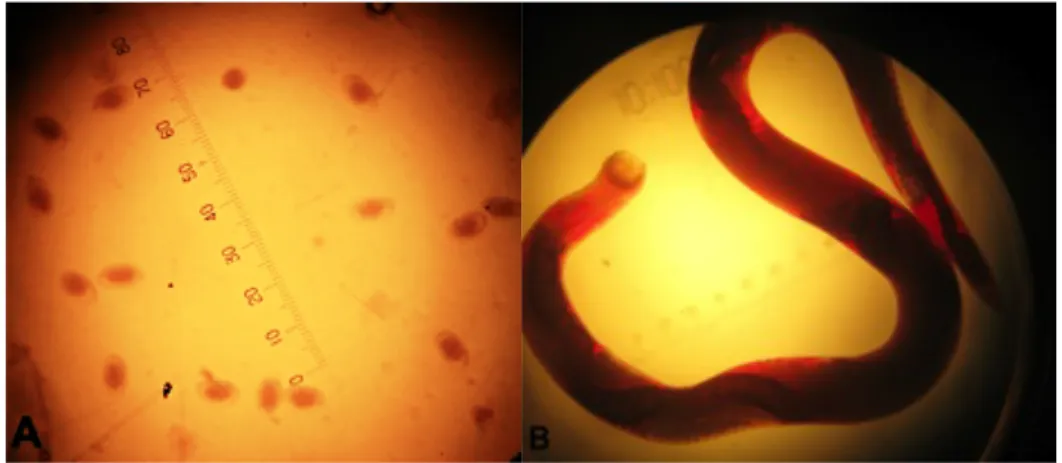
Figure 7: Nematode eggs after fecal float test; Syngamus trachea worm under optical microscope magnification ... 52
Figure 8: Placement of an oesophageal stetoscope on a Anodorhynchus hyacinthinus with SCUD lesions. ... 59
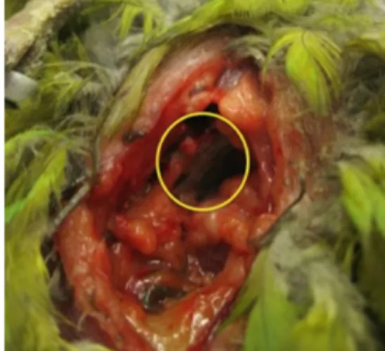
Figure 9: Detail of the SCUD lesions on an Anodorhynchus hyacinthinus ... 60
Figure 10: "Ekky" (Bubo bubo), hospitalised at GWR...61
Figure 11: Corneal reflex evaluation during GA ... 62
Figure 12: "Sonny" with a buster collar ... 66
Figure 13: Air sac tube placement on "Sonny"; Tracheoscopy and aspiration of exudate ... 67
Figure 14: Nebulizator pressurized gas source; Nebulization and O2 into patient´s cage ... 68
Figure 15: Surgical preparation and positioning of "Sonny"; Skin incision ... 69
Figure 16: After removal of the affected traqueal segment; Lone Star Retractor System™. . 69
Figure 17: Affected segment of the trachea with a decreased luminal diameter. ... 69
Figure 18: Injured Cygnus olor; GA. ... 72
Figure 19: Abscess removal; Closure ... 72
Figure 20: Removed abscess; Recovery. ... 72
Figure 21: Positioning for examination of the oral cavity; Mass ... 74
Figure 22: Removing a sample from the mass for histopathology ... 74
Figure 23: Diarrhoea of "Laura"; IV catheterization of the basilic vein ... 77
Figure 24: Positioning "Laura" for x-ray; Egg yolk removed by ovocentesis ... 78
Figure 25: Post-mortem exam of "Laura" with multiple haemorrhagic foci ... 78
Figure 26: Microscopic examination of the impression smears from "Laura" ... 79
Figure 27: The bird´s leg after the traumatic event; Material used for splinting. ... 82
Figure 28: The splint after molding it to the bird´s leg and filling with Technovit®6091; Applying the splint ... 83
Graphics
Graphic 1: Mammal species seen at practice...3 Graphic 2: Reptile species seen at practice...3 Graphic 3: Avian orders seen at practice...4
Abbreviations
2-Pam 2-pyridine aldoxime methyl chloride ACE Angiotensin converting enzyme ACh Acetylcholine
A&E Avian and exotic AGP African grey parrot
APA Air sac perfusion anaesthesia ARF Acute renal failure
AST Aspartate aminotransferase ATP Adenosine tri-phosphate BC Biochemistry
BID Bis in die
BMR Basal metabolic rate BP Blood pressure bpm Beats per minute BW Body weight
CBC Complete blood count CK Creatine kinase
CNS Central nervous system CO Cardiac output
CPCR Cardiopulmonary-cerebral resuscitation CRT Capillary refill time
CS Clinical signs
CSF Cerebral spinal fluid CT Computed tomography ECG Electrocardiogram
EDTA Ethylenediamine tetraacetic acid e.g. Exempli gratia
EPH Protein electrophoresis ET Endotracheal et al. Et alia FB Foreign body g Gram G Gauge GA General anaesthesia GI Gastro-intestinal
GWR Great Western Referrals h Hour
HR Heart rate i.e. Id est
IM Intramuscular IO Intraosseous
IPPV Intermittent positive pressure ventilation IU International units
IV Intravenous
IZVG International zoo vetgroup pathology Kg Kilogram
LRS Lactated Ringer´s solution
MAC Minimum anaesthetic concentration
MER Maintenance energy requirement min Minute
mm Millimeter
MM Mucous membranes
MRI Magnetic ressonance imaging
nd No date No. Number
NSAIDs Nonsteroidal anti-inflammatory drugs PBFD Psittacine beak and feather disease PCV Packed cell volume
PDD Proventricular dilatation disease PDT Photodynamic therapy
PDV Pacheco´s disease virus PE Physical examination PGE2 Prostaglandin E2
PGF2α Prostaglandin factor 2 alpha QID Quaque die
RAAS Renin-angiotensin-aldosterone system RR Respiratory rate
s Second SC Subcutaneous
SCC Squamous cell carcinoma
SCUD Septicaemic cutaneous ulcerative disease SID Semel in die
SVR Systemic vascular resistance TID Ter in die
TP Total protein UK United Kingdom US Ultrasonography WBC White blood cells
Chapter 1. Externship report
The following report will describe the activities developed during a 4 month training externship in the field of exotic animal medicine and surgery as well as wildlife casualities. The externship took place at Great Western Referrals Hospital (GWR), Swindon, United Kingdom (UK), in the Avian and Exotic (A&E) department between the 20th of September 2010 and the 30th of January 2011 with a total duration of 800 hours, having Dr. Neil Forbes as my supervisor and Prof. Sandra Jesus as my coordinator.
During my externship I was enrolled in a training programme that covered nursing, hospitalisation, therapeutics, fluid and nutritional support of hospitalised patients. I performed procedures such as clinical pathology sample collection and testing, anaesthesia monitoring, diagnostic imaging and endoscopy. I also observed and assisted with consultations and surgical procedures. Not so frequently, I was involved in the small animal department for similar procedures with dogs and cats. I also accompanied Dr. Neil Forbes in some home visits to raptors and inspections to wildlife parks such as the Slimbridge Wetland Centre, Gloucestershire.
I had the chance of attending 6 CPD (Continuing Professional Development) courses on the subjects of "Update on rabbit medicine", "Veterinary Care of Backyard Poultry", "Avian cardiology", "Canine knees & hips", "The diagnosis & management of cognitive dysfunction" and "Wildlife casualities". I also participated in 2 practical trainning sessions performed by Dr. Neil Forbes to the veterinary staff of the A&E department on avian orthopaedic and soft tissue surgery and a day course designed for falconers on "Management of raptors for health and longevity" as well as some small animal meetings with specialists on dermatology, surgery and diabetes mellitus. I was also provided the oportunity to attend the 11th European Association of Avian Veterinarians (AAV) conference and 1st European College of Zoological Medicine (ECZM) meeting in Madrid, Spain, April 26-30, 2011.
GWR is recognized by the European College of Veterinary Surgeons (ECVS), European College of Avian Medicine and Surgery (ECAMS) and Royal College of Veterinary Surgeons (RCVS) as a veterinary training center for surgery and avian medicine.
GWR offers a small animal referral service in internal medicine and surgery and an avian and exotic service. GWR works mostly as a referral center so first opinion cases are not accepted in the small animal department (except for emergency cases). In the A&E department both referral and first opinion cases are accepted.
The hospital is open 24 hours a day, 7 days a week with an out of hours (OOH) service working with one veterinary nurse and one veterinary intern always available for emergencies and hospitalised patients.
The hospital building has 2 floors. The first floor comprises the small animal department, reception with a waiting room, 1 office, 4 consult rooms, an ultrasonography (US) room, a
pharmacy, a x-ray room and a small room for the x-ray developer. There are 3 hospitalisation wards for dogs, cats and infectious diseases, 2 surgical theatres (one for soft tissue surgery and the other for orthopaedic surgery), a laundry room, a sterilization room and a store house, known as Unit 14, for storage of medical and surgical material, 2 freezers and a fluoroscopy machine.
The second floor consists of the A&E department with a preparation room used for most procedures, one surgical theatre, a x-ray room and another room used for all the small mammal dental procedures or if the main room is occupied. There are also 3 hospitalisation wards, one isolation ward for birds where there are also reptile vivariums, another for psittacines and small mammals and other for raptors and ferrets. GWR has its own in-house laboratory also on the second floor where haematology, citology, parasitology, microbiology or biochemistry procedures are performed. There is also an autoclave room, staff room with kitchen, 2 bedrooms, an auditorium for conferences, 4 offices for the administrative, specialists, residents and common office for interns and externs with a veterinary library. A dermatology specialist gives consultations in the hospital twice a week and a cardiology specialist gives consultations twice a month. Two times a week, there is a Magnetic Ressonance Imaging (MRI) mobile service operating at GWR.
The A&E veterinary staff consists of 1 specialist, 2 residents, 1 intern and 2 nurses.
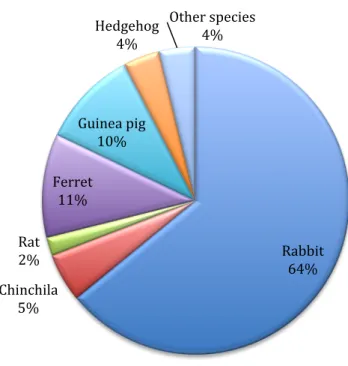
During my externship a total of 383 avian and exotic animals were observed including birds, mammals, reptiles, 3 Koi fish and 1 tarantula. A total of 156 mammals were seen, mostly rabbits (100) but also chinchilas (8), rats (3), ferrets (17), guinea pigs (16), hedgehogs (6) and other species including chipmunks (2), meerkats (2) and gerbils (2) (Graphic 1). Most common causes of mammal presentation included vaccination/health checks, dental disease, gut stasis, dermatology, referrals, ovariohysterectomy, castration and nail/hair trims.
Graphic 1: Mammal species seen at practice
A total of 76 reptiles were seen including testudines (38), serpents (9) and lizards (29) (Graphic 2). Common causes of presentation included pre-hibernation checks, dewormings, nutritional problems or trauma.
Graphic 2: Reptiles seen at practice
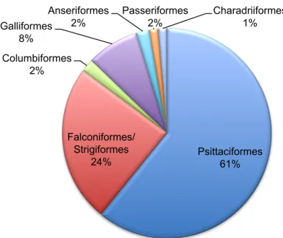
A total of 148 birds were seen mostly Psittaciformes (90) and Falconiformes/Strigiformes (36) but also Anseriformes (3), Passeriformes (2), Galliformes (12), Columbiformes (3) and Charadriiformes (2) (Graphic 3). Most common causes of presentation included health checks, nail and beak trims, trauma, emergencies, gastrointestinal (GI) disease and referral cases. Rabbit 64% Chinchila 5% Rat 2% Ferret 11% Guinea pig 10% Hedgehog 4% Other species 4% Testudines 73% Serpents 17% Lizards 10%
Graphic 3: Avian orders seen at practice
Psittaciformes 61% Falconiformes/ Strigiformes 24% Columbiformes 2% Galliformes 8% Anseriformes 2% Passeriformes 2% Charadriiformes 1%
Chapter 2. Literature review
Increasing numbers of exotic animals are being kept as pets and owners expect to receive the same high quality medical care as given to dogs and cats (Lichtenberger, 2007a).
The veterinarian dealing with avian emergencies must be able to perform a fast and effective judgement of each case, knowing when it is appropriate to carry out a physical examination (PE) or when it is prudent to stabilise a critical patient first, and then be able to formulate a logical approach to diagnostics/treatments and anticipate potential complications (Harris, 2003).
A change of approach is necessary when treating exotic pets in general practice and value should be given to the bond between owner and pet, regardless of the species involved (Lichtenberger, 2007a).
2.1. Taxonomy, morphology and function
Among the Animalia kingdom, the largest class is Aves with many orders including Galliformes (e.g. turkey, chicken); Anseriformes (e.g. swan, duck); Psittaciformes (e.g. cockatiel, macaw); Strigiformes (e.g. owls); Columbiformes (e.g. dove, pigeon); Passeriformes (e.g. finch, canary) and others. There are over 9000 different species with a large variation in size (Sibley & Monroe, 1990).
Some particular considerations of morphology and function include a skeleton that reflects adaptations to flight with pneumatised bones reducing weight and trabeculae that maintain strength (Harcourt-Brown, 2005). Birds have a very high metabolic rate to permit a high level of activity and maintain a constant temperature (Hernandez-Divers, 2005). The average adult bird has a core body temperature of 38-42.5°C. Regulation of internal body temperature relies upon many factors, such as feather condition, adipose and muscle condition, hydration, food intake and respiration (Whittow, 1986 cited by Dorrestein, 2000).
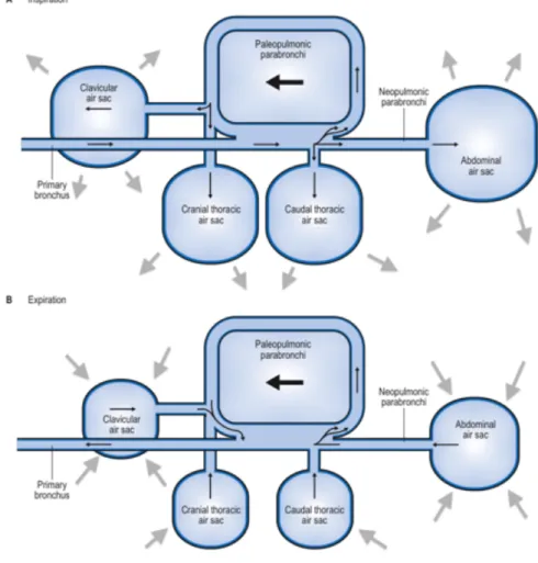
The avian respiratory system is unique with the syrinx at the tracheal bifurcation involved in sound production and usually 9 air sacs: cervicocephalic (1-2), interclavicular (1), cranial thoracic (2), caudal thoracic (2) and abdominal (2) (Brown & Pilny, 2007). Air sacs are expansions of the bronchial wall, acting as bellows to the lungs and make up to 80% of the volume of the respiratory tract. Functions include cooling, reducing weight and aiding buoyancy in swimming birds, not being involved in gas exchange (Harcourt-Brown, 2005). In the lungs, air flows through the paleopulmonic parabronchi, minimally anastomosing with uni-directional airflow and neopulmonic parabronchi, a meshwork of anastomosing parabronchi with bi-directional airflow. Airflow is crosscurrent to the blood flow in the blood capillaries, optimising oxygen (O2) exchange. Air flows through the lung in the neoplumonic parabronchi to the caudal air sacs, from here to the paleopulmonic parabronchi (absorptive surface) and to the cranial air sacs, meaning that the air passes across the absorptive surface continually and unidirectionally (Fig. 1) (Macwhirter, 2000). The flow through air
capillaries within the lungs is a much more efficient gas exchange mechanism than the dead end alveoli found in mammalian lungs (Mitchell & Tully Jr., 2009).
Figure 1: Schematic representation of the air flow in the avian respiratory system. (A) Inspiration; (B) Expiration (Adapted from Longley, 2008).
Other particular features of avian morphology and function include: i) a heart proportionally larger and more efficient due to increased stroke volumes, decreased heart rate (HR) and increased cardiac output (CO) (Harcourt-Brown, 2005); ii) avian white blood cells (WBC) similar to mammalian WBC but avian neutrophil equivalents with heterophilic granules are termed heterophils (Fudge, 2000); iii) a gastro-intestinal (GI) system with a crop that carries out storage function, not involved in digestion (Macwhirter, 2000); iv) excretory system with trilobed retroperitoneal kidneys and ureters that feed directly into the urodeum of the cloaca, without a urinary bladder; v) nitrogenous waste excreted as non-toxic, semi-solid uric acid; vi) droppings passed as a mix of urine, urate and faeces (Harcourt-Brown, 2005); vii) blood flow through the kidney controlled by the renal portal valve and a complex system of shunts (King & McLelland, 1979 cited by Coles, 2007), and viii) a female reproductive system with (usually) a single functional left ovary and absent or poorly developed right ovary (Macwhirter, 2000).
Birds are oviparous and sexual dimorphism varies between groups. For sexing, the clinician can use rigid endoscopy under general anaesthesia (GA) for direct visualisation of the testes/ovary. DNA testing from feather pulp or blood is also used and since feather sampling is non-invasive, many laboratories offer testing directly to owners and breeders (Harcourt-Brown, 2005).
2.2. Emergency and critical care of the avian patient
2.2.1. Emergency stabilisation
2.2.1.1. Arrival of the avian patient and preliminary evaluation
When birds arrive to the veterinarian they are usually in 1 of 3 conditons: healthy, injured or ill. Injured birds need to be stabilised with supportive care before the injury can be successfully treated (Harris, 2003). It is part of a bird´s natural behaviour to evade predators by hiding illness, so owners frequently present them only when advanced clinical disease overcomes this evasion behaviour (Hernandez-Divers, 2005 and Matos & Morrisey, 2005). Treatment and diagnosis must be performed in a step-wise fashion, with constant re-evaluation of the patient´s condition and ability to tolerate further procedures although a critically ill bird may not allow diagnostic tests to be performed because either the stress of sample collection or the time needed to process them is more than the patient can endure (Lightfoot, 2008).
Birds that display signs of illness for several days often compensate while those with acute serious clinical signs (CS) may be decompensated (Harris, 2003). History should include age, sex (if known), source of the bird, other pets in the household, diet (e.g. type, proportions, changes in food/water intake), exposure to trauma, toxins, droppings appearance, reproductive activity or other behavioural changes and environmental history (e.g. type of cage, ability to leave the cage unsupervised) (Echols, 2007).
After obtaining a brief history, the bird should be observed before handling it, if possible from a distance because the bird will often seem more alert and responsive when the clinician stands close giving an illusion of wellness. The cage should be examined for regurgitated material, blood/other discharges, faecal appearance and potential sources of toxins (Bowles, Lichtenberger & Lennox, 2007).
Before handling, general demeanor and overall appearance should be evaluated (e.g. fluffed feathers, unconscious, unable to perch, falling to one side, circling, wings hanging down, hanging onto perch/cage with the beak), respiration (Echols, 2007), droppings (i.e. if scant and green to black may suggest anorexia; many birds display stress-related polyuria; lime green urates, blood or no droppings) and any other gross abnormalities (Bowles et al., 2007). Common CS of critical illness include tail bobbing, severe weakness, emaciation, changes of vocalisation and closed or slitty/"lemon" eyes (Echols, 2007).
The bird that brightens when approached may be stronger than one that continues to show significant signs of illness. After gathering all the information from history/observation the clinician can decide to: i) place the bird in a supportive intensive care chamber; ii) directly administer basic supportive treatment before proceeding, or iii) perform the physical examination with or without collecting diagnostic samples (Harris, 2003).
2.2.1.2. Physical examination
PE must be complete and thorough as in any other species. The crop should be palpated, and the choanal slit of the oropharynx checked for inflammatory debris, foreign material or blunted papillae. The beak is inspected for bleeding, asymmetry, altered keratin quality and fractures (Bowles et al., 2007). Perfusion status should be evaluated by determination of the capillary refill time (CRT) of the basilic vein and hydration by skin turgor. A CRT greater than 1-2s indicates more than 7% dehydration as normal veins are turgid and refill immediately (Echols, 2007). Hydration and temperature evaluation are major points on PE (Echols, 2007) and it can be presumed that a critical patient (due to illness, not trauma) is 10% dehydrated and probably hypothermic so dehydration and hypothermia should be treated before any further manipulations (Harris, 2003).
Auscultation is a challenge because of patient size, rigid lungs and unique respiratory system, but since respiratory sounds are minimal in birds, crackles, wheezes and clicks are usually abnormal. The coelomic space, between the ventral keel and pelvis, should be palpated. Overall body condition is evaluated by palpation of the pectoral muscle mass. The vent and cloaca are inspected for lesions (e.g. mass, swelling, prolapsed tissue) and the surrounding feathers should be clean (Bowles et al., 2007). Weight should be taken and the bird observed in an incubator after the PE to determine the impact of handling on overall condition. Sick birds should always be weighed in grams at least once daily, preferably at the same time of day, while hospitalised (Echols, 2007). In cases where PE is not recommended, a rapid examination is performed while transferring the bird from its cage to the incubator, palpating the pectoral muscle mass and coelomic space during transfer and observing the bird carefully after release into the incubator (Bowles et al., 2007).
2.2.1.3. Hospitalization
Heat, humidity and oxygen should be provided. Moist air appears to safely aid in correction of hypothermia by reducing evaporation from the extensive internal respiratory surface area, in addition, the oxygen drying tendency is nullified. The patient should be closely monitored while in the chamber in order to prevent overheating (Harris, 2003).
Some common avian infectious diseases include chlamydophilosis, Psittacine Beak and Feather Disease (PBFD), Proventricular Dilatation Disease (PDD), aspergillosis, tuberculosis or salmonellosis so multiple birds sharing the same airspace should be avoided to decrease
contamination across patients. Many zoonoses affect birds so adequate protective measures for staff should also be considered (Graham & Heatley, 2007).
2.2.1.4. Medical management
There is a distinction between a critically ill bird and a trauma patient because the first will invariably die without immediate medical attention, so the bird should be evaluated in order to know if injury management takes precedence over medical support (i.e. if haemorrhage is present it must be controlled, if an airway obstructed it must be cleared and simple injuries like lacerations or fractures should be managed only after the patient is assessed and stabilised) (Harris, 2003).
Before handling the bird, a plan should be created. Because the patient needing most support is the one least capable of tolerating stress a "conservative progression" approach is the best way to deal with it, one procedure at a time, prioritizing needs, then waiting for the clinical effect before proceeding. General techniques for supportive care can be employed on almost any critical patient without delay providing stabilisation (e.g. thermal support, fluid therapy, catheterisation, blood transfusion, nutritional support, pain relief) (Hess, 2002; Wilson, 2005).
2.2.2. Emergency procedures
2.2.2.1. Oxygen administration
Indications for O2 therapy include respiratory/cardiac disease, shock, post-surgical recovery and stress. Critical patients may have a diminished CO and O2 maximizes cardio-respiratory efficiency. For any dyspnoeic bird, O2 is recommended and hyper-oxygenating the patient prior to handling may also decrease its risk. Some patient’s don´t tolerate masks and if an oxygen cage is not available, most incubators, cages and aquariums can be modified so that supplemental O2 can be provided into them. For most critical patients kept in a closed clear chamber, a 40-50% O2 saturation level is recommended (Echols, 2007). Pure O2 is acceptable for short-term use given via facemask or in an enclosed environment. Prolonged exposure to high O2 levels was shown to be toxic in budgerigars (Wilson, 2005).
Low dose sedation may reduce some of the stress associated with upper airway obstruction (Graham, 2004). Restraint may increase the respiratory rate (RR) 1.5-2 times the resting rate, so it should be avoided, if possible, for dyspnoeic patients (Wilson, 2005).
2.2.2.2. Nebulization
Nebulizers convert liquids to aerosol particles appropriately sized for inhalation. They consist of a disposable/reusable nebulizer and a pressurized gas source and can be used to deliver moisture or topical medications to the mucosa of the respiratory system (Bowles et al., 2007) reducing the stress of handling (Harcourt-Brown, 2005).
The systemic drug dose is usually diluted in 5-10ml saline and delivered over a single 15-30 minute session. The most frequently used antibiotics for nebulization are aminoglycosides. Parenteral bronchodilators such as terbutaline have been empirically used in birds with lower airway disease via nebulization. Initially terbutaline (0.01mg/Kg) is given IM or SC while the bird is placed into an oxygen-enriched environment and then is nebulized (0.01mg/Kg diluted into 5-10ml of saline, TID) (Bowles et al., 2007).
2.2.2.3. Injection sites and sampling
Subcutaneous (SC), intramuscular (IM), intravenous (IV) or intraosseous (IO) routes can be used for injection and sampling of avian patients. SC sites include the featherless inguinal and/or axillary skin fold, taking care to aspirate prior to injection to confirm the needle has not penetrated the air sacs (Tully Jr. & Beaufrère, 2011). For IM administration the pectoral muscles are used while for IV administration, the right jugular vein, basilic vein or dorsal metatarsal vein in larger birds are the choice (Harcourt-Brown, 2005). For IO route the distal ulna or proximal tibiotarsus are the options (Tully Jr. & Beaufrère, 2011).
In birds, blood volume is around 10% bodyweight (BW) (Harcourt-Brown, 2005) and it will be safe to collect up to 1% of the BW from passerines and psittacines (Ritchie, Harrison & Harrison, 1994). In compromised birds, this should be reduced to 0.5% total BW. Samples for haematology and biochemistry (BC) are obtained before treatment for best diagnostic value, however, the patient´s needs must be prioritized (Graham & Heatley, 2007).
2.2.2.4. Intravenous catheterization
IV catheters can be placed in the right jugular, basilic or metatarsal veins. The jugular vein can be used in birds as small as 75g but it can be difficult to maintain the catheter in place (Bowles et al., 2007). There is no need for plucking at this site because there is an area of apterylae over it. A 20-22G catheter of sufficient length to reach the thoracic inlet can be used. The basilic vein (over the medial ulna) is easy to visualize and catheterise. Once the catheter is secured, the wing can be wrapped in a figure 8 bandage (Hess, 2002). The dorsal metatarsal vein (along the dorso-medial aspect of the tarsometatarsus) allows easy visualization of the catheter and the thick skin of the feet helps holding the catheter in place. This vein can be used in birds over 300g (Bowles et al., 2007).
2.2.2.5. Intraosseous catheterization
In many cases (small or hypovolaemic birds) IO catheterization may be preferable to IV catheterization, as the latter is often more technically difficult in terms of placement/maintenance and has a higher risk of significant blood loss if removed by the bird (Bowles et al., 2007). IO catheters can be placed in the distal ulna (larger birds) or proximal ulna, proximal tibiotarsus and lateral femur (young/smaller birds) (Tully Jr. & Beaufrère, 2011). Short spinal needles, 20-22G, can be used for larger birds and 22-25G for smaller
birds (Bowles et al., 2007). IO catheters require more maintenance than IV catheters and should be flushed daily to maintain patency (Tully Jr. & Beaufrère, 2011).
Birds with IO/IV catheters should be monitored daily for complications such as a cardiac murmur, local pain, swelling or heat that might suggest infection (Echols, 2007) and analgesia should be provided (Lichtenberger & Ko, 2007). All catheters should be removed after 48-72h even if no complications occur and a new catheter placed in a different site if needed (Echols, 2007).
2.2.3. Emergency avian surgery
The actual procedures are too numerous to list here, however, the basics of emergency avian surgery techniques are discussed in this section. Microsurgery and magnification instruments are often required due to small patient size (N. A. Forbes, personal communication, December 21, 2010).
2.2.3.1. Air sac tube placement
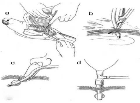
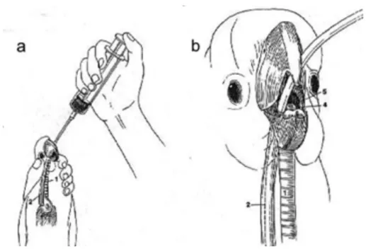
The unique respiratory system of birds allows direct delivery of oxygen/anaesthetics through the air sacs, bypassing the trachea (Chavez & Echols, 2007). Placement of an air sac tube is an emergency technique that can be used if the oral cavity or trachea are occluded, causing acute respiratory distress (Brown & Pilny, 2007). Anaesthesia via air sac breathing tubes has been successfully used in patients as small as zebra finches (Nilson, Teramitsu & White, 2005). Sterile different sized air sac tubes should be available at all times. Commercial avian air sac cannulas exist but endotracheal (ET) tubes can be shortened for this purpose. Material should be rigid enough not to collapse but flexible enough to avoid damage of internal organs. For very small birds, 20 or 22G IV catheters can be used temporarily (Graham & Heatley, 2007). It is advised to use a tube 25% larger than the patient´s trachea, inserted to ⅓ of the width of the coelom at the point of insertion. The tube should have a length to suit patient size and multiple holes in the side as well as the end, with pre-placed sutures at the level of the coelomic wall (N. A. Forbes, personal communication, December 21, 2010). It is always recommended to radiograph patients prior to placement as presence of organomegaly or ascites can lead to complications (Graham & Heatley, 2007).
Air sac tubes can be left in place for up to 7 days, however they should be ideally removed within 4 days because of the risk of air sacculitis associated with prolonged tube placement (Brown & Pilny, 2007). Complications include coelomic organ damage, life threatening blood loss, air sac damage and SC emphysema (malpositioning, excessive perfusion volume, obstruction) (Korbel, 1999).
Figure 2: Placement of an air sac tube. Making a skin incision in the area of the sternal notch (a) then passing a pair of haemostats through the abdominal muscle (b) and suturing an ET tube to the body
wall (c,d) (Adapted from Ritchie et al., 1994).
2.2.3.2. Oesophagostomy tube placement
Oesophageal tubes are used to bypass the upper GI tract, allow enteral medication and/or provide food to the crop/proventriculus of anorexic patients (Huynh & Forbes, 2011). The tube is measured from the midcervix (midneck) to the distal edge of the keel and cut on the proximal end to the measured point. The tube is then sutured into place, both at the cervical insertion site and on the skin of the dorsum. Liquid food can be given by a syringe, followed by flushing of the tube to maintain patency and hygiene, with minimal restraint (Chavez & Echols, 2007). The tube can remain in place for several days until the bird is able to feed sufficiently to maintain its weight without assistance. The sutures can be removed, the tube retracted and the wound left to heal by secondary intention (Echols, 2007). Complications include penetration of the oesophageal wall opposite to the incision site and oesophageal strictures (Ritchie et al., 1994).
2.2.3.3. Proventriculectomy
Proventriculectomy is used to access the proventriculus and ventriculus to remove foreign bodies (FB) or for surgical biopsy. The ventriculus is a muscular organ with a white tendinous lateral aspect (N. A. Forbes, personal communication, December 21, 2010).
Since birds have no mesentery, an enterotomy has a higher risk of postoperative peritonitis so the liver can provide some of the protective function of the absent mesentery (N. A. Forbes, personal communication, December 21, 2010). Exteriorized organs should be kept moist with warm saline but care should be taken to minimise fluid use as the air sacs are exposed (Chavez & Echols, 2007). Complications include contamination of the abdominal cavity with gastric contents so meticulous attention to proper closure is vital to prevent
leakage. A small, atraumatic needle should be used with a continuous suture pattern to provide the best seal. If the proventricular wall appears thin and friable, the potential for postoperative incisional leakage may warrant placement of a duodenal feeding tube that will allow enteral alimentation of the patient while bypassing the gastric incision (Ritchie et al., 1994).
2.2.3.4. Ingluviotomy
Ingluviotomy is indicated for retrieval of crop, proventricular or ventricular FB, placement of an ingluviotomy/proventriculotomy tube or rapidly emptying the crop (e.g. sour crop, ingluviolith) (N. A. Forbes, personal communication, December 21, 2010). Because of the ability of the ingluvies to stretch, the incision should be made only about half the size necessary to accomplish the procedure, however, having adequate exposure is more important than having a small incision, and retrieval of large FB through small ingluviotomy incisions should not be attempted to avoid serious tissue damage. Following the procedure, closure is done with 2 layers of continuous inversion sutures since this is less compromising on the size of the crop lumen. The crop may be inflated with saline or air to check for leakage prior to skin closure and avoid complications such as contamination of the abdominal cavity (Ritchie et al., 1994).
2.2.3.5. Tracheotomy and trachectomy
Tracheotomy is commonly indicated for treatment of syringeal aspergillomas or retrieval of a tracheal FB (N. A. Forbes, personal communication, December 21, 2010). Air sac tube placement for anaesthesia is recommended for both procedures (Harrison & Lightfoot, 2006). Trachectomy is used in cases of severe tracheal stenosis due to trauma, infection or stricture, when tracheal resection with removal of the affected tissue is performed (N. A. Forbes, personal communication, December 21, 2010). Not all species can tolerate a shortening of the tracheal length (Harrison & Lightfoot, 2006) and unless absolutely mandatory, the trachea should not be completely transected in order to maintain its alignment, reduce tension on the closure and prevent complete disruption of the blood supply (Ritchie et al., 1994). Postoperative care including analgesia, is best achieved with carprofen (4mg/Kg, IM), which may be augmented with butorphanol, although meloxicam (0.2-0.4mg/Kg) may be safely used. Fluid therapy, warmth, nutritional supplementation and good nursing are as important as the surgical skills (N. A. Forbes, personal communication, December 21, 2010).
Complications for both procedures include iatrogenic trauma during manipulation and stricture formation. Stay sutures placed around the tracheal rings adjacent to the incision site allow atraumatic manipulation of the trachea. When closing the incision, knots should be
placed external to the tracheal lumen because intraluminal granuloma formation at the suture site is common. This is a difficult procedure that should be only used as a life-saving technique, when all other methods have failed (Ritchie et al., 1994).
2.2.4. Diagnostic testing
Diagnostic testing techniques for avian emergency and critical care cases should include blood work, cytology, histology, microbiology, urinalysis, evaluation of faeces and crop specimens, nasal flush and endoscopy. Routine haematology and chemistry analysis should include a complete blood count (CBC) and at least aspartate aminotransferase (AST), creatine kinase (CK), glucose, uric acid, bile acids, serum calcium (Ca), phosphorus (P), albumin and electrolyte measurements. Packed cell volume (PCV) evaluates the presence of anaemia. A WBC count and blood smear should be performed. Other measurements include blood lead (Pb) level, plasma zinc (Zn) level and protein electrophoresis (EPH) (Bowles et al., 2007). Lichtenberger (2005) recommends for a critically ill bird, to collect no more than 0.4 to 0.5ml/100g BW. Imaging techniques include radiography, US, MRI and computed tomography (CT) scan. Fluoroscopy is used for GI motility studies or detection of metal FB without GA (Lichtenberger, 2005).
2.2.5. Frequently seen emergencies
Common emergency presentations of pet birds will be discussed on this topic. Knowledge of each disease, diagnostic tests and treatment protocols are essential to provide immediate and efficient/effective care to critical patients (Lightfoot, 2008).
2.2.5.1. Collapse/profound weakness
Stabilise the bird with O2 therapy, IV/IO fluids containing dextrose and consider broad-spectrum bactericidal antibiotics such as enrofloxacin (7.5-15mg/Kg, IM/PO, q12h), cefotaxime (75-100mg/Kg, IM/IV, q4-8h), amikacin (hydrated birds, 10-15mg/Kg, IM/IV, q12h) or piperacillin (100-200mg/Kg, IM/IV, q6-8h) (Hess, 2002).
2.2.5.2. Acute respiratory distress
This is potentially life threathening and all patients should be gently handled to minimise stress and supplemental O2 provided before handling (Echols, 2007). Some drugs can be administered to birds in respiratory distress before placement in an oxygen chamber, such as bronchial dilators (terbutaline 0.01mg/Kg, IM, q6-8h) (Plumb, 2005) and analgesics with antianxiety properties (butorphanol 1-2 mg/Kg, IM, q2-3h) (Echols, 2007).
It is important to differentiate decreased oxygenation from tracheal obstruction (which may require placement of an air-sac tube) from other conditions such as air sacculitis/pneumonia, sarcocystosis, internal haemorrhage, inhaled toxins, allergic pneumonitis, cardiac disease or abdominal masses (Lightfoot, 2008). History, auscultation and PE allow the clinician to reach
diagnosis (i.e. a bird from the genus Amazona with acute onset dyspnoea and abrupt loss of voice may have syringeal disease; a Psittacus erithacus fed only sunflower seeds may have hypovitaminosis A; and a Saker falcon - Falco cherrug - is likely to succumb to aspergillosis after stress) (Echols, 2007). Palpation of the abdomen allows detection of fluid, masses or displaced viscera. Auscultation of the lungs may not reveal subtle respiratory disease because of their rigidity but can help localizing and detecting abnormal sounds. Moist or harsh expiratory sounds are suggestive of bronchopulmonary disease while clicking and crackles indicate thickened air sacs touching (air sac disease). The heart should be auscultated for murmurs that may be evident with cardiac disease and an echocardiogram or US performed (Bowles et al., 2007).
Respiratory disease may affect the upper airways (sinus), large airways (glottis to syrinx), small airways (primary bronchi), parenchyma (lungs to air sacs) or intracoelomic cavity resulting in compression of the lungs (Echols, 2007).
2.2.5.3. Traumatic presentations
Traumatic injuries are common in the pet bird. The initial protocol for injured birds includes haemorrhage control, O2, heat support, analgesics and parenteral antibiotics. External haemorrhage should be stopped immediately (Bowles et. al., 2007). The clinician should distinguish between haemorrhage from an external site and blood without active bleeding. Continued frank haemorrhage requires intervention while haemorrhage that has ceased is best left undisturbed. The color of the tissue in the choanal slit can be used to estimate anaemia. Hypovolaemia is the initial concern and rehydration should be performed rapidly. Moribund patients should have an IO/IV catheter placed, however, if the bird is still conscious this may be detrimental due to stress (Lightfoot, 2008). Warm SC fluids with hyaluronidase should be administered. Birds in septic shock or septicaemia from extensive wounds, should receive parenteral antibiotics (e.g. piperacillin, 100-200mg/Kg IM, IV q6-8h; trimethoprim-sulpha, 8-30 mg/Kg, IM, BID) (Hess, 2002 and Harrison & Lightfoot, 2006).
The goal of treatment is patient survival first and then accessing traumatized tissue. A bird that has been struggling for hours with a trapped wing may have a fractured bone but is in more danger of dying from stress related to prolonged struggling than complications from the fracture itself. Temporary stabilisation of the traumatized tissue is enough until the bird is stable. If the bleeding comes from the vent, the clinician should determine its origin (Lightfoot, 2008).
Birds with broken blood feathers often present as emergencies. The feather should be ligated or pulled at the base with a haemostat, extracting it from the follicle and pressure applied. In the case of large primary wing feathers, wing bones should be supported during extraction to avoid fractures. Haemostatic agents such as silver nitrate and styptic powder shouldn´t be applied on the feather follicle because they may damage it (Bowles et al., 2007) but can be used to stop the bleeding on broken nails (Hess, 2002).
The greater challenge is to recognize internal blood loss. The rate that blood is lost from circulation is the determining factor for mortality. Losing 20-25% of blood over minutes may be fatal while losing the same volume for hours is not. Discoloration or bruising of the abdominal wall, along the ventral midline and caudal abdomen, may suggest internal haemorrhage and radiology/endoscopy may help locating it (Jenkins, 2005).
Analgesia is necessary in birds that have suffered fractures or soft tissue trauma. Post-traumatic administration of nonsteroidal anti-inflammatory drugs (NSAIDs) may decrease soft tissue sensitization and reduce the dose of opioid drug required. Steroids are contra-indicated (Bowles et al, 2007).
2.2.5.3.1. Wounds (lacerations, bite wounds) and open fractures
The lesions should be flushed with chlorhexidine solution and parenteral β-lactams antibiotics (e.g. piperacillin 100-200mg/Kg, IM/IV, q6-8h), trimethoprim-sulpha or fluoroquinolones. Piperacillin/tazobactam or cefotaxime/amikacin are good choices and should be continued for a minimum of 5 days. Large recent wounds should be flushed with sterile saline solution and sutured partially closed and older lacerations flushed, debrided and bandaged with wet to dry bandages that should be changed twice daily. These wounds may be closed later when infection is minimized (Hess, 2002). Other option is to apply 50% dextrose, sugar and honey as a wound dressing in large contaminated wounds. These are inexpensive and readily available materials considered to be bactericidal because of their high osmolality and additionally provide the wound with moisture and a local nutrient source to promote granulation tissue. The high osmolality is also responsible for moving oedematous fluid out and drawing macrophages into the wound. The wound should be debrided and flushed with saline, then sugar/honey applied to it and covered by a wet-to-dry bandage. Some topical wound treatments use products that promote wound healing by stimulating the production of cytokines or growth factors within the wound (e.g. D-glucose, polysaccharide, hydrolyzed bovine collagen, porcine collagen products, tripeptide-copper complex) (Bowles et al., 2007).
Bite wounds from carnivores can lead to fatal septicaemia if not treated aggressively with systemic antibiotics (Jenkins, 2005) and silver sulfadiazine cream can be applied on contaminated wounds. Nonadherent bandaging materials are recommended to avoid disruption of the healing surface of the wound. Self-adherent bandages can tighten when wet, so the access of bandaged birds to open containers of water should be limited or monitored (Bowles et al., 2007).
In the case of self-mutilation, topical antibiotics such as silver sulfadiazine cream may be applied and the bird temporarily collared/bandaged with a moisture-permeable dressing while treating the underlying cause (Bowles et al., 2007). Ensure that the diet and home environment are appropriate. Psychotropic drugs (haloperidol, 0.2mg/Kg, q12h for birds
weighing less than 1Kg and 0.15mg/Kg, q12-24h for birds above 1Kg) may be used in cases of severe mutilation unresponsive to other therapies.
Diagnostic tests should include a CBC, plasma BC panel, EPH, faecal Gram´s stain and direct smear, polyoma and PBFD virus testing, radiographs and skin/feather biopsies (Hess, 2002).
2.2.5.3.2. External burn injuries
Burn injuries are classified by severity into superficial, partial thickness and full thickness burns.
For superficial burns only the epidermis is affected, with a transient erythema, desquamation and the site is very sensitive and hyperaesthetic. Such burns should be gently cleansed with sterile saline with 5% povidone iodine or chlorhexidine solution (Jenkins, 2005).
Partial thickness burns extend to the mid-dermis, loss of epidermis is complete, capillaries and venules in the dermis are dilated, congested and exude plasma (Bowles et al., 2007). The site may be painful but sensitivity is decreased (Jenkins, 2005).
Full thickness burns result in coagulation of epidermis/dermis so its no longer vital, a severe oedema of the subcutis develops because of the increased permeability of deep vessels and necrosis of the damaged tissues, resulting in dry, leathery eschar. Feathers and skin may be easily pulled or peeled, respectively (Jenkins, 2005).
Partial to full-thickness burns should be cleansed gently, debrided daily and treated topically with a water-soluble antibiotic dressing such as silver sulfadiazine. Due to associated pain, GA and analgesia should be provided (Bowles et al., 2007). If more than 50% of the body surface is involved (partial to full-thickness burns) prognosis is grave and euthanasia should be considered (Hess, 2002).
Diagnostics include radiography in case of exposure to smoke to evaluate pulmonary injury, and in severe/extensive burns haemogram and serum electrolyte testing are advised. Dyspnoeic birds often have laryngeal oedema and upper airway excretions so O2 via an air sac tube may be provided. Systemic bactericidal antibiotics (e.g. Piperacillin) should be initiated in cases complicated by infection or treatments done outside a sterile environment. If the burn is recent, it should be treated with cold water or compresses for 20-30min (Bowles et al., 2007) to minimize coagulation and extent of burn depth by dissipating heat. Blood, protein loss and renal function should also be monitored (Hess, 2002).
Complications include circulatory collapse, decreased renal function (oliguria), renal failure, pneumonia, scarring/healing complications (especially in areas where tissue is mobile or under tension) and sepsis in the first 24-48h. Infection is a common cause of death for birds surviving the initial injury and can be caused by opportunistic bacteria (Pseudomonas, Streptococcus, Proteus) (Jenkins, 2005).
2.2.5.3.3. Fractures
Fractures alone are not usually life threatening. The bird should be evaluated for open or closed fractures and soft tissue trauma (Bowles et al., 2007) and the tissue immobilised. When splinting, the rule is to immobilise the joint above and below the fracture (Chavez & Echols, 2007). Bandages should be used to immobilise the fracture and supportive care provided until further fracture stabilisation (e.g. surgery, splinting) is attempted (Hess, 2002). It is preferable to let the bird stabilise than performing immediate surgery on a debilitated bird. Once stable, delays should be minimised to limit muscle contracture, which complicates surgical repair. Closed reduction and repair is associated with significantly less tissue damage (Bowles et al., 2007). Birds have little SC fat or other tissues, so immediate wound coverage decreases further damage and desiccation. If immediate wound closure is not possible, packing the wound with moistened sterile gauze sponges or a water-soluble lubricating gel (K-Y® Jelly, Johnson and Johnson Products) limits desiccation. The use of a water-based preparation is preferred and topical steroids avoided. A hydrogel containing acemannan (Carravet® Wound Dressing, Carrington Laboratories) stimulates healing and maintains moisture (Graham & Heatley, 2007). The selection of an appropriate technique for definitive repair will depend on several variables, including the size of the patient, the degree of postoperative return to function required, cost, the skill of the surgeon and concurrent medical conditions (Harrison & Lightfoot, 2006).
2.2.5.3.4. Ophthalmological injury
Ophthalmologic examination of anterior and posterior segments should be carried out in all trauma cases. Posterior segment examination should include evaluation of the pecten for signs of disruption. Retinal detachment and cataract formation are common sequelae of head trauma. Topical or systemic antibiotics are indicated for corneal ulceration or perforation. Topical ophthalmic NSAIDs can be used to treat uveitis in raptors without corneal laceration (Graham & Heatley, 2007).
2.2.5.3.5. Electrocution
It is not uncommon, especially in immature raptors, with a higher incidence during the winter. CS may not be evident for several days after injury. Consequences include cardiac arrest, pericardial effusion, neurogenic pulmonary oedema with acute respiratory distress and thermal burns, especially in the feet, head and carpi. Feather damage of the bars and barbules without damage to the rachis is a classic sign of electrocution. Injury can be so severe that euthanasia may be necessary (Graham & Heatley, 2007). Treatment is aimed at addressing the symptoms (Hess, 2002). Neurogenic pulmonary oedema may respond to a single dose (2mg/Kg) of furosemide (Lasix®, Sanofi Aventis, Paris, France) and supportive care with O2 and sedation. This oedema is secondary to a capillary permeability-induced alveolar injury so continued administration of diuretics is not advised. The prognosis varies
from fair to grave depending on the extent and severity of the damage. Even if injuries are limited to the feathers, it may prevent releasing a wild bird from a moult cycle for up to 1 year (Graham & Heatley, 2007).
2.2.5.4. Egg binding (dystocia)
Cockatiels, budgerigars, lovebirds, finches and canaries are the most commonly and severely affected species, possibly because of their small size. It can occur from low Ca intake or overproduction of eggs. CS include acute depression, abdominal straining, blood-stained faeces, persistent tail wagging, wide stance, failure to perch, abdominal distension, dyspnoea and/or sudden death. If abdominal structures are compressed, circulatory disorders, lameness, paresis or paralysis can be present and pressure necrosis of the oviductal wall may occur (Bowles et al., 2007).
Presence of an egg is confirmed by radiography (with the bird standing awake on the radiographic cassette) unless the egg is low enough making palpation possible (Lightfoot, 2008). US can be useful and may identify soft-shelled eggs that are hard to see in radiographs. Blood work and serum BC can help determining secondary diseases (e.g. hypocalcaemia) (Stanford, 2005; Bowles et al., 2007).
The prognosis is guarded if the patient has paraparesis/paralysis, decreased BW, depression or labored breathing. Supportive care must be provided before extraction of the egg is attempted, including fluid therapy, injectable Ca gluconate (25-100mg/Kg, IM), adequate warmth/humidity and nutritional support (Lightfoot, 2008). If an infectious aetiology is suspected or the integrity of the oviduct compromised, broad-spectrum antibiotics are indicated. Analgesics are often required. Supportive care is usually enough for non-obstructive dystocia but care must be taken with monitoring the bird closely for any deterioration that may require further intervention (Bowles et al., 2007). If supportive care doesn´t lead to oviposition, further treatment with oxytocin and the avian equivalent, arginine vasotocin for uterine contraction, may be attempted (Lightfoot, 2008). Prostaglandin and hormonal therapy may be used to induce oviductal contraction which can result in extraction of the egg but only if: i) the contractility of the oviduct is sufficient; ii) the uterus is intact; iii) the egg is within the oviduct, and iv) there is no obstruction or egg adhered to the oviduct (Bowles et al., 2007). Oxytocin is more readibly available, but it does not dilate the uterovaginal sphincter, so oviduct rupture is possible (Lightfoot, 2008). PGF2α and PGE2 may also be administered. Neither PGF2α or oxytocin relax the uterovaginal sphincter while inducing oviductal contractions, so the sphincter should be opened in order to avoid reverse peristalsis, severe pain and rupture of the uterus. It should also be noted that prostaglandin and hormonal therapy require exogenous Ca to be effective so supplemental Ca may be necessary prior to administration of these medications. PGE2 gel can be applied topically to the uterovaginal sphincter (0.1ml/100g BW), causing relaxation of the sphincter and oviductal contractions while decreasing the incidence of systemic side effects. PGF2α is administered
parenterally, rather than locally increasing the risk of systemic side-effetcs (e.g. hypertension, bronchoconstriction, general smooth muscle stimulation) (Bowles et al., 2007). If even after medical therapy the egg doesn’t pass, GA and manual extraction may be necessary (Lightfoot, 2008).
Complications of these procedures include retroperistalsis of the egg out of the oviduct into an ectopic position within the ceolom, egg rupture, oviductal trauma/laceration, cloacal or oviductal prolapse and displacement of the egg or fragments out of the uterus and into the coelom which may cause an egg-related peritonitis (Bowles et al., 2007). If fertilisation has occurred and the egg is removed intact, it can be incubated. If any haemorrhage has occurred, antibiotics are indicated. Ovocentesis may aid in the passage of the egg, being performed through the cloaca if the egg is located distally or by lateral coeliotomy if more cranial (Lightfoot, 2008).
Radiography should be used to confirm that all the eggshell pieces have been expelled. It may be advised to flush the uterus postoviposition with saline, chlorhexidine or iodine to remove any shell fragments and decrease the incidence of metritis. In the case of ectopic eggs and dystocia or if after all the procedures described oviposition doesn´t occur salpingohysterectomy may be considered and secondary diseases corrected (Bowles et al., 2007).
2.2.5.5. Neurologic disease
2.2.5.5.1. Head trauma
If MRI and CT are not available, history and CS can guide diagnosis and treatment protocol. If there are no fractures, radiographs are usually unrewarding (Bowles et al., 2007). Birds should be placed in a dark, quiet area at approximately 24ºC (Hess, 2002). Analgesics are provided to restless birds. Seizures can be controlled with phenobarbital (2-7mg/Kg, PO, BID) or potassium bromide (25-75mg/Kg, PO, SID) (Carpenter, 2005 and Bowles et al., 2007). The mainstay of head trauma treatment is O2 and maintenance of a normal blood pressure (BP). Systemic hypotension with intracranial hypertension (e.g. vascular bleed, mass) may worsen neurologic injuries and outcome (Bowles et al., 2007). If intracranial haemorrhage has occurred, fluids should be given judiciously at ½ to ⅔ the replacement volumes (Hess, 2002) (0.9% sodium chloride bolus, IO/IV, 10ml/Kg) until indirect systolic BP is 90-120mmHg. Mannitol (0.5mg/Kg, IV slowly) is used if there is no response to therapy and worsening of neurological status (i.e. stupor to comatose) (Bowles et al., 2007).
2.2.5.5.2. Seizures
Seizures can occur as a result of hypocalcaemia (e.g. Psittacus erithacus vitamin D3 deficiency), hypoglycaemia (e.g. neonates, raptors) (Hess, 2002), hypertension, toxin exposure, trauma or idiopathic epilepsy (e.g. Agapornis roseicollis, Nymphicus hollandicus) (Bowles et al., 2007). Work up includes PE and evaluation of blood glucose, Ca and