C A P I L L A R O S C O P I C PAT T E R N S
I N R H E U M AT I C D I S E A S E S
Sara Cortes
Maurizio Cutolo
A B S T R A C T
Nailfold capillaroscopy (NVC) is a simple, non-invasive, inexpensive and useful method for the analysis of microvascular abnormalities found in several rheumatic disorders. The well-known Raynaud’s pheno-menon is a clinical condition that should promptly lead to a microvascular analysis, in order to distinguish its primary form (functional, not disease associated) from the secondary Raynaud’s phenomenon (disea-se associated). NVC has an exceptional predictive value in this early distinction, and this may be the best advantage this technique can offer.
Microvascular damage is a typical feature of Systemic Sclerosis (SSc) and more than 95% of the patients present architectural disorganization, giant capillaries, haemorrhages, loss of capillaries, avascular areas and neovascularization, as main microvascular abnormalities. These sequential capillaroscopic changes characterize the «scleroderma pattern» and reflect the SSc microangiopathy. In dermatomyositis and undifferentiated connective tissue disease the capillaroscopic aspects are generally named as «scleroder-ma-like pattern». Capillaroscopy changes have also been found in other systemic rheumatic diseases such as Systemic Lupus Erythematosus, Antiphospholipid Syndrome and Sjögren’s Syndrome, further epide-miological and clinical studies are needed to better characterize and standardize nailfold capillaroscopy patterns in these disorders.
Keywords: Capillaroscopy; Raynaud’s Phenomenon; Systemic Sclerosis; Microangiopathy.
R E S U M O
A capilaroscopia do leito ungueal é um método simples, não-invasivo, económico e muito útil para estu-do das alterações da microcirculação encontradas em várias estu-doenças reumáticas. A presença estu-do fenóme-no de Raynaud deve conduzir a uma avaliação imediata da microcirculação para distinguir a sua forma primária (funcional, não associada a doença) da forma secundária (associada a outra patologia). Uma das grandes vantagens da capilaroscopia do leito ungueal é o seu excepcional valor predictivo na distin-ção precoce destas duas formas.
As lesões microvasculares ocorrem tipicamente na esclerose sistémica e mais de 95% dos doentes apresenta desorganização da arquitectura dos capilares, megacapilares, hemorragias, perda de capilares, áreas avasculares e neovascularização como alterações mais frequentemente encontradas. Estas altera-ções sequenciais caracterizam o «padrão de esclerodermia» e reflectem a microangiopatia da esclerose sistémica. Na dermatomiosite e na doença indiferenciada do tecido conjuntivo os aspectos capilaroscó-picos são denominados genericamente como «padrão esclerodermia-like». As alterações capilaroscópi-cas também têm sido encontradas noutras doenças reumáticapilaroscópi-cas sistémicapilaroscópi-cas como o lúpus eritematoso sis-témico, a síndrome dos anticorpos antifosfolípidos e a síndrome de Sjögren, mas são necessários mais estudos clínicos e epidemiológicos para melhor caracterizar padrões capilaroscópicos nestas patologias. Palavras-Chave: Capilarocopia; Fenómeno de Raynaud; Esclerose Sistémica; Microangiopatia.
A R T I G O D E R E V I S Ã O
C A P I L L A R O S C O P I C P A T T E R N S I N R H E U M A T I C D I S E A S E S
Sara Cortes
*, Maurizio Cutolo
**Introduction and historical review
The story of the capillaroscopy «started» from the observations of an Italian physician, Giovanni Ra-sori (1766-1837), who described the close rela-tionship between conjunctival inflammation and the presence of an «inextricable knot of capillary lo-ops» by a magnifying glass.
During the beginning of the 20th century Brown and O’Leary used the capillaroscopic analysis to show in detail the abnormalities that characterize involvement of the microvasculature during Ray-naud’s phenomenon (RP) in Systemic Sclerosis (SSc).1In 1973 Hildegard Maricq and Carwyle
Le-Roy published the first paper in Arthritis & Rheu-matism describing the specific capillaroscopic pat-terns in SSc, as well as the modification of the ca-pillary blood flow during cold exposure both in pri-mary and secondary RP.2,3In the 21st century,
Cutolo et al. defined three major nailfold videoca-pilaroscopy (NVC) patterns for SSc, which reflect the progression of the microvascular abnormali-ties in this condition: «early», «active» and «late» patterns.4NVC represents the best method to
ana-lyze microvascular abnormalities in rheumatic di-seases. In normal conditions, the microvascular pattern is characterized by a regular array of micro-vessels with large intra/inter-individual variability. However, absolute absence of capillary loss and gi-ant capillaries is expected in normal pattern.
How to perform a capillaroscopy
The observation of the capillary bed is usually made at nailfold, because here capillaries run parallel to the skin surface, whereas in other areas they are perpendicular to skin surface. The room
tempera-patient should be kept inside for at least 15 minu-tes before the exam. All ten fingers should be exa-mined, except those affected by recent local trau-ma. The observation is more accurate in the fourth and fifth fingers due to the greater skin transpa-rency in these fingers. A drop of immersion oil is placed at nailfold of each finger before observa-tion, in order to improve image resolution.
NVC is a simple, non-invasive, cheap and useful tool for the study of microvasculature, and in cli-nical practice it is usually performed through an incident light microscope. More recently, the vide-ocapillaroscopic systems have demonstrated to be a sophisticated method of microvascular analysis, which can also detect blood flow in the micro-vessels.
Capillaroscopy: when to perform?
RP is the hallmark of microvascular involvement in several rheumatic diseases, and it is particularly important in SSc (Figure 1). The occurrence of this phenomenon should promptly lead to a microvas-cular analysis through capillaroscopic examina-tion. Several criteria are helpful in distinguishing primary RP (functional, not disease associated)
C A P I L L A R O S C O P I C PAT T E R N S I N R H E U M AT I C D I S E A S E S
from secondary RP (disease associated):5
Primary RP
• Symmetric attacks
• Absence of tissue ulceration, necrosis or gan-grene
• Absence of serum antinuclear antibodies • Normal erythrocyte sedimentation rate (ESR) • Normal aspect of nailfold capillaries
Secondary RP
• Age of onset above 30 years
• Intense, symmetrical, painful and/or associated with ischemic skin lesions
• Presence of specific autoantibodies
• Clinical features suggestive of connective tissue disease
• Evidence of microvascular alterations in capilla-roscopic examination
The mean age of onset of primary RP is 14 years, with 27% beginning at 40 or later.6In a
meta-ana-lysis of 10 articles7about 10% of people suffering
from RP who seeked medical help developed a con-nective tissue disease (CTD)(n=639). These data suggest that it is essential to identify the patients at significant risk of developing a CTD from the wide population with RP. One of the best advantages that rheumatologists can have with NVC is its high ne-gative predictive value for CTD (>90%) in subjects with RP. On the other hand, its positive predictive value is only about 50%, but this is higher than any other single screening test.8
Approximately 15 to 20% of individuals with RP who have microcirculatory alterations (assessed by NVC) and/or autoantibodies will develop a con-nective tissue disease within two years (not mee-ting criteria at baseline).9
The normal aspect of the capillary bed
Nailfold’s capillaries in the healthy subject usually show a regular architecture, uniform shape, distri-bution and diameter, and most of them show a hair-pin or U shaped aspect (Figure 2). An early morpho-logical feature of SSc’s microangiopathy is a strik-ing modification of the normal capillary architec-ture, which can be seen in capillaroscopy. The early microvascular alterations in patients with recent onset of RP may be patchy, unilateral or even in a single finger, and that is why all the fingers should be examined.
Capillaroscopy patterns: the scleroderma
pattern
Microangiopathy, which is an important feature of SSc, is characterized by a sequence of events occur-ring in the microvessels, that can be easily asses-sed by nailfold capillaroscopic examination, or even better by videocapillaroscopic examination: this technique is safe, non invasive and has both diagnostic and prognostic value in the presence of RP.10,11
Previous capillaroscopy studies have graded and classified microvascular damage in SSc into two major capillaroscopy scleroderma patterns, na-mely the «active» (presence of definite loss of ca-pillaries – moderate or extensive) and the «slow» (capillary telangiectasias and/or extremely enlar-ged nailfold capillaries with no or minimal loss of capillaries) patterns.12
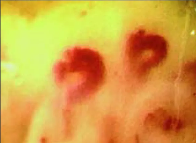
Recently, the scleroderma microangiopathy has been reclassified into three major patterns: «early», «active» and «late» pattern.4The «early»
scleroder-ma NVC pattern is characterized by relatively well--preserved capillary distribution, few enlarged/ /giant capillaries, few capillary haemorrhages and no evident loss of capillaries (Figure 3). Frequent giant capillaries, frequent haemorrhages, mild di-sorganization of the capillary architecture, mode-rate loss of capillaries, absent or mild ramified capillaries characterizes the «active» pattern (Figu-re 4). In the «late» pattern seve(Figu-re loss of capillaries with extensive avascular areas, together with disor-Figure 2. The nailfold’s capillaries in the healthy subject
usually show a regular architecture, uniform shape, distribution and diameter, and most of them show a hairpin or U shaped aspect. (magnification 200x)
ganization of the normal capillary array, ramified/ /bushy capillaries are commonly found, as well as irregular capillary enlargement and few or absent giant capillaries or haemorrhages (Figure 5).
This classification stresses the sequential pro-cess of SSc microangiopathy, beginning with a pre-dominantly microvascular damage which progres-ses into a fibrotic phase with loss of capillaries and
the possible evolution of the disease process.4
The NVC patterns have been correlated with dif-ferent clinical features of the disease, thus playing an important part in the overall study of the
disea-se.13,14Recently, the relationship between specific
autoantibodies and the patterns of microcircula-tory damage in patients with SSc (limited cutane-ous and diffuse cutanecutane-ous forms) was investigated: the positivity for Scl 70 antibody was more com-S A R A C O R T E com-S E C O L.
Figure 4. The «active» pattern characterized by frequent
giant capillaries, frequent hemorrhages, mild disorganization of the capillary architecture, moderate loss of capillaries, absent or mild ramified capillaries (magnification 200x)
Figure 5. In the «late» pattern severe loss of capillaries
with extensive avascular areas, disorganization of the normal capillary array, ramified/bushy capillaries are commonly found, as well as irregular capillary enlargement and few or absent giant capillaries or hemorrhages (magnification 200x)
Figure 6.The hallmark of the scleroderma pattern is the
presence of megacapillaries (magnification 200x)
Figure 3. The «early» scleroderma NVC pattern
characterized by a relatively well-preserved capillary distribution, few enlarged/giant capillaries, few capillary haemorrhages, no evident loss of capillaries (magnification 200x)
Capillaroscopic patterns and rheumatic diseases
The hallmark of the scleroderma pattern is the pre-sence of megacapillaries (Figure 6) and decreased capillary density. In a large recent study, 186 pa-tients with secondary RP (65 with undifferentiated connective tissue disease (UCTD), 47 with syste-mic lupus erythematosus (SLE), 26 with derma-to/polymyositis, 14 with rheumatoid arthritis (RA), 7 with primary Sjögren’s syndrome (SS) and 102 with SSc) were investigated.16A sclerodermapat-tern was found in 14 of 16 patients diffuse cutane-ous SSc (87,5%), and in 53 of 86 patients with limi-ted cutaneous form of SSc (61,6%). A scleroderma pattern was identified in 13,8% of UCTD, 8,5% of the SLE and 26,4% of the patients with derma-to/polymyositis. None of the patients with RA or primary SS had this capillaroscopic pattern.
In conclusion, the scleroderma pattern is often
present in dermato/polymyositis, and can occa-sionally be found in patients with RP and UCTD, and therefore, NVC helps in the early selection of patients more prone to develop scleroderma spec-trum disorders.
Dermatomyositis
A well defined pattern has been described in pa-tients with dermatomyositis,17and it includes two
or more of the following findings in, at least, 2 nail-folds: enlargement of capillary loops, loss of capil-laries, disorganization of the normal distribution of capillaries, «budding» («bushy») capillaries (seen in Figure 7), twisted enlarged capillaries, and ca-pillary haemorrhages (seen in Figure 8).18This
pat-tern is often associated with scleroderma patpat-tern and is also called «scleroderma-like pattern». SLE
The microvascular alterations found in SLE inclu-de morphological changes in capillary loops, venu-lar visibility and sludging of blood with variability of capillary loop length (Figure 9).19One hundred
patients with SLE were evaluated in a recent study.20In this study, both the presence of altered
NVC was associated with the presence of RP (p<0,001) or anti U1 RNP antibodies (p<0,01), and also with the simultaneous presence of RP and U1 RNP antibodies (p<0,001). These authors also found a negative association between the presen-ce of anticardiolipin antibodies and scleroderma pattern (p<0,05). Therefore, this study demonstra-tes a strong association between capillaroscopic C A P I L L A R O S C O P I C PAT T E R N S I N R H E U M AT I C D I S E A S E S
Figure 7. «Budding» («bushy») capillaries related to the
neovascularization (magnification 200x)
Figure 8. Capillary microhemorrages (magnification 200x)
Figure 9. Microvascular alterations found in SLE include
morphological changes in capillary loops, venular visibility and sludging of blood with variability of capillary loop length (magnification 200x)
abnormalities and either U1 RNP antibodies or RP in SLE patients.
A recent retrospective study21in patients with
SLE (n=123) analysed NVC alterations and possi-ble association with clinical features and labora-tory parameters: 35,8% of the patients showed ma-jor capillary abnormalities but no specific pattern was noted. Also, there was no association between the presence of anticardiolipin antibodies and mi-crovascular changes. However, there was a signifi-cant correlation between SLEDAI index and the se-verity of capillary abnormalities (p<0,0001). It was also found that pathologic capillary abnormalities were increased in the presence of anti U1-RNP an-tibodies (p<0,05). These data suggest that NVC exa-mination could be useful in identifying the more severe forms of the disease.
Sjögren’s Syndrome
Microvascular abnormalities have been reported in primary SS.22In a recent controlled study, 40
pa-tients with primary SS (16 with RP, 14 without RP and 10 with anti-centromere antibodies (ACA)), 20 with scleroderma (disease control group) and 40 healthy controls were evaluated. Patients with SS and RP showed a higher frequency of microvascu-lar abnormalities than those without RP. 80% of the patients with ACA showed scleroderma-type findings. Thus, nailfold capillaroscopy can be used to easily assess the microvascular changes in SS patients, especially in those with RP and those with ACA.23
Antiphospholipid Syndrome
Microcirculatory changes were recently described in patients with primary antiphospholipid syndro-me (PAPS). In a recent study,24the percentage of
subjects with morphological microvascular altera-tions (mainly tortuosities) was 78%, compared to 21% of healthy controls. Capillary diameter was found to be significantly smaller in PAPS patients than in controls, although no difference was found in capillary density or blood flow velocity. Another study reported the presence of symmetrical mi-crohemorrhages at NVC analysis, and these altera-tions were found to be particularly significant in patients with both IgG and IgM anticardiolipin
an-ful technique for microvascular analysis in rheu-matic diseases and has a highly negative predicti-ve value in the evaluation of patients presenting with RP.26 Its value is unquestionable in the
as-sessment of SSc microangiopathy. In SLE minor microcirculatory alterations have been reported but the scleroderma pattern seems to be associa-ted with the presence of RP and U1 RNP antibo-dies, as mentioned above. In dermatomyositis a scleroderma-like pattern has been well defined and in SS scleroderma-like findings have been as-sociated with the presence of ACA. In PAPS micro-circulatory changes (mainly tortuosities) are com-mon and symmetrical haemorrhages have been reported specially in patients with anticardiolipin antibodies and thrombotic manifestations.
Further studies in this area are needed to better characterize NVC patterns in rheumatic diseases. NVC opens a window to the systemic microvascu-lar damage that occurs in certain rheumatic dise-ases and plays an important role in the diagnosis and prognosis of SSc.
Aknowledgements
We woud like to thank Dr. Alberto Sulli and Dr. Car-men Pizzorni for providing the pictures.
S A R A C O R T E S E C O L.
Figure 10. Symmetrical microhemorrhages at
capillaroscopic analysis are found particularly significant in patients with both IgG and IgM anticardiolipin antibodies (magnification 200x)
Maurizio Cutolo
Research Laboratory and Division of Rheumatology, University of Genova
Viale Benedetto XV, 6 Genova E-mail: [email protected]
References
1. Brown GE, O’Leary PA. Skin capillaries in scleroder-ma. Arch Intern Med 1925;36:73-88.
2. Maricq HR, LeRoy EC. Patterns of finger capillary ab-normalities in connective tissue disease by widefield microscopy. Arthritis Rheum 1973;16:619-628. 3. Maricq HR, Downey JA, LeRoy EC. Standstill nailfold
capillary blood flow during cooling in scleroderma and Raynaud’s syndrome. Blood Vessels 1976;13:338--349.
4. Cutolo M, Sulli A, Pizzorni C, Accardo S. Nailfold vi-deocapillaroscopy assessment of microvascular da-mage in systemic sclerosis. J Rheumatol 2000; 27:155-160.
5. Le Roy EC, Medsger TA Jr. Raynaud’s phenomenon: a proposal for classification. Clin Exp Rheumatol 1992;10:485-488.
6. Planchon B, Pistorius MA, Beurrier P, De Faucal P. Pri-mary Raynaud’s phenomenon: age of onset and pa-thogenesis in a prospective study of 424 patients. An-giology 1994; 45:677-686.
7. Spencer-Green G. Outcomes in primary Raynaud’s phenomenon. Arch Intern Med 1998; 158:595-600. 8. Luggen M, Belhorn L, Evans T, Fitzgerald O, Spencer
Green G. The evolution of Raynaud´s phenomenon: a longterm prospective study. J Rheum 1995; 22:2226-2232.
9. Zuffery P, Depairon M, Chamot AM, Monti M. Prog-nostic significance of nailfold capillary microscopy in patients with Raynaud’s phenomenon and scleroder-ma-pattern abnormalities: a six-year follow-up study. Clin Rheumatol 1992;11:536-541.
10. Carpentier PH, Maricq HR. Microvasculature in Systemic Sclerosis. Rheum Dis Clin North Am 1990;16:75-91.
11. Mannarino E, Pasqualini L, Fedeli F et al. Nailfold ca-pillaroscopy in the screening and diagnosis of Ray-naud’s phenomenon. Angiology 1994; 45:37-42. 12. Maricq HR, Le Roy EC, D’Angelo WA et al. Diagnostic
potential of in vivo capillary microscopy in scleroder-ma and related disorders. Arthritis Rheum 1980; 23:183-189.
13. Nobili F, Cutolo M, Sulli A et al. Impaired quantitative cerebral blood flow in scleroderma patients. J Neurol Sci 1997;152:63-71.
14. Distler O, Del Rosso A, Giacomelli R et al. and
angio-genic factors in systemic sclerosis: increased levels of vascular endothelial growth factor are a feature of the earliest disease stages and are associated with the ab-sence of fingertip ulcers. Arthritis Res 2002;4:R11. 15. Cutolo M, Pizzorni C, Tuccio M et al. Nailfold
video-capillaroscopic patterns and serum autoantibodies in systemic sclerosis. Rheumatology (Oxford) 2004;43:719-726.
16. Nagy Z, Czirjak L. Nailfold digital capillaroscopy in 447 patients with connective tissue disease and Ray-naud’s disease. J Eur Acad Dermatol Venereol. 2004;18:62-68.
17. Klyscz T, Bogenschutz O, Junger M, Rassner G. Micro-angiopathic changes and functional disorders of nail fold capillaries in dermatomyositis Hautarzt. 1996;47:289-293.
18. Bergman R, Sharony L, Schapira D et al. The hand-held dermatoscope as a nail-fold capillaroscopic ins-trument. Arch Dermatol. 2003;139:1027-1030. 19. Candela M, Pansoni A, De Carolis ST et al. Nailfold
capillary microscopy in patients with antiphospholi-pid syndrome. Recenti Prog Med 1998;89:444-449. 20. Furtado RN, Pucinelli ML, Cristo VV et al.
Scleroder-ma-like nailfold capillaroscopic abnormalities are as-sociated with anti-U1-RNP antibodies and Raynaud’s phenomenon in SLE patients. Lupus 2002;11:35-41. 21. Ingegnoli F, Zeni S, Meani L et al. Evaluation of
nail-fold videocapillaroscopic abnormalities in patients with systemic lupus erythematosus. J Clin Rheum 2005; 11:295-298.
22. Tektonidou M, Kaskani E, Skopouli FN, Moutsopou-los HM. Microvascular abnormalities in Sjogren’s syndrome: nailfold capillaroscopy. Rheumatology (Oxford). 1999;38:826-830.
23. Capobianco KG, Xavier RM, Bredemeier M, Restelli VG, Brenol JC. Nailfold capillaroscopic findings in primary Sjogren’s syndrome: clinical and serological correlations. Clin Exp Rheumatol. 2005;23:789-794. 24. Vaz JL, Dancour MA, Bottino DA, Bouskela E.
Nail-fold videocapillaroscopy in primary antiphospholi-pid syndrome (PAPS). Rheumatology (Oxford) 2004; 43:1025-1027.
25. Sulli A, Pizzorni C, Cutolo M. Nailfold videocapilla-roscopy abnormalities in patients with antiphospho-lipid antibodies. J Rheumatol 2000;27:1574-1576. 26. Cutolo M, Sulli A, Secchi ME, Paolino S, Pizzorni C.
Nailfold capillaroscopy is useful for the diagnosis and follow-up of autoimmune rheumatic diseases. A fu-ture tool for the analysis of microvascular heart in-volvement? Rheumatology (Oxford) 2006;45 Suppl 4:iv43-iv46.