A Role for MicroRNA-155 Expression in
Microenvironment Associated to
HPV-Induced Carcinogenesis in K14-HPV16
Transgenic Mice
Isabel Paiva1,2, Rui M. Gil da Costa3,4, Joana Ribeiro1,5, Hugo Sousa1,5, Margarida Bastos3, Ana Faustino Carlos Rocha7,4, Paula A Oliveira6,7, Rui Medeiros1,2,5,8,9*
1 Molecular Oncology and Viral Pathology Group, CI-IPOP, Portuguese Institute of Oncology of Porto, Rua Dr. António Bernardino de Almeida, 4200–072 Porto, Portugal, 2 ICBAS, Abel Salazar Institute for the Biomedical Sciences, University of Porto, Rua de Jorge Viterbo Ferreira 228, 4050–313, Porto, Portugal, 3 LEPABE, Faculty of Engineering, University of Porto, Rua Dr. Roberto Frias s/n, 4200–465, Porto, Portugal, 4 Experimental Pathology and Therapeutics Group, CI-IPOP, Portuguese Institute of Oncology, Rua Dr. António Bernardino de Almeida, 4200–072 Porto, Portugal, 5 Virology Service, Portuguese Institute of Oncology of Porto, Rua Dr. António Bernardino de Almeida, 4200–072 Porto, Portugal, 6 Veterinary Sciences Department, University of Trás-os-Montes and Alto Douro, UTAD, Quinta de Prados, 5001–801, Vila Real, Portugal, 7 Center for the Research and Technology of Agro-Environmental and Biological Sciences (CITAB), University of Trás-os-Montes and Alto Douro, UTAD, Quinta de Prados, 5001–911, Vila Real, Portugal, 8 CEBIMED, Faculty of Health Sciences of Fernando Pessoa University, Porto, Portugal, 9 Portuguese League Against Cancer (Liga Portuguesa Contra o Cancro–Núcleo Regional do Norte), Estrada Interior da Circunvalação, nº6657, 4200–177 Porto, Portugal
Abstract
Human Papillomavirus cause a number of diseases most notably cervical cancer. K14-HPV16 transgenic mice expressing the K14-HPV16 early genes in squamous epithelial cells provide a suitable experimental model for studying these diseases. MicroRNAs are small non-coding RNAs that play an important role in regulating gene expression and have been suggested to play an important role in cancer development. The role of miR-155 in cancer remains controversial and there is limited evidence linking this miRNA to HPV- associated diseases. We hypothesized that miR-155 expression modulates each tissue’s susceptibility to develop HPV-associated carcinogenesis. In this study, we analyzed miR-155 expression in ear and chest skin samples from 22-26 weeks old, female K14-HPV16 transgenic (HPV16+/-) and wild-type (HPV-/-) mice. Among wild-type mice the expression of miR-155 was lower in ear skin compared with chest skin (p = 0.028). In transgenic animals, in situ carcinoma was present in all ear samples whereas chest tissues only showed epidermal hy-perplasia. Furthermore, in hyperplastic chest skin samples, miR-155 expression was lower than in normal chest skin (p = 0,026). These results suggest that miR-155 expression may modulate the microenvironmental susceptibility to cancer development and that high miR155 levels may be protective against the carcinogenesis induced by HPV16. OPEN ACCESS
Citation: Paiva I, Gil da Costa RM, Ribeiro J, Sousa H, Bastos M, Rocha AFC, et al. (2015) A Role for MicroRNA-155 Expression in Microenvironment Associated to HPV-Induced Carcinogenesis in K14-HPV16 Transgenic Mice. PLoS ONE 10(1): e0116868. doi:10.1371/journal.pone.0116868 Academic Editor: George Calin, University of Texas, MD Anderson Cancer Center, UNITED STATES Received: August 19, 2014
Accepted: December 15, 2014 Published: January 27, 2015
Copyright: © 2015 Paiva et al. This is an open access article distributed under the terms of the Creative Commons Attribution License, which permits unrestricted use, distribution, and reproduction in any medium, provided the original author and source are credited.
Data Availability Statement: All relevant data are in the Supporting Information file (Supporting Information(Table S1).
Funding: This study was supported by Liga Portuguesa Contra o Cancro. Rui M. Gil da Costa is supported by grant SFRH/BPD/85462/2012 from the Portuguese Foundation for Science and Technology, financed by the Portuguese Government and the Social European Fund. The funders had no role in study design, data collection and analysis, decision to publish, or preparation of the manuscript.
Introduction
Human papillomaviruses (HPVs) are the most common sexually transmitted agents [1]. High-risk human papillomavirus, such as HPV16 and HPV18, are the causative agents of virtually all cases of cervical cancer and a significant proportion of other anogenital cancers, as well as some head and neck cancers [2–4].
The K14-HPV16 transgenic mouse model is particularly useful to study the development of HPV-associated squamous cells carcinomas. In this model, the expression of HPV16 early region genes (E2-E8) is driven by the cytokeratin 14 (K14) promoter/enhancer, specifically tar-geting epithelial basal cells [5]. Basal cells are mitotically active and thus may develop further mutations in response to a proliferative stimulus, and the expression of K14 has been shown to persist in well-differentiated squamous carcinomas [6]. The expression of the HPV oncogenes E6 and E7 induces epithelial carcinogenesis through multiple premalignant stages [7]. Accord-ingly, the K14-HPV16 transgenic mice develop epidermal hyperplasia that progresses to dyspla-sia and in situ carcinoma (CIS) lesions and, ultimately, to invasive cancer. This animal model simulates the multistep carcinogenesis process and thus facilitates the study of epigenetic and the genetic factors that coordinate malignant conversion and regulate neoplastic progression.
MicroRNAs (miRNAs) are small noncoding RNAs that regulate gene expression promoting inhibition of messenger RNA (mRNA) translation or its degradation [8]. In normal cells, miR-NAs control several processes including proliferation, differentiation and apoptosis. These molecules are also described as key regulators in many diseases including neurological disor-ders, cardiovascular diseases, viral infections and cancer [9]. During carcinogenesis, some miR-NAs are lost whereas others are upregulated, and in fact, previous data indicates that miRmiR-NAs may be important to distinguish subtypes of cancers, where the histological diagnosis is com-plex and difficult [10].
MicroRNA-155 (miR-155) plays a role in many of the above oncogenic processes. This microRNA is overexpressed in many types of cancer cells and accumulating evidence shows that miR-155 is an oncogenic microRNA. However, recent studies claim that miR-155 may display anti-oncogenic properties or promote an adequate immunological response to cancer [11,12]. MiR-155 has emerged as an essential regulator of cellular physiology, particularly important in the mammalian immune system [13–15]. Thus, a possible link between miR-155 and inflamma-tion in cancer has been reported [16]. Moreover, miR-155 transgenic mice develop B-cell lym-phoma [15], and miR-155-knock-out mice exhibit impaired immune function [17].
In HPV-associated cancers, the interplay between miR-155 and HPV genes remains elusive and poorly understood. The tumor microenvironment associated to miRNAs plays an increas-ingly appreciated role in cancer [18], but the microenvironment of normal tissues and its role in tumorigenesis remains poorly studied.
In this study, we aimed to evaluate the expression of miR-155 in skin samples with or with-out the presence of integrated HPV DNA and with different HPV-associated lesions. For this purpose, we have used K14-HPV16 transgenic mice, [19], to analyze miR-155 expression in ear and chest skin samples, evaluating its correlation with tissue microenvironment and HPV-induced carcinogenesis.
Materials and Methods
Transgenic mice
Generation of K14-HPV mice has been previously reported [19]. K14-HPV16 mice on a FVB/n background were generously donated by Drs. Jeffrey Arbeit and Douglas Hanahan, from the University of California, through the USA National Cancer Institute Mouse Repository. The
Competing Interests: Rui Medeiros, Molecular Oncology and Viral Pathology Group, Portuguese Institute of Oncology Asoociate Professor, ICBAS, Instituto Ciências Biomédicas Abel Salazar (University of Porto) is a PLOS ONE Editorial Board member. This does not alter the authors’ adherence to all the PLOS ONE policies on sharing data and materials.
animal experiments were approved by the University of Trás-os-Montes and Alto Douro Ethics committee, University of Trás-os-Montes and Alto Douro, UTAD, Quinta de Prados, 5001-801, Vila Real, Portugal. Adequate environmental enrichment was provided for each cage and health checks were performed daily. Before collecting the samples, all animals were anesthetized by using sodium pentobarbital, followed by intracardiac punction and exsanguination, as indicated by the Federation for Laboratory Animal Science Associations (FELASA). After one week quar-antine, the animals were maintained and bred in accordance with Portuguese (Portaria 1005/92 dated October the 23rd) and European (EU Directive 2010/63/EU) legislation, under controlled conditions of temperature (23±2ºC), light-dark cycle (12 h light/12 h dark) and relative humidity (50±10%), using hardwood bedding. A standard diet (4RF21 GLP, Mucedola, Italy) and water were provided ad libitum. A total of 15 female mice from consecutive litters were selected for the study.
Genotyping of HPV16-E6 and E2
The animals were genotyped at weaning, using tail tip samples. Tissue lysis was performed by adding 300μL of MagnaPure DNA Tissue Lysis Buffer (Roche, Indianapolis, USA) and 20 μL of Proteinase K and incubating overnight (aproximately 16h) at 65ºC. Nucleic acids were extracted using the High Pure Viral Nucleic Acid kit (Roche, Indianapolis, USA) following the manufacturer’s instructions. DNA quality was assessed by measuring the absorbance at 260 nm, using an UV/Visible spectrophotometer. DNA purity was assessed by the ratio of the absorbance values at 260/280 nm, using the NanoDrop spectrophotometer v3.7 (Thermo Scientific, Wilmington DE, USA). The presence of amplifiable genomic DNA was tested by poly-merase chain reaction (PCR) amplification of mouseβ-globin using specific primers (Table 1) [20]. The PCR reaction was performed in a 50μl solution with 1x Taq buffer, 2.0 mM MgCL2, 0.2 mM DNTP’S, 0.50 μM of each primer, 1 U de Taq DNA Polimerase and 0.2 μg of genomic DNA. The amplification conditions were as following: denaturation of DNA template at 94ºC for 3 min, followed by 35 cycles at 94ºC for 30 s 60ºC for 45 s, 72ºC for 90 s, and a final extension step at 72ºC for 10 min. The amplified fragment of 494 base pairs (bp) was analyzed by electrophoresis in 1.5% (w/v) agarose gels stained with ethidium bromide and visualized under UV light.
The presence of integrated HPV was assessed by amplification of E6 and HPV16-E2 genes with specific primers (Table 1), which amplify a region of 130 bp and 184 bp, respec-tively, (adapted from a protocol described by Cañadas et al. and Ribeiro et al [21,22]). The PCR amplification reaction with HPV16-E6 and HPV16-E2 primers was carried in a 50μl reaction mixture with 1x PCR Buffer, 2.5 mM MgCl2, 0.2 mM DNTP’S, 0.30 μM of each primer, 1 U of Taq DNA polymerase and 0.2μg of genomic DNA. Thermal cycling was performed as follows:
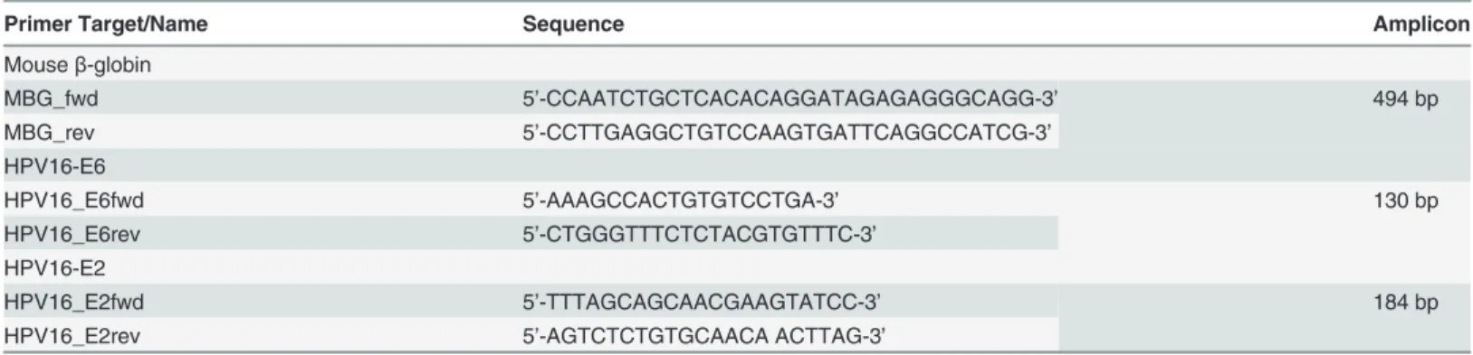
Table 1. Primer sequences.
Primer Target/Name Sequence Amplicon
Mouseβ-globin MBG_fwd 5’-CCAATCTGCTCACACAGGATAGAGAGGGCAGG-3’ 494 bp MBG_rev 5’-CCTTGAGGCTGTCCAAGTGATTCAGGCCATCG-3’ HPV16-E6 HPV16_E6fwd 5’-AAAGCCACTGTGTCCTGA-3’ 130 bp HPV16_E6rev 5’-CTGGGTTTCTCTACGTGTTTC-3’ HPV16-E2 HPV16_E2fwd 5’-TTTAGCAGCAACGAAGTATCC-3’ 184 bp
HPV16_E2rev 5’-AGTCTCTGTGCAACA ACTTAG-3’
initial denaturation of DNA template at 94ºC for 2 min, followed by 35 cycles at 94ºC for 1 min, 60ºC for 1 min, and 72ºC for 1 min and a final extension step at 72ºC for 5 min. The amplified fragment was analyzed by electrophoresis in 1.5% (w/v) agarose gels stained with ethidium bromide and visualized under UV light.
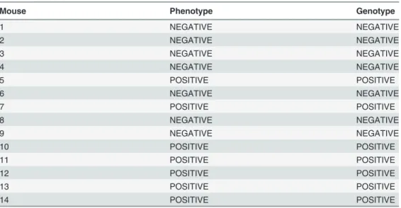
The resulting genotypes were compared with the respective phenotypes (Table 2).
Sample collection
Seven hemizygous (+/-) and seven wild-type (-/-) females were sacrificed at 22 to 26 weeks of age. Ear and chest skin samples from each animal (14 samples from transgenic and 12 samples from wild-type animals) were collected into TriPure reagent (Roche Applied Science), macerat-ed, and kept at−80ºC until processing. Matched samples were collected into 10% neutral buff-ered formalin for routine histological processing. Histological sections (2μm-thick) were stained with haematoxylin and eosin (H&E) for examination on light microscopy. Samples were classified as normal skin, epidermal hyperplasia and epidermal CIS by two independent researchers (CL and RGC), as previously described [19].
miRNA expression analysis
Extraction of total RNA from samples preserved in TriPure reagent was performed using the High Pure Viral Nucleic Acid kit (Roche, Indianapolis, USA), according to manufacturer’s instructions. RNA quality was assessed using NanoDrop spectrophotometer v3.7 (Thermo Scientific, Wilmington DE, USA).
Mmu-miR-155_002571 and snoRNA-202_001232 were analyzed using two-step real-time PCR protocols with TaqMan MicroRNA Assays (Applied Biosystems, Foster CA, USA). The conversion of miRNA to cDNA was performed using TaqMan MicroRNA Reverse Transcrip-tion Kit (Applied Biosystems, Foster CA, USA) in a 15μL of total volume reaction mix with: 7μL of a master mix containing 1x RT buffer, 1.0 mM of total dNTPs, 50U MultiScribe Reverse Transcriptase Enzyme and 0.25U of RNase inhibitor; 3μL of RT primers (Applied Biosystems, Foster CA, USA); and 5μL of RNA sample. The amplification conditions were as follows:
Table 2. Association between the characteristic phenotype of HPV–associated lesions and genotype of HPV E6/E2 DNA of mice.
Mouse Phenotype Genotype
1 NEGATIVE NEGATIVE 2 NEGATIVE NEGATIVE 3 NEGATIVE NEGATIVE 4 NEGATIVE NEGATIVE 5 POSITIVE POSITIVE 6 NEGATIVE NEGATIVE 7 POSITIVE POSITIVE 8 NEGATIVE NEGATIVE 9 NEGATIVE NEGATIVE 10 POSITIVE POSITIVE 11 POSITIVE POSITIVE 12 POSITIVE POSITIVE 13 POSITIVE POSITIVE 14 POSITIVE POSITIVE doi:10.1371/journal.pone.0116868.t002
30 min at 15ºC, 52 min at 42ºC and finally 10 min at 85ºC. All reverse transcriptase reactions included two non-template controls using double distilled water to replace template RNA.
qPCRs were performed on a StepOne Real-time PCR System (Applied Biosystems, Foster CA, USA) with a 20μl final volume: 1× TaqMan Universal PCR Master Mix II (Applied Biosystems, Foster City, California USA); 1x MicroRNA Assay (Applied Biosystems, Foster City, California USA); and 2μL cDNA from RT snoRNA-202 was used as endogenous control. Thermal cycling conditions were: 10 min at 95ºC followed by 45 cycles of 15 s at 95ºC and 1 min at 60ºC. All reactions included two-template controls using double distilled water to re-place template cDNA.
Statistical Analysis
Data analysis was performed using the computer software IBM SPSS Statistics for Windows (Version 20.0). The t-Student test was used to evaluate statistical differences in the normalized expression of the miR-155. In order to analyze the normalized relative expression (-ΔCt) of the different groups, we considered the results corresponding to a 99% representation of the popu-lation (X¯ ± 2SD).
Results
Genotyping/phenotyping and histological analysis
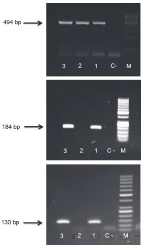
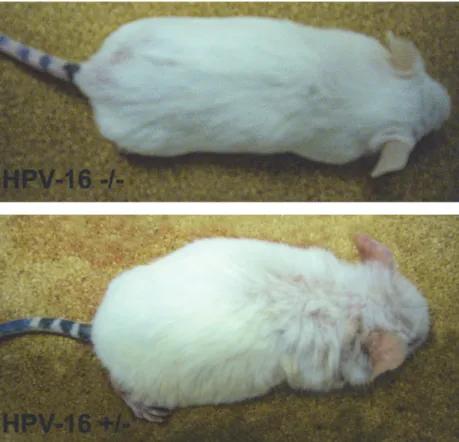
We observed the presence of HPV16 integration in 7 out of 14 animals (Table 2andFig. 1). While wild-type mice did not develop any skin lesion, all mice with integrated HPV DNA dem-onstrated, phenotypically, various degrees of persistent epidermal squamous hyperplasia and hyperkeratosis, characteristic lesions associated with HPV infection as previously described [19] (Fig. 2). After histologic evaluation, we observed that, in all cases with integrated HPV16, the ear tissues presented CIS, while the chest tissues showed only epidermal hyperplasia, while wild-type mice showed normal skin histology (Fig. 3).
MiRNA-155 expression profile in tissues from wild-type mice
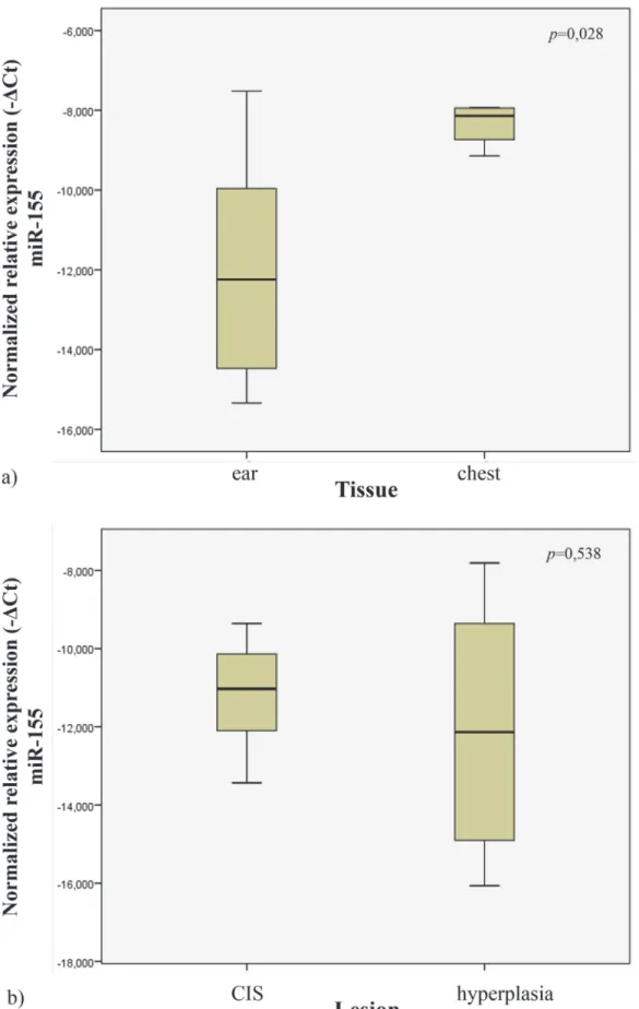
In order to study the miR-155 expression profile in normal tissues, we quantified its expression in the ear and chest skin samples of the wild-type mice. We observed that the ear tissues had lower expression levels when compared to chest tissues (p = 0.028) (Fig. 4A).
Mir-155 expression profile in tissues of transgenic mice
In order to study miR-155 expression in tissues from transgenic mice, we analyzed the samples from ear and chest skin. We observed no statistical significant difference in miR-155 expression levels between these groups (histologically presenting with CIS vs hyperplasia) (p = 0.538) (Fig. 4B).
MiRNA-155 expression profile in normal chest skin versus hyperplastic
skin
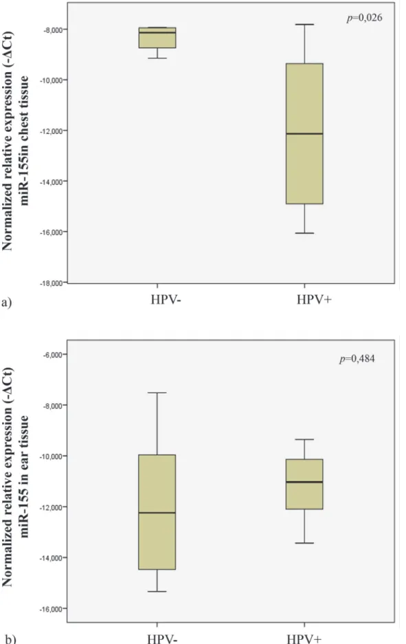
In order to explore the possible influence of HPV16 on miR-155 during the early phases of skin carcinogenesis, we studied its expression levels in wild-type (histologically normal) and HPV16-transgenic (hyperplastic) chest skin samples. When comparing wild-type with trans-genic chest skin, we observed that, transtrans-genic skin had lower expression levels of miR-155 than normal skin (p = 0,026) (Fig. 5A).
MiRNA-155 expression profile in normal ear skin versus CIS
We also compared the relative miR-155 expression levels on ear tissues of transgenic (showing CIS) and wild-type (showing normal skin histology) mice. Our data showed no statistical sig-nificant difference in miR-155 expression levels between these groups (p = 0.484) (Fig. 5B).
Discussion
A large number of different biomarker microRNAs have previously been reported to be con-nected to cellular transformation. Deregulation of miRNAs is intimately associated with the de-velopment and progression of cancer [23,24].
In this context, microRNA profiling studies indicate that deregulation of miR-155 is fre-quently linked with a wide range of malignancies, including various forms of lymphoma and carcinomas of breast, lung, pancreas, head and neck, and kidney [25–28]. Furthermore, miR-155 is detected during the immune response in activated mature B and T lymphocytes [29], germinal centers B cells [17], and monocytes [16]. BIC/miR-155 knock-out mice exhibited im-paired immune response and cytokine production [17], further supporting the vital role of
Figure 1. Mice genotyping. The presence of integrated HPV was assessed by amplification of HPV-E2 (b) and HPV-E6 (c) genes by polymerase chain reaction methodology (PCR) in-house. Samples 1 and 3 are HPV+; sample 2 is HPV-. Mouse-β-globin gene was used as endogenous control (a). M: molecular weight size marker: (a)100 bp, (b,c)50 bp; C-: negative control.
miR-155 in immunology. Much of the current research in the field has implicated miR-155 in promoting oncogenesis. Controversially, recent studies report anti-oncogenic effects of miR-155 [11,12]. Interestingly, these authors found that miR-155 knockdown in myeloid cells facili-tated breast cancer development in mice. Some novel concepts that arose from the analysis of these papers were that miR-155 is not only a promoter of some cancers, but may also act to pre-vent cancer by promoting proper immune function.
It is accepted that HPV infection is the most important factor for transition from normal cervical epithelium to cervical preneoplastic lesions, intraepithelial neoplasia and, subsequent-ly, to invasive cervical cancer [30,31]. The influence of others factors, including the host micro-environment, remains poorly defined. Specifically, in cervical cancer, the most important HPV-associated tumor, no conclusive evidence concerning the relation between HPV and miR-155 expression has been reported. There have been studies on miRNA expression in head and neck cancers reporting miR-155 to be upregulated in oral cancer compared to normal oral tissue [32–34]. However, when compared HPV-positive with HPV-negative squamous cell car-cinoma of the head and neck cell lines, this miRNA was downregulated in the presence of HPV-16 DNA [35].
K14-HPV16 transgenic mice are a useful experimental model for studying progressive, multistep HPV-induced carcinogenesis. The FVB/n mouse strain has been shown to be partic-ularly prone to HPV-driven carcinogenesis, as other mouse strains (e.g. Balb/c, C57Bl/6, SSIN/ SENCAR) bearing an identical transgene, failed to develop invasive carcinomas [5]. This early
Figure 2. Wild-type (-/-) and K14-HPV16 transgenic (+/-) mice. Transgenic mice show a hunched position, partial thoracic and cephalic alopecia, together with extensive hyperkeratosis and auricular erythema. doi:10.1371/journal.pone.0116868.g002
observation already highlighted the key role of host factors in the development of HPV-induced cancers. Thus, the characterization of miRNA expression levels in this model animals may be a useful strategy for understanding the mechanisms carcinogenesis associated with HPV.
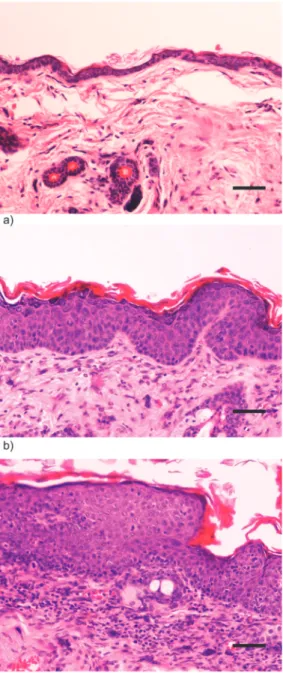
Figure 3. Histology of wild-type and transgenic mice. a) Wild-type (-/-) female FVB/n mouse. Chest skin showing normal histology. H&E, 200x bar = 50μm b) K14-HPV16 transgenic (+/-) female FVB/n mouse. Chest skin showing epidermal hyperplasia and orthokeratotic hyperkeratosis. Note increased number of epidermal strata with conserved orderly squamous differentiation. c) K14-HPV16 transgenic (+/-) female FVB/n mouse. Ear skin showing in situ carcinoma. Note loss of epidermal stratification and progressive differentiation, presence of suprabasal mitotic figures and anisocytosis and abrupt parakeratotic keratinization with hyperkeratosis. The underlying stroma exhibits intense mixed inflammatory cell infiltration and neovascularization.
Figure 4. Normalized relative expression of miR-155 in ear and chest normal tissue (a). Normalized expression of miR-155 in different lesions of K14-HPV16 transgenic mice (CIS and hyperplasia) (b).
Cutaneous squamous cell carcinoma develops in multiple locations through well-defined steps. In the present study 22–26 weeks-old transgenic animals (HPV16+/-), showed CIS in all ear skin samples, whereas chest skin samples only showed epidermal hyperplasia. This provid-ed an opportunity to study miR-155 expression levels in different phases HPV16-inducprovid-ed car-cinogenesis. Our results (Fig. 6) indicate that, among wild-type mice (HPV-/-), the expression of miR-155 is lower in ear skin tissue compared with chest skin (p = 0.028). Also, we observed that hyperplastic chest skin presented lower levels of miR-155 compared with normal chest skin (p = 0,026). Based on these results, miR-155 expression levels appear to be a significant microenvironmental factor involved in the development of HPV-associated lesions. Specifical-ly, these results suggest that downregulation of miR-155 may be involved in HPV16-driven early hyperplastic lesions.
In agreement with our findings, a recent study reports that miR-155 acts as a tumor sup-pressor in human Caski cervical cancer cells (carrying HPV16 DNA). Moreover, it was demon-strated that p53 expression is upregulated by miR-155 overexpression [36]. Recent study indicates that miR-155 overexpression results in decreased cyclin D1 to p21 ratio, suggesting a role in inhibiting cell proliferation [37,38].
Previous reports concluded that interleukin 10 (IL-10) downregulates miR-155 expression post-transcriptionally [39]. Also, women who are genetically programmed to produce high or moderate levels of IL-10 are more likely to develop cervical cancer, compared to individuals ge-netically predisposed to present low IL-10 production [40]. Our results are in accordance with these reports, suggesting that miR-155 may direct HPV16-induced pathological processes to-wards hyperplasia rather than malignant transformation.
Other study indicates that miR-155 acts as a positive regulator of interferon gamma (IFN-γ) production [41] and the increase of IFN-γ enhances susceptibility of cervical cancer cells to
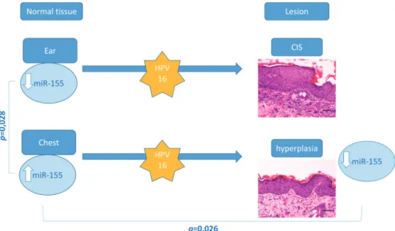
Figure 6. Overview of genotyping, histological and miR-155 profiling results. MiR-155 levels are significantly higher in normal chest skin compared with ear skin samples. Targeted expression of HPV-16 oncogenes to basal keratinocytes leads to multistep skin carcinogenesis–transgenic ear skin samples showed CIS while chest samples showed epidermal hyperplasia. Hyperplastic (chest) skin samples showed a significant miR-155 downregulation compared with matched wild-type samples. No differences were observed between wild-type and transgenic ear samples or between transgenic ear and chest samples. doi:10.1371/journal.pone.0116868.g006
lysis by tumor-specific cytotoxic T cells [42]. Also, previous reports concluded that miR-155 targets important oncogenes such as B-cell lymphoma 2 (BCL2), which regulates apoptosis [43]. These data may explain the relation between low miR-155 expression levels and cancer development.
Our results showing the decrease of miR-155 expression levels in hyperplastic skin com-pared with normal chest skin may be explained in the light of these previous reports, including the loss of p53 and the increased of p21 and BCL2, the upregulation of IL-10 and the decrease of IFN-γ levels, promoting an epidermal hyperplasia.
Despite the acknowledged functions of miR-155 in various cancers, further studies are needed to clarify its contribution of theses miRNAs in immunomodulation, and its interaction with cell signaling pathways. Our results suggested that it might be related to the induction of a microenvironment less favorable for HPV-induced carcinogenesis. In conclusion, the data dis-cussed in this article relates a possible anti-oncogenic effect of miR-155. These findings are im-portant in determining the possible role of miR-155 expression in differential tissue
predisposition to cancer development.
Supporting Information
S1 Table. MiR-155 expression analysis of ear and chest tissue of transgenic (HPV+) and wild-type (HPV-) mice.
(DOC)
Author Contributions
Conceived and designed the experiments: IP RGC RM. Performed the experiments: IP JR HS RGC CL AFR. Analyzed the data: IP RGC RM. Contributed reagents/materials/analysis tools: MB PO RM. Wrote the paper: IP RGC HS RM.
References
1. Kjaer SK, Chackerian B, van den Brule AJ, Svare EI, Paull G, et al. (2001) High-risk human papillomavirus is sexually transmitted: evidence from a follow-up study of virgins starting sexual activity (intercourse). Cancer Epidemiol Biomarkers Prev 10: 101–106. PMID:11219765
2. Walboomers JM, Jacobs MV, Manos MM, Bosch FX, Kummer JA, et al. (1999) Human papillomavirus is a necessary cause of invasive cervical cancer worldwide. J Pathol 189: 12–19. PMID:10451482 3. Watson M, Saraiya M, Ahmed F, Cardinez CJ, Reichman ME, et al. (2008) Using population-based cancer registry data to assess the burden of human papillomavirus-associated cancers in the United States: overview of methods. Cancer 113: 2841–2854. doi:10.1002/cncr.23758PMID:18980203 4. Major T, Szarka K, Sziklai I, Gergely L, Czegledy J (2005) The characteristics of human papillomavirus
DNA in head and neck cancers and papillomas. J Clin Pathol 58: 51–55. PMID:15623482
5. Coussens LM, Hanahan D, Arbeit JM (1996) Genetic predisposition and parameters of malignant pro-gression in K14-HPV16 transgenic mice. Am J Pathol 149: 1899–1917. PMID:8952526
6. Stoler A, Kopan R, Duvic M, Fuchs E (1988) Use of monospecific antisera and cRNA probes to localize the major changes in keratin expression during normal and abnormal epidermal differentiation. J Cell Biol 107: 427–446. PMID:2458356
7. Masset A, Maillard C, Sounni NE, Jacobs N, Bruyere F, et al. (2011) Unimpeded skin carcinogenesis in K14-HPV16 transgenic mice deficient for plasminogen activator inhibitor. Int J Cancer 128: 283–293. doi:10.1002/ijc.25326PMID:20232379
8. Garzon R, Calin GA, Croce CM (2009) MicroRNAs in Cancer. Annu Rev Med 60: 167–179. doi:10. 1146/annurev.med.59.053006.104707PMID:19630570
9. Chen CZ (2005) MicroRNAs as oncogenes and tumor suppressors. N Engl J Med 353: 1768–1771. PMID:16251533
10. Paranjape T, Slack FJ, Weidhaas JB (2009) MicroRNAs: tools for cancer diagnostics. Gut 58: 1546– 1554. doi:10.1136/gut.2009.179531PMID:19834118
11. Huffaker TB, Hu R, Runtsch MC, Bake E, Chen X, et al. (2012) Epistasis between microRNAs 155 and 146a during T cell-mediated antitumor immunity. Cell Rep 2: 1697–1709. doi:10.1016/j.celrep.2012. 10.025PMID:23200854
12. Zonari E, Pucci F, Saini M, Mazzieri R, Politi LS, et al. (2013) A role for miR-155 in enabling tumor-infiltrating innate immune cells to mount effective antitumor responses in mice. Blood 122: 243–252. doi:10.1182/blood-2012-08-449306PMID:23487026
13. Ceppi M, Pereira PM, Dunand-Sauthier I, Barras E, Reith W, et al. (2009) MicroRNA-155 modulates the interleukin-1 signaling pathway in activated human monocyte-derived dendritic cells. Proc Natl Acad Sci U S A 106: 2735–2740. doi:10.1073/pnas.0811073106PMID:19193853
14. O’Connell RM, Taganov KD, Boldin MP, Cheng G, Baltimore D (2007) MicroRNA-155 is induced during the macrophage inflammatory response. Proc Natl Acad Sci U S A 104: 1604–1609. PMID:17242365 15. Costinean S, Zanesi N, Pekarsky Y, Tili E, Volinia S, et al. (2006) Pre-B cell proliferation and
lympho-blastic leukemia/high-grade lymphoma in E(mu)-miR155 transgenic mice. Proc Natl Acad Sci U S A 103: 7024–7029. PMID:16641092
16. Taganov KD, Boldin MP, Chang KJ, Baltimore D (2006) NF-kappaB-dependent induction of microRNA miR-146, an inhibitor targeted to signaling proteins of innate immune responses. Proc Natl Acad Sci U S A 103: 12481–12486. PMID:16885212
17. Thai TH, Calado DP, Casola S, Ansel KM, Xiao C, et al. (2007) Regulation of the germinal center re-sponse by microRNA-155. Science 316: 604–608. PMID:17463289
18. Whiteside TL (2008) The tumor microenvironment and its role in promoting tumor growth. Oncogene 27: 5904–5912. doi:10.1038/onc.2008.271PMID:18836471
19. Arbeit JM, Munger K, Howley PM, Hanahan D (1994) Progressive squamous epithelial neoplasia in K14-human papillomavirus type 16 transgenic mice. J Virol 68: 4358–4368. PMID:7515971
20. Konkel DA, Tilghman SM, Leder P (1978) The sequence of the chromosomal mouse beta-globin major gene: homologies in capping, splicing and poly(A) sites. Cell 15: 1125–1132. PMID:569555
21. Canadas MP, Darwich L, Sirera G, Cirigliano V, Bofill M, et al. (2010) New molecular method for the de-tection of human papillomavirus type 16 integration. Clin Microbiol Infect 16: 836–842. doi:10.1111/j. 1469-0691.2009.02964.xPMID:19840031
22. Ribeiro J, Teixeira D, Marinho-Dias J, Monteiro P, Loureiro J, et al. (2014) Characterization of human papillomavirus genotypes and HPV-16 physical status in cervical neoplasias of women from northern Portugal. Int J Gynaecol Obstet 125: 107–110. doi:10.1016/j.ijgo.2013.10.011PMID:24513260 23. Garzon R, Marcucci G, Croce CM (2010) Targeting microRNAs in cancer: rationale, strategies and
challenges. Nat Rev Drug Discov 9: 775–789. doi:10.1038/nrd3179PMID:20885409
24. Nelson KM, Weiss GJ (2008) MicroRNAs and cancer: past, present, and potential future. Mol Cancer Ther 7: 3655–3660. doi:10.1158/1535-7163.MCT-08-0586PMID:19074842
25. Liu X, Chen Z, Yu J, Xia J, Zhou X (2009) MicroRNA profiling and head and neck cancer. Comp Funct Genomics: 837514.
26. Volinia S, Calin GA, Liu CG, Ambs S, Cimmino A, et al. (2006) A microRNA expression signature of human solid tumors defines cancer gene targets. Proc Natl Acad Sci U S A 103: 2257–2261. PMID: 16461460
27. Iorio MV, Ferracin M, Liu CG, Veronese A, Spizzo R, et al. (2005) MicroRNA gene expression deregula-tion in human breast cancer. Cancer Res 65: 7065–7070. PMID:16103053
28. Eis PS, Tam W, Sun L, Chadburn A, Li Z, et al. (2005) Accumulation of miR-155 and BIC RNA in human B cell lymphomas. Proc Natl Acad Sci U S A 102: 3627–3632. PMID:15738415
29. Turner M, Vigorito E (2008) Regulation of B- and T-cell differentiation by a single microRNA. Biochem Soc Trans 36: 531–533. doi:10.1042/BST0360531PMID:18481999
30. Bosch FX, de Sanjose S (2007) The epidemiology of human papillomavirus infection and cervical can-cer. Dis Markers 23: 213–227. PMID:17627057
31. zur Hausen H (2002) Papillomaviruses and cancer: from basic studies to clinical application. Nat Rev Cancer 2: 342–350. PMID:12044010
32. Chang SS, Jiang WW, Smith I, Poeta LM, Begum S, et al. (2008) MicroRNA alterations in head and neck squamous cell carcinoma. Int J Cancer 123: 2791–2797. doi:10.1002/ijc.23831PMID:18798260 33. Wong TS, Liu XB, Chung-Wai Ho A, Po-Wing Yuen A, Wai-Man Ng R, et al. (2008) Identification of
py-ruvate kinase type M2 as potential oncoprotein in squamous cell carcinoma of tongue through micro-RNA profiling. Int J Cancer 123: 251–257. doi:10.1002/ijc.23583PMID:18464261
34. Wong TS, Liu XB, Wong BY, Ng RW, Yuen AP, et al. (2008) Mature miR-184 as Potential Oncogenic microRNA of Squamous Cell Carcinoma of Tongue. Clin Cancer Res 14: 2588–2592. doi:10.1158/ 1078-0432.CCR-07-0666PMID:18451220
35. Wald AI, Hoskins EE, Wells SI, Ferris RL, Khan SA (2011) Alteration of microRNA profiles in squamous cell carcinoma of the head and neck cell lines by human papillomavirus. Head & Neck 33: 504–512. doi:10.1016/j.ophtha.2014.11.008PMID:25556115
36. Lei C, Wang Y, Huang Y, Yu H, Huang Y, et al. (2012) Up-regulated miR155 reverses the epithelial-mesenchymal transition induced by EGF and increases chemo-sensitivity to cisplatin in human Caski cervical cancer cells. PLoS One 7: e52310. doi:10.1371/journal.pone.0052310PMID:23284982 37. Das LM, Torres-Castillo MDLA, Gill T, Levine AD (2013) TGF-[beta] conditions intestinal T cells to
ex-press increased levels of miR-155, associated with down-regulation of IL-2 and itk mRNA. Mucosal Immunol 6: 167–176. doi:10.1038/mi.2012.60PMID:22785227
38. Fan X, Liu Y, Chen JJ (2005) Down-regulation of p21 contributes to apoptosis induced by HPV E6 in human mammary epithelial cells. Apoptosis 10: 63–73. PMID:15711923
39. Cheung ST, So EY, Chang D, Ming-Lum A, Mui AL (2013) Interleuk10 inhibits lipopolysaccharide in-duced miR-155 precursor stability and maturation. PLoS One 8: e71336. doi:10.1371/journal.pone. 0071336PMID:23951138
40. Stanczuk GA, Sibanda EN, Perrey C, Chirara M, Pravica V, et al. (2001) Cancer of the uterine cervix may be significantly associated with a gene polymorphism coding for increased IL-10 production. Int J Cancer 94: 792–794. PMID:11745479
41. Trotta R, Chen L, Ciarlariello D, Josyula S, Mao C, et al. (2012) miR-155 regulates IFN-gamma produc-tion in natural killer cells. Blood 119: 3478–3485. doi:10.1182/blood-2011-12-398099PMID:
22378844
42. Street D, Kaufmann AM, Vaughan A, Fisher SG, Hunter M, et al. (1997) Interferon-gamma enhances susceptibility of cervical cancer cells to lysis by tumor-specific cytotoxic T cells. Gynecol Oncol 65: 265–272. PMID:9159336
43. Willimott S, Wagner SD (2012) miR-125b and miR-155 contribute to BCL2 repression and proliferation in response to CD40 ligand (CD154) in human leukemic B-cells. J Biol Chem 287: 2608–2617. doi:10. 1074/jbc.M111.285718PMID:22139839