Cell Cycle- and Cancer-Associated Gene
Networks Activated by Dsg2: Evidence of
Cystatin A Deregulation and a Potential Role
in Cell-Cell Adhesion
Abhilasha Gupta1, Daniela Nitoiu2, Donna Brennan-Crispi1,3, Sankar Addya4, Natalia A. Riobo3, David P. Kelsell2, MỹG. Mahoney1,3*
1Department of Dermatology and Cutaneous Biology, Thomas Jefferson University, Philadelphia, Pennsylvania, United States of America,2Center for Cutaneous Research, Blizard Institute, Barts and the London School or Medicine and Dentistry, Queen Mary University of London, London, United Kingdom, 3Department of Biochemistry and Molecular Biology, Thomas Jefferson University, Philadelphia,
Pennsylvania, United States of America,4Kimmel Cancer Center, Department of Cancer Biology, Thomas Jefferson University, Philadelphia, Pennsylvania, United States of America
Abstract
Cell-cell adhesion is paramount in providing and maintaining multicellular structure and signal transmission between cells. In the skin, disruption to desmosomal regulated intercellular con-nectivity may lead to disorders of keratinization and hyperproliferative disease including can-cer. Recently we showed transgenic mice overexpressing desmoglein 2 (Dsg2) in the epidermis develop hyperplasia. Following microarray and gene network analysis, we demon-strate that Dsg2 caused a profound change in the transcriptome of keratinocytesin vivoand altered a number of genes important in epithelial dysplasia including: calcium-binding proteins (S100A8 and S100A9), members of the cyclin protein family, and the cysteine protease inhibi-tor cystatin A (CSTA). CSTA is deregulated in several skin cancers, including squamous cell carcinomas (SCC) and loss of function mutations lead to recessive skin fragility disorders. The microarray results were confirmed by qPCR, immunoblotting, and immunohistochemis-try. CSTA was detected at high level throughout the newborn mouse epidermis but dramati-cally decreased with development and was detected predominantly in the differentiated layers. In human keratinocytes, knockdown of Dsg2 by siRNA or shRNA reduced CSTA ex-pression. Furthermore, siRNA knockdown of CSTA resulted in cytoplasmic localization of Dsg2, perturbed cytokeratin 14 staining and reduced levels of desmoplakin in response to mechanical stretching. Both knockdown of either Dsg2 or CSTA induced loss of cell adhesion in a dispase-based assay and the effect was synergistic. Our findings here offer a novel path-way of CSTA regulation involving Dsg2 and a potential crosstalk between Dsg2 and CSTA that modulates cell adhesion. These results further support the recent human genetic findings that loss of function mutations in the CSTA gene result in skin fragility due to impaired cell-cell adhesion: autosomal-recessive exfoliative ichthyosis or acral peeling skin syndrome.
OPEN ACCESS
Citation:Gupta A, Nitoiu D, Brennan-Crispi D, Addya S, Riobo NA, Kelsell DP, et al. (2015) Cell Cycle- and Cancer-Associated Gene Networks Activated by Dsg2: Evidence of Cystatin A Deregulation and a Potential Role in Cell-Cell Adhesion. PLoS ONE 10(3): e0120091. doi:10.1371/ journal.pone.0120091
Academic Editor:Johanna M Brandner, University Hospital Hamburg-Eppendorf, GERMANY
Received:September 5, 2014
Accepted:February 2, 2015
Published:March 18, 2015
Copyright:© 2015 Gupta et al. This is an open access article distributed under the terms of the Creative Commons Attribution License, which permits unrestricted use, distribution, and reproduction in any medium, provided the original author and source are credited.
Data Availability Statement:All relevant data are within the paper and its Supporting Information files. Microarray data was submitted to Gene Expression Omnibus (Accession number: GSE62814).
Introduction
Desmosomes are major adhesion structures localized to the cell-cell borders of epithelial cells where the cytoplasmic plaque components, including the plakin (desmoplakin) and keratin families, assemble with the armadillo (plakoglobin and plakophilins) and cadherin (desmogle-ins and desmocoll(desmogle-ins) protein families [1,2]. These adhesion structures are essential not only for the maintenance of cell structure and integrity, but also for tissue development and mor-phogenesis. Mutations within the desmosome are the underlying cause of many skin fragility disorders with or without heart abnormalities [3]. Additionally, desmosomes also serve as“ sig-naling centers”playing an active role in modulating several important pathways, including the Wnt/β-catenin and the T-cell factor/lymphoid enhancer factor [4]. Mounting evidence sup-ports their participation in modulating cell fate and survival. Desmosomal proteins may acti-vate intracellular signaling through the modulation of expression levels and patterns, both of which can dramatically alter adhesion and cell proliferation [5,6]. In the interfollicular epider-mis, Dsg2 is normally expressed at very low level and restricted to the proliferative basal cell layer. Recently, we developed a transgenic mouse model overexpressing desmoglein 2 (Dsg2) in the skin [5]. We determined that ectopic expression of Dsg2 activates multiple growth and survival pathways that may promote cancer development and progression. Although the Inv-Dsg2 transgenic mice developed precancerous papillomas and were more susceptible to chemi-cally induced carcinogenesis, the mechanism by which Dsg2 induces these changes remains unclear. We recently showed that Dsg2 associates with caveolin-1 providing a mechanism for regulating mitogenic signaling and modulating the cell surface presentation, both of which may contribute to malignant transformation and tumor progression [7]. In this report, we sought to identify genes associated with the hyperproliferative phenotype by comparing the ex-pression profile of Inv-Dsg2 transgenic mice with cDNA from wild-type mice as a control, via microarray analysis.
Specifically, we found Dsg2 was associated with the regulation of cystatin A (CSTA; mouse Csta1–3), also referred to as stefin A, acid cysteine protease inhibitor, keratolinin or“epidermal SH-protease inhibitor", a member of the Type 1 cysteine protease inhibitors [8–11]. CSTA is expressed primarily in epithelial and lymphoid tissues where it protects the proteolytic process-ing of cytoplasmic and cytoskeletal proteins by inhibitprocess-ing cathepsins, the papain-like, lysozo-mal cysteine proteases [12–14]. It is therefore no surprise that CSTA possesses a number of biological functions, including a bacteriostatic role to protect tissues from cysteine proteases that are produced by invading pathogens [15]. In the skin, CSTA was originally identified in the cornified cell envelope and is suggested to play a role in barrier function targeting dust mite proteases [16]. More recently, we discovered that mutations in theCSTAgene are the underly-ing genetic cause of the skin fragility condition known as exfoliative ichthyosis with impaired cell-cell adhesion in the lower layers of the epidermis [17]. Additionally, recessive CSTA muta-tions can be associated with an acral peeling skin condition [18,19]. Here, we describe a num-ber of studies investigating Dsg2 and CSTA in keratinocyte adhesion.
Materials and Methods
Ethics statement
All animal experiments were approved by the ethics committee that operates under Thomas Jefferson University Internal Animal Care and Use Committee (IACUC) approved protocols (642B and 642D).
Generation of Inv-Dsg2 transgenic mice
We previously established transgenic mice expressing Dsg2 in the differentiating layers of the epidermis under the control of the involucrin (Inv) promoter (Inv-Dsg2) [5]. Briefly, the mouseDsg2.FlagcDNA was subcloned into the pH3700-pL2 parental vector epitope at the
NotI restriction site downstream of the involucrin promoter. Genotyping and characterization of the transgenic mice were previously described in detail [5]. Wild-type control and Inv-Dsg2 transgenic littermates of approximately 6 weeks old were used for these studies.
Microarray analysis
Mouse skin tissues, stored in RNAlater (Qiagen, Valencia, CA), were pulverized with a mortal and pestle and homogenized in a Dounce homogenizer. Total RNA was isolated using TRIzol (Invitrogen, Carlsbad, CA) and RNeasy (Qiagen) according to the manufacturers’protocols. Complementary DNA (cDNA) was synthesized from 2μg RNA using hexa-random primers and M-MLV reverse transcriptase (SuperScriptIII System, Invitrogen). First strand cDNA were synthesized from 5μg total RNA (2 wild-type and 2 transgenic samples) by reverse transcrip-tion and used to generate biotin-labeled cRNA byin vitrotranscription. The microarrays were then processed using Streptavidin-Alexa 647 conjugate. After hybridization, washed slides were scanned to acquire fluorescent signals for each spot with a ScanArray XL-5000 confocal scanner (PerkinElmer, Boston, MA). To quantify the fluorescence intensities for each spot on the array, 16-bit images, were analyzed by Quant Array 3.0 software (PerkinElmer). After image processing, data were analyzed by GeneSpring 11.5 software (Agilent technologies, Santa Clara, CA). Background values based on signal intensities around each spot were sub-tracted, and results were normalized by quantile normalization and using baseline transforma-tion median of all samples. Microarray data was submitted to Gene Expression Omnibus (Accession number: GSE62814).
Volcano plots were used to identify differentially expressed genes (greater than equal to 2 fold and statistical significance of p<0.05) using the Student’st-test (unpaired) and no multiple
testing corrections. The differentially expressed gene list was loaded into Ingenuity Pathway Analysis (IPA 9.0) software for network analysis.
RT-PCR and quantitative real-time PCR (qRT-PCR) analysis of
Dsg2
,
Csta1
,
Csta2
,
Csta2l1
, and
Csta3
Gene expression levels were assayed by qRT-PCR analysis by using 1μl of the cDNA and SsoFast EvaGreen Supermix (Bio-Rad Laboratories, Hercules, CA) according to a standard am-plification protocol on an ABI Prism 7900 Sequence Detection System (Applied Biosystems).
GAPDHexpression was used to normalize data. The primer sequences employed for qRT-PCR forDsg2,Csta1,Csta2,Csta2l1, andCsta3are included above.
Immunohistochemistry and Immunoblotting
Unless otherwise indicated, all chemicals were from either Sigma (St. Louis, MO) or Fisher (Waltham, MA). For histology, skin tissues were fixed at room temperature overnight in a 10% formalin solution. Tissues were embedded in paraffin, sectioned (4μm), mounted on glass slides, and stained with Hematoxylin and Eosin. For immunostaining, formalin-fixed paraffin-embedded tissues were heated to 60°C for 1 hr and deparaffinized in xylene (3 times), 100% EtOH (2 times), 95% EtOH (2 times), 75% EtOH, 50% EtOH, and H2O [20]. Antigens were
re-trieved by microwaving in Tris-EDTA Buffer (10 mM Tris Base, 1 mM EDTA solution, 0.05% Tween 20, pH 9.0). Tissues were then blocked in blocking buffer (5% normal goat serum, 1% BSA, 0.1% Ttriton X-100 in PBS) for 1 hr at room temperature (RT) then incubated with cysta-tin A antibody (1:50; AB4065, Millipore, Temecula, CA), Dsg2 Ab10 (1:10,000) and Flag M2 (1:500, Sigma) in blocking buffer, overnight at 4°C. Tissues were washed in PBS (2 times) and incubated with Alexa Fluor anti-Rabbit 488 IgG and anti-Mouse 594 IgG (1:400; Molecular Probes, Oregon) for 1 hr, at RT in the dark. Nuclei were stained with DAPI (1:1000, Sigma) for 1 min at RT in the dark followed by washing in PBS.
For Western blots, skin tissues were flash frozen in liquid nitrogen, pulverized with mortal and pestle and homogenized in RIPA buffer (50 mM Tris-HCl (pH 7.5), 150 mM NaCl, 1% Nonidet P-40, 0.5% deoxycholate, 0.1% SDS, protease inhibitor (PI) cocktail (Roche Diagnos-tics, Indianapolis, IN), 1 mM phenylmethylsulfonyl fluoride (PMSF) and phosphatase inhibitor cocktail (Fisher). Protein concentration was determined (Pierce BCA kit, Pierce Biotech, Rock-ford, IL) and immunoblotting was performed as described previously [21] with ~20μg of pro-tein in each lane resolved over 4–20% SDS-PAGE (Bio-Rad Laboratories, Hercules, CA). Primary antibodies used were: Flag M2 (1:2000, Sigma); Actin (1:5000; Calbiochem, San Diego, CA); Cystatin A (1:1000; Millipore, Temecula, CA); human Dsg2 (1:100; monoclonal Ab 10D2); DSP I/II (1:250; Clone 5–11F; [22]); vinculin (1:1000; AbCAM, Cambridge, UK). Sig-nals were detected with the addition of secondary antibody HRP-conjugated (1:5000; Jackson Labs, Bar Harbor, ME), visualized by chemiluminescence (ECL; Amersham Biosciences, Pis-cataway, NJ). Alternatively, signals were detected using specific goat anti-mouse IR-Dye 800 CW (1:20,000; LI-COR Cat. 926–32210) or goat anti-rabbit IR Dye 800 CW (1:20,000; LI-COR Cat. 926–32211) and further analyzed and quantified by Odyssey IR imaging system (LI-COR Biosciences, Lincoln, NE). Western blots analyzed by chemiluminescence were quantified using ImageJ software developed at the National Institutes of Health (website:http://rsbweb. nig.gov/ij/). Statistical analysis was examined using Student’sttest with apvalue of<0.05
con-sidered significant (p<0.05;p<0.01;p<0.001), whereby quantified measurements from
three independent experiments were normalized to Actin loading control.
Cell culture and siRNA knockdown of
Dsg2
and
CSTA
(L-011645–00, Dharmacon) andCsta(L-010020–00, Dharmacon) using Lipofectamine RNAi-Max (Invitrogen, Carlsbad, CA) and Opti-Mem medium (Invitrogen) according to the manu-facturer’s protocol. Cells were returned to normal medium after 18 hr and incubated for 72 hr. Cells were lysed in 9 M Urea then prepared for Western blot analysis as described above. HaCaT cells were treated with siRNA by reverse transfection as per manufacturer’s instructions using Lipofectamine RNAiMAX. For immunofluorescence, cultured cells were fixed in MeOH -20°C for 10 min and permeabilized in 1% TX-100 for 5 min. After non-specific sites were blocked, cells were incubated with antibody 10D2 for Dsg2 or CSTA antibody (see above). In some experiments, cells treated with scrRNA orCSTAsiRNA were seeded on BioFlex 6-well plates containing a flexible rubber membrane coated with pronectin (Flexplates, Flexcell International, Hillsborough, NC, USA). Cells were grown to ~80% confluency, and the mono-layers were stressed mechanically with a Flexcell FX-4000 Tension System (Flexcell Interna-tional, Hillsborough, NC, USA) as previously described in detail [17] (Blaydon et al., 2011). Cells were stretched for 0–4 hr, and then fixed and stained for Dsg2 (1:500; Rabbit Ab10; [23]) and keratin 14 (1:100; Clone LL001; Santa Cruz Biotechnology, Inc.). Cells were also lysed in Laemmli buffer for Western blotting.
Knockdown of Dsg2 by shRNA
A431 cells stably transfected with the pSuper Retro-puro vector carrying specific shRNA tar-geting nucleotides to GFP (shGFP) or Dsg2 (shDsg2) were established as described previously in detail [24]. Briefly, two oligos (‘5-GAT CCC CGA GAG GAT CTG TCC AAG AAT TCA
AGA GAT TCT TGG ACA GAT CCT CTC TTT TT-3’and‘5-AGC TTA AAA AGA GAG
GAT CTG TCC AAG AAT CTC TTG AAT TCT TGG ACA GAT CCT CTC GGG-3’) were
synthesized, annealed, and ligated into pSuper puro (Oligo-Engine, Seattle, WA). Retro-virus production and infection was performed in Phoenix cells. Transfected A431 cells were maintained in DMEM supplemented with 10% FBS and puromycin (2μg/ml).
Dispase-based cell dissociation assay
To assess the effects of cystatin A and Dsg2 on cell adhesion, Mock and shDsg2 A431 cells were plated on six-well culture dishes at a density of 2 X 10⁵cells/well and then treated with 50 nM scrambled siRNA control or siRNA toCstaas described above. After 72 hr, cells were washed with Hank’s Balanced Salt Solution and incubated with dispase (BD Biosciences, San Diego, CA, USA) for 5 min. The lifted cell sheets were subjected to dispase-based dissociation assay by pipetting five times by using a 1-ml pipette. Cell fragments were fixed in 3% formalin solution and stained with crystal violet. Experiments were repeated at least 3 times. We note here, that these experiments were performed without adding extra calcium.
Results
Microarray analysis comparing wild-type and Inv-Dsg2 transgenic
mouse skin
background. We opted to use full-thickness skin in order to observe any changes in gene ex-pression in the dermis that may have been introduced by the overlying epidermis. Thus, back skin from littermates at 6 weeks after birth was used.
H&E-stained tissue sections of transgenic skin samples exhibited extensive hyperplasia compared to that of wild-type skin (S1A Fig.). Expression of the transgene was confirmed by Western blot analysis whereby the Flag antibody detected the 160 kDa Dsg2.Flag protein in the transgenic but not in the wild-type mouse skin (S1B Fig.). In addition, immunofluoresence mi-croscopy verified the expression of the Flag-tagged Dsg2 protein in the superficial epidermis of the transgenic, but not wild-type mice (S1C Fig.). These results demonstrate, as previously de-scribed [5], that ectopic expression of Dsg2 induces epidermal hyperplasia without altering the endogenous expression of Dsg2. In both mouse and human interfollicular epidermis, Dsg2 is expressed at significantly low level in the basal cell layer, and that expression level decreases further with development [5,25]. In adult tissues, only a minute signal is detected when the ex-posure level is enhanced. Attempts were made to co-immunostain for Dsg2 and Csta using anti-Dsg2 antibodies. However, due to the nature of those antibodies, low expression of endog-enous Dsg2, and the background being too high (data not shown), we opted to use anti-Flag antibodies to detect the Flag-tagged Dsg2 transgene. Dsg2-Flag is expressed in the superficial epidermis under the control of the involucrin promoter of the transgenic mice as previously de-scribed in detail [5].
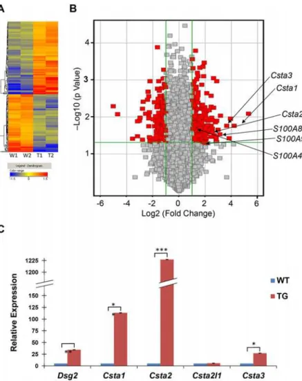
Total RNA samples isolated from the skin of two transgenic and two wild-type mice were used to generate biotin-labeled cRNA probes and then hybridized to oligonucleotide microar-rays representing 21,619 mouse genes (Fig. 1A). A comparison of the average gene signals re-vealed approximately 492 genes altered by more than two fold in response to Dsg2 expression (Table 1andS1 Table). A volcano plot revealed that 275 transcripts were upregulated and 217 downregulated with statistical significance (p<0.05, shown in red,Fig. 1B). These 492 genes
modulated in response to Dsg2 were further analyzed by the Ingenuity Analysis Program (Qia-gen), which describes the top 5 gene networks according to their degree of relevance to the Net-work Eligible Molecules in our dataset (S2 Table). This analysis revealed strong association with the Cell Cycle (S2A Fig.) and Cancer Regulation pathways (S2B Fig.). A significant num-ber of genes involved in different phases of cell cycle regulation were upregulated (red) in re-sponse to Dsg2 (and further summarized inS3 Table). Similarly, many genes involved in tumorigenesis were also upregulated in response to Dsg2 including, the mitotic checkpoint protein kinase (BUB1) (S1 Table) and the distal-less 4-homeobox (DLX4) and the cancer sus-ceptibility protein (BRCA1) (S2B Fig.). Interestingly however, two members of the forkhead family of transcription factors,FOXC1(-9.0 fold) andFOXC2(-11.7 fold), were downregulated in the Inv-Dsg2 transgenic mice. FOXC2, in particular, induces epithelial-mesenchymal transi-tion and plays a role in eyelid closure [26,27]. Knockdown of FOXC2 expression in breast can-cer cells abrogates their metastatic potential [28], implicating its role in cancer development. The role of FOXC2 in the skin homeostasis and cancer development is not known.
Fig 1. Differential gene expression between Inv-Dsg2 transgenic and wild-type mouse skin.(A) Total RNA was isolated from the skin of 2 wild-type and 2 Inv-Dsg2 transgenic mice, reverse transcribed, biotin-labeled and applied to a mouse cDNA microarray. The dendogram (heat map) shows that 492 genes were either up-regulated (red/orange) or down-regulated (blue/green) in transgenic (T1 and T2) and control (W1 and W2) mice. (B) Volcano plot shows the log2 (fold change) in x-axis versus the—log10 (p value) in the y-axis. The points having a fold-change less than 2 (log2 = 1) are shown in gray. The vertical green lines demarcate where the fold change equals 2 (right line) or equals—2 (left line). The horizontal green line demarcates where the p value is 0.05, with points above the line having p<0.05 and points below the line having p>0.05. Depicted in red are the genes that exhibit a greater than 2 fold change with a p>0.05 in transgenic epidermis as compared to control. The arrows indicate genes of interest. (C) Quantitative real-time RT-PCR analysis reveals an average of 34.33±1.32 fold increase in Dsg2 RNA expression in Inv-Dsg2 transgenic (Tg) compared to that of wild-type (WT). In addition, RNA expression for transgenic relative to control were:Csta1, 113.24±2.23;Csta2, 1227.04±1.26;Csta2l1, 1.11±0.47;Csta3, 26.97±0.44 (Bar = mean±s.d.; (*p<0.05;**p<0.01;***p<0.001; Student’sttest).
doi:10.1371/journal.pone.0120091.g001
Table 1. Differentially expressed genes in response to Dsg2.
Fold Gene Mapped Un-mapped Up Down
>2.0 492 468 24 275 217
>2.5 247 234 13 144 103
>3.0 152 144 8 54 98
whilst expression of one member, cyclin A1 (Ccna1, -2.0 fold) was found to be decreased (Table 3). The cyclin family of proteins regulates cyclin-dependent kinases and controls the progression of cells through the cell cycle [30]. This finding is consistent with our previous results showing that Dsg2 enhances cell proliferation. CSTA has been shown to possess anti-apoptotic activity [31], is upregulated in several epithelial-derived malignancies including SCC [32], and is mutated in inherited cell-cell adhesion defective epidermal disorders [17–19]. Fur-thermore, CSTA inhibits cathepsins [33,34], which are also deregulated in skin cancer [35–37]. Table 2. Top ten genes up- or down-regulated in response to Dsg2.
GenBank Accession # Gene Gene Product Fold p-value Functions
Top ten genes up-regulated
NM_021480 TDH L-threonine dehydrogenase 39.18 8.1E-03 Enzyme
M92417 Csta1/StfA1 Cystatin A1/Stefin A1 18.39 1.7E-02 Cysteine proteinase inhibitor D45850 AKR1C4 Aldo-keto reductase family 1 member C4 15.18 1.1E-02 Enzyme
AK007978 Unknown Unidentified EST 14.06 3.7E-02 Unknown
M92419 Csta3/StfA3 Cystatin A3/Stefin A3 12.37 1.7E-02 Cysteine proteinase inhibitor AJ251685 GPNMB Glycoprotein (transmembrane) nmb 9.09 9.1E-03 Pigmentation
NM_009638 CRISP3 Cysteine-rich secretory protein 3 8.79 2.9E-02 Sperm regulation M83218 S100A8 S100 calcium binding protein A8 7.86 2.2E-02 Casein kinase inhibitor
AK010010 Unknown Unidentified EST 7.40 4.8E-02 Unknown
AJ237585 Ncapg Non-SMC condensin I complex, subunit G 7.39 1.3E-02 Chromatin condensation Top ten genes down-regulated
NM_008791 PCP4 Purkinje cell protein 4 -6.58 3.9E-03 Calmodulin-mediated signaling
AK019744 Unknown Unidentified EST -6.84 6.1E-03 Unknown
AF070470 SMOC1 SPARC related modular calcium binding 1 -6.89 4.1E-02 Matrix assembly and cell adhesiveness
NM_007568 BTC Betacellulin -7.75 1.0E-02 EGF receptor ligand
AK018865 Unknown Unidentified EST -8.22 1.3E-02
NM_008592 FOXC1 Forkhead box C1 -8.95 3.3E-02 Transcription regulation
BC014714 HMGCS2 3-Hydroxy-3-methylglutaryl-CoA synthase 2 -8.98 1.3E-02 Enzyme
NM_013519 FOXC2 Forkhead box C2 -11.69 3.8E-02 Transcription regulation
AK004289 Unknown Unidentified EST -25.73 8.3E-03 Unknown
AK009582 PBRM1 Protein polybromo 1 -32.82 5.1E-03 Chromatin-remodeling
doi:10.1371/journal.pone.0120091.t002
Table 3. Upregulation of cystatin A, S100 and cyclin genes in Dsg2 transgenic mice.
Gene symbol Name Fold p-value
Csta1/Stfa1 Cystatin A1 (Stefin A1) 18.39 1.7E-02
Csta3/Stfa3 Cystatin A3 (Stefin A3) 12.37 1.7E-02
Csta2/Stfa2 Cystatin A2 (Stefin A2) 5.71 2.6E-02
S100A8 S100 calcium binding protein A8 7.86 2.2E-02
S100A9 S100 calcium binding protein A9 4.08 5.0E-02
S100A4 S100 calcium binding protein A4 2.28 2.2E-02
Ccna2 Cyclin A2 5.8 2.1E-02
Ccnb2 Cyclin B2 5.4 1.7E-02
Ccnb1 Cyclin B1 4.6 1.8E-02
Ccne1 Cyclin E1 2.6 3.3E-03
Ccna1 Cyclin A1 -2.0 2.9E-02
For those reasons, we focused the remainder of this study on CSTA. The functional relevance of the S100 and cyclin proteins will be examined in detail in a future study.
Confirmation of Dsg2-modulation of CSTA expression
The microarray results were confirmed by examining the expression of mouse Csta mRNA (1, 2, 2L1 and 3) by RT-PCR (S3 Fig.) and real-time qPCR (Fig. 1C). First, total RNA was iso-lated from the skin of two individual representative wild-type and Inv-Dsg2 transgenic mice, cDNA was generated and used as a template for PCR confirming the upregulation ofDsg2,
Csta1,Csta2, andCsta3in transgenic as compared to control mice (S3 Fig.). No change in RNA expression was observed withCsta2l1andGAPDH. Csta2l1 is highly expressed in the fetal liver, bone marrow, and spleen and was used here as a negative control for skin [38]. The RT-PCR data was further validated by real-time qPCR using skin biopsies from 3 wild-type and 3 transgenic mice (Fig. 1C). Again, up-regulation ofDsg2(34.33±1.32) was observed in skin from transgenic mice as compared to skin from control animals. By real-time qPCR, we observed an increase inCsta1(113.24±2.23),Csta2(1227.04±1.39), andCsta3(26.97±0.44), but notCsta2l1(1.11±0.47). With the exception ofCsta2l1, all changes from transgenic com-pared to control samples were statistically significant. Interestingly, the fold change difference obtained by real-time qPCR was significantly different than obtained from microarray analysis. This is not unusual and has been observed in other systems [39]. In summary, qPCR analysis confirmed the microarray data demonstrating that ectopic expression of Dsg2 in the epidermis caused an increase in the RNA expression of the mouseCstafamily of proteins and that this enhanced expression is regulated by the overexpression of Dsg2 in the skin.
Next, skin lysates from newborn and adult control mice (n = 3 each) were immunoblotted for Csta revealing high level in newborn skin, but dramatically decreased in adult skin
(Fig. 2A), corroborating with previous observations [40]. Interestingly, we observed both cyto-plasmic and nuclear staining for Csta in newborn mouse skin by immunofluorescence (Fig. 2B). No significant difference in Csta level was observed between the wild-type and Inv-Dsg2 transgenic newborn mice (not shown). However, since Csta was low in the skin of adult mice, ectopic expression of the Dsg2 dramatically enhanced Csta level (Fig. 2C). The Flag anti-body detected the Flag-tagged Dsg2 protein in the transgenic but not the wild-type mice (Fig. 2C). Similar results were observed using the Dsg2 specific antibody 10D2 demonstrating that ectopic expression of Dsg2 in the superficial epidermis did not alter the endogenous Dsg2 level (data not shown). Due to the nature of the antibodies, we were unable to perform co-immunostaining for Dsg2 and Csta. However, conditions were optimized to immunostain for the Flag-tagged Dsg2 (anti-Flag antibodies) and Csta (Fig. 2D). In the Dsg2 Tg skin, Csta was expressed in cells regardless of transgene expression, thus suggesting a non-autonomous effect. Staining for Csta was also detected in the corneocytes of wild-type mice and increased in the transgenic mice (Fig. 2D). Although Csta was not detected by Western blot using total skin ly-sates, we cannot rule out that in the cornified envelope, Csta may be highly cross-linked and thus resistant to lysis in SDS/DTT lysis buffer.
Fig 2. Dsg2 enhances cystatin A expressionin vivo.(A) Western blot analysis of skin lysates from 3 newborn and 3 adult C57Bl6 mice shows high expression of Csta in newborn but virtually undetectable in adult skin. Actin was used as a control for equal loading. (B) Immunofluorescent staining confirms the Western blotting results showing high level of Csta in newborn wild-type mouse skin. Enlarged image in inset shows cytoplasmic as well as nuclear staining for Csta. (C) Western analysis for Dsg2 and Csta in adult wild-type and Inv-Dsg2 transgenic mouse skin. The results showed expression of the Flag-tagged Dsg2 and Csta in the transgenic but not type mice. Actin showed equal loading. (D) Immunofluorescence was performed on adult skin of wild-type and transgenic mice revealing increased levels CSTA in transgenic skin. Nuclei were counter-stained with DAPI (blue).
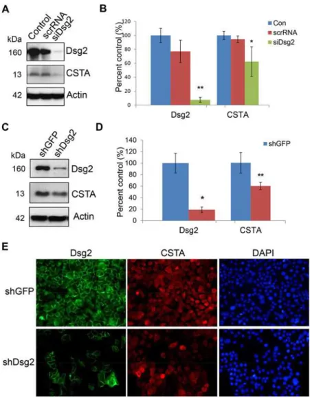
Dsg2 modulates CSTA expression
To determine if Dsg2 modulates human CSTA in keratinocytes, A431 cells were treated with siRNA specific for Dsg2 (siDsg2), CSTA (siCSTA) or scrambled RNA (scrRNA). Three days post-transfection, with scrRNA and siCSTA, Western blot analysis showed that knockdown of CSTA did not affect the expression of Dsg2 (S4 Fig.). In contrast, a dramatic reduction of Dsg2 by siDsg2 but not scrRNA, resulted in small but reproducible reduction of CSTA (Fig. 3A,B). To further confirm the effect of Dsg2 on CSTA expression, stable A431 cell lines with knock-down of Dsg2 using short hairpin RNA (shRNA) were established [24]. Immunoblotting re-vealed that Dsg2 was significantly down regulated in shDsg2 cells, as compared to shGFP cells (Fig. 3C,D). Similar to the results observed using siRNA, knockdown of Dsg2 by shRNA slight-ly suppressed the expression of CSTA (Fig. 3C,D). A431-shGFP and A431-shDsg2 were immu-nostained for Dsg2 and CSTA showing knockdown of Dsg2 in the A431-shDsg2 cells resulted in reduction of CSTA (Fig. 3E). In summary, these results suggest that CSTA is modulated in part by Dsg2.
Effect of CSTA on cell-cell adhesion
In the autosomal recessive disorder exfoliative ichthyosis, loss-of-function mutations inCSTA
results in coarse peeling of the skin on the palms and soles and detachment occurring in the lower epidermis with abnormal desmosomes [17]. Here, we wanted to assess whether Dsg2 plays a role exfoliative ichthyosis by synergizing with CSTA to modulate cell adhesion. Normal human back skin and palm were immunostained for Dsg2 showing low levels in the basal layer of the interfollicular epidermis but high levels in the both the basal and superficial layers in pal-moplantar epidermis (Fig. 4A). These results are similar to our previous findings [23].
Next, the impact of Dsg2 and CSTA on cell-cell adhesion was assessed. We attempted multi-ple times to express CSTA in keratinocytes using different expression plasmids (His or HA tag) but were unsuccessful possibly due to CSTA being negatively regulated by the Ras/Raf-1/ MEK1/ERK pathway [42]. Hence, we opted to knockdown CSTA. shGFP and A431-shDsg2 cells were subjected to the in vitro dispase-based keratinocyte dissociation assay 72 hr post-treatment with scrRNA or siCSTA to knockdown CSTA. Sheets of cells were lifted from the culture dish using dispase and disrupted by pipetting. Cell fragments were photographed and counted. Independent knockdown of either Dsg2 (Fig. 4Aiv, Biv) or CSTA (Fig. 4Aiii, Biii) resulted in an increase in fragmentation when compared to untreated cells (Fig. 4Ai, Bi) or scrRNA treated cells (Fig. 4Aii, Bii). In addition, the dual loss of Dsg2 and CSTA (Fig. 4Avi, Bvi) further enhanced fragmentation.
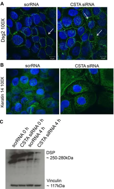
Finally, to demonstrate that loss of CSTA leads to decreased cell-cell adhesion by disrupting desmosomal structures, we treated HaCaT keratinocytes with scrRNA or CSTA siRNA, sub-jected to 4 hr mechanical stretching and then immunostained for Dsg2 (Fig. 5A) and keratin 14 (Fig. 5B). CSTA knockdown led to increase in cytoplasmic localization of Dsg2 and retrac-tion of cytoskeletal organizaretrac-tion. Furthermore, Western blotting showed a decrease in desmo-plakin levels in CSTA knockdown stretched monolayers compared to control cells stretched for the same number of hours (Fig. 5C). The changes observed in components of the desmo-some suggest that the cell-cell breakage occurs at the desmodesmo-somes. In summary, CSTA plays a role in epithelial cell adhesion as loss of CSTA rendered the cells susceptible to mechanical dis-ruption by destabilizing the desmosomal structures.
Discussion
dramatically altered the expression of many genes involved in cell cycle regulation and cancer development, including the expression of the cysteine protease inhibitor, CSTA. This study demonstrates for the first time that a desmosomal cadherin can modulate the expression of CSTA bothin vivoandin vitro.
CSTA was originally identified as a precursor protein of the cornified cell envelope [43] and reduced expression of CSTA contributes to a defective epidermal barrier in the skin condition Fig 3. Modulation of CSTA expression by Dsg2.(A) A431 cells were treated for 72 hr with 100 nM of scrambled RNA orDsg2siRNA. Western blot analysis for Dsg2 and cystatin A shows that knockdown of Dsg2 reduced CSTA level. Immunoblotting for Actin showed equal loading. (B) The Western blot results were quantified and expression level of each band was normalized against Actin. Values shown are percentage of expression against control untreated. The results showed significant reduction in Dsg2 in response toDsg2
siRNA but not scrambled siRNA, while knockdown of Dsg2 slightly reduced the expression of CSTA. The change was statistically significant. Bar = mean±s.e.m.*p<0.05 and***p<0.001 using Student’sttest. (C) A431 cells were stably transfected with shRNA to GFP (shGFP) or Dsg2 (shDsg2) and selected in
puromycin. Immunoblotting showed loss of Dsg2 reduced CSTA expression in the shDsg2 cells, as compared to the shGFP cells. Actin was used as a loading control. (D) Quantification of the Western blot results showed reduction in Dsg2 in the shDsg2 cell as compared to the shGFP cells and knockdown of Dsg2 reduced CSTA expression. Bar = mean±s.e.m.*p<0.05 and***p<0.001 using Student’sttest. E) Immunofluorescence of Dsg2 and CSTA showed that knockdown of Dsg2 reduced the expression of Dsg2 and CSTA in the A431-shDsg2 as compared to A431-shGFP cells. Nuclei were counter-stained with DAPI (blue).
known as atopic dermatitis [44,45,16]. However,CSTAmessage has been localized to the lower epidermis [46]. Here we show that Csta, similar to Dsg2, was expressed at high level through-out the newborn epidermis but decreased during development. In the epidermis of adult mice, minor signal was detected in the differentiated layers [20]. Ectopic expression of Dsg2 in trans-genic mice enhanced Csta expression suggesting perhaps that Csta may be regulated in part by Dsg2 [5]. Concomitant with the expression level of Dsg2 and Csta, the skin of newborn mice is hyperplastic (~5–6 layers) as compared to the 2–3 layers in adult mice. An increase in Dsg2 with upregulation of Csta in the Inv-Dsg2 transgenic mice also induced hyperplasia [5]. In human keratinocytes, CSTA displays anti-apoptotic activity through its inhibition of UVB-induced caspase 3 activation, thereby suppressing UVB-UVB-induced apoptosis suggesting that CSTA may play an important role in controlling cell growth, differentiation and survival [44]. Fig 4. Modulation of cell adhesion by CSTA and Dsg2.(A) Immunofluorescence of normal human skin (A) and palm (B) showing low levels of Dsg2 in the basal layer (arrows) of the normal skin (inset: enlarged image) but high levels in both the basal and differentiated layers (arrow head) in the palm. Note: Immunostaining was performed at the same time and images were captured at the same exposure. (B) shGFP and A431-shDsg2 cells were treated with scrambled RNA or siRNA toCSTAfor 72 hr and then subjected to thein vitro
Our results complement the recent findings showing that loss-of-function mutations in the
CSTAgene is the underlying cause of the skin condition exfoliative ichthyosis [17]. The skin of affected individuals displayed hyperkeratosis and superficial exfoliation. Interestingly however, loss of CSTA did not affect barrier function or terminal differentiation. Instead, histology of Fig 5. Loss of CSTA leads to destabilized intercellular connections.Cells were treated with non-targeting pool scrRNA or withCSTAsiRNA (CSTA KD) followed by mechanical stretching for 4 hr. Cells were allowed to adhere, fixed, and immunostained for Dsg2 (A) and cytokeratin 14 (B) or lysed in Laemmli buffer and immunoblotted for desmoplakin (C). Knockdown of CSTA in keratinocytes resulted in cytoplasmic relocalization of Dsg2, breakage of cytokeratin intercellular connections, and loss of the desmosomal protein, desmoplakin.
the exfoliative ichthyosis palmoplantar skin exhibited loss of cell-cell adhesion in the deep dermis. Particularly, the basal and immediate suprabasal layers showed signs of disrupted epi-dermal structure and disorganization of desmosomes. Here, we have shown that loss of CSTA and Dsg2 enhanced cell-cell disadhesion (Fig. 4B,C) and that CSTA modulated desmosome stability (Fig. 5). These results suggest that Dsg2 may play a role in the skin fragility phenotype of exfoliative ichthyosis as it is highly expressed in both the basal and superficial layers in pal-moplantar tissues (Fig. 4A). We note here that we cannot rule out the role of other desmosomal proteins, such as desmoglein 3 and desmocollins 2 and 3, that are also expressed in the deep epidermis and that may affect the expression and/or function of CSTA [20,47]. The crosstalk between these other cadherins and CSTA will be the subject of future studies.
The impact of the Dsg2-CSTA influence on cell adhesion may play a yet unrecognized role in the skin fragility condition pemphigus. Pemphigus is a group of autoimmune skin blistering diseases caused by loss of cell-cell adhesion due to autoantibodies binding to Dsg1 and Dsg3 re-sulting in the internalization of desmosomes [48]. Passive transfer of pathogenic pemphigus IgG into neonatal mice produces epidermal blister formation similar to those observed in pa-tients [49,50]. Our previous study showed that compared to wild-type mice, ectopic expression of Dsg2 in the superficial epidermis rendered the Inv-Dsg2 transgenic mice more resistant to blister formation by pemphigus foliaceus IgG [51]. Here, we show that loss of Dsg2 disrupted cell-cell adhesion and this effect was further amplified with loss of CSTA. Thus, we speculate that in the Inv-Dsg2 transgenic mice, forced expression of Dsg2 increased the CSTA level thereby enhancing cell-cell adhesion and possibly protecting the epidermis from acantholysis-associated PF blister formation. Dsg2 is often upregulated in the skin of affected patients [48] and keratinocytes from Dsg3 knockout mice upregulate Dsg2 [52]. These findings support the notion that induction of Dsg2 in lesional skin of pemphigus patients could be a compensatory mechanism to enhance cell-cell adhesion and thus protect the patients from blister formation. It would be interesting to assess the expression of CSTA in pemphigus patients’skin.
Although evidence supports a causal role for proteases in malignant progression of human cancers, the role of CSTA is somewhat complicated and controversial [53]. In head and neck SCC, expression of CSTA has been reported to be down-regulated in some patients while up-regulated in others [54,55]. Overexpression of CSTA has been detected in a variety of human cancers including lung, breast, head and neck, vulva, cervix, esophagus and prostate, and in some mouse sarcomas. In some forms of highly malignant and metastasizing breast cancer, there is a correlation between increased CSTA expression and poor prognosis [56]. Upregula-tion of CSTA is also detected in another transgenic mouse model overexpressing the early gene region of the human papillomavirus type 8, these mice develop papillomas similar to our Inv-Dsg2 mice [57]. The role of Dsg2 in cancers is equally controversial and the expression level is dependent on the tumor type. We, and others have shown that while Dsg2 expression in the interfollicular epidermis is demonstrably low [20], it is markedly increased in skin, prostate, and colon cancers [23,58–61]. Interestingly, in diffuse-type gastric cancers, decreased expres-sion of Dsg2 is associated with poor prognosis suggesting a complex role for Dsg2 in oncogene-sis, serving as a tumor enhancer or suppressor [62].
The mechanism by which Dsg2 modulates CSTA expression remains to be determined and future studies to assess the Dsg2-DNA interactions and map the Dsg2 binding sites are neces-sary. However, we recently demonstrated that Dsg2 might have an impact on signaling by binding to the scaffolding protein caveolin-1, the major components of lipid rafts, caveolae [7]. Caveolins and caveolae have been implicated as regulators of key cellular functions by
compensatory mechanism to offset the increase in activity of proteases such as cathepsin that modulate matrix remodeling during disease progression. Alternatively, an increase in CSTA activity in tumors may counter the activation of apoptosis induced by the tumor necrosis factor alpha and cathepsin B [64]. Thus, whether CSTA is an oncogene or a tumor suppressor is unre-solved. However, the general consensus is that in many cancers, there is an imbalance between the proteases and their respective cystatin inhibitors [65–67].
Conclusions
In this report, we demonstrated that Dsg2 plays an active role in modulating epithelial cell growth and survival by altering the epithelial gene transcriptome and modulating the expres-sion of proteins that are markers or prognostic indicators of the dysplastic phenotype includ-ing, CSTA.
Supporting Information
S1 Fig. Upregulation of Dsg2 in the Inv-Dsg2 transgenic mice.(A) H&E-staining shows epi-dermal hyperplasia in the Inv-Dsg2 transgenic skin compared to wild-type control. (B) Western blot analysis of Flag shows the Flag-tagged Dsg2 in the transgenic but not wild-type skin. Immu-noblot with anti-Actin antibody served as loading control for protein lysates. (C) Immunofluores-cent analysis reveals expression of Dsg2 in the differentiated layers of the transgenic epidermis. Nuclei were stained with DAPI (blue). Scale bar, 200μm. Tg, transgenic; WT, wild-type. (TIF)
S2 Fig. Functional gene networks identified using Ingenuity Pathway Analysis (IPA) soft-ware from differentially expressed genes between wild-type and Inv-Dsg2 transgenic mice. The IPA analysis revealed the top functional gene networks to be cell-cycle (A) and cancer (B) composed of multiple genes, many of which are involved in skin cancers (Cyclins, S100 family, FOXC1/2 and BRCA1 genes) to be most differentially expressed by Dsg2 compared to wild-type. Nodes represent genes and their level of color intensity is related to its level of expression (red, up-regulation;green, down-regulation). Uncolored nodes means these genes were not identified as differentially expressed and were integrated as part of the network analysis based on the information in the IPA databases.
(TIF)
S3 Fig. RT-PCR analysis ofDsg2andCstaexpression.RT-PCR showed that mRNA expres-sion ofDsg2,Csta1,Csta2andCsta3were relatively higher in the Inv-Dsg2 transgenic skin compared to that of wild-type.Csta2l1expression was used as a control.
(TIF)
S4 Fig. Effect ofCSTAknockdown on Dsg2.A431 cells were treated for 72 hr with 100 nM of scrambled RNA orCSTAsiRNA and total protein lysate was immunoblotted for Dsg2 showing that knockdown of CSTA had no effect on Dsg2 expression.
(TIF)
S1 Table. Dsg2-dependent Gene Changes. (PDF)
S2 Table. Associated Network Functions. (PDF)
Acknowledgments
We thank James K. Wahl III (University of Nebraska, Lincoln, NE) for the A431-shGFP and A431-shDsg2 cell lines and antibody 10D2. We thank David Garrod (University of Manches-ter, UK) for the DP antibody Clone 5–11F.
Author Contributions
Conceived and designed the experiments: AG DN DPK MGM. Performed the experiments: AG DN DBC MGM. Analyzed the data: AG DN DBC SA DPK MGM. Contributed reagents/ materials/analysis tools: SA NAR DPK MGM. Wrote the paper: AG DN DBC DPK MGM.
References
1. Delva E, Tucker DK, Kowalczyk AP. The desmosome. Cold Spring Harb Perspect Biol. 2009; 1: a002543. doi:10.1101/cshperspect.a002543PMID:20066089
2. Pasdar M, Krzeminiski KA, Nelson WJ. Regulation of desmosome assembly in MDCK epithelial cells: coordination of membrane core and cytoplasmic plaque domain assembly at plasma membrane. J Cell Biol. 1991; 113: 645–655. PMID:1707884
3. Petrof G, Mellerio JE, McGrath JA. Desmosomal genodermatoses. Br J Dermatol. 2012; 166: 36–45. doi:10.1111/j.1365-2133.2011.10640.xPMID:21929534
4. Green KJ, Getsios S, Troyanovsky S, Godsel LM. Intercellular junction assembly, dynamics, and ho-meostasis.Cold Spring Harb Perspect Biol. 2010; 2: a000125. doi:10.1101/cshperspect.a000125 PMID:20182611
5. Brennan D, Hu Y, Joubeh S, Choi YW, Whitaker-Menezes D, O’Brien T, et al. Suprabasal Dsg2 expres-sion in transgenic mouse skin confers a hyperproliferative and apoptosis-resistant phenotype to kerati-nocytes. J Cell Sci. 2007; 120: 758–771. PMID:17284515
6. Merritt AJ, Berika MY, Zhai W, Kirk SE, Ji B, Hardman MJ, Garrod DR. Suprabasal desmoglein 3 ex-pression in the epidermis of transgenic mice results in hyperproliferation and abnormal differentiation. Mol Cell Biol. 2002; 22: 5846–5858. PMID:12138195
7. Brennan D, Peltonen S, Dowling A, Medhat W, Green KJ, Wahl JK, et al. A role for caveolin-1 in desmo-glein binding and desmosome dynamics. Oncogene. 2012; 31: 1636–1648. doi:10.1038/onc.2011.346 PMID:21841821
8. Brown WM, Dziegielewska KM. Friends and relations of the cystatin superfamily-new members and their evolution. Protein Sci. 1997; 6: 5–12. PMID:9007972
9. Dubin G. Proteinaceous cysteine protease inhibitors. Cell Mol Life Sci.2005; 62: 653–669. PMID: 15770418
10. Magister S, Kos J. Cystatins in immune system. J Cancer. 2013; 4: 45–56. doi:10.7150/jca.5044 PMID:23386904
11. Rawlings ND, Barrett AJ. Evolution of proteins of the cystatin superfamily. J Mol Evol. 1990; 30: 60–71. PMID:2107324
12. Barrett AJ. The cystatins: a diverse superfamily of cysteine peptidase inhibitors. Biomed Biochim Acta. 1986; 45: 1363–1374. PMID:3555466
13. Strauss M, Stollwerk J, Lenarcic B, Turk V, Jany KD, Gassen HG. Chemical synthesis of a gene for human stefin A and its expression in E. coli. Biol Chem Hoppe Seyler. 1988; 369: 1019–1030. PMID: 3067731
14. Vray B, Hartmann S, Hoebeke J. Immunomodulatory properties of cystatins. Cell Mol Life Sci. 2002; 59: 1503–1512. PMID:12440772
15. Takai T, Kato T, Hatanaka H, Inui K, Nakazawa T, Ichikawa, et al. Modulation of allergenicity of major house dust mite allergens Der f 1 and Der p 1 by interaction with an endogenous ligand. J Immunol. 2009; 183: 7958–7965. doi:10.4049/jimmunol.0713276PMID:19933866
16. Zettergren JG, Peterson LL, Wuepper KD. Keratolinin: the soluble substrate of epidermal transglutami-nase from human and bovine tissue. Proc Natl Acad Sci U S A. 1984; 81: 238–242. PMID:6141559 17. Blaydon DC, Nitoiu D, Eckl KM, Cabral RM, Bland P, Hausser I, et al. Mutations in CSTA, Encoding
18. Kavaklieva S, Yordanova I, Bruckner-Tuderman L, Has C. Acral peeling skin syndrome resembling epi-dermolysis bullosa simplex in a 10-month-old boy. Case Rep Dermatol. 2013; 5: 210–214. doi:10. 1159/000354572PMID:24019772
19. Krunic AL, Stone KL, Simpson MA, McGrath JA. Acral peeling skin syndrome resulting from a homozy-gous nonsense mutation in the CSTA gene encoding cystatin A. Pediatr Dermatol. 2013; 30: e87–88. doi:10.1111/pde.12092PMID:23534700
20. Mahoney MG, Hu Y, Brennan D, Bazzi H, Christiano AM, Wahl JK. Delineation of diversified desmo-glein distribution in stratified squamous epithelia: implications in diseases. Exp Dermatol. 2006; 15: 101–109. PMID:16433681
21. Brennan D, Hu Y, Kljuic A, Choi Y, Joubeh S, Bashkin M, et al. Differential structural properties and ex-pression patterns suggest functional significance for multiple mouse desmoglein 1 isoforms. Differenti-ation. 2004; 72: 434–449. PMID:15606502
22. Parrish EP, Steart PV, Garrod DR, Weller RO. Antidesmosomal monoclonal antibody in the diagnosis of intracranial tumours. J Pathol. 1987; 153: 265–273. PMID:3323433
23. Brennan D, Mahoney MG. Increased expression of Dsg2 in malignant skin carcinomas: A tissue-microarray based study. Cell Adh Migr. 2009; 3: 148–154. PMID:19458482
24. Keim SA, Johnson KR, Wheelock MJWahl, JK. Generation and characterization of monoclonal antibod-ies against the proregion of human desmoglein-2. Hybridoma. 2008; 27: 249–258. doi:10.1089/hyb. 2008.0020PMID:18707543
25. Hartlieb E, Kempf B, Partilla M, Vigh B, Spindler V, Waschke J. Desmoglein 2 is less important than desmoglein 3 for keratinocyte cohesion.PLoS One. 2013; 8: e53739. doi:10.1371/journal.pone. 0053739PMID:23326495
26. Battula VL, Evans KW, Hollier BG, Shi Y, Marini FC, Ayyanan A, et al. Epithelial-mesenchymal transition-derived cells exhibit multilineage differentiation potential similar to mesenchymal stem cells. Stem Cells. 2010; 28: 1435–1445. doi:10.1002/stem.467PMID:20572012
27. Kuracha MR, Siefker E, Licht JD, Govindarajan V. Spry1 and Spry2 are necessary for eyelid closure. Dev Biol. 2013; 383: 227–238. doi:10.1016/j.ydbio.2013.09.014PMID:24055172
28. Mani SA, Yang J, Brooks M, Schwaninger G, Zhou A, Miura N, et al. Mesenchyme Forkhead 1 (FOXC2) plays a key role in metastasis and is associated with aggressive basal-like breast cancers. Proc Natl Acad Sci U S A. 2007; 104: 10069–10074. PMID:17537911
29. Sorenson BS, Khammanivong A, Guenther BD, Ross KF, Herzberg MC. IL-1 receptor regulates S100A8/A9-dependent keratinocyte resistance to bacterial invasion. Mucosal Immunol. 2012; 5: 66–75. doi:10.1038/mi.2011.48PMID:22031183
30. Galderisi U, Jori FP, Giordano A. Cell cycle regulation and neural differentiation. Oncogene. 2003; 22: 5208–5219. PMID:12910258
31. Takahashi H, Komatsu N, Ibe M, Ishida-Yamamoto A, Hashimoto Y, Iizuka H. Cystatin A suppresses ul-traviolet B-induced apoptosis of keratinocytes. J Dermatol Sci. 2007; 46: 179–187. PMID:17412564 32. Butler MW, Fukui T, Salit J, Shaykhiev R, Mezey JG, Hackett NR Crystal RG. Modulation of cystatin
A expression in human airway epithelium related to genotype, smoking, COPD, and lung cancer. Can-cer Res. 2011; 71: 2572–2581. doi:10.1158/0008-5472.CAN-10-2046PMID:21325429
33. Estrada S, Nycander M, Hill NJ, Craven CJ, Waltho JP, Björk I. The role of Gly-4 of human cystatin A (stefin A) in the binding of target proteinases. Characterization by kinetic and equilibrium methods of the interactions of cystatin A Gly-4 mutants with papain, cathepsin B, and cathepsin L. Biochemistry. 1998; 37: 7551–7560. PMID:9585570
34. Pavlova A, Björk I. Grafting of features of cystatins C or B into the N-terminal region or second binding loop of cystatin A (stefin A) substantially enhances inhibition of cysteine proteinases. Biochemistry. 2003; 42: 11326–11333. PMID:14503883
35. Bitu CC, Kauppila JH, Bufalino A, Nurmenniemi S, Teppo S, Keinänen M, et al. Cathepsin K is present in invasive oral tongue squamous cell carcinoma in vivo and in vitro. PLoS One. 2013; 8:e70925. doi: 10.1371/journal.pone.0070925PMID:23951042
36. Ishida M, Kojima F, Okabe H. Cathepsin K expression in basal cell carcinoma. J Eur Acad Dermatol Venereol. 2013; 27: e128–130. doi:10.1111/j.1468-3083.2011.04436.xPMID:22220587
37. Ruffell B, Affara NI, Cottone L, Junankar S, Johansson M, DeNardo DG, et al. Cathepsin C is a tissue-specific regulator of squamous carcinogenesis. Genes Dev. 2013; 27: 2086–2098. doi:10.1101/gad. 224899.113PMID:24065739
39. Morey JS, Ryan JC, Van Dolah FM. Microarray validation: factors influencing correlation between oligo-nucleotide microarrays and real-time PCR. Biol Proced Online. 2006; 8: 175–193. PMID:17242735 40. Scott DK, Lord R, Muller HK, Malley RC, Woods GM. Proteomics identifies enhanced expression of
ste-fin A in neonatal murine skin compared with adults: functional implications. Br J Dermatol. 2007; 156: 1156–1162. PMID:17441952
41. Tsui FW, Tsui HW, Mok S, Mlinaric I, Copeland NG, Gilbert DJ, et al. Molecular characterization and mapping of murine genes encoding three members of the stefin family of cysteine proteinase inhibitors. Genomics. 1993; 15: 507–514. PMID:8468045
42. Takahashi H, Honma M, Ishida-Yamamoto A, Namikawa K, Kiyama H, Iizuka H. Expression of human cystatin A by keratinocytes is positively regulated via the Ras/MEKK1/MKK7/JNK signal transduction pathway but negatively regulated via the Ras/Raf-1/MEK1/ERK pathway. J Biol Chem. 2001; 276: 36632–36638. PMID:11451947
43. Steven AC, Steinert PM. Protein composition of cornified cell envelopes of epidermal keratinocytes. J Cell Sci. 1994; 107: 693–700. PMID:8006082
44. Takahashi H, Kinouchi M, Wuepper KDIizuka H. Cloning of human keratolinin cDNA: keratolinin is iden-tical with a cysteine proteinase inhibitor, cystatin A, and is regulated by Ca2+, TPA, and cAMP. J Invest Dermatol. 1997; 108: 843–847. PMID:9182808
45. Vasilopoulos Y, Cork MJ, Teare D, Marinou I, Ward SJ, Duff GW, Tazi-Ahnini R. A nonsynonymous substitution of cystatin A, a cysteine protease inhibitor of house dust mite protease, leads to decreased mRNA stability and shows a significant association with atopic dermatitis. Allergy. 2007; 62: 514–519. PMID:17441792
46. Hawley-Nelson P, Roop DR, Cheng CK, Krieg TM, Yuspa SH. Molecular cloning of mouse epidermal cystatin A and detection of regulated expression in differentiation and tumorigenesis. Mol Carcinog. 1988; 1: 202–211. PMID:2471537
47. Nuber UA, Schäfer S, Stehr S, Rackwitz HR, Franke WW. Patterns of desmocollin synthesis in human epithelia: immunolocalization of desmocollins 1 and 3 in special epithelia and in cultured cells. Eur J Cell Biol. 1996; 71: 1–13. PMID:8884173
48. Iwatsuki K, Han GW, Fukuki R, Ohtsuka M, Kikuchi S, Akiba H, Kaneko F. Internalization of constitutive desmogleins with the subsequent induction of desmoglein 2 in pemphigus lesions. Br. J. Dermatol. 1999; 140: 35–43. PMID:10215765
49. Amagai M, Tsunoda K, Zillikens D, Nagai T, Nishikawa T. The clinical phenotype of pemphigus is de-fined by the anti-desmoglein autoantibody profile. J Am Acad Dermatol. 1999; 40: 167–170. PMID: 10025740
50. Mahoney MG, Wang Z, Rothenberger K, Koch PJ, Amagai M, Stanley JR. Explanations for the clinical and microscopic localization of lesions in pemphigus foliaceus and vulgaris. J Clin Invest. 1999; 104: 461–468.
51. Brennan D, Hu Y, Dowling A, Medhat W, Mahoney MG. Superficial Dsg2 expression limits epidermal blister formation mediated by Pemphigus Foliaceus antibodies and exfoliative toxins. Dermatol Res Pract. 2010 June 9;.
52. Hartlieb E, Rötzer V, Radeva M, Spindler V, Waschke J. Desmoglein 2 compensates for desmoglein 3 but does not control cell adhesion via regulation of p38 mitogen-activated protein kinase in keratino-cytes.J Biol Chem. 2014; 289(24): 17043–17053. doi:10.1074/jbc.M113.489336PMID:24782306 53. Rinne A. Epidermal SH-protease inhibitor (ACPI, cystatin A) in cancer. A short historical review. Pathol
Res Pract. 2010; 206: 259–262. doi:10.1016/j.prp.2009.12.005PMID:20116931
54. Parker BS, Ciocca DR, Bidwell BN, Gago FE, Fanelli MA, George J, et al. Primary tumour expression of the cysteine cathepsin inhibitor Stefin A inhibits distant metastasis in breast cancer. J Pathol. 2008; 214: 337–346. PMID:17985332
55. Strojan P, Anicin A, Svetic B, Pohar M, Smid L, Kos J. Stefin A and stefin B: markers for prognosis in operable squamous cell carcinoma of the head and neck. Int J Radiat Oncol Biol Phys. 2007; 68: 1335–1341. PMID:17418975
56. Kuopio T, Kankaanranta A, Jalava P, Kronqvist P, Kotkansalo T, Weber E, Collan Y. Cysteine protein-ase inhibitor cystatin A in breast cancer. Cancer Res. 1998; 58: 432–436. PMID:9458085
57. LazićD, Alborzi F, Marcuzzi GP, Angel P, Hess J, Pfister H, Akgül B. Enhanced StefinA and Sprr2 ex-pression during papilloma formation in HPV8 transgenic mice. J Dermatol Sci. 2011; 62: 84–90. doi:10. 1016/j.jdermsci.2011.02.006PMID:21458245
59. Biedermann K, Vogelsang H, Becker I, Plaschke S, Siewert JR, Höfler H, Keller G. Desmoglein 2 is ex-pressed abnormally rather than mutated in familial and sporadic gastric cancer. J Pathol. 2005; 207: 199–206. PMID:16025435
60. Kurzen H, Munzing I, Hartschuh W. Expression of desmosomal proteins in squamous cell carcinomas of the skin. J Cutan Pathol. 2003; 30: 621–630. PMID:14744087
61. Teh MT, Parkinson EK, Thurlow JK, Liu F, Fortune F, Wan H. A molecular study of desmosomes identi-fies a desmoglein isoform switch in head and neck squamous cell carcinoma. J Oral Pathol Med. 2011; 40: 67–76. doi:10.1111/j.1600-0714.2010.00951.xPMID:20923451
62. Yashiro M, Nishioka N, Hirakawa K. Decreased expression of the adhesion molecule desmoglein-2 is associated with diffuse-type gastric carcinoma. Eur J Cancer. 2006; 42: 2397–403. PMID:16890424 63. Galbiati F, Volonte D, Brown AM, Weinstein DE, Ben-Ze'ev A, Pestell RG, Lisanti MP. Caveolin-1
ex-pression inhibits Wnt/beta-catenin/Lef-1 signaling by recruiting beta-catenin to caveolae membrane do-mains. J Biol Chem. 2000; 275: 23368–23377. PMID:10816572
64. Foghsgaard L, Wissing D, Mauch D, Lademann U, Bastholm L, Boes M, et al. Cathepsin B acts as a dominant execution protease in tumor cell apoptosis induced by tumor necrosis factor. J Cell Biol. 2001; 153: 999–1010. PMID:11381085
65. Gelb BD, Shi GP, Chapman HA, Desnick RJ. Pycnodysostosis, a lysosomal disease caused by cathep-sin K deficiency. Science. 1996; 273: 1236–1238. PMID:8703060
66. Pennacchio LA, Lehesjoki AE, Stone NE, Willour VL, Virtaneva K, Miao J, et al. Mutations in the gene encoding cystatin B in progressive myoclonus epilepsy (EPM1). Science. 1996; 271: 1731–1734. PMID:8596935