UNIVERSIDADE DA BEIRA INTERIOR
Ciências da Saúde
Modulação da inflamação e da neurogénese no
hipocampo: desvendar uma nova interação entre
a histamina e o lipopolissacarídeo
Sandra Raquel Pinto Barata Antunes
Dissertação para a obtenção do Grau de Mestre em
Ciências Biomédicas
(2º ciclo de estudos)
Orientadora: Prof. Doutora Liliana Bernardino
Coorientadora: Doutora Raquel Ferreira
iii
Acknowledgments
Firstly, I would like to thank my supervisor, Prof. Dr. Liliana Bernardino, for the opportunity to integrate this project and for her guidance and unconditional support during this year. Her helpful recommendations greatly contributed for my development as a scientific researcher. I would also like to thank my co-supervisor, Dr. Raquel Ferreira, for her kind help and support throughout this work and for always encouraging my critical thinking.
I am also grateful to my research group for all the help and for sharing their knowledge during this work.
I want to thank specially to Prof. Dr. Elisabete Ferreiro and Prof. Dr. Jorge Valero for their fundamental help in the hippocampal neurogenesis analysis performed in this thesis.
I am also thankful to my friends, specially to Diana, Tânia and my best friend Marta, for all the support and understanding over this year.
I would also like to thank my boyfriend Jorge for all the support and love.
And finally, I would like to thank my family, specially my lovely sisters, Ana and Cláudia, for all the help, affection and encouragement. The person I am today is because of them.
v
Resumo
A histamina é uma amina biogénica endógena que atua como neurotransmissor no Sistema Nervoso Central e regula uma variedade de funções cerebrais. Vários estudos têm demonstrado que a histamina pode ter efeitos contraditórios na modulação da neuroinflamação mediada pela microglia, uma das principais características patológicas presente em várias doenças neurodegenerativas. Contudo, a função desta amina no hipocampo ainda não é completamente conhecida. Assim, o principal objetivo deste trabalho foi avaliar o efeito da histamina, por si só e na presença de um mediador inflamatório, na neuroinflamação e neurogénese do hipocampo in vivo. Para tal, foram utilizados murganhos, os quais foram injetados intraperitonealmente com lipopolissacarídeo (LPS; 1 ou 2 mg/Kg), seguido de uma injeção estereotáxica de histamina (100μM), no giro dentado do hipocampo. Quatro dias após a injeção com LPS, procedeu-se à avaliação dos níveis proteicos de marcadores de reatividade glial, fatores pro-inflamatórios e marcadores de funcionalidade neuronal e sinática através da técnica de western blot. Os resultados demonstraram que a histamina por si só aumentou a expressão dos marcadores de reatividade glial (Iba1, do inglês
ionized calcium binding adaptor molecule 1; e GFAP, do inglês glial fibrillary acidic protein).
Por outro lado, diminuiu significativamente a reatividade glial induzida pelo LPS. Curiosamente, a histamina não alterou os níveis de expressão dos mediadores inflamatórios (IL-1β, do inglês interleukin-1 beta; e HMGB1, do inglês high mobility group box 1), mas conseguiu inibir o aumento da expressão de ambos os mediadores induzido pelo LPS. Esta amina conseguiu também prevenir o decréscimo na expressão de ambos os marcadores de funcionalidade neuronal (CREB, do inglês cyclic-AMP-response element binding protein) e pós-sinática (PSD-95, do inglês postsynaptic density protein 95) induzido pelo estímulo inflamatório. Posteriormente, foi contado, no giro dentado, o número total de células positivas para Bromodeoxiuridina (BrdU)/Doublecortin (DCX) e BrdU/Neuronal Nuclei (NeuN), como medida da proliferação e da sobrevivência das novas células neuronais, respetivamente. Os resultados revelaram que a histamina por si só e, quando administrada em conjunto com o estímulo inflamatório, aumentou a proliferação celular (células BrdU+) bem como a sobrevivência a longo prazo das novas células (células BrdU+ e BrdU+/NeuN+) no giro dentado. Em suma, estes resultados apotam para o potencial terapêutico da histamina no tratamento ou melhoria de condições neuronais associadas a neuroinflamação e neurodegeneração no hipocampo.
Palavras-chave
vii
Resumo alargado
A histamina é uma amina biogénica que regula uma variedade de funções ao nível do Sistema Nervoso. Vários estudos têm demonstrado o seu papel na regulação da neuroinflamação mediada pela microglia. Este processo é de particular importância uma vez que, quando desregulado, causa danos graves no cérebro podendo mesmo culminar em neurodegeneração. De facto, esta é uma característica patológica presente em várias doenças neurodegenerativas. No contexto da neuroinflamação, estudos recentes revelaram que a histamina pode ter efeitos contraditórios dependendo do contexto e de qual dos seus recetores é ativado. Esta amina por si só induz um perfil pró-inflamatório nas células da microglia e compromete a sobrevivência neuronal. Pelo contrário, num contexto inflamatório, esta protege os neurónios das respostas tóxicas da microglia. Contudo, o papel da histamina no hipocampo, ainda não é completamente conhecido. Esta é uma região cerebral responsável por funções cognitivas e comportamentais que se encontra disfuncional em várias patologias tais como a Doença de Alzheimer e a epilespia. Adicionalmente, esta é uma região particularmente vulnerável à neuroinflamação mediada pela microglia. Tendo em conta estas evidências, o principal objetivo deste trabalho foi avaliar o efeito da histamina, por si só e num contexto inflamatório, na neuroinflamação e neurogénese do hipocampo in vivo. Para tal, foram utilizados murganhos adultos, os quais foram injetados intraperitonelmente com lipopolissacarídeo (LPS; 1 ou 2 mg/Kg), uma endotoxina frequentemente utilizada para induzir neuroinflamação. Dois dias após a indução do estímulo inflamatório, procedeu-se à administração da histamina no hipocampo através de uma injeção estereotáxica. Inicialmente, procedeu-se à avaliação dos níveis proteicos de marcadores de reatividade glial (Iba1, do inglês ionized calcium binding adaptor molecule 1, para a reatividade da microglia; e GFAP, do inglês glial fibrillary acidic protein, para a reatividade astroglial), fatores pro-inflamatórios (IL-1β, do inglês interleukin-1 beta; e HMGB1, do inglês high mobility group box
1) e marcadores de funcionalidade neuronal (CREB do inglês cyclic-AMP-response elemento binding protein) e sinática (sintaxina como marcador pré-sinático; e PSD-95, do inglês postsynaptic density protein 95, como marcador pós-sinático), recorrendo à técnica de western blot, quatro dias após o estímulo inflamatório. Posteriormente, procedeu-se à
avaliação da neurogénese no giro dentado do hipocampo, sendo que esta foi avaliada em duas fases: i) a curto prazo, avaliou-se a proliferação celular, e ii) a longo prazo a sobrevivência das novas células. Para a avaliação da proliferação celular, foi contado no giro dentado do hipocampo o número total de células positivas para a Bromodeoxiuridina (BrdU; marcador de proliferação celular) e para BrdU/Doublecortin (DCX; marcador de neurónios imaturos), 5 dias após a injeção com LPS e/ou histamina. Para a avaliação da sobrevivência das novas células, foi contado o número total de células positivas para BrdU e para BrdU/Neuronal Nuclei (NeuN; marcador de neurónios maduros), 6 semanas após a injeção com LPS e/ou histamina.
Os resultados demonstraram que a histamina conseguiu inibir significativamente o aumento da expressão dos marcadores de reatividade glial (Iba-1 e GFAP) assim como a expressão de fatores pró-inflamatórios (IL-1β e HMGB1), induzida pelo LPS. Adicionalmente, também preveniu o decréscimo na expressão de ambos os marcadores de funcionalidade neuronal (CREB) e pós-sinatica (PSD-95) induzido pelo estímulo inflamatório. De notar que os efeitos protetores da histamina foram mais significativos quando esta foi administrada em conjunto com uma maior concentração de LPS. Notavelmente, a administração de histamina por si só apenas aumentou significativamente os níveis proteicos dos marcadores de reatividade glial e pré-sinático (sintaxina). Quanto aos resultados da neurogénese, a histamina por si só e quando administrada com o estímulo inflamatório, aumentou a proliferação celular (células BrdU+), assim como a sobrevivência a longo prazo das novas células (células BrdU+ e BrdU+/NeuN+) no giro dentado. Curiosamente, o LPS não provocou uma diminuição significativa no número de neuroblastos proliferativos e não alterou a sobrevivência dos novos neurónios neste nicho neurogénico.
Em geral, este trabalho revela o potencial da histamina como um promissor agente terapêutico para condições neuronais associadas a neuroinflamação e neurodegeneração no hipocampo.
ix
Abstract
Histamine is an endogenous biogenic amine that acts as a neurotransmitter in the Central Nervous System and controls a variety of brain functions. Increasing evidences have demonstrated a dual role of histamine in the modulation of microglial-mediated neuroinflammation, a main pathological feature of several neurodegenerative conditions. Yet, the role of this amine on hippocampus is not yet fully recognized. Therefore, the aim of this work was to evaluate the effects of histamine per se or in the presence of an inflammatory context, namely in hippocampal neuroinflammation and neurogenesis in vivo. To address this aim, mice were injected intraperitoneally with lipopolysaccharide (LPS; 1 or 2 mg/Kg) and further challenged with a stereotaxic injection of histamine in the dentate gyrus (DG) of the hippocampus. First, protein levels of glial reactivity markers, pro-inflammatory factors and neuronal and synaptic function markers were assessed by western blot analysis 4 days after LPS injection. We found that histamine per se increased the expression of glial reactivity markers (ionized calcium binding adaptor molecule 1, Iba1; and glial fibrillary acidic protein, GFAP) while it was able to significantly decrease LPS-induced glial reactivity. Interestingly, histamine per se did not change the expression levels of pro-inflammatory mediators (interleukin-1 beta, IL-1β; and high mobility group box 1, HMGB1) yet, it was able to counteract the increased expression of the same factors induced by LPS. Histamine was also able to prevent LPS-induced decrease in the expression of both neuronal (cyclic-AMP-response element binding protein, CREB) and postsynaptic (postsynaptic density protein 95, PSD-95) functional markers. Then, the total number of Bromodeoxyuridine (BrdU)/Doublecortin (DCX) and BrdU/Neuronal Nuclei (NeuN)-positive cells were counted in the DG, as a measure of proliferation and survival of newborn mature cells, respectively. We found that histamine per
se or upon LPS challenge, increased cell proliferation (BrdU+ cells) and long-term survival of newborn cells (both BrdU+ and BrdU+/DCX+ cells) in the DG niche. Collectively, our results highlight histamine as promising therapeutic agent to treat or improve neuronal conditions associated with hippocampal neuroinflammation and neurodegeneration.
Keywords
xi
Table of contents
Chapter 1 - Introduction 1
1.1. Neuroinflammation 1
1.1.1. Microglia-mediated neuroinflammation 1 1.1.2. Astrocytes in the modulation of neuroinflammation 4 1.1.3. Lipopolysaccharide challenge: an experimental model of
neuroinflammation 5
1.1.4. Role of neuroinflammation in conditions associated with hippocampal
dysfunction 7
1.2. Neurogenesis 7
1.2.1 Neurogenesis in the dentate gyrus of the hippocampus 8
1.3. Histamine: a brief overview 10
1.3.1. Role of histamine in neuroinflammation 13 1.3.2. Role of histamine in neurogenesis 15
Chapter 2 - Aims 17
Chapter 3 - Materials and Methods 19
3.1. Animals 19
3.2. Intraperitoneal and stereotaxic injections 19
3.3. Western Blotting 21
3.3.1. Preparation of the brain tissue extracts 21
3.3.2. Immunoblot assay 21
3.4. Immunohistochemistry 23
3.4.1. Preparation of the brain tissue 23
3.4.2. Immunostaining assay 23
3.5. Cell counting, area and volume analysis 24 3.5.1. Neuroblast proliferation analysis 24 3.5.2. Survival of newborn neurons analysis 24 3.5.3. Area and volume quantification 24
3.6. Data analysis 25
Chapter 4 - Results 27
4.1. Effects of histamine in mouse hippocampal neuroinflammation 27 4.1.1. Effect of histamine on glial reactivity 27 4.1.2. Effect of histamine on the expression of inflammatory factors 28 4.1.3. Effect of histamine on hippocampal neuronal functionality 29 4.1.4. Effect of histamine on hippocampal synaptic function 30 4.2. Effect of histamine in hippocampal neurogenesis 31 4.2.1. Effect of histamine on hippocampal neuroblast proliferation 32 4.2.2. Effect of histamine on the survival of hippocampal newborn neurons 34
Chapter 5 - Discussion 37
Chapter 6 - Conclusions 43
xiii
List of Abbreviations
AD Alzheimer’s disease BBB Blood brain barrier BrdU Bromodeoxyuridine CNS Central Nervous System
CREB cyclic-AMP-response element binding protein DCX Doublecortin
DG Dentate gyrus
HMGB1 High mobility group box 1 HR Histamine receptor
Iba-1 Ionized calcium binding adaptor molecule 1 IL Interleukin
IL-1β Interleukin-1 beta i.p. intraperitoneal injection LPS Lipopolysaccharide NeuN Neuronal Nuclei NSCs Neural stem cells
NF-κB Nuclear factor kappa-light-chain-enhancer of activated B cells ROS Reactive oxygen species
RT Room temperature PBS Phosphate buffered-saline PD Parkinson's disease
PSD-95 Postsynaptic density protein 95 SN Substantia nigra
SVZ Subventricular zone Th T helper
xv
Work presented in this thesis has resulted in:
Oral communication on the XI ANNUAL CICS-UBI SYMPOSIUM 2016:
Barata-Antunes S, Cristóvão AC, Machado-Pereira M, Santos T, Saraiva C, Ferreira R, Bernardino L, Unveiling a novel crosstalk between histamine and lipopolysaccharide in the modulation of inflammation and neurogenesis in the hippocampus. (30th June) CICS-UBI – Health Sciences Research Centre, University of Beira Interior, Covilhã, Portugal.
The following manuscript was submitted:
Barata-Antunes S, Cristóvão AC, Pires J, Rocha MS, Bernardino L, Dual role of histamine on microglia-induced neurodegeneration - BBA Molecular Basis of Diseases (submitted).
1
Chapter 1 – Introduction
1.1. Neuroinflammation
Neuroinflammation is the term frequently given to the innate immune response occurring in the Central Nervous System (CNS) as a consequence of harmful signals, such as infection, traumatic injury, toxins, or autoimmunity. It is a complex and integrated response that involves the action of diverse cell types (1). Glial cells, namely microglia and astrocytes, have a predominant role in this process. These cells, together with neurons, peripheral immune cells, vascular cells, and several immune modulators (e.g., cytokines, chemokines, and complement system) constitute the orchestrated response that represents the basis of neuroinflammation (2). Acute and moderate neuroinflammation is believed to be beneficial, with the initial purpose of repairing and regenerating the damaged brain region. However, when deregulated, the neuroinflammatory response can become chronic and trigger neurodegeneration (2, 3). Indeed, chronic neuroinflammation is a pathological feature of diverse neurodegenerative diseases such as Alzheimer's disease (AD), Parkinson's disease (PD), multiple sclerosis (MS), Huntington's disease (HD) and amyotrophic lateral sclerosis (ALS) (3, 4). For this reason, the role of neuroinflammation in the CNS deserves particular attention. Hence, the main cellular players and mediators of this process will be discussed in the next sections.
1.1.1. Microglia-mediated neuroinflammation
Microglia are the immunocompetent cells residing in the CNS that monitor the brain for invading pathogens and other toxic insults (5). In contrast to neurons and other glial cells (e.g. astrocytes and oligodendrocytes), microglia have a hematopoietic origin (6). Additionally, microglia share several macrophage functional features such as the production and release of pro-inflammatory mediators, antigen presentation, recruitment of other immune cells and phagocytosis, contributing to maintain and restore CNS homeostasis upon lesion (7). Microglia are largely dispersed throughout the CNS and show different morphology and density depending on the region and species, representing about 5–20% of the total adult cells and approximately 20% of the total glial cell population (8, 9). In physiological conditions, “resting” or “surveillant” microglia, also known as M0 phenotype, display a highly ramified morphology and are constantly patrolling the extracellular CNS parenchyma (10, 11). “Resting” microglia maintain brain homeostasis by interacting with neurons and other cells and by modulating several functions such as cell death, survival, proliferation, neurogenesis, synaptic formation, pruning and function (10, 12-15), synaptic integration of newborn neuronal cells (16) and cerebrovascular angiogenesis (17). When an insult occurs, microglial
cells became activated, switch their morphology and migrate to the injured region (9). At that point, microglia can rapidly polarize into distinct phenotypes depending on the environmental factors. Until now, in vitro and in vivo experimental studies led to the characterization of two different polarization states, namely M1 (or classical) and M2 (or alternative) activated phenotypes. M2 phenotype can be still subdivided in three different subtypes: 2a, 2b and 2c. Whereas M1 cells have higher soma area and shorter processes, M2 cells are more ramified (18, 19). The anti- and pro-inflammatory mediators secreted by differently activated microglia and their typical polarization inducers are detailed in figure 1.
Figure 1 - Microglial phenotypes. Several stimuli promote polarization of microglia towards specific phenotypes. The molecules that induce the classical (M1) or the alternative (M2) activation phenotypes as well as the identifying markers for each specific state are herein indicated. The resting phenotype (M0) may be included within the M2 polarized cell group (likely as an attenuated protective phenotype). Abbreviations: Arg1, arginase 1; BDNF, brain-derived neurotrophic factor; CCL, chemokine (C-C motif) ligand; CD, cluster of differentiation; COX2, cyclooxygenase 2; CXCL, chemokine (C-X-C motif) ligand; DAB2, disabled homolog 2; G-CSF, granulocyte colony-stimulating factor; GM-CSF, granulocyte-macrophage colony-stimulating factor; HMGB1, high mobility group box 1; IFN-γ, interferon gamma; IGF-1, insulin growth factor-1; IL, interleukin; IL-1β, IL-1 beta; IL-1R, IL-1 receptor; IL-1RA, IL-1R antagonist; IL-4Rα, IL-4 receptor alpha; iNOS, inducible nitric oxide synthase; LPS, lipopolysaccharide; MHC-II, major histocompatibility complex type II receptor; NGF, nerve growth factor; PPARγ, peroxisome proliferator-activated receptor gamma; SOCS3, suppressor of cytokine signaling 3; Sphk1/2, sphingosine kinase 1/2; SR-A1/B1, scavenger receptor class A1/B1; TGF-β, transforming growth factor-beta; TLR-4, toll-like receptor 4; TNF-α, tumor necrosis factor-alpha; VEGF, vascular endothelial growth factor. Adapted from (5, 18-20).
3
In broad terms, M1 phenotype is the cytotoxic state responsible for generating a powerful inflammatory response to fight against invading organisms, through the activation of downstream pro-inflammatory signaling cascades, such as the nuclear factor kappa-light-chain-enhancer of activated B cells (NF-κB) pathway (5, 18, 21). Additionaly, M1 cells also remove pathogens and debris of the injured area by phagocytosis. On the other hand, M2a cells suppress inflammation through the inhibition of the pro-inflammatory NF-κB isoforms and expression of anti-inflammatory molecules. Furthermore, they contribute to repair and regeneration via action of several extracellular matrix factors (18, 20, 21). M2b is a mixed activation state, since it can stimulate or inhibit the secretion of pro-inflammatory cytokines, as well as stimulate anti-inflammatory cytokines secretion (18, 20). Finally, M2c is the acquired-deactivating phenotype that is able to turn off the microglial immune response, for instance by decreasing microglial response to antigens and by inhibiting inflammatory cytokines secretion (18, 20).
Of note, in vivo, microglia respond to injury with different activated states simultaneously (19) as an attempt to recovery from injury. However, as disease progresses, microglia can adopt a permanent partially activated state. Importantly, this “primed” microglia become more reactive to secondary insults (22, 23). The permanent high levels of pro-inflammatory molecules, such interleukin (IL-)1β, have a prominent role in the establishment and maintenance of an M1-like microglial phenotype. Remarkably, under chronic insult, IL-1/NF-κB signaling pathway leads to the expression of pro-inflammatory cytokines, therefore perpetuating inflammation (24, 25), which ultimately may leads to neurodegeneration (4). Furthermore, necrotic cells secret endogenous molecules, such as high mobility group box 1 (HMGB1), that triggers the M1 phenotype (26). Moreover, HMGB1 can also induce a reactive profile on astrocytes characterized by the release of mediators that facilitates local leukocyte infiltration (27). Therefore, the prolonged exposure to danger signals such as HMGB1, together with other disease-associated factors, generates a vicious cycle that sustains microglial activation, culminating in further neurodegeneration (figure 2) (5).
Figure 2 - Microglial phenotypes in acute versus chronic inflammation. Depending on the stimulus, microglia can be polarized towards one end of the spectrum and be more M1- or M2-like (left board). Upon prolonged or chronic inflammation, an overabundance of inflammatory cytokines and other chronic disease-associated factors trigger microglial polarization towards the M1 phenotype. M1 microglia, in turn, produce additional inflammatory mediators, generating a cycle that further induces inflammation and maintains the M1 state. This skewed population of M1 microglia exhibits impaired phagocytosis and is cytotoxic, leading to neurodegeneration in several neuronal conditions (e.g. Alzheimer’s disease and aging) (right board). Abbreviations: IFNγ, interferon gamma; IL-, interleukin; ROS, reactive oxygen species; TGFβ, transforming growth factor-beta; TNFα, tumor necrosis factor alpha. Adapted from (21).
1.1.2. Astrocytes in the modulation of neuroinflammation
Astrocytes represent 20 to 40% of the total number of cells in mammalian brains, varying according to CNS regions and species (28). Parenchymal astrocytes derive from neurogenic radial glia cells at different regions of the developing forebrain and migrate along the radial glia trajectories to diverse CNS regions throughout development (29, 30). The different origins of astrocytes may explain their molecular, morphological, density and proliferation rate heterogeneity in the adult, which strongly contributes to delineate the cytoarchitecture of the CNS (31).
Astrocytes were initially recognized as supporting cells to the CNS, providing metabolic and structural sustenance for neurons. However, now it is recognized that astrocytes may play other important functions, such as the regulation of blood flow (32), modulation of neural repair and axon regrowth after injury (33), induction and maintenance of the blood brain barrier (BBB) (34), control of synaptic function (35), and regulation of adult neurogenesis (36). Notably, similarly to microglial cells, astrocytes also play important roles in the regulation of innate and adaptive immune responses of the CNS (37). Once activated, astrocytes undergo several changes such as hypertrophy, process elongation (38) overexpression of cytoplasmic intermediate filaments (e.g. glial fibrillary acidic protein, GFAP) (39), as well as up-regulated expression of immune receptors (40) and alterations in
5
inflammatory gene expression (41, 42). Furthermore, in severe cases, astrocytes can also proliferate and lead to scar formation. These astrocytic changes, frequently termed as “reactive astrogliosis”, are very important, particularly in acute phases of injury to limit damage (43). Moreover, astrocytes can also counteract neuroinflammatory responses through secretion of neuroprotective mediators (e.g. NGF; transforming growth factor-beta, TGF-β; and prostaglandin E2) (44-47) and through preferential stimulation of regulatory T and T helper (Th) type 2 cells over Th1 and Th17 cells (47).
However, under sustained inflammation astrocytes can gain (maladaptive astrogliosis) or lose (astrodegeneration) some of their normal functions (43). Astrodegeneration is characterized by astroglial atrophy with loss of function. In contrast, maladaptive astrogliosis is considered a dysfunctional astrogliosis that leads to exacerbation of injury through the gain of astroglial injurious functions. These harmful effects comprise the impairment of BBB function (48), overproduction of reactive oxygen species (ROS), release of excitotoxic glutamate and secretion of cytokines that aggravate inflammation (49, 50). In fact, similarly to microglia, astrocytes seem to be primed in the context of chronic neurodegeneration to produce exaggerated inflammatory responses (51). Thus, under sustained inflammation the profiles of glial cells seem to be altered, leading preferentially to the loss of their protective functions, and, ultimately, to the progression and aggravation of neurodegenerative diseases.
1.1.3.
Lipopolysaccharide
challenge:
an
experimental
model
of
neuroinflammation
Lipopolysaccharide (LPS) administration is one of the most used and well-characterized approaches to induce neuroinflammation. LPS is present in the outer membrane of Gram-negative bacteria and it signals mainly through toll-like receptor 4 (TLR-4), which is located on the surface of some mammalian cells (52). For efficiently LPS/TLR-4 binding, it is required the interaction with several other proteins including the LPS binding protein (LBP), CD14, and MD-2. The detailed signaling cascade is depicted in figure 3.
Figure 3 - LPS-TLR4 signaling pathways and subsequent cellular events. A) LPS recognition is mediated by TLR4/MD-2 receptor complex and is facilitated by LBP and CD14. B) MyD88-dependent pathway. C) MyD88-independent (TRIF-dependent) pathway. Abbreviations: AP-1, activator protein 1; IκBζ, nuclear factor of kappa light polypeptide gene enhancer in B-cells inhibitor, zeta; IKKs, IκB kinases; IRAKs, interleukin-1 receptor-associated kinases; IRFs, interferon regulatory factors; LBP, LPS binding protein; MyD88, myeloid differentiation primary response gene 88; NF-κB, nuclear factor kappa-light-chain-enhancer of activated B cells; RIP1, receptor-interacting protein 1; TANK, TRAF family member-associated NF-kappa-B activator; TAK1, transforming growth factor beta-activated kinase 1; TBK1, TANK binding kinase 1; TIRAP, toll-interleukin-1 receptordomain-containing adaptor protein; TLR-4, toll-like receptor 4; TRAFs, TNF receptor associated factors; TRAM, TRIF-related adaptor molecule; TRIF, TIR domain-containing adaptor inducing IFN-β. Adapted from (53).
In the CNS, TLR4 are expressed by microglia, astrocytes and endothelial cells (54). Nevertheless, microglia were identified as the major LPS-responsive cells (55). In fact, LPS exposure is able to promote all the conventional microglial responses observed under inflammatory injury, such as the secretion of inflammatory mediators, phagocytosis, proliferation and migration (56-58). Astrocytes express lower levels of TLR4 and lack the expression of CD14, suggesting that these cells are less sensitive to TLR4-mediated LPS activation (5). Still, LPS-treated purified rodent brain astrocyte cultures were able to trigger TLR4 activation and downstream signaling (59). Thus, even to a less extension, astrocytes seem to contribute to the neuroinflammatory environment induced by LPS through TLR-4-mediated signaling. Importantly, systemic LPS challenge also induces robust neuroinflammatory response in the brain, featured by enhanced TLR-4 mRNA levels together with microglial activation, inflammatory cytokines release, peripheral cells recruitment, and reduced animal food intake, body weight and locomotion (56-58, 60). Notably, a single
7
systemic LPS injection was able to initiate a persistent and self-propelling chronic neuroinflammation, culminating in progressive neurodegeneration in the substantia nigra (SN) (61) as well as long-term impairment on hippocampal neurogenesis and memory (62). Although it is not clear how peripheral LPS administration induces these effects on brain, there are multiple ways to translate a peripheral inflammatory stimulus into a CNS corresponding one (63, 64). Both central and peripheral LPS challenge could mimic at least some of the cellular pathways and microenvironment occurring in inflamed and neurodegenerative brain, representing a good experimental model for neurodegenerative diseases, such as PD and AD (65, 66).
1.1.4. Role of neuroinflammation in conditions associated with hippocampal
dysfunction
The hippocampus is a brain region essential for cognitive functions, such as learning and memory (67). Notably, several authors suggest that systemic inflammation induces hippocampal neuroinflammation, which results in increased seizure susceptibility and negative implications in cognitive function (68-70). Importantly, CNS vulnerability at the time of the systemic inflammatory insult determines the degree of lesion severity. For instance, during aging, the hippocampus is particularly vulnerable to degeneration (71). Herein, immune cells (as microglia) remain into a mild chronic inflammatory activation state. Moreover, aging is associated with impaired inhibitory control of microglial activation (72, 73), less responsiveness to the inducing M2 phenotype signals (74), impaired BBB function (75) and an imbalance between pro- and anti-inflammatory cytokine levels (76). All of these changes leads to a greater brain susceptibility following an immune challenge such as LPS (76, 77). Additionally, AD is a progressive neurodegenerative disease in which systemic inflammatory challenge also seems to aggravate its neuropathology (62, 78). Similar to aging, but to a greater extent, AD is featured by primed microglial responses (73), as well as impaired inhibitory control of microglial activation (72) and increased BBB permeability (75). Thus, counteracting inflammation seems to be imperative due to its ability to worsen hippocampal functions that are already impaired by aging or pre-existing neurodegenerative diseases.
1.2. Neurogenesis
Neurogenesis is a biological process that leads to the production of functional newborn neurons from neural stem/progenitor cells (NSCs). It comprises different developmental steps such as proliferation, differentiation, migration, maturation and functionally integration. In the past, it was thought that neurogenesis only occurred during embryonic and perinatal
stages in the mammalian brain (79, 80). However, the adult mammalian brain maintains the ability to generate new neurons throught life. The formation of new neurons during adulthood is believed to be essential for brain plasticity (80). Adult neurogenesis is limited to two specific brain niches: the ventricular–subventricular zone (V-SVZ) lining the lateral ventricles (81, 82) and the subgranular zone (SGZ) of the dentate gyrus (DG) of the hippocampus (83). Nevertheless, in addition to these main neurogenic niches, this process has also been observed in the hypothalamus (84, 85) and others have suggested that it can also occur in other adult CNS regions upon injury (86). In the next section DG neurogenesis will be discussed in greater detail.
1.2.1. Neurogenesis in the dentate gyrus of the hippocampus
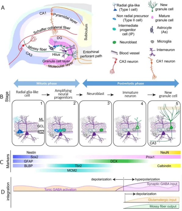
The DG of the hippocampus is able to generate functional newborn neurons that arise from NSCs located in the SGZ (87). The exact function of these new cells has yet to be fully clarified, but it is believed that are particularly important for dentate-dependent memory, learning and emotional processes (87, 88). The radial glia-like cells represent the primary precursor to new neurons (79). In addition to neurons, the NSCs in the DG can also give rise to further stem cells and non-stem astrocytes, therefore having self-renewal and multipotency properties (88). The developmental stages occurring during adult hippocampal neurogenesis are highlighted in figure 4B. During the maturation process, new granule neurons project their dendritic arbor into the adjacent molecular layer (ML) and send their axons to the target cells present in the hilus and in the CA3 area (89). Importantly, for synaptic integration into the pre-existing neuronal network, the new neurons need to be previously activated by GABAergic synaptic inputs from local interneurons and, lastly, by glutamatergic synaptic inputs (79) (figure 4D). Of note, during neurogenesis, the newborn cells have to pass through critical developmental phases where they are more vulnerable to apoptosis, and the majority of these newborn granule neurons die before being integrated into the neuronal network (90). Several cells, including astrocytes, microglia, endothelial cells and mature neurons contribute for the maintenance of the DG neurogenic niche. For example, endothelial cells seem to regulate adult neural precursor proliferation (91). Furthermore, astrocytes express membrane factors and secret molecules that modulate not only proliferation and fate specification of adult neural precursors but also regulate migration, maturation and synapse formation and integration of newly neurons (36, 92, 93). Additionally, these cells, which are closely associated with the vasculature and its basal lamina in the adult DG, can modulate the accessibility of mediators (e.g. cytokines and growth factors) to the basal lamina as well as the effects of the endothelial-released factors and circulation-derived molecules (94, 95). Microglial cells are also active regulators of the adult DG neurogenesis. Under physiological conditions, these cells quickly phagocytose apoptotic bodies of newborn neurons, therefore maintaining the homeostasis of the neurogenic niche (96). Additionally, resting microglia can
9
stimulate stem cell proliferation and migration through the secretion of anti-inflammatory mediators and neurotrophic factors, such as brain-derived neurotrophic factor (BDNF) and insulin growth factor-1 (IGF-1) (97), as well as modulate synaptic integration of newborn neurons (16).
Notably, neurogenesis is not a static process, instead it is a phenomenon that confers adaptive advantages to the DG, since it can be modulated by a variety of extrinsic environmental signals (89). These cues comprise exercise/physical activity, new stimuli given in an enriched environment, and hippocampus-dependent learning, which seems to modulate the formation, survival, maturation, and integration of newborn DG cells (87, 89). In addition to the positive influence of the supporting factors referred above, adult neurogenesis can also be modulated by repressive factors, such as stress, aging and inflammation (89). Importantly, impaired hippocampal neurogenesis has been associated with the cognitive decline frequently observed in a large number of neurological conditions (e.g. aging, depression, AD and other neurodegenerative diseases) (98-100), most of which are associated with neuroinflammation.
Figure 4 - Overview of hippocampal adult neurogenesis. A) Coronal section of the hippocampus highlighting the neurogenic niche found in the DG. B) Schematization of the developmental stages during adult hippocampal neurogenesis in the dentate gyrus (DG): 1) activation of quiescent radial glia-like cell in the subgranular zone (SGZ); 2) proliferation of amplifying neural progenitors; 3) generation of neuroblasts; 4) integration of immature neurons; 5) maturation of adult-born dentate granule cells. C) Expression of the specific markers of each cellular development stage. D) Schematization of the sequential process of synaptic integration. Abbreviations: BLBP, brain lipid-binding protein; DCX, doublecortin; GCL, granule cell layer; GFAP, glial fibrillary acidic protein; ML, molecular layer; NeuN, Neuronal Nuclei; Prox1, prospero homeobox protein 1; SGZ, subgranular zone; Sox2, sex-determining regionY-box 2; Tbr2, T-box brain protein 2. Adapted from (79).
1.3. Histamine: a brief overview
Histamine (4-imidazolyl-2-ethylamine) is an endogenous biogenic amine present in several mammalian organs, including in the brain (101). In the peripheral system, histamine is mainly secreted and stored by mast cells, basophils, monocytes/macrophages, dendritic cells,
11
enterochromaffin-like cells, gastrin-containing cells, neutrophils and platelets (102). This amine also plays essential functions in both peripheral and central nervous systems. In addition to immunomodulation, it acts as a neurotransmitter and controls several functions, such as energy and endocrine homeostasis, sleep–waking cycle, appetite, behavior and motor and cognitive performance (103-106). In the CNS, histamine is released by neurons, microglia as well as mast cells located in the meninges, and circumventricular organs (104, 107). Histamine can trigger the activation of four different G protein-coupled receptors (GPCRs): H1R, H2R, H3R and H4R, which can activate distinct signaling pathways (figure 5). H1R, H2R and H3R are highly expressed in the CNS, while H4R are expressed mainly in peripheral tissues. In the CNS, they are expressed with distinct density and patterns in endothelial cells, neurons, astrocytes and microglia cells (105, 108).
H1Rs are mainly expressed in regions responsible for the modulation of behavioral, nutritional and neuroendocrine states (103, 106). H1R signaling has an excitatory action on neurons in most brain regions (e.g. hypothalamus, thalamus, hippocampus, olfactory bulb, cortex, amygdala and septum), except on hippocampal pyramidal neurons where the activation of potassium channels leads to a decrease in cell excitability (103). H2Rs are predominantly expressed in the hippocampus, cortex, basal ganglia and amygdala, where they mediate several postsynaptic actions (103). Similar to H1Rs, their action is usually excitatory (109). Specifically, H2Rs actions seem to be particularly relevant for cognitive performance, since they modulate neuronal plasticity and synaptic transmission in the hippocampus (110). Of note, H1Rs can have opposite or synergistic effects with H2Rs depending on the timing and context of receptor activation (103). H3Rs are the most prominent HRs in the CNS and are located on the somata, dendrites and axonal varicosities of histaminergic and other neurons (110). They are predominantly found in the tuberomamillary nucleus (TMN), cerebral cortex (anterior parts), hippocampus, SN, striatum, amygdala, nucleus accumbens, olfactory tubercles, cerebellum and brain stem. H3Rs play pivotal roles in the modulation of brain functions such as axonal and synaptic plasticity. H3Rs act as autoreceptors, inhibiting cell firing as well as synthesis and release of histamine. Acting as presynaptic heteroreceptors, H3Rs also modulate the release of several other neurotransmitters (e.g. acetylcholine, GABA and glutamate) (103). H4Rs, the last identified HR, are predominantly expressed on peripheral immune cells and microglia and are mainly involved in the modulation of immune responses under inflammatory context (110, 111). In fact, we have previously descrived its dual role in neuroinflammation (108); these effects are further detailed in the next section.
Figure 5 – HRs signaling pathways. A) H1R signaling pathway. B) H2R signaling pathway. C) H3R signaling pathway. D) H4R signaling pathway. Abbreviations: AC, adenylyl cyclase; cAMP, 3’-5’-cyclic adenosine monophosphate; cGMP, cyclic guanosine monophosphate; Ch, channel; DAG, diacylglycerol; CREB, cyclic-AMP-response element binding protein; ER, endoplasmic reticulum; IAHP, small conductance, Ca2+
-dependent K+ current; I
h, hyperpolarization-activated cationic channel; IK, intermediate-conductance
calcium-activated potassium channels; IP3 or Ins(1,4,5)P3, inositol (1,4,5)-trisphosphate; Kv3,
voltage-gated potassium channels; NMDA, N-methyl-D-aspartate; NO, nitric oxide; GC, guanylyl cyclase; NCX, Na+–Ca2+ exchanger; PKA, protein kinase A; PKC, protein kinase C; PLA, phospholipase A; PLC,
phospholipase C; PLC-β, phospholipase Cβ; PtdIns(4,5)P2, phospholipid phosphatidylinositol
(4,5)-bisphosphate; VACCs, voltage-activated Ca2+ channels. Adapted from (111, 112).
In the adult vertebrate brain, histaminergic neurons are restrained to the TMN of the posterior hypothalamus from where they send their projections to basically all areas of the CNS (103) (figure 6). The rate of histamine production in the CNS is defined by the bioavailability of its precursor, l-histidine, which is taken up into the cerebrospinal fluid and neurons through L-aminoacid transporters and is converted to histamine through the enzyme l-histidine decarboxylase. Then, histamine is kept in synaptic vesicles by the vesicular monoamine-transporter (VMAT)-2, being secreted by exocytosis. After release, histamine is kept inactivated in the extracellular space through its methylation into tele-methylhistamine by the enzyme histamine N-methyltransferase that is located postsynaptically and in glial cells. Notably, the turnover rate for histamine is relatively high (approximately 30 minutes), but can vary depending on neuronal activity (104, 105, 109)
.
13 Figure 6 - The histaminergic system in the human brain: origin (green) and projections (red). Histaminergic neurons are located in the tuberomamillary nucleus of the human brain and innervate the major regions of the cerebrum, cerebellum, posterior piuitary and the spinal cord. Adapted from (109).
In the cerebrospinal fluid and parenchyma of the healthy brain, histamine is present at very low concentrations (113). Notably, alterations in histamine levels and density of its receptors have been observed in aging and several neurological diseases (e.g. AD, PD, and schizophrenia), some of which are accompanied by neuroinflammation (101, 104, 105). These data suggest that a dysfunctional histaminergic system could contribute to the pathogenesis of these diseases, highlighting histamine as a potential target to develop novel therapeutic approaches.
1.3.1 Role of histamine in neuroinflammation
Histamine has been suggested as a mediator of neuroinflammation mainly through its ability to regulate microglial cell activity (figure 7). In fact, all four types of HRs are expressed by microglia (108) and a subpopulation of microglial cells particularly sensitive to this amine was identified (114). The effects of histamine in microglial function comprise: increased cell motility through H4R activation by a mechanism that involves α5β1 integrins, p-38 and Akt signaling pathways (108), induction of inducible nitric oxide synthase (iNOS) production (115), induction of ROS production by H1R and H4R activation, through a mechanism involving the Nox1 signaling pathway (116), induction of pro-inflammatory cytokines release (e.g. TNF-α, IL-6) through H1R and H4R activation, loss of mitochondrial membrane potential (117, 118), as well as induction of microglial phagocytosis by H1R activation (116). Moreover, histamine-induced microglial activation ultimately compromises dopaminergic neuronal survival in rodents both in vitro (115) and in vivo (116). Thus, under a physiological context, histamine challenge seems to induce microglia into a pro-inflammatory phenotype that leads to harmful consequences to neuronal function/survival. However, this amine could also inhibit
LPS-induced microglial cytotoxicity. For example, in vitro histamine could counteract LPS-LPS-induced microglial migration through H4R activation, as well as LPS-induced IL-1β (108) and prostaglandin E2 secretion (119). Moreover, histamine significantly inhibited microglial phagocytosis and ROS production induced by LPS in vitro (unpublished data, submitted). Notably, histamine was able to significantly prevent the decrease of dopaminergic neurons induced by LPS both in vitro and in vivo (unpublished data, submitted). Overall, these data suggest that histamine has a dual role in the modulation of microglial responses and neuronal survival. Interestingly, the anti-inflammatory effects of histamine under LPS challenge have also been observed at the peripheral system mainly through H2R signaling (120-122). Moreover, the dual role of histamine has been reported in neuronal injuries accompanied by microglia-induced neuroinflammation. Specifically, histamine has been shown to aggravate MS pathophysiology by potentiating neuroinflammation through H1R activation. However, there are also evidences demonstrating the protective role of histamine in this condition particularly through H2R activation (reviewed in (123)). Furthermore, post-ischemic administration of L-histidine significantly prevented ischemia-induced injury (124), which was accompanied by an inhibition in microglia activation through H2R activation (125). Moreover, L-histidine treatment also promoted astrocytic migration into the infarct core through H2R signaling, which led to long-term neurological recovery (126). Thus, the effect of histamine under neuroinflammation seems to be dependent on the environment context and which receptor is activated. Overall, these data open a new perspective for the therapeutic use of histamine in neuronal conditions associated with neuroinflammation.
Figure 7 - Effects induced by histamine on microglial functions, under a physiological context and lipopolysaccharide (LPS) challenge, which ultimately affects dopaminergic neuronal survival in the
substantia nigra (SN). In a physiological state, histamine enhances microglia cell motility, phagocytosis
activity and NADPH oxidase (Nox) activation with subsequent increase of reactive oxygen species (ROS) production. Consequently, these microglial actions remarkably compromise dopaminergic neuronal survival in the SN. Notably, when histamine is administrated under LPS challenge, it inhibits microglial inflammatory action induced by this inflammogen insult and subsequently prevents dopaminergic neurodegeneration. Adapted from (unpublished data, submitted).
15
1.3.2. Role of histamine in neurogenesis
There are strong evidences that histamine plays an important role in neurogenesis during development, regulating processes such as neuronal differentiation and migration, neurite elongation and synaptogenesis. Furthermore, the neurogenic peak matches the highest level of histamine in the developing brain, suggesting this amine as a key player in this process (112). Additionally, in vitro studies have shown that histamine induces proliferation and differentiation of neural progenitors through H2R and H1R signaling, respectively (127-129). Specifically, histamine induces neuronal differentiation in early postnatal SVZ precursor cells from mouse through H1R by triggering histone H3 trimethylation on lysine K4 on the promoter regions of the proneurogenic genes (129). Notably, Bernardino and colleagues also demonstrated that pre-treatment with histamine-loaded microparticles facilitated neuronal differentiation of SVZ precursor cells grafted in hippocampal slices and in in vivo mouse brain in the neurogenic (hippocampal DG) and non-neurogenic (striatum) niches (129). Interestingly, a study showed that H1R deficiency in mouse caused a reduced number of proliferative cells in the hippocampal DG, which was accompanied by pronounced deficits in spatial learning and memory, suggesting that histamine signaling through H1R could be required for adult neurogenesis, probably by modulating survival and/or proliferation in this neurogenic niche (130). Generally, these data highlight histamine as a key soluble factor released in the neurogenic niches that favors neuron commitment.
Interestingly, histamine is present at lower concentrations in the brain under physiologic conditions, but its levels are increased in the cerebrospinal fluid and brain parenchyma following brain injury mainly due to mast cell degranulation, consequently increasing BBB permeability (131). Thus, it is crucial to study the effects of increased levels of histamine on the brain, namely on the neurogenesis process. In this sense, Eiriz and collaborators showed that the intraventricular infusion of histamine in the lateral ventricles induced a significant increase in the number of total (doublecortin - DCX+ cells) and proliferative neuroblasts (Bromodeoxyuridine - BrdU+/DCX+ cells) in the SVZ, which were able to migrate towards the olfactory bulb where they differentiate into mature neurons. Interestingly, histamine infusion did not alter the number of BrdU+DCX− cells in both SVZ and olfactory bulb regions, suggesting that histamine preferentially triggers neuronal commitment and/or induces neuroblast proliferation, instead of inducing an overall increase in cell proliferation (131). Overall, these studies demonstrate that histamine can greatly modulate NSCs dynamics and could be a promising target for brain regenerative therapies.
17
Chapter 2 – Aims
Histamine seems to have a dual role in the CNS, playing cytotoxic or anti-inflammatory effects, depending on the microenvironment and on which histamine receptor is activated. Above all, there is a lack of information regarding the effects of increased histamine levels in the hippocampus, a brain region that play key roles in behavior and cognitive performance and that is compromised under neuroinflammatory conditions. In this sense, we aim to evaluate the effects of histamine, per se or under an inflammatory context mimicked by LPS, on:
hippocampal neuroinflammation, by assessing protein expression of inflammatory mediators as well as neuronal and synaptic function markers;
hippocampal neurogenesis, by evaluating newborn cell proliferation, differentiation and long-term survival.
19
Chapter 3 - Materials and Methods
3.1. Animals
All experiments related to the use of experimental animal models were conducted in agreement with protocols approved by the national ethical requirements for animal research and the European Convention for the Protection of Vertebrate Animals Used for Experimental and Other Scientific Purposes (European Union Directive number 192 2010/63/EU). In this study, a total of 41 adult (2 to 5 months-old) male C57BL/6J mice were used. All animals were maintained in appropriate and similar cages in the same room, under temperature (22 ºC) and light (12 hours light/dark cycle) controlled environment with open access to food and water. All efforts were made to minimize the suffering and the number of animals used.
3.2. Intraperitoneal and stereotaxic injections
As shown in figure 8, mice were initially subjected to an intraperitoneal injection (i.p.) of LPS (from Escherichia coli 055:B5, Sigma-Aldrich), at 1 mg/Kg (62) and 2 mg/Kg (57), diluted in 0.1 M sterile phosphate buffered-saline (PBS) at pH 7.4. Mice intraperitoneally injected with 0.1 M of sterile PBS were considered the control condition. Two days after LPS administration, the mice were anesthetized with a mixture of ketamine and xylazine (90 mg/kg and 10 mg/kg of mouse weight, respectively) before proceeding to intracerebral histamine administration. Then, animals were positioned in the digital stereotaxic frame (51900 Stoelting, Dublin, Ireland) and their scalp was disinfected with Betadine. An incision was made, using a scalpel, along the midline to expose the mouse skull and define the coordinates after setting the zero at the bregma point. An intracerebral injection of 2 µL of sterile histamine dihydrochloride (100 μM in PBS, Sigma) was performed in the DG of the hippocampus (anteroposterior: -1.9 mm, mediolateral: -1.2 mm, and dorsoventral: -1.8 mm from bregma (129)) using a Hamilton syringe at a speed of 0.2 µL/min for 10 minutes. After intracerebral injection, the incision was sutured and mice were kept warm (37 ºC) until they recovered from surgery. To unveil the effects of histamine in hippocampal neuroinflammation, a set of animals were euthanized 2 days after histamine stereotaxic injection and brains were removed for further immunoblotting assays. To evaluate the effects of histamine in neuroblast proliferation in the DG, another set of animals was also injected with BrdU (BrdU; 100 mg/kg of animal weight, Sigma) dissolved in a sterile saline solution (0.9% NaCl) to label dividing cells. BrdU administration was performed through an i.p. injection in the following 2 days (every 12 hours) after the histamine stereotaxic injection. Animals were maintained for 3 days after histamine treatment before being euthanized for further immunohistochemistry analysis (immunostaining against BrdU and DCX). Lastly, to
uncover the effects of histamine in the survival of newborn neurons in the DG, another group of animals was also intraperitoneally injected with BrdU (50 mg/kg of animal weight in 0.9% NaCl) during the first 3 days after the histamine stereotaxic injection, twice a day (every 12 hours). Six weeks after this experimental procedure, mice were euthanized for further immunohistochemistry analysis (immunostaining against BrdU and Neuronal Nuclei, NeuN). Animal weight was controlled from the day of LPS injection till recovery. Animals showed no significant weight changes during all experiments.
Six experimental conditions were tested: i) contralateral hemisphere of mice intraperitoneally injected with PBS (control condition - Ctr), ii) ipsilateral hemisphere of mice intraperitoneally injected with PBS and stereotactically injected with 100 μM histamine (His), iii) contralateral hemisphere of mice intraperitoneally injected with 1 mg/Kg LPS (LPS1), iv) ipsilateral hemisphere of mice intraperitoneally injected with 1 mg/Kg LPS and stereotactically injected with 100 μM histamine (LPS1 + His), v) contralateral hemisphere of mice intraperitoneally injected with 2 mg/Kg LPS (LPS2), and vi) ipsilateral hemisphere of mice intraperitoneally injected with 2 mg/Kg LPS and stereotactically injected with 100 μM histamine (LPS2 + His) (figure 9).
Figure 8 - Schematic representation of the in vivo experimental assays. Abbreviations: BrdU, bromodeoxyuridine; CREB, cAMP response element binding; DCX, Doublecortin; GFAP, glial fibrillary acidic protein; HMGB1, high mobility group box 1; Iba-1, ionized calcium binding adaptor molecule 1; IL-1β, interleukin-1 beta; LPS, lipopolysaccharide; NeuN, Neuronal Nuclei; PSD-95, postsynaptic density protein 95.
21 Figure 9 – Representative scheme of the experimental conditions in vivo. Abbreviations: CL, contralateral cerebral hemisphere; Ctr, control; His, histamine; IPSI, ipsilateral cerebral hemisphere; LPS, lipopolysaccharide.
3.3. Western Blotting
3.3.1. Preparation of the brain tissue extracts
To unveil the effect of histamine on hippocampal neuroinflammation, mice were euthanized 2 days after the histamine stereotaxic injection, brains were removed, frozen in liquid nitrogen, and stored at -80 °C. Hippocampal tissues were mechanically dissociated and lysed on ice in RIPA buffer (0.15 M NaCl, 0.05 M Tris, 5 mM ethylene glycol tetraacetic acid, 1% Triton X-100, 0.5% deoxycholic acid, 0.1% sodium dodecyl sulphate, 10 mM dichlorodiphenyltrichloroethane, containing a cocktail of proteinase inhibitors). The soluble fraction was obtained (centrifugation at 12000 rpm, 20 minutes, 4 °C) and, after vortex homogenization, the total protein concentration from the lysates was determined using the bicinchoninic acid assay (Thermo Scientific, MA, USA). Protein samples were treated with SDS-PAGE buffer (6x concentrated: 350 mM Tris, 10% (w/v) SDS, 30% (v/v) glycerol, 0.6 M DTT, 0.06% (w/v) bromophenol blue) boiled for 5 minutes at 95 °C.
3.3.2. Immunoblot assay
First, equal amounts of protein lysate (40-80 μg of total protein) were loaded into each lane of an 8-12% bisacrylamide gel (Applichem, Darmstadt, Germany). Proteins were separated by SDS-PAGE electrophoresis in the following conditions: 90-100 V, 90-120 minutes, in a Tris-glycine running buffer solution (1x concentrated: 25 mM Tris, 190 mM Tris-glycine pH 8.3, 0.1% SDS) at room temperature (RT). Then, proteins were transferred to a polyvinylidene difluoride (PVDF) membrane (GE Healthcare, Little Chalfont, UK) through semi-dry transfer in the following conditions: 1.0 A, 25 V, 15-30 minutes, using Towbin transfer buffer (1x concentrated: 25 mM Tris, 192 mM glycine pH 8.3, 20% methanol) at RT. To block non-specific
binding, the membranes were incubated with a tris-buffer saline (TBS) containing 0.1% Tween 20 (Thermo Fisher Scientific, Waltham, MA, USA), and 5% low-fat milk or 5% BSA (Amresco LLC, Solon, USA) or 0.1% gelatin (Fluka, St Louis, MO, USA), depending on the antibody used, for 20 minutes at RT. Membranes were then incubated overnight at 4 °C with appropriate primary antibodies (Table 1) and, after washing three times with TBS-T, they were further incubated with the respective secondary antibodies conjugated with horseradish peroxidase at RT for 2 hours (Table 2). To normalize the expression of the target proteins, the membranes were further incubated with a housekeeping antibody (1.5 hours) and the respective secondary antibody (1 hour), both at RT. Protein levels were detected by enhanced chemiluminescence and densitometric analyses, using the software ImageLab (Bio-Rad, Hercules, CA, USA).
Table 1 - Primary antibodies used for Wester Blotting (CREB, cAMP response element binding; GAPDH, glyceraldehyde 3-phosphate dehydrogenase; GFAP, glial fibrillary acidic protein; HMGB1, high mobility group box 1; Iba-1, ionized calcium binding adaptor molecule 1; IL-1β, interleukin-1 beta; LPS, lipopolysaccharide; PSD-95, postsynaptic density protein 95).
Table 2 - Secondary antibodies used for Wester Blotting.
Primary antibody Dilution Band molecular weight (kDa) Company
Mouse anti-Iba-1 1:200 17 Santa Cruz Biotechonology
Mouse anti-GFAP 1:5000 50 Santa Cruz Biotechonology
Rabbit anti-IL-1β 1:200 17 HMGBiotech
Mouse anti-HMGB1 1:500 29 Cell Signaling
Rabbit anti-CREB 1:1000 43 Cell Signaling
Mouse anti-Syntaxin 1:5000 35 Sigma
Mouse anti-PSD-95 1:1000 100 Millipore
Mouse anti-Actin (housekeeping) 1:1000 42 BD Mouse anti-GAPDH (housekeeping) 1:5000 37 Millipore Mouse anti-Tubulin (housekeeping) 1:5000 50 Sigma
Secondary antibody Dilution Company
Goat anti-Mouse 1:5000 Santa Cruz Biotechonology
23
3.4. Immunohistochemistry
3.4.1. Preparation of the brain tissue
To unveil the effects of histamine on hippocampal newborn cells proliferation and survival, 3 days and 6 weeks after the histamine injection respectively, the mice were anesthetized with a mixture of ketamine and xylazine (90 mg/kg and 10 mg/kg of mouse weight, respectively), and then perfused intracardially with NaCl 0.9%, followed by 4% paraformaldehyde (PFA, Sigma). The brains were removed and post-fixed in 4% PFA overnight at 4ºC, followed by immersion in a 30% sucrose solution (Fisher Scientific) in 0.1 M PBS at 4ºC to cryoprotect tissues. After sinking, brains were frozen in liquid nitrogen and maintained at -80˚C until sectioning. Thereafter, the brains were embedded in optimal cutting temperature solution and were cut in coronal sections (40 μm) using a cryostat-microtome (Leica CM3050S, Leica Microsystems, Nussloch, Germany) at -20 ºC. The slices (spaced 240 µm from each other) corresponding to the hippocampus of each animal were collected sequentially in six wells of 24-well plates, and were left freefloating in a cryopreservation solution (30% glycerol, 30% ethylene glycol and 10% phosphate buffer (0.2 M)) at -20°C until immunostaining assay.
3.4.2. Immunostaining assay
The immunostaining assays were performed using an adapted protocol described in (132). First, tissue sections were rinsed three times in PBS for 5 minutes to remove the cryopreservation solution. Then, brain sections were incubated with 2 M HCl for 25 minutes at 37 ºC to induce DNA denaturation. After washing with PBS, tissue sections were further incubated in a blocking solution containing 2% of horse serum (Life Technologies) and 0.3% Triton X-100 (Fisher Scientific, Pittsburgh, PA, USA) diluted in 0.1 M PBS for 2 hours at RT. After the blocking procedure, tissue sections were incubated for 72 hours at 4ºC in the following primary antibodies (diluted in the blocking solution): rat monoclonal anti-BrdU (1:500, AbD Serotec, Raleigh, NC, USA), goat polyclonal anti-DCX (1:500, Santa Cruz Biotechnology, Inc.), or mouse monoclonal anti-NeuN (1:500, Merck Millipore). After primary antibody incubation, sections were rinsed in PBS and then incubated with Hoechst (1:1000; Sigma) and the respective secondary antibodies: Alexa Fluor-488 donkey anti-rat (1:500; Life Technologies), Alexa Fluor-546 donkey anti-goat or anti-mouse (1:500; Life Technologies), diluated in a solution containing 0.3% Triton X-100 in 0.1 M PBS, for 2 hours at RT. Finally, sections were rinsed in PBS and mounted in Fluoroshield Mounting Medium (Abcam Plc.) for further cell counting.
3.5. Cell counting, area and volume analysis
3.5.1. Neuroblast proliferation analysis
To assess neuroblast proliferation, fluorescence immunostaining z-stack projections of the DG were acquired in serial sections at 480 µm rostrocaudal intervals along the entire hippocampus (from bregma -3.88 mm to bregma -0.94 mm (133)) using an AxioObserver LSM 710 confocal microscope (Carl Zeiss) under a 40× oil immersion objective. BrdU+ and BrdU+/DCX+ cells were counted in these serial sections using ImageJ software (NIH Image, Bethesda, MD, USA). Total number of BrdU+ and BrdU+/DCX+ cells was estimated using the Abercrombie formula: T = (N × V)/(t +D), where T is the total number of cells, N is cell density, V is the total volume of the considered area, t is slice thickness (40 µm) and D is average cellular diameter (cell diameters from 6 random cells per experimental condition) (134). The quantification of the area and the volume is explained in the section 3.5.3.
3.5.2. Survival of newborn neurons analysis
To assess survival of newborn neurons, BrdU+ and BrdU+/NeuN+ cells were counted in serial sections at 240 µm rostrocaudal intervals along the entire hippocampus, using an AxioObserver LSM 710 confocal microscope under a 63× oil immersion objective. Total number of BrdU+ and BrdU+/NeuN+ cells was estimated by applying the Abercrombie formula, as described in the previous section.
3.5.3. Area and volume quantification
To estimate areas and volumes, images of the DG were taken in serial sections at 240 or 480 μm rostrocaudal intervals along the entire hippocampus. The images were obtained using an AxioObserver LSM 710 confocal microscope under a 10x objective. As schematized in figure 10A, the areas were estimated delineating a line around DG using the FIGI software (NIH, Bethesda, MD, USA). The volume was estimated through the equation: V (µm3) = ∑n
i=1 Ai x d (134), where A is the area of each section and d corresponds to the interval between slices (240 or 480 μm) (figure 10B).
25 Figure 10 – Schematic figure of the quantification of DG area and volume. A) Each DG slice was delineated as schematized in A, and its area was estimated by FIGI software. B) DG volume was calculated as the total sum of the product of the area of each DG slice by the distance between two consecutives slices, which corresponds to 240 µm (quantification of newborn neuronal survival), or to 480 µm (quantification of proliferating neuroblasts).
3.6. Data analysis
Data are shown as the mean ± standard error of the mean (SEM), expressed as percentages of values obtained in control condition. Statistical analysis was performed using one-way ANOVA, followed by Bonferroni’s Multiple Comparison Test. Values of P<0.05 were considered significant. *P<0.05, **P<0.01, ***P<0.001 and ****P<0.0001 when compared to control condition; #P<0.05, ##P<0.01, ###P<0.001 and ####P<0.0001 when compared to LPS-treated condition. All statistical analysis was achieved using GraphPad Prism 5 Demo (GraphPad Sotware, San Diego, CA, USA).
27
Chapter 4 – Results
4.1. Effect of histamine on mouse hippocampal neuroinflammation
4.1.1. Effect of histamine on glial reactivity
Neuroinflammation is mainly mediated by glial cells and their reactivity is enhanced after an injury or infection (2). First, the expression of activated microglia (Iba-1) and astrocytes (GFAP) markers was assessed in the hippocampus of mice challenged with LPS and/or histamine, by western blot (see methodology in figure 8). Histamine administration per se significantly increased Iba-1 (meanHIS=186.7±20.3, n=7; figure 11A) and GFAP expression (meanHIS= 161.7±8.8, n=6; figure 11B) when compared to control condition. Then, LPS was used as a classic stimulus to trigger TLR4-mediated neuroinflammation. As expected, LPS administration significantly enhanced Iba-1 (meanLPS1=192.7±18.3, n=4; meanLPS2=204.4±39.6, n=7; figure 11A) and GFAP (meanLPS1=150.6±21.9, n=4; meanLPS2=176.0±15.6, n=6; figure 11B) expression when compared with control condition. Then, to disclose the modulatory role of histamine in LPS-induced neuroinflammation, a group of mice were treated with LPS for 2 days and then challenged with histamine for two further days (see methodology in figure 8). Interestingly, histamine was only able to counteract LPS-induced glial reactivity when the higher dose of LPS (2 mg/kg) was used (meanHIS+LPS2=85.6±22.1, n=7; figure 11A (Iba-1); meanHIS+LPS2=130.8±8.9, n=7; figure 11B (GFAP)).