Inhibition of brain citrate synthase activity in an
animal model of sepsis
Inibição da atividade da citrato sintase cerebral em um modelo
animal de sepse
INTRODUCTION
Sepsis and its related complications, such as multiple organ dysfunction syndrome (MODS), are the most frequent causes of morbidity and mortality in intensive care units, contributing to 750,000 cases per year in the United States alone with an average mortality of 29%.(1) Sepsis is a complex
syndrome deined as the host’s reaction to infection characterized by systemic inlammation(2) and an imbalance between pro- and anti-inlammatory
responses to pathogens.(3)
Evidence from the literature has demonstrated that reactive oxygen species (ROS) play an important role in the development of multiple organ
Giselli Scaini1,3, Natália Rochi1,3,
Joana Benedet1,3, Gabriela
Kozuchovski Ferreira1,3, Brena
Pereira Teodorak1,3, Clarissa
Martinelli Comim2,3, Larissa
de Souza Constantino1,3,
Francieli Vuolo1,3, Leandra Celso
Constantino2,3, João Quevedo2,3,
Emilio Luiz Streck1,3, Felipe
Dal-Pizzol1,3
1. Experimental Pathophysiology Laboratory, Health Sciences Post-Graduation Program, Universidade do Extremo Sul Catarinense – Criciúma (SC), Brazil.
2. Neurosciences Laboratory, Health Sciences Post-Graduation Program, Universidade do Extremo Sul Catarinense – Criciúma (SC), Brazil. 3. Instituto Nacional de Ciência e Tecnologia Translacional em Medicina – Porto Alegre (RS), Brazil.
ABSTRACT
Objective: An extensive body of evidence from experimental studies indicates that sepsis is associated with increased reactive oxygen species production, depletion of antioxidants, and accumulation of markers of oxidative stress. Moreover, mitochondrial dysfunction has been implicated in the pathogenesis of multiple organ dysfunction syndrome (MODS). Citrate synthase is an enzyme localized in the mitochondrial matrix and an important component of the Krebs cycle; consequently, citrate synthase has been used as a quantitative enzyme marker for the presence of intact mitochondria. hus, we investigated citrate synthase activity in the brains of rats submitted to a cecal ligation puncture model of sepsis.
Methods: At several times points (3, 6, 12, 24 and 48 hours) after the cecal ligation puncture operation, six rats were killed by decapitation. heir brains were removed, and the hippocampus, striatum, cerebellum, cerebral cortex and prefrontal cortex were dissected and used
to determine citrate synthase activity.
Results: We found that citrate synthase activity in the prefrontal cortex was inhibited 12, 24 and 48 hours after cecal ligation puncture. In the cerebral cortex, citrate synthase activity was inhibited 3, 12, 24 and 48 hours after cecal ligation puncture. Citrate synthase was not afected in the hippocampus, striatum or cerebellum up to 48 hours after cecal ligation puncture.
Conclusion: Considering that energy impairment due to mitochondrial dysfunction in sepsis has been well described and that oxidative stress plays a crucial role in sepsis development, we believe that energy impairment may also be involved in these processes. If citrate synthase inhibition also occurs in a sepsis model, it is tempting to speculate that a reduction in brain metabolism may be related to the pathophysiology of this disease.
Keywords: Citrate (si)-synthase/ metabolism; Sepsis; Mitochondria; Brain; Rats, Wistar; Models, animal
his study was conducted at Experimental Pathophysiology Laboratory – Universidade do Extremo Sul Catarinense – Criciúma (SC), Brazil.
Conlicts of interest: None.
Submitted on March 4, 2011 Accepted on March 28, 2011
Corresponding author: Felipe Dal-Pizzol
Laboratório de Fisiopatologia Experimental
Universidade do Extremo Sul Catarinense
Zip Code: 88806-000 – Criciúma (SC), Brazil.
failure and septic shock.(4-8) Treatments that reduce the
generation or that prevent or reverse the efects of ROS have shown beneicial efects in a variety of models of endotoxic and septic shock.(9-15)
Mitochondrial dysfunction has been strongly implicated in the pathogenesis of multiple organ dysfunction syndrome (MODS) and a wide variety of disease states.(3,16-18)
he hypothesis of cytopathic hypoxia postulates that impairment in mitochondrial oxidative phosphorylation reduces aerobic adenosine triphosphate (ATP) production and potentially induces MODS.(19) In this context, some
studies have reported deiciencies within the electron transport chain in models of sepsis.(19-22)
Citrate synthase (EC 4.1.3.7) is localized in the mitochondrial matrix and catalyzes the condensation of oxaloacetate and the acetyl group of acetyl coenzyme-A (acetyl CoA), the irst step of the Krebs cycle. In this step, oxaloacetate reacts with acetyl CoA and H2O to yield citrate and CoA. Citrate synthase is inhibited by high levels of ATP, acetyl-CoA and NADH, which are present when the cell energy supply is high. his regulation ensures that the Krebs cycle does not oxidize an excess of pyruvate and acetyl CoA when cellular ATP concentrations are high.(23) In addition, citrate synthase
has been used as a quantitative enzyme marker for the presence of intact mitochondria.(24)
Considering that citrate synthase plays an important role in brain energy metabolism and that mitochondrial dysfunction has been implicated in the pathogenesis of MODS, in the present study, we investigated whether sepsis induced by cecal ligation and puncture (CLP) modiies citrate synthase activity in the rat brain.
METHODS
Animals
Male adult Wistar rats (60 days old) were obtained from the Universidade do Extremo Sul Catarinense (UNESC) breeding colony. he animals were housed ive per cage with food and water available ad libitum and were maintained on a 12 h light/dark cycle (lights on at 7:00 AM). All experimental procedures involving animals were performed in accordance with the National Institutes of Health Guide for the Care and Use of Laboratory Animals and the Brazilian Society for Neuroscience and Behavior (SBNeC) recommendations for animal care.
Cecal ligation and perforation surgery
he animals were subjected to CLP as previously
described by Ritter et al.(25) Briely, rats were anesthetized
with a mixture of ketamine (80 mg/kg) and xylazine (10 mg/kg) given intraperitoneally. Under aseptic conditions, a 3 cm midline laparotomy was performed to expose the cecum and the adjoining intestine. he cecum was tightly ligated with a 3.0-silk suture at its base below the ileocecal valve and was perforated once with a 14-gauge needle. he cecum was then gently squeezed to extrude a small amount of feces from the perforation site and then returned to the peritoneal cavity. he laparotomy was then closed with 4.0-silk sutures. All animals received isotonic saline solution (50 mL/kg s.c.) immediately after the procedure. All animals were returned to their cages with free access to food and water. In the sham-operated group, the rats were submitted to all surgical procedures and received isotonic saline solution (50 mL/ kg s.c.) immediately afterwards.
he rats were randomly allocated into the sham and CLP groups just prior to the procedure. At several time points (3, 6, 12, 24 and 48 hours) after the CLP or sham operation, six rats were killed by decapitation, and brain structures (prefrontal cortex, hippocampus, striatum, cerebellum and cerebral cortex) were immediately isolated and stored at -80ºC. All animals showed signs of encephalopathy at 6 h after sepsis (lethargy, mild ataxia, lack of spontaneous movement, and loss of righting relex) and gradually returned to their normal waking status 24–36 h after CLP.(26)
To minimize the possibility of animals not developing sepsis, the CLP procedure was always performed by the same investigators. In addition, all animals were observed after CLP to determine signs of infection (pyloerection, lethargy, tachypnoea and weight loss), and the number of animals that survived is in accordance with our previous reports.(25)
Tissue and homogenate preparation
Twelve hours after the last injection, the rats were killed by decapitation, the brain was removed, and the prefrontal cortex, hippocampus, striatum, cerebellum and cerebral cortex were homogenized (1:10, w/v) in SETH bufer, pH 7.4 (250 mM sucrose, 2 mM EDTA, 10 mM Trizma base, 50 IU/ml heparin). he homogenates were centrifuged at 800 × g for 10 min, and the supernatants were kept at −70°C until used for enzymatic activity determination. he maximal period between homogenate preparation and enzyme analysis was always less than 5 days. Protein content was determined by the method described by Lowry et al.(27)
Results are expressed as the mean ± S.D. (n=6). Diferent from control group, *p<0.05 (One-way ANOVA followed by Tukey test).
Figure 1 - Citrate synthase activity in the prefrontal cortex of rats 3, 6, 12, 24 and 48 hours after cecal ligation puncture (CLP).
Citrate synthase activity
Citrate synthase activity was assayed according to the method described by Srere et al.(28) he reaction mixture
contained 100 mM Tris, pH 8.0, 0.1 mM acetyl-CoA, 0.1 mM 5,5’-di-thiobis-(2-nitrobenzoic acid), 0.1% triton X-100, and 2–4 mg supernatant protein. he reaction was initiated with 0.2 mM oxaloacetate and monitored at 412 nm for 3 min at 25°C.
Statistical analysis
he data were analyzed by one-way analysis of variance (ANOVA) followed by the Tukey test when F was signiicant. All analyses were performed using the Statistical Package for the Social Science (SPSS) software. Diferences were considered signiicant when P<0.05.
RESULTS
Our results demonstrate that citrate synthase activity in the prefrontal cortex was not afected 3 and 6 hours after CLP. However, the enzyme was signiicantly
Results are expressed as the mean ± S.D. (n=6). Diferent from control group, *p<0.05 (One-way ANOVA followed by Tukey test).
Figure 2 - Citrate synthase activity in the cerebral cortex of rats 3, 6, 12, 24 and 48 hours after cecal ligation puncture (CLP).
Results are expressed as the mean ± S.D. (n=6). Diferent from control group, *p<0.05 (One-way ANOVA followed by Tukey test).
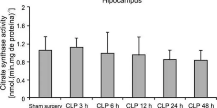
Figure 3 - Citrate synthase activity in the hippocampus of rats 3, 6, 12, 24 and 48 hours after cecal ligation puncture (CLP).
Results are expressed as the mean ± S.D. (n=6). Diferent from control group, *p<0.05 (One-way ANOVA followed by Tukey test).
Figure 4 -Citrate synthase activity in the striatum of rats 3, 6, 12, 24 and 48 hours after cecal ligation puncture (CLP).
Results are expressed as the mean ± S.D. (n=6). Diferent from control group, *p<0.05 (One-way ANOVA followed by Tukey).
Figure 5 - Citrate synthase activity in cerebellum of rats 3, 6, 12, 24 and 48 hours after cecal ligation puncture (CLP).
DISCUSSION
Cell death within the central nervous system (CNS) during sepsis has been described in rodent models and humans.(29,30) An extensive body of evidence from
experimental and clinical studies indicates that sepsis is associated with increased ROS production, depletion of antioxidants, and accumulation of markers of oxidative stress.
Direct evidence for free radical production in sepsis comes from studies using spin traps to detect hydroxyl radicals and electron paramagnetic resonance spectroscopy to analyze NO production in rats as well as studies detecting ascorbate radical production in septic patients.(31,32) Increased xanthine oxidase (XO) activity,
an important ROS producer, has been reported in patients with sepsis.(33) In addition to showing elevated
ROS production, studies have also found decreased antioxidant defenses leading to redox imbalance in sepsis patients. Glutathione replenishment by N-acetylcysteine and glutathione has been shown to decrease oxidative stress in patients with sepsis.(34) he increased ROS
production and decreased antioxidant levels have been accompanied by increased lipid peroxidation in patients with sepsis compared with controls.(35)
In addition, the respiratory chain is an important pillar in sepsis pathophysiology. Because mitochondria play a critical role in cellular energy production via electron transport chain-dependent synthesis of ATP through oxidative phosphorylation and are the main site of ROS production, inlammatory insult results in mitochondria being damaged functionally and structurally. We previously performed a time-course experiment to evaluate the activities of mitochondrial respiratory chain complexes I, II, III and IV and creatine kinase after CLP; we demonstrated that brain energy metabolism is altered six and twelve hours after CLP.(36)
More speciically, we observed that animals submitted to CLP presented decreased mitochondrial respiratory chain activity in complexes I and II but not in complexes III and IV at 24, 48 and 96 h post-CLP. A previous study showed that succinate dehydrogenase activity (an important enzyme in the Krebs cycle) was decreased at 48 and 96 h post-CLP in all analyzed structures.(37) In
addition, other works indicate that mitochondrial free radical generation is increased in sepsis.(38,39)
Production of ROS perpetuates and propagates mitochondrial injury, leading to mitochondrial swelling and diminution of cytochrome c levels in the mitochondria.(40)
Consequently, derangements in mitochondrial function
primarily afect cells that have a high energy demand, such as neurons,(41) and brain energy impairment has
been linked to neuronal death and neurodegeneration.(42)
In this context, the brain may be one of the irst organs afected during sepsis, and encephalopathy is a frequent association but infrequently recognized.(43,44) Acute
encephalopathy has demonstrated in animal models of polymicrobial sepsis, and human sepsis survivors present cognitive impairment that could be a secondary efect of CNS damage.(26) In addition, survivors from critical
care units, including sepsis patients, may show persistent brain-related morbidity, including neurocognitive deicits and development of psychiatric disorders.(45-49)
In the present study, we demonstrate that citrate synthase activity is inhibited in the prefrontal cortex and cerebral cortex of adult rats after sepsis induced by CLP. Considering that energy impairment resulting from mitochondrial dysfunction in sepsis has been well described and that citrate synthase has been used as a quantitative enzyme marker for the presence of intact mitochondria, we therefore believe that energy impairment may also be involved in these processes. If the inhibition of citrate synthase also occurs in a sepsis model, it is tempting to speculate that a reduction of brain metabolism may be related to the pathophysiology of this disease.
Prefrontal cortex lesions are associated with social disinhibition; impulse dyscontrol; organizational, planning, and attentional dysfunctions; dysluency and slowing of spontaneous behaviors.(50) Konarska et
al.(51) reported that regional deicits in the frontal lobe,
particularly in the anterior cingulate and the orb to the frontal cortex, consistently delineate subjects with central nervous system disorders from the general population. We hypothesize that mitochondrial dysfunction may be related to CNS damage in sepsis.
CONCLUSION
In conclusion, we have demonstrated that citrate synthase is decreased by CLP in the prefrontal cortex and cerebral cortex. hese data corroborate those from other studies, suggesting that mitochondrial dysfunction is implicated in the pathogenesis of CLP.
ACKNOWLEDGEMENTS
RESUMO
Objetivo: Um amplo corpo de evidência oriundo de estudos experimentais indica que a sepse se associa com um aumento da produção de espécies de oxigênio reativo, depleção de antioxidan-tes, e acúmulo de marcadores de estresse oxidativo. Além disto, a disfunção mitocondrial foi implicada na patogênese da síndrome de disfunção de múltiplos órgãos. A citrato sintase é uma enzima que se localiza no interior das células, na matriz mitocondrial, sen-do uma etapa importante sen-do ciclo de Krebs; esta enzima foi utili-zada como um marcador enzimático quantitativo da presença de mitocôndrias intactas. Assim, investigamos a atividade da citrato sintase no cérebro de ratos submetidos ao modelo sepse com de ligadura e punção do ceco.
Métodos: Em diferentes horários (3, 6, 12, 24 e 48 horas) após cirurgia de ligadura e punção do ceco, seis ratos foram sacriicados por decapitação, sendo seus cérebros removidos e dissecados o hi-pocampo, estriato, cerebelo, córtex cerebral e córtex pré-frontal, e
utilizados para determinação da atividade de citrato sintase.
Resultados: Veriicamos que a atividade de citrato sintase no córtex pré-frontal estava inibida após 12, 24 e 48 horas da ligadura e punção do ceco. No córtex cerebral, esta atividade estava inibida após 3, 12, 24 e 48 horas da ligadura e punção do ceco. Por outro lado a citrato sintase não foi afetada no hipocampo, estriato e cere-belo até 48 horas após a ligadura e punção do ceco.
Conclusão: Considerando-se que é bem descrito o compro-metimento da energia decorrente da disfunção mitocondrial na sepse, e que o estresse oxidativo desempenha um papel essencial no desenvolvimento da sepse, acreditamos que o comprometimento da energia pode também estar evolvido nestes processos. Se a ini-bição da citrato sintase também ocorre em um modelo de sepse, é tentador especular que a redução do metabolismo cerebral pode provavelmente estar relacionada com a isiopatologia desta doença.
Descritores: Citrato (si)-sintase/metabolismo; Sepse; Mitocôndrias; Ratos Wistar; Modelos animais
REFERENCES
1. Sands KE, Bates DW, Lanken PN, Graman PS, Hibberd PL, Kahn KL, Parsonnet J, Panzer R, Orav EJ, Snydman DR, Black E, Schwartz JS, Moore R, Johnson BL Jr, Platt R; Academic Medical Center Consortium Sepsis Project Working Group. Epidemiology of sepsis syndrome in 8 academic medical centers. JAMA. 1997;278(3):234-40. 2. Vandijck D, Decruyenaere JM, Blot SI. he value of
sepsis deinitions in daily ICU-practice. Acta Clin Belg. 2006;61(5):220-6.
3. Hotchkiss RS, Karl IE. he pathophysiology and treatment of sepsis. N Engl J Med. 2003;348(2):138-50. Review.
4. Basu S, Eriksson M. Oxidative injury and survival during endotoxemia. FEBS Lett. 1998;438(3):159-60.
5. Zhang H, Slutsky AS, Vincent JL. Oxygen free radicals in ARDS, septic shock and organ dysfunction. Intensive Care Med. 2000;26(4):474-6.
6. Kozlov AV, Szalay L, Umar F, Fink B, Kropik K, Nohl H, et al. EPR analysis reveals three tissues responding to endotoxin by increased formation of reactive oxygen and nitrogen species. Free Radic Biol Med. 2003;34(12):1555-62.
7. Ritter C, Andrades M, Frota Júnior ML, Bonatto F, Pinho RA, Polydoro M, et al. Oxidative parameters and mortality in sepsis induced by cecal ligation and perforation. Intensive Care Med. 2003;29(10):1782-9.
8. Barichello T, Fortunato JJ, Vitali AM, Feier G, Reinke A, Moreira JC, et al. Oxidative variables in the rat brain after sepsis induced by cecal ligation and perforation. Crit Care Med. 2006;34(3):886-9.
9. Powell RJ, Machiedo GW, Rush BF Jr, Dikdan GS. Efect of oxygen-free radical scavengers on survival in sepsis. Am Surg. 1991;57(2):86-8.
10. Villa P, Ghezzi P. Efect of N-acetyl-L-cysteine on sepsis in mice. Eur J Pharmacol. 1995;292(3-4):341-4.
11. Sprong RC, Winkelhuyzen-Janssen AM, Aarsman CJ, van Oirschot JF, van der Bruggen T, van Asbeck BS. Low-dose N-acetylcysteine protects rats against endotoxin-mediated oxidative stress, but high-dose increases mortality. Am J Respir Crit Care Med. 1998;157(4 Pt 1):1283-93.
12. Kong CW, Tsai K, Chin JH, Chan WL, Hong CY. Magnolol attenuates peroxidative damage and improves survival of rats with sepsis. Shock. 2000;13(1):24-8.
13. Vulcano M, Meiss RP, Isturiz MA. Deferoxamine reduces tissue injury and lethality in LPS-treated mice. Int J Immunopharmacol. 2000;22(8):635-44.
14. Salvemini D, Cuzzocrea S. herapeutic potential of superoxide dismutase mimetics as therapeutic agents in critical care medicine. Crit Care Med. 2003;31(1 Suppl):S29-38. Review. 15. hiemermann C. Membrane-permeable radical scavengers
(tempol) for shock, ischemia-reperfusion injury, and inlammation. Crit Care Med. 2003;31(1 Suppl):S76–84. 16. Brealey D, Singer M. Mitochondrial dysfunction in sepsis.
Curr Infect Dis Rep. 2003;5(5):365-71.
17. Streck EL, Delwing D, Tagliari B, Matté C, Wannmacher CM, Wajner M, Wyse AT. Brain energy metabolism is compromised by the metabolites accumulating in homocystinuria. Neurochem Int. 2003;43(6):597-602. 18. Wallace DC. A mitochondrial paradigm of metabolic
and degenerative diseases, aging, and cancer: a dawn for evolutionary medicine. Annu Rev Genet. 2005;39:359-407. 19. Fink MP. Bench-to-bedside review: cytopathic hypoxia. Crit
Care. 2002;6(6):491-9.
20. Singer M, Brealey D. Mitochondrial dysfunction in sepsis. Biochem Soc Symp. 1999;66:149-66.
Fourrier F, Chopin C. Escherichia coli endotoxin reduces cytochrome aa3 redox status in pig skeletal muscle. Crit Care Med. 2000;28(10):3491-7.
22. Chen HW, Hsu C, Lu TS, Wang SJ, Yang RC. Heat shock pretreatment prevents cardiac mitochondrial dysfunction during sepsis. Shock. 2003;20(3):274-9.
23. Shepherd D, Garland PB. he kinetic properties of citrate synthase from rat liver mitochondria. Biochem J. 1969;114(3):597-610.
24. Marco R, Pestaña A, Sebastian J, Sols A. Oxaloacetate metabolic crossroads in liver. Enzyme compartmentation and regulation of gluconeogenesis. Mol Cell Biochem. 1974;3(1):53-70.
25. Ritter C, Andrades ME, Reinke A, Menna-Barreto S, Moreira JC, Dal-Pizzol F. Treatment with N-acetylcysteine plus deferoxamine protects rats against oxidative stress and improves survival in sepsis. Crit Care Med. 2004;32(2):342-9.
26. Barichello T, Martins MR, Reinke A, Feier G, Ritter C, Quevedo J, Dal-Pizzol F. Cognitive impairment in sepsis survivors from cecal ligation and perforation. Crit Care Med. 2005;33(1):221-3; discussion 262-3.
27. Lowry OH, Rosebrough NJ, Farr AL, Randall RJ. Protein measurement with the Folin phenol reagent. J Biol Chem. 1951;193(1):265-75.
28. Srere PA. Citrate synthase. Methods Enzymol. 1969;13:3-11. 29. Semmler A, Okulla T, Sastre M, Dumitrescu-Ozimek L,
Heneka MT. Systemic inlammation induces apoptosis with variable vulnerability of diferent brain regions. J Chem Neuroanat. 2005;30(2-3):144-57.
30. Sharshar T, Annane D, de la Grandmaison GL, Brouland JP, Hopkinson NS, Françoise G. he neuropathology of septic shock. Brain Pathol. 2004;14(1):21-33.
31. Sato K, Kadiiska MB, Ghio AJ, Corbett J, Fann YC, Holland SM, et al. In vivo lipid-derived free radical formation by NADPH oxidase in acute lung injury induced by lipopolysaccharide: a model for ARDS. FASEB J. 2002;16(13):1713-20.
32. Linares E, Nakao LS, Augusto O, Kadiiska MB. ERP studies of in vivo radical production by lipopolysaccharide: potential role of iron mobilized from iron-nitrosyl complexes. Free Radic Biol Med. 2003;34(6):766-73.
33. Batra S, Kumar R, Seema, Kapoor AK, Ray G. Alterations in antioxidant status during neonatal sepsis. Ann Trop Paediatr. 2000;20(1):27-33.
34. Ortolani O, Conti A, De Gaudio AR, Moraldi E, Cantini Q, Novelli G. he efect of glutathione and N-acetylcysteine on lipoperoxidative damage in patients with early septic shock. Am J Respir Crit Care Med. 2000;161(6):1907-11.
35. Bayir H. Reactive oxygen species. Crit Care Med. 2005;33(12 Suppl):498-501.
36. Comim CM, Rezin GT, Scaini G, Di-Pietro PB, Cardoso MR, Petronilho FC, et al. Mitochondrial respiratory chain and creatine kinase activities in rat brain after sepsis induced by cecal ligation and perforation. Mitochondrion.
2008;8(4):313-8.
37. Comim CM, Constantino LC, Barichello T, Streck EL, Quevedo J, Dal-Pizzol F. Cognitive impairment in the septic brain. Curr Neurovasc Res. 2009;6(3):194-203.
38. Boczkowski J, Lisdero CL, Lanone S, Samb A, Carreras MC, Boveris A, et al. Endogenous peroxynitrite mediates mitochondrial dysfunction in rat diaphragm during endotoxemia. FASEB J. 1999;13(12):1637-46.
39. Callahan LA, Stofan D, Szweda L, Nethery DE, Supinski GS. Free radicals alter maximal diaphragmatic oxygen consumption in endotoxin-induced sepsis. Free Radic Biol Med. 2001;30(1):129-38.
40. Crouser ED. Mitochondrial dysfunction in septic shock and multiple organ dysfunction syndrome. Mitochondrion. 2004;4(5-6):724-41.
41. Enns GM. he contribution of mitochondria to common disorders. Mol Genet Metab. 2003;80(1-2):11-26. Review. 42. Navarro A, Boveris A. he mitochondrial energy transduction
system and the aging process. Am J Physiol Cell Physiol. 2007;292(2):C670-86. Review.
43. Milbrandt EB, Angus DC. Bench-to-bedside review: critical illness-associated cognitive dysfunction-mechanisms, markers, and emerging therapeutics. Crit Care. 2006;10(6):238. 44. Ebersoldt M, Sharshar T, Annane D. Sepsis associated
delirium. Intensive Care Med. 2007; 33(6):941-50.
45. Hopkins RO, Weaver LK, Pope D, Orme JF, Bigler ED, Larson-LOHR V. Neuropsychological sequelae and impaired health status in survivors of severe acute respiratory distress syndrome. Am J Respir Crit Care Med. 1999;160(1):50-6. 46. Angus DC, Musthafa AA, Clermont G, Griin MF,
Linde-Zwirble WT, Dremsizov TT, Pinsky MR. Quality-adjusted survival in the irst year after the acute respiratory distress syndrome. Am J Respir Crit Care Med. 2001;163(6):1389-94.
47. Granja C, Dias C, Costa-Pereira A, Sarmento A. Quality of life of survivors from severe sepsis and septic shock may be similar to that of others who survive critical illness. Crit Care. 2004;8(2):R91-8.
48. Granja C, Lopes A, Moreira S, Dias C, Costa-Pereira A, Carneiro A; JMIP Study Group. Patients’ recollections of experiences in the intensive care unit may afect their quality of life. Crit Care. 2005;9(2):R96-109.
49. Hopkins RO, Weaver LK, Collingridge D, Parkinson RB, Chan KJ, Orme JF Jr. Two-year cognitive, emotional, and quality-of-life outcomes in acute respiratory distress syndrome. Am J Respir Crit Care Med. 2005;171(4):340-7. 50. Lou H. Etiology and pathogenesis of attention-deicit
hyperactivity disorder (ADHD): signiicance of prematurity and perinatal hypoxic-haemodynamic encephalopathy. Acta Paediatr. 1996;85(11):1266-71.