Manuela Ermelinda
Lopes Lago
Caracterização e funcionalização de hidrogeis para
cultura de células
Characterization and functionalization of hydrogels
for cell culture
Manuela Ermelinda
Lopes Lago
Caracterização e funcionalização de hidrogeis para
cultura de células
Characterization and functionalization of hydrogels
for cell culture
Tese apresentada à Universidade de Aveiro para cumprimento dos requisitos necessários à obtenção do grau de Mestre em Biotecnologia, ramo Biotecnologia Molecular, realizada sob a orientação científica do Doutor Mário Grãos, Investigador Principal da Unidade de Biologia Celular do Biocant e do Professor Doutor José António Teixeira Lopes da Silva, Professor Auxiliar do Departamento de Química da Universidade de Aveiro.
Este trabalho é financiado por Fundos FEDER através do Programa Operacional Fatores de Competitividade – COMPETE e por Fundos Nacionais através da FCT – Fundação para a Ciência e a Tecnologia no âmbito do projeto FCOMP-01-0124-FEDER-021150 (referência FCT: PTDC/SAU-ENB/119292/2010),
o júri
presidente Prof. Dr. Luísa Alexandra Seuanes Serafim Martins Leal
Professora Auxiliar Convidada do Departamento de Química da Universidade de Aveiro
Dr. Joana Paes de Faria Monteiro
Investigadora Principal no Instituto de Biologia Molecular e Celular
Dr. Mário Martins Rodrigues Grãos
agradecimentos Em primeiro lugar, gostaria de agradecer ao Doutor Mário Grãos, pela oportunidade que me deu de integrar este projecto e pela forma como me recebeu. Obrigada pelo tempo despendido comigo; pelos conselhos, pela paciência, enorme compreensão e motivação que sempre me transmitiu. Obrigada por toda a partilha de conhecimentos e experiência. As nossas interacções são um enorme progresso para a minha evolução.
Em segundo lugar, um sincero agradecimento ao Dr. José Lopes da Silva, pela disponibilidade, pelos conselhos e toda a compreensão.
Gostaria também de agradecer a todos os meus colegas do Biocant pelo carinho e alegria com que me receberam.
Da Unidade de Biologia Celular, gostaria de agradecer, em especial à Tânia Lourenço pela orientação, pelos conhecimentos teóricos e práticos transmitidos. Pelo apoio nas dificuldades e dúvidas; pelas horas que dispôs para me ajudar. Não poderiam faltar o Plácido Pereira, que com as suas “maluqueiras” anima o dia de qualquer um; e a Heloísa, que apesar de curto o contacto, estás sempre disposta para me ajudar, ouvir e tranquilizar. À ex-colega Cataria Domingues, pelas viagens, boa disposição e amizade. Da Unidade de Proteómica e Metabolómica gostaria de agradecer especialmente à Sandra Anjo, Matilde Melo e Cátia Santa pelo apoio, conselhos e conhecimentos que me transmitiram
Pedro, Francisco, Lúcia, Liliana Pedro, Hugo, Dário, Gil, Carlos, Nadine, Patrícia, Andreia, André, Nuno, David, Rodrigo, gostaria de vos agradecer por tudo que aconteceu ao longo destes 5 anos. Apesar de todos os seus altos e baixos, serão sempre indescritíveis. Francisco, Lúcia e Liliana, obrigada pelas viagens, pelas partilhas, pelas conversas… Basicamente por me ouvirem.
Gostaria ainda de agradecer à minha família pelo apoio incondicional, pela paciência e disponibilidade. Pai… Mãe… Sem vocês não seria possível. À minha irmã, Paula, queria agradecer o apoio, as confidências, a paciência. A ti, Joaninha, quero que saibas que deixas saudade, que estarás para sempre no nosso coração e és um exemplo de coragem para os teus pais, irmãs e toda a família. Obrigada pelo vosso amor, compreensão, confiança e paciência. Obrigada ao Filipe pela amizade, pela presença, por me ouvir, pela força que
palavras-chave Mecanotransdução, Rigidez, Matriz extracelular, Substratos sintéticos, Oligodendrócitos
resumo Mecanotransdução é a resposta e/ou a produção de um estímulo mecânico
exercido sobre ou por células, que é acoplado a sinais bioquímicos. As células estão rodeadas por matriz extracelular (ECM) que tem propriedades mecânicas e de composição específicas, dependendo do tecido. Estes componentes ligam-se a integrinas e activam-nas, resultando em sinalização intracelular que envolve o citoesqueleto de actina e proteínas motoras. Em doenças neurodegenerativas, são observadas modificações na composição da matriz extracelular e da sua rigidez que pode resultar na inibição de diferenciação de oligodendrócitos e de remielinização das áreas afectadas. Os oligodendrócitos (OLs) são células do sistema nervoso central (CNS) responsáveis pela produção de mielina. A sua diferenciação é modulada por, entre outros fatores, proteínas presentes na matriz extracelular como laminina e fibronectina e pela rigidez do substrato.
As células são também sensíveis à rigidez do substrato quando cultivadas in vitro. De forma a mimetizar essa componente mecânica, foram criadas plataformas de poli-acrilamida como substrato com rigidez definida, tendo em consideração o tecido que pretendemos mimetizar - o cérebro. Estas plataformas foram funcionalizadas com proteínas da ECM ou pequenos péptidos presentes nessas mesmas proteínas, permitindo estudar e modular a influência destes mesmos fatores na diferenciação celular, em contraste com condições de cultura standard.
A principal novidade deste estudo consiste na manutenção e diferenciação de oligodendrócitos in vitro utilizando um substrato compatível e definido. Foram para isso utilizados péptidos derivados da laminina-alfa2, que promoveram a adesão e diferenciação das células. Este estudo exploratório sugere que os péptidos sob estudo têm potencial para ser utilizados no futuro na modulação da diferenciação de células primárias e perceber qual o papel destes nas vias bioquímicas intracelulares envolvidas.
keywords Mechanotransduction, Stiffness, Extracellular matrix, Synthetic Substrates, Oligodendrocytes
abstract Mechanotransduction is the response to and/or the production of mechanical
stimuli exerted upon, or by cells, that is coupled to biochemical signals. Cells are surrounded by extracellular matrix (ECM) which has specific mechanical properties and composition depending on the tissue. These components bind to and activate integrins, which results in intracellular signaling that involves the actin cytoskeleton and myosin motor proteins. In neurodegenerative diseases, modifications occur in the ECM composition and rigidity that seem to inhibit oligodendrocyte differentiation and remyelination of the affected area. Oligodendrocytes (OLs) are the myelin-producing cells of the central nervous system (CNS). OL differentiation is modulated by, among other factors, ECM proteins like laminin and fibronectin and by substrate rigidity.
Cells also sense substrate stiffness when cultured in vitro. In order to mimic this mechanical component, polyacrylamide platforms were created with defined stiffness, considering the stiffness of the target tissue relevant for this study – the brain. These platforms were functionalized with ECM proteins or small peptides (derived from ECM proteins), that allow to study the impact of these factors on cellular differentiation, in contrast with standard cell culture conditions.
The main achievement in this study was to maintain and differentiate oligodendrocytes using a fully defined compliant substrate. Several peptides derived from the laminin-alpha2 chain were used, to provide adhesion to the cells and allow their differentiation. This exploratory study suggests that the peptides under study have a potential to be explored in the future using primary cells and fully evaluate their capacity to modulate oligodendrocyte differentiation, namely to understand which biochemical pathways are involved.
Table of contents
Table of contents ... i
List of abbreviations ... iii
I. Introduction ... 1
I.1. Central Nervous System (CNS) ... 3
I.1.1. Overview ... 3
I.1.2. Oligodendrocyte Development and differentiation ... 3
I.1.3. Demyelinating diseases ... 6
I.2. Characterization of the Cell Microenvironment ... 9
I.2.1. The Niche and extracellular matrix composition ... 9
I.2.2. Cell adhesion molecules, receptors and signal transduction involved in oligodendrocyte maturation ... 10
I.3. Mechanotransduction ... 14
I.3.1. Mechanisms of mechanotransduction ... 15
I.4. Mimicking the cellular microenvironment ... 17
I.4.1. ECM proteins and soluble factors ... 18
I.4.2. ECM stiffness and biomaterials ... 19
I.4.2.1. Polyacrylamide hydrogel-based synthetic substrates ... 21
I.5. Objectives ... 22
II. Material and Methods ... 23
II.1. Cell culture ... 25
II.1.1. Human Oligodendroglioma (HOG) cell line culture ... 25
II.1.2. CG4 cell line culture ... 26
II.1.3. B104 cell culture and preparation of conditioned medium ... 27
II.2. Preparation of polyacrylamide hydrogels ... 27
II.2.1. Crosslinking of ECM proteins and peptides on polyacrylamide hydrogels ... 29
II.3. PAA hydrogels rheological characterization ... 30
II.4. Coating on coverslips ... 31
II.5. Fluorescence microscopy and immunocytochemistry ... 32
II.6. Image and fluorescence intensity analysis ... 33
III.1. Optimization of polyacrylamide hydrogels ... 37
III.2. Human oligodendroglioma cell culture and differentiation ... 38
III.2.1. Adhesion study of human oligodendroglial cell line (HOG) ... 38
III.2.2. HOG differentiation and Fractal dimension analysis ... 41
III.3. CG4- cells differentiation ... 45
III.4. Adhesion study of CG-4 cell line to laminin-α2 derived peptides ... 47
III.5. CG-4 cells differentiation ... 49
IV. Discussion ... 55
V. Conclusion ... 65
VI. Bibliography... 69
List of abbreviations
A A Area
Ac Acrylamide
AEP Anterior peduncular area APTMS 3-aminopropiltrimethoxisilane APS Ammonium persulfate
B bHLH Basic helix-loop-helix
Bis-Ac Bis-acrylamide
BSA Bovine serum albumin
C Cbp Csk-binding protein (Cbp)
CGE Caudal ganglionic eminence CNS Central nervous system Csk C-terminal Src kinase
D D Asparagine (Arg)
DAPI 4’,6- Diamidino-2-phenylindole dihydrochloride DM Differentiation medium
dbcAMP 2'-O-dibutyryladenosine 3':5' cyclic monophosphate
E E Young’s modulus or compressive modulus
ECM Extracellular matrix EtOH Ethanol
F F Force
FA Focal adhesion FAK Focal adhesion kinase FBS Fetal bovine serum FGF Fibroblast growth factor FN Fibronectin
G G Glycine (Gly)
G Shear modulus
GDP Guanosine diphosphate GF Growth factor
GFAP Glial fibrillary acidic protein GPCR G-protein-coupled receptor GTP Guanosine-5’-triphosphate
H HBS Hepes buffer saline
HOG Human oligodendroglioma
HSPGs Heparin/Heparan Sulfate Proteoglycan
I IBMX 3-Isobutyl-1-methylxanthine
K kPA kilo Pascal
L LGE Lateral ganglionic eminence
MBP Myelin basic protein
MFI Mean fluorescence intensity MGE Medial ganglionic eminence min minutes
MN Merosin
MSCs Mesenchymal stem cells MT Microtubules
MW Molecular weight
N N Asparagine (Asp)
NCAM Neural cell adhesion molecule NEP Neuroepithelial precursor cell NHS N- acryloxusuccinimide
O OL Oligodendrocyte
ON Overnight
OPC Oligodendrocyte precursor cell O-2A Oligodendrocyte type-2 astrocytes
P PAA Polyacrylamide
PAH Polyacrylamide hydrogels Pax Paired-box
PBS Phosphate buffered saline PDGF Platelet-derived growth factor PDL Poly-D-lysine
PEGDA Poly(ethylene glycol) diacrylate PI3K Phosphoinositide 3-Kinase
PIP2 Phosphatidylinositol (4,5) bis-phosphate PLP Proteolipid protein Ptc Patched PM Proliferation medium R R Arginine (Arg) RT Room temperature S Shh Sonic hedgehog Smo Smoothened SFK Src family kinases SVZ Subventricular zone T T3 Triiodo-L-thyronine T4 Thyroxine TEMED Tetramethylethylenediamine TH Thyroid hormone U UV Ultraviolet Y Y Tyrosine
I.1. Central Nervous System (CNS)
I.1.1.
OverviewNeurons and glial cells are the constituents of the central nervous system (CNS). The neurons are the cells responsible for the nerve impulse transmission. Glial cells include ‘macroglia’ (derived from the neural tube) and ‘microglia’ (derived from hematopoietic precursors). The resident macrophages of the CNS, known as ‘microglia’, are essential for immune surveillance and defense. The two major macroglia cells types are astrocytes and oligodendrocytes (Kessaris et al., 2008). The first visualization of these cells was a century ago by Andriezen and colleagues (Andriezen, 1893). Furthermore, it is known that glial cells play an important role in the homeostasis of the CNS. Oligodendrocytes are responsible for formation of myelin sheaths around CNS axons. On the other hand, astrocytes are responsible to provide structural support to CNS neurons, interact with blood vessels and the formation of the blood-brain barrier and also for the regulation of the CNS synaptogenesis and synaptic transmission (Kessaris et al., 2008).
I.1.2. Oligodendrocyte Development and differentiation
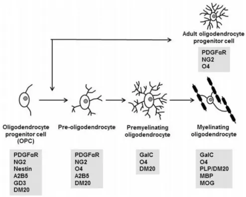
Martin Raff and colleagues in the early 1980s identified oligodendrocytes precursor cells (OPCs) (Hart et al., 1989). OPCs are proliferating cells that could differentiate into oligodendrocyte type-2 astrocytes (O-2A cells), immature oligodendrocytes or myelinating mature oligodendrocyte (Figure I.1) (Franklin and ffrench-Constant, 2008; Kessaris et al., 2008).
Figure I.1 - The oligodendrocyte (OL) lineage commitment. Schematics of morphological features and expression of specification markers from progenitor cells to myelinating oligodendrocytes. (Adapted from Schumacher et al., 2012)
Oligodendrocytes (OLs) are essential for myelinating events of the CNS (Bradl and Lassmann, 2010; Kessaris et al., 2008). In the spinal cord, oligodendrocyte precursor cells (OPCs) are derived from neuroepithelial precursor cells (NEPs) named motor neuron precursors (pMN) at a specific domain of the ventral ventricular zone (Bradl and Lassmann, 2010; Kessaris et al., 2008). OPCs migrate and differentiate into myelin-forming oligodendrocytes. Another source of OPCs is the dorsal spinal cord, but from there, production of OPCs occurs 2 days later than in the ventral zone (Kessaris et al., 2008).
The first wave of OPCs produced in the forebrain appears in the anterior peduncular area (AEP) and medial ganglionic eminence (MGE). Two more waves are produced from more dorsal regions [the lateral and caudal ganglionic eminences (LGE/CGE)] and finally from within the postnatal cortex (Bradl and Lassmann, 2010; Kessaris et al., 2008). During postnatal life, the first waves of OPCs generated in the ventral forebrain (MGE/AEP)
practically disappear and are replaced by other populations including the LGE/CGE population (Kessaris et al., 2008; Wen et al., 2009).
A program of NEP cell fate occurs, in the spinal cord, when Sonic hedgehog (Shh) is secreted and diffused, from the notochord, that creates a morphogenetic gradient and complementary signals from the roof plate. Activation or repression by Shh involves activation or repression of a set of homeobox, paired-box (Pax) and basic helix-loop-helix (bHLH) transcription factor genes. The combinatorial expression of these genes, influenced by Shh, generates different classes of spinal cord neurons. Shh signaling in vertebrates is still unclear. Shh interacts with transmembrane receptor Patched (Ptc) (Rivera et al., 2010), causing disinhibition of its co-receptor Smoothened (Smo) - Figure I.2, a seven-pass transmembrane G-protein-coupled receptor (GPCR), eventually promoting nuclear translocation of the full-length form of the transcription factor of the Gli family (Gli1–Gli3) (Kessaris et al., 2008).
Figure I.2 - The Shh signalling pathway. Two transmembrane receptors are involved: Patched (Ptch) and Smoothened (Smo). Binding of Shh inhibits Ptch function and so Smo is no longer inhibited. Smo represses the cleavage of Gli that is translocated to the nucleus, acting as a transcription modulator. SUFU act as a repressor of the transcriptional activity of intact Gli. (Adapted from Scotting et al., 2005)
Gli transcription factor allows expression of Olig1 and Olig2, effectors of oligodendroglial differentiation and myelination. Another factor that stimulates oligodendrogenesis is platelet-derived growth factor (PDGF), which stimulates OPC proliferation and survival (Rivera et al., 2010; Wen et al., 2009).
Likewise, thyroid hormone (TH) induces proliferation and differentiation of OPCs and enhances morphological and functional maturation of post-mitotic oligodendrocytes. Myelination is delayed in hypothyroid animals (Rivera et al., 2010).
Genes relevant for oligodendrocyte maturation and consequently myelination are inhibited by Notch downstream targets (Hes1 and Hes5). The Notch signaling pathway inhibits oligodendroglial differentiation and promotes astrocytic fate (Rivera and Aigner, 2012).
The oligodendrogenic process may be activated by intrinsic activators, the Sox genes. For example, Sox9 is essentially for glial fate decision; Sox17 expression increases in differentiating OPCs, which enhances myelin gene expression. Sox genes are required for oligodendrogenesis and promote OL development and myelination. Oligodendrogenesis is also dependent on extracellular matrix components that regulate cell adhesion, migration and differentiation (Rivera et al., 2010; Wen et al., 2009).
I.1.3. Demyelinating diseases
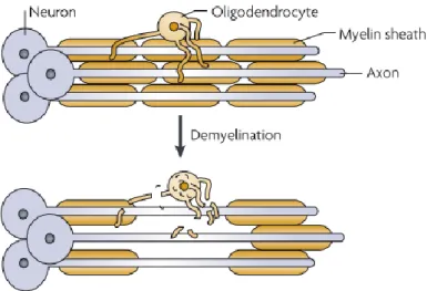
In the CNS, demyelinating diseases are characterized by the pathological process in which the myelin sheath formed around axons is damaged - Figure I.3 (Franklin and ffrench-Constant, 2008; O'Meara et al., 2011). Myelin is a major protein present in the multilamellar sheath synthesized by oligodendrocytes in the central nervous system that
Albers and White, 2011; Kramer et al., 1997). The unique composition of that membrane is ~70% lipid (for example, galactocerebroside) and ~30% protein, which contrasts with other common membranes that have 30-50% lipid.
Figure I.3 - The demyelination of axons. This pathological process is characterized by the loss of myelin sheaths around axons (Franklin and ffrench-Constant, 2008).
There are three major causes for demyelination of the CNS: (i) genetic abnormalities that affect glia; (ii) inflammatory damage; or (iii) acute traumatic injuries to the CNS (e.g. contusion or compression of the spinal cord). The loss of the myelin sheath that protects nerve fibers causes anomalous insulation of the CNS neuronal axons resulting in abnormalities of the nerve impulse transmission and neuronal death (Franklin and ffrench-Constant, 2008). Astrocytes influence demyelination by enhancing the immune response. They are responsible for the activation of autoreactive T cells through expression of MHC class II; the production of chemokines that recruits T cells, macrophages and microglia to inflammatory lesions; secretion of cytokines IL-12 and IL-23 (McFarland and Martin, 2007; Nair et al., 2008).
Remyelination is a process in which entire myelin sheaths are restored to demyelinated axons, reinstating salutatory conduction (Franklin and ffrench-Constant, 2008; Smith et
al., 1979) and resolving functional deficits (Franklin and ffrench-Constant, 2008). However, this process is normally not very efficient in humans.
Oligodendrocyte precursor cells typically have a simple and bipolar morphology and are responsive to soluble growth factors (GFs), which promote their proliferation and survival. While differentiating, OPCs initiate contact with multiple axons and when matured, oligodendrocytes extend a complex meshwork of processes. When axo-glial contact are established, OLs produce large amounts of myelin sheaths that insulate axons (O'Meara et al., 2011).
OPCs have an important contribution for remyelination, due to their ability to proliferate, migrate and terminally differentiate into newly formed OLs. During remyelination, these cells can be influenced to proliferate and migrate by the growth factors PDGF and fibroblast growth factor (FGF). There are four relevant evidences suggesting that OPCs are the major source of remyelinating oligodendrocytes: (i) retroviral and autoradiographic tracing indicate that dividing cells in normal adult white matter give rise to remyelinating oligodendrocytes; (ii) transplanted OPCs remyelinate areas of demyelination with great efficiency; (iii) focal areas of demyelination are repopulated by OPCs suggesting that OPCs are the source of the remyelinating cells; (iv) cells with transitional expression of OPC and oligodendrocyte markers can be identified at the beginning of remyelination (Franklin and ffrench-Constant, 2008).
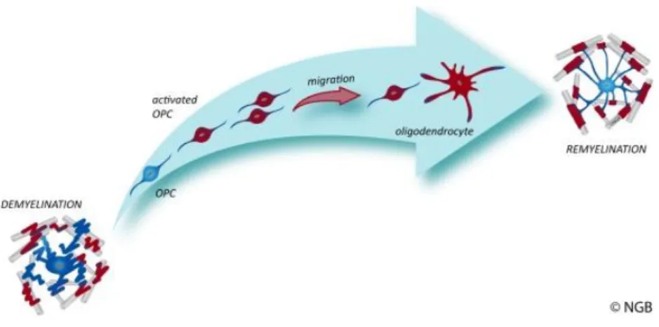
Following demyelination, microglia and astrocytes become activated, and induce the rapid proliferative response of OPCs to the injury site. During the remyelination process, local OPCs change from an essentially quiescent state to a regenerative phenotype. The first step in this process involves changes in morphology and up-regulation of several genes related with the development of oligodendrocytes (that encode the transcription factors Olig2, NKX2.2, MYT1 and Sox2). Following recruitment, the differentiation phase of OPCs into remyelinating oligodendrocytes involves a contact with the axon and the formation of a wrap and compact myelin to form the sheath. These steps are represented in Figure I.4. There are many factors which affect the efficiency of remyelination like age, gender and genetic background (Franklin and ffrench-Constant, 2008).
Figure I.4 - The remyelination process. In demyelinating events, the myelin sheath or the oligodendrocytes are lost. Through biochemical signals, oligodendrocytes progenitor cells are activated, proliferate and are recruited to the affected region. Then, they differentiate into remyelinating oligodendrocytes. (http://www.crm.ed.ac.uk/research/group/myelination-and-repair-cns, 2013).
I.2. Characterization of the Cell Microenvironment
I.2.1. The Niche and extracellular matrix compositionR. Schofield proposed, in 1978, the concept of the stem cell niche showing that is essential to the cell fate (Schofield, 1978). The niche encompasses interactions between soluble molecules, other cells and extracellular matrix (ECM). The capacity of the cell to sense all the biophysical and biochemical cues in their surrounding microenvironment is crucial to regulate the cell and tissue maintenance and development (Discher et al., 2009; Eyckmans et al., 2011; Gobaa et al., 2011; Morrison and Spradling, 2008).
The ECM of the CNS has a unique composition, since the major element of the niche is a complex mixture of large glycoproteins (such as fibronectins, collagens, laminins), proteoglycans and glycosaminoglycans (such as hyaluronan, chondroitin sulphate and heparin sulfate). This structure provides not only a scaffold for cellular support, but also the triggers of regulatory signals through the activation of transmembrane receptors, like the integrins (Eyckmans et al., 2011; Ma et al., 2008).
I.2.2. Cell adhesion molecules, receptors and signal transduction involved in oligodendrocyte maturation
The protein complex of an integrin was first characterized in 1986 (Tamkun et al., 1986). Integrins are fundamental in cellular processes like adhesion, proliferation, migration, cell survival and differentiation in a variety of tissues (Campbell and Humphries, 2011; Danen and Sonnenberg, 2003).
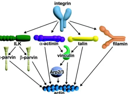
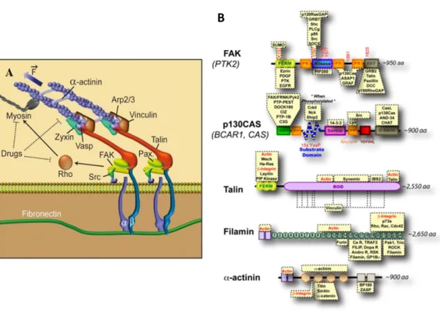
Integrins are heterodimeric transmembrane protein receptors (Brakebusch and Fassler, 2003; Campbell and Humphries, 2011; Hynes, 2002) and constitute the major group of receptors for ECM constituents (Montgomery et al., 1996; Ruppert et al., 1995). There are 18α and 8β subunits that result in 24 different combinations of receptors with different distribution (Hynes, 2002; Ma et al., 2008). Integrins are composed by two non-covalently associated subunits (Hynes, 2002), α and β, which are both involved in the binding to extracellular matrix proteins and in the coordination of the actin cytoskeleton and cellular response to growth factors (Leone et al., 2005; Tamkun et al., 1986) that allow for regulation of cell motility, cell polarity, cell growth and survival (Brakebusch and Fassler, 2003; O'Meara et al., 2011). The β subunits of integrins have longer cytoplasmic tails that can bind to adaptor proteins such as talin, α-actinin or filamin (Figure I.5), which in turn recruit other players involved in the formation of focal adhesions (FAs).
FAs are complex structures, constituting recruitment sites for actin filaments and cytoplasmic tails of integrins, contributing heavily for cellular adhesion and mechanotransduction signaling events (Brakebusch and Fassler, 2003; Moore et al., 2010).
Figure I.5 - Different pathways through which integrins can link to the actin cytoskeleton. ILK bins to the cytoplasmic tails β-integrin subunits and can recruit a family of F-actin binding proteins. α-actinin connects actin fibrils to the cytoplasmic tails of transmembrane receptors such as integrins, cadherins and inter-cellular adhesion molecules. α-actinin can also recruit actin filament by the interaction with vinculin. Talin binds to integrin, focal adhesion kinases (FAK), phosphatidylinositol phosphate kinase, phosphatidylinositol (4,5) bis-phosphate (PIP2), vinculin and Arp 2/3 complex. When PIP2 interacts with vinculin is replaced by actin filaments. Filamin is a dimeric protein and has a head domain containing the actin binding site (Brakebusch and Fassler, 2003).
Expression of integrins is dependent on oligodendrocyte development stage, but is also dependent on the surrounding environment. Moreover, changes in ECM constituents results in alteration in oligodendrocyte integrin expression (ffrench-Constant and Colognato, 2004). αvβ1-integrin and αvβ3-integrin (among others) are strongly expressed during OL precursor phases contributing to migration and proliferation, respectively. However, αvβ5-integrin and α6β1 are strongly expressed in late stages of development, contributing to differentiation. α6β1 also contributes to the survival of newly formed oligodendrocytes (O'Meara et al., 2011).
The integration of external factors is dependent on the activation of both integrins and growth factor receptors, that activate Src family kinases (SFKs) that are non-receptor tyrosine kinases (Colognato et al., 2004; O'Meara et al., 2011). The amplification of GF-signaling is dependent on GF-signaling pathways involving phosphotidylinositide 3-kinase
(PI3K) and mitogen-activated protein kinase (MAPK) cascades, involved in proliferation, survival, signal transduction and cytoskeletal reorganization (O'Meara et al., 2011).
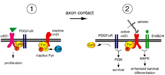
One SFK is Lyn that is expressed by oligodendrocyte progenitors. Integrin αvβ3 promotes activatation of Lyn by phosphorylation in catalitic Y397 which contributes to OPCs proliferation (Colognato et al., 2004). Another one is Fyn that is expressed in the brain and its activity is correlated with myelination and oligodendrocyte differentiation process. Activation of Fyn is dependent on axonal contact and ligation by laminin-2 to integrin α6β1 (Figure I.6). Laminin induces dephosphorylation of the inhibitory tyrosine residue (Y531) of Fyn. To become completely activated, Fyn is phosphorylate in tyrosine residue Y420, a process mediated by contacting with axonal neural cell adhesion molecule L1 (NCAM-L1). Laminin regulates elevated levels of C-terminal Src kinase (Csk) and Csk-binding protein (Cbp), promoting Fyn activity and OL differentiation (Colognato et al., 2004; Relucio et al., 2009).
Figure I.6 - Model for regulation of SFK activity by integrins during oligodendrocyte differentiation. In proliferating stages (1), Lyn is associated with the PDGF-αVβ3 integrin complex and Fyn is maintained inactive by Csk-mediated phosphorylation (in residue Y531). After axonal contact (2) and ligation of α2 chain laminin to α6β1 integrin, Lyn is dissociated from the integrin-growth factor complex and Csk is dowregulated, activating Fyn-α6β1 complexes. This complex can trigger PI3K and MAPK signalling involved in oligodendrocyte survival (depending on the ligand of PDGFαR) and differentiation (depending on the ligand of ErbB2/4), respectively. (Adapted from Colognato et al., 2004)
Active Fyn modulates factors such as Rho family GTPases (Rho, Rac1 and Cdc42). GTPases are active when bound to GTP, and inactive when bound to GDP. Cdc42 and Rac1 are activated by Fyn and build filamentous actin while GTP-bound Rho depolymerizes actin filaments, thus, these proteins have influence on cell morphology (O'Meara et al., 2011). Futhermore, Wang et al. demonstrated that depletion of activated Fyn in oligodendrocytes depletes GTP-bound Rac1, GTP-bound Cdc42, and GDP-bound Rho, resulting in morphological deffects in OL differentiation and myelination, demonstrating the importance of these proteins in the cytoskeleton rearregement and process extension (Huveneers and Danen, 2009; Wang et al., 2009).
Another function of Fyn is to phosphorylate rhoGAPs, more specifically p190RhoGAP, that increases with oligodendrocyte differentiation (Huveneers and Danen, 2009; Wolf et al., 2001). That event promotes the formation of Rho GDP-bound, which is the inactive form of RhoA, and when active stimulates myosin II-dependent actomyosin contractility [intracellular forces generated by the dynamic interaction of myosin motors and actin filaments (Sun et al., 2012)] and MBP expression increase (Burgstaller and Gimona, 2004; Wang et al., 2012). Therefore Fyn-mediated inactivation of RhoA promotes oligodendrocytes diffentiation (Wang et al., 2008).
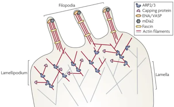
All these proccesses are crucial for the dynamic of the cytoskeleton. Actin polymerization, and the subsequent invasion of microtubules (MT) are processes that mediate reorganization of the cytoskeleton with the formation of filopodia and lamellipodia (Figure I.7), resulting in formation and extension of processes in oligodendrocytes (Bauer et al., 2009). Cdc42 and Rac1 activation are the crucial steps to filopodia and lamellipodia formation, respectively (Huveneers and Danen, 2009). Inibition of myosin II regulates actin cytoskeleton dynamics, myelin formation and potentiates OLs branching (Wang et al., 2008).
Figure I.7 - Oligodendrocyte process formation. Extracellular signals mediate polymerization of actin filaments, resulting in the protrusion of the plasma membrane and the formation of filopodia. Actin branching mediated by the Wiskott-Aldrich syndrome protein (N-WASP)/actin-related protein-2/3 (Arp2/3) complex causes filopodia to enlarge. Microtubules migrate into the widen membrane protrusions and convert them into lamellipodia. (Adapted from Heasman and Ridley, 2008)
I.3. Mechanotransduction
Cells are sensitive to the mechanical properties of their microenvironment (Eyckmans et al., 2011; Saha et al., 2008). Cellular processes such as cell adhesion, actin flow, retraction forces or gene expression are influenced by substrate rigidity (Engler et al., 2006; Gobaa et al., 2011; Jagielska et al., 2012; Moore et al., 2010). The composition and mechanical properties of the extracellular matrix (ECM) are essential for cellular proliferation, fate and differentiation (Cameron et al., 2011; Saha et al., 2008). Hence, the microenvironment can influence cells by the presence of not only biochemical, but also physical and mechanical stimuli (Saha et al., 2008). Wang and colleagues demonstrated that integrins interact with the extracellular matrix influencing cell behavior by transducing mechanical signals to the cytoskeleton (Wang et al., 1993). Mechanotransduction may be defined as the response to and/or the production of a
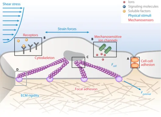
mechanical stimulus exerted upon, or by the cells, that is coupled to biochemical signals that influence their behaviour and phenotype (Figure I.8).
Figure I.8 - Mechanotransduction in a Cell-ECM Unit. A cell connected to the ECM and a neighbor cell. (A) Mechanotransduction at adherents junctions. (B) Mechanoreceptors at the cell membrane. (C) Mechanotransduction at the nucleus. (D) Mechanotransduction at the focal adhesion. (Adapted from Sun et al., 2012)
I.3.1. Mechanisms of mechanotransduction
Mechanotransduction involves the conversion of mechanical stimuli into changes in protein activity (Giannone and Sheetz, 2006). One of the key proteins involved in this event is focal adhesion kinase (FAK), whose activity increases with mechanical forces (Moore et al., 2010). This enzyme interacts with integrins and phosphorylates tyrosine residues of intracellular proteins, like paxillin (Pax), promoting their recruitment to focal adhesion (Figure I.9) (Eyckmans et al., 2011; Moore et al., 2010). Focal adhesions (FA) are regions of cellular attachment to the extracellular matrix, which are formed by the
coordinated recruitment of intracellular adaptor and catalytic (mostly kinases) proteins linked to the extracellular matrix through integrins (Figure I.9-A).
Mechanical stimuli increase tyrosine phosphorylation activity of Src, a tyrosine kinase. In mechanotransduction, there is evidence that cell stretching and matrix rigidity increases tyrosine phosphorylation and exposes protein-protein binding domains of important proteins. For instance, tyrosine phosphorylation of the substrate domain of p130CAS, a downstream target of Src, is observed upon stretch. Also, Talin exposes 11 potential vinculin binding sites, as well as for actin filaments, integrins and other proteins. Mechanical forces stretch Talin, which then results in reinforcement of early adhesions of focal adhesions, through the recruitment of additional actin-binding proteins. Filamins have an actin-binding domain followed by 24 immunoglobulin repeats, and bridge integrins, through their C-terminal domain, to actin – Figure I.9-B (Moore et al., 2010).
Figure I.9 - Proteins involved in integrin-mediated rigidity sensing. (A) Mechanical stimuli induce conformational changes in focal adhesions and enzyme kinetics (Adapted from Eyckmans et al., 2011) (B) FAK its kinase activity is regulated by mechanical forces, removal of the FERM domain from the kinase could play a role. Upon stretching, the substrate domain of p130Cas contains 15 tyrosine residues that become exposed. Stretching of talin’s rod domain exposes vinculin binding sites. Extension of filamin immunoglobulin repeats (labelled 1-24) could regulate the binding of proteins. Mechanical forces could regulate this dimerization or its association with other proteins (Adapted from Moore et al., 2010).
Thus, integrins bind to ECM proteins and focal adhesions (FAs) are formed. Through actomyosin contraction, cells exert force on the substrate allowing movement of actin fibers. If the substrate is very soft, talin is not stretch and the matrix deforms. In case of a stiffer substrate, actomyosin induces tension, talin stretches and recruits vinculin and other FA proteins (Moore et al., 2010).
Mechanotransduction, through several mechanisms, allows cells to sense and respond to their surrounding microenvironment. Many details of mechanotransduction are still unexplored and need more elucidation (Moore et al., 2010).
Recent studies have been showing, in vitro, the influence of many factors which mimic the native tissue during OL differentiation. Kippert et al. showed that physical properties of the matrix regulate the cell surface area of oligodendrocytes, influenced by actomyosin contractility. The inhibition of actomyosin contractility using different factors, such as blebbistatin (an inhibitor of myosin II) or Y27632 [an inhibitor of the Rho/Rho-kinase (ROCK) pathway] suggests that the low contractility favors OL maturation (Kippert et al., 2009). Jagielska and colleagues demonstrated that OPCs from the CNS are mechanosensitive. Using polyacrylamide hydrogels they showed that cell survival, proliferation, migration and differentiation are dependent on the mechanical properties of the substrate where the cells are seeded (Jagielska et al., 2012).
I.4. Mimicking the cellular microenvironment
these cells, because it implies invasive procedures to the CNS. So, alternatives are being investigated. To obtain these cells, in vitro, the conditions of native microenvironment have a special attention. For that, it is important to consider the stiffness and the characteristics of the biomaterial, ECM proteins (or peptides) and soluble factors.
I.4.1. ECM proteins and soluble factors
ECM proteins, such as fibronectin and laminin, are a crucial component for cell adhesion, maintenance and differentiation.
Fibronectin (FN) is a polypeptide that contains a large number of binding regions. There are binding domains for cellular integrins [including the characteristic amino acid sequence Arg-Gly-Asp (RGD)] and protein-protein interactions sites [with heparin/heparan sulfate proteoglycan (HSPGs), collagen] (Li et al., 2013).
Laminin, another ECM glycoprotein, composed by the combination by one of the 5 types of α chain, one β chain of the three possible and one of 3 types of γ chains (Aumailley et al., 2005; Buttery and ffrench-Constant, 1999; Urushibata et al., 2010). The isoform laminin α2β1γ1 (merosin-MN) is known to bind the integrin receptor α6β1, crucial for oligodendrocyte survival and differentiation (Colognato et al., 2004). Furthermore, there are also heparin-binding active sites that are involved in interactions with HSPGs, such as syndecans and α-dystroglycans (Colognato et al., 2007; Urushibata et al., 2010).
Oligodendrogenesis is a process that may be stimulated by many soluble factors. PDGF, a survival and mitogen factor, stimulates the proliferation and survival of oligodendrocyte progenitors (Colognato et al., 2004; Rivera et al., 2010). Thyroid hormones (TH) are also crucial for the proliferation of OPCs and their differentiation (Rivera et al., 2010).
I.4.2. ECM stiffness and biomaterials
Cells are influenced by physical and mechanical factors like ECM stiffness in vivo, or when cultured on a synthetic matrix, in vitro. Stiffening of the microenvironment has an important consequence in cell spreading, morphology and function (Engler et al., 2006). The stiffness of the materials is measured based on the force required to deform the matrix. Cells may be cultured in vitro on synthetic substrates, which are viscoelastic materials. These materials have separable shear elastic (storage, G’) and viscous (loss, G’’) modulus components. The shear modulus (G) of a crosslinked network system can be related to the Young’s modulus or compressive modulus (E), through Equation 1.1, where 𝜈 is the Poisson ratio of the material (Cameron et al., 2011).
( ) Equation (1.1)
The shear modulus (G) and Young’s modulus (also known as Elastic modulus - E) are distinct and depend on the direction of the force that is applied. To quantify the shear modulus (G), the force is applied parallel to the material’s surface, however for elastic modulus (E), the force is applied perpendicular to the surface – Figure I.10 (Moore et al., 2010). Nevertheless, for practical reasons, for several techniques it is easier to measure the shear modulus (force applied parallel to the surface of the material). In these cases, the elastic modulus (which is a widely used measure of the stiffness of a material) may then be calculated from the shear modulus using Equation 1.1. For hydrogels, the formula may be simplified to the ratio that the compressive modulus (E) of is thus approximately 3 times that of the shear modulus (G), assuming that the Poisson ration of materials that do not change their volume under stretch (as it is in the case of hydrogels) is approximately 0.5 (Moore et al., 2010).
Figure I. 10 - Rigidity moduli. Elastic and shear moduli are the ratio of the amount of force applied per area (F/A) and strain [which reflects the displacement in the direction of the force applied relative to the initial length (Δx/L or ΔL/L)]. (Adapted from Moore et al., 2010)
Engler et al. showed that the microenvironment is an important element influencing the cellular phenotype (Engler et al., 2006). Solid tissues exhibit a range of stiffness (Table I.1) from 0.1 to 30,000,000 nN/μm2 (nN/μm2 may also be expressed as kPa) and when mesenchymal stem cells are cultured on soft matrices (coated with collagen to promote cell adhesion) with stiffness that mimics the stiffness of the brain, muscle or bone, the cells acquire a neurogenic, myogenic or osteogenic phenotype, respectively (Engler et al., 2006; Tse and Engler, 2011).
Table I.1 - Elasticity of some human solid tissues. Range of stiffness measured by the
Elastic modulus , E. (Adapted from Moore, S.W., et al. 2010)
Tissue type Elastic modulus (nN/μm2)
Brain 0.1 – 10
Muscle 12 – 100
Fat 20
Furthermore, Saha and colleagues demonstrated that neural stem cells cultured on soft matrices (~100-500 Pa) differentiated preferentially into neurons, whereas when cultured on stiffer matrices (~1,000-10,000Pa) glial fates were favoured (Saha et al., 2008). This observation seems to recapitulate the developmental fate of neural stem cells, since neurons differentiate first, on a softer environment, followed by glial cells, which encounter a more rigid niche later on.
I.4.2.1. Polyacrylamide hydrogel-based synthetic substrates
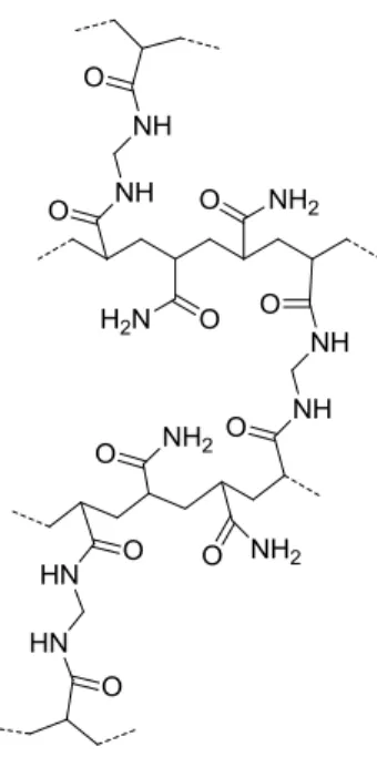
Cells in living tissues establish contacts with other cells and with the extracellular matrix components. To understand the mechanotransduction properties of cells and tissues and how to tailor more appropriate materials to be used as in vitro platforms (for mechanotransduction studies) or implants for tissue engineering, model microenvironments are required. This will allow researchers to create biomimetic environments that will contribute to our knowledge of cellular biology in a more realistic context. In vitro, hydrogels functionalized with ECM proteins (or peptides), associated with soluble factors, have been widely used as a model for ECM (Moshayedi et al., 2010). Polyacrylamide (PAA) hydrogels (Figure I.11) are artificial gel matrices that constitute a widespread platform used in cell biology. They are cheap, biologically inert, capable of modeling different ranges of stiffness and ‘anti-adhesive’ materials prepared by the co-polimerization of different percentages of acrylamide (Ac) and bis-acrylamide (Bis-Ac) (Cretu et al., 2010; Moshayedi et al., 2010). PAA hydrogels need to be activated and covalently grafted with adhesion proteins or peptides in order to enable them for cell culture (Moshayedi et al., 2010).
Figure I.11 - Schematic representation of the molecular structure of a polyacrylamide hydrogel.
The development of new hydrogels has the goal to imitate aspects of the ECM of native tissues and should mimic the mechanical and biological features of the tissues being replicated (Hutson et al., 2011).
I.5. Objectives
There is an increase of studies showing the crucial impact of mechanical and physical forces and mechanotransduction on cell behavior, shape and fate. For this purpose, to approximate cell culture closer to the native microenvironment of the cells, we propose to use a synthetic tunable platform which mimics the stiffness of the native tissue and may be conjugated with extracellular proteins, to differentiate oligodendrocytes in vitro. The main objective of this work was to functionalize a synthetic platform (polyacrylamide hydrogels) with ECM proteins and small peptides derived from the laminin-aplpha2 chain to study and understand the effect of stiffness and ECM-like composition on the differentiation of oligodendrocytes, by addressing the expression of specific differentiation markers and morphology analysis.
II.1.
Cell culture
II.1.1. Human Oligodendroglioma (HOG) cell line culture
HOG cells (kindly provided by Dr. José Antonio López-Guerrero, Centro de Biologia Molecular Severo Ochoa, CSIC-UAM, Madrid, Spain) are a human oligodendrocyte cell line derived from a surgically removed human oligodendroglioma (Post and Dawson, 1992) which express a 15kDa form of myelin basic protein (MBP), myelin-specific lipids galactosylceramide and sulfogalactosylceramide (sulfatide) and high level of the maturation marker CNP. Moreover, do not express astrocyte markers glial fibrillary acidic protein (GFAP) or glutamine synthetase activity (Buntinx et al., 2003; Post and Dawson, 1992). To proliferate, HOG cells were maintained in proliferation medium (PM): DMEM low glucose (Gibco) with 3.57g/L HEPES, 1.5g/L sodium bicarbonate, 10%FBS (Gibco), 1% Penicillin/Streptomycin (Gibco) and 1% Amphotericin B (Gibco) (Bello-Morales et al., 2009). Cells were kept in an incubator at 37°C, 5% CO2/95% air and 95% humidity.
For differentiation experiments performed on coverslips and hydrogels, cells were plated on these two platforms at 6,400 cells/cm2 in proliferation medium for two days. The medium was then replaced by one of two different differentiation media: N1 (Louis et al., 1992) supplemented with T3 and T4 [composed by 5μg/mL apo-transferrin, 10ng/mL biotin, 5ng/mL sodium selenite, 5μg/mL insulin, 6.3ng/mL progesterone, 16μg/mL putrescine, 30ng/mL triiodo-L-thyronine (T3) and 40ng/ml Thyroxin (T4)], 1% Penicillin/Streptomycin (Gibco) and 1% Amphotericin B (Gibco) or the medium described by Bello Morales (Bello-Morales et al., 2011) composed by: DMEM with high glucose (4,500 mg/mL – Thermo Hyclone) supplemented with 50μg/mL apo-transferrin, 30nM sodium selenite, 0.5μg/mL insulin, 16.1mg/mL putrescine, 30nM T3, 0.5mM dbcAMP and IBMX and 1.5g/L sodium bicarbonate (all the supplements were from Sigma-Aldrich), 1% Penicillin/Streptomycin (Gibco) and 1% Amphotericin B (Gibco), for more two days. To determine the best conditions of coating on PAA hydrogels and coverslips, HOG cells were plated at a density of 6,000cells/cm2 for 1 dayin proliferation medium. As a control, HOG cells were plated in glass coverslips with same conditions of cellular density, timing,
II.1.2. CG4 cell line culture
The CG4 cell line (kindly provided by Dr. Adil J. Nazarali, College of Pharmacy and Nutrition, University of Saskatchewan, Saskatoon, Canada) is a rat oligodendrocyte cell line characterized by a bipotential oligodendrocyte-type 2 astrocyte (O2A progenitor cell) morphology. The cells were maintained as described by Louis and colleagues (Louis et al., 1992). In detail, CG-4 cells were washed 2 or 3 times using a sterile Puck’s solution (80g/L NaCl, 4g/L KCl, 0.6g/L KH2PO4, 0.9g/L Na2HPO4.H2O, 10g/L D-glucose). Then, the cells were detached using Trypsin-EDTA 0.05% (Gibco). After dissociation and detachment, the trypsin was inactivated using serum-supplemented recovery medium [DMEM high glucose (Hyclone) supplemented with 2mM sodium pyruvate (Sigma-Aldrich), 5% FBS (Gibco), 5μg/mL insulin (Sigma-Aldrich), 1% Penicillin/Streptomycin (Gibco) and 1% Amphotericin B (Gibco)]. Then, the cells were centrifuged (20oC, 201g, 5 minutes), counted and seeded at the density of 2,500 cells/cm2 in serum-supplemented recovery medium on cell culture dishes (Corning-Costar), coated with poly-D-Lysine (Sigma-Aldrich) at 100μg/ml. When the cells were attached (approximately 30 minutes at 37°C) the recovery medium was replaced by CG4 proliferation medium [DMEM high-Glucose (Hyclone-Thermo) supplemented with 50μg/mL apo-tranferrin (Sigma-Aldrich), 9.8ng/mL biotin (Sigma-Aldrich), 40ng/mL sodium selenite (Sigma-Aldrich), 30% of B104-conditioned medium, 1% Penincilin/Streptomycin (Gibco) and 1% Amphotericin B (Gibco)]. Then, the CG4 cultures were maintained in an incubator at 37°C, 5% CO2/95% air and 95% humidity and the medium was change every two days (Ji et al., 2011).
Hydrogels and coverslips were placed in 12 well plates and 24 well plates, respectively, and then CG4 cells were seeded at a density of 6,400cell/cm2 or 10,000cells/cm2 if we intend to proliferate or differentiate, respectively. Cells were allowed to attach in recovery medium for 1h, at 37°C and the medium was changed for proliferation, for 2 days or differentiation medium – N1+T3+T4, for 3 days.
II.1.3. B104 cell culture and preparation of conditioned medium
B104 neuroblastoma cells were kindly provided by Adil J. Nazarali, College of Pharmacy and Nutrition, University of Saskatchewan, Saskatoon, Canada. Cells were cultured in B104 proliferation medium constituted by DMEM high-glucose/F12 (Gibco) [1:1] containing 10% FBS, 1%Penicillin/Streptomycin and 1% Amphotericin B. For the preparation of conditioned medium, cells were detached using trypsin-EDTA 0.05% (trypsin was then inhibited using proliferation medium), centrifuged (290g for 5 minutes, at room temperature), counted and seeded at a density of 15,000cells/cm2 in B104 proliferation medium. After 24 hours, the cells were washed 3 times with Puck’s solution and the cells were maintained in defined medium [DMEM high-glucose/F12 (1:1), 10μg/mL holo-transferin (Sigma-Aldrich), 5ng/mL sodium selenite (Sigma-Aldrich), 16μg/mL putrescine (Sigma-Aldrich), 6.3ng/mL progesterone (Sigma-Aldrich), 1% Penicillin/Streptomycin and 1% Amphotericin B] for 3 days in an incubator at 37°C, 5% CO2/95% air and 95% humidity. Then, the medium was collected and added 1μg/ml of PMSF (phenylmethanesulfonylfluoride). Finally the medium was centrifuged (1,000g at 4°C, for 10 minutes) and the supernatant filtered (using a 0.22μm Cellulose Acetate filter from VWR) and stored at -20°C.
II.2.
Preparation of polyacrylamide hydrogels
Reactive glass coverslips (15x15 mm) allow covalent links between the hydrogel and the coverslip (Cretu et al., 2010). For this, coverslips were treated for 3 minutes with a solution that consisted of 3-(Trimethoxysilyl)propyl methacrylate diluted in ethanol (1:200) and 3% (v/v) of diluted acid acetic (1:10 glacial acetic acid: water) (Figure II.1) (Hoffecker et al., 2011). Finally, coverslips were washed with absolute ethanol twice and then dried.
Figure II.1 - Reaction of activation of glass coverslips with 3-(trimethoxysilyl) propyl methacrylate..
Acrylamide – 40% (Ac - purchased from AppliChem), bis-acrylamide – 2% (Bis-Ac - purchased from AppliChem), mQ water and tetramethylethylenediamine (TEMED – purchased from Fluka) were mixed, according to Table II.1. The pH of the solution was adjusted to 7.5-8 using HCl 2N. Next, the solution was degassed for 30 minutes using a vacuum pump. Then, N-acryloxysuccinimide (NHS, 20 mg/mL in toluene, Santa Cruz Biotechnology) and 10 % ammonium persulfate (APS, Sigma-Aldrich) were added to the hydrogel solution according to Table II.1 and vortexed briefly (Figure II.2).
Table II.1 - Hydrogel formulation with 6.5kPa- volume added (µL) per milliliter of hydrogel solution (Lourenço, 2012). PAA hydrogel formulation - μL (10%Ac/0.3%Bis-Ac) Ac 40% 250 Bis – Ac 2% 150 NHS 220 APS 10% 3 TEMED 1 Water 376
Hydrogel polymerization was carried out using a Mini-Protean III system from BioRad with 1mm spacers. Polymerization of the hydrogels was allowed to occur on the treated coverslips during 30 minutes. Hydrogels were then washed three times with PBS, five
minutes each, on a rocker and sterilized by exposure to UV light for 30 minutes in an air flow cabinet.
Figure II.2 - Reaction of PAA gel production. Co-polymerization of acrylamide with N-acryloxysuccinimide crosslinked with bis-acrylamide.
II.2.1. Crosslinking of ECM proteins and peptides on polyacrylamide hydrogels
To mimic the extracellular matrix and allow cell attachment, hydrogels can be functionalized with ECM proteins or peptides representing epitopes of ECM proteins. NHS acts as a crosslinker in this approach (Cretu et al., 2010), allowing covalent binding of primary amines of proteins or peptides to the hydrogels (Figure II.3).
In order to functionalize the hydrogels, either individual proteins or a mixture of proteins [such as Laminin-2/Merosin (MN) or Fibronectin (FN)] were diluted in PBS at different concentrations in the presence of poly-D-Lysine (PDL). The Fibronectin and merosin were isolated from human plasma and placenta from Roche and Millipore, respectively. Poly-D-Lysine was from Sigma-Aldrich. The concentrations and combinations of proteins/peptides used are indicated in the Results section, according to each
with PBS. Hydrogels were blocked with a solution of 1mg/mL of heat-inactivated BSA in DMEM low glucose at 37°C for 30 minutes. Finally, hydrogels were washed once with PBS and incubated at 37°C for 4 hours to equilibrate. In case of functionalization of the hydrogels using peptides the blocking step was not performed.
Figure II.3 - Reaction of functionalization of PAA gels. The functional group – NHS allows the covalent bond of the hydrogel with primary amines of proteins or polypeptides.
II.3.
PAA hydrogels rheological characterization
The rheological characterization of polyacrylamide hydrogels was done by Tânia Lourenço (Lourenço, 2012). Briefly, the stiffness of hydrogels was measured by rheology using a Kinexus Pro rheometer and rSpace software. According to the literature, the elastic modulus (E) is determined using the formula E=2G(1+𝜈), where 𝜈 is the Poisson ratio, that is assumed 0.5 (at 1Hz) for materials do not change volume under stretch, resulting in an elastic modulus that will be three times its shear modulus (G) (Moore et al., 2010; Saha et al., 2008). The measurements of G were performed after an equilibration of hydrogels, overnight, in PBS. The distance between the top and bottom plates (gap) was defined as 1mm, and was then fine-tuned until the top plate applied a normal force of 0.1N on the hydrogel. The measurements were then carried out using 2mstrain, at 1Hz, at 37°C
II.4.
Coating on coverslips
In order to functionalize with ECM proteins and PDL, the glass coverslips (12mm diameter) were immersed in 65% nitric acid, during 24 hours with agitation. Then, the acid was removed and the coverslips were washed several times with an excess of milliQ water and allowed for 3 hours with agitation in milliQ water. Lastly, they were sterilized with dry heat (for 15 minutes at 121oC).
However, to obtain a longer spacer between the glass coverslip and the peptides (several peptides with short sequences of about 15 aminoacids or less were used), a different treatment was done. First, the glass coverslips were incubated in a 1N NAOH (from J. T . Baker) solution for 30 minutes, at room temperature (RT). Then, the NaOH was aspirated and the coverslips were treated with 200μL of 10% 3-aminopropiltrimethoxisilane (3-APTMS from Sigma-Aldrich) in 96% ethanol (EtOH from Merck), for 30 minutes at room temperature. The coverslips were washed (3 times, 10 minutes) with abundant MilliQ water (H2O mQ) with agitation, in order to minimize the excess of 3-APTMS that are able to react with glutaraldehyde- from Merck (Chung and Min, 2009). The coverslips were then immersed with 3% glutaraldehyde in PBS (1x) for 20 minutes at room temperature and washed 3 times with H2O mQ, with agitation – Figure II.4 (Cretu et al., 2010; Wipff et al., 2009).
Figure II.4 – Formulation of reactive coverslips. The coverslips were incubated with NaOH (1) followed by de addition of 3-APTMS (2). Glutaraldehyde (3) was used to crosslink the 3-APTMS and the polypeptide.
In both cases, the coverslips were coated with 50μL of protein or peptide solution (in PBS) for 4 hours at 37°C in an incubator. The cell culture in this platform was done using the same procedure as in section II.1.2.
II.5.
Fluorescence microscopy and immunocytochemistry
To evaluate morphological features and differentiation markers of cells before and after the differentiation protocol, a fluorescence microscopy approach was performed. Cells were washed once with Puck’s solution (HOG cells were washed with PBS) and fixed with 4% paraformaldehyde for 20 min at room temperature. After washing with PBS, cells were incubated either with FITC-labeled agglutinin (Invitrogen – Molecular Probes) or antibodies against oligodendroglial lineage markers expressed at distinct differentiation stages.
In order to evaluate morphological features, cells were stained with FITC-labelled agglutinin (5µg/mL) in HBSS for one hour. To stain cells using antibodies, cells were permeabilized using PBS-triton 0.1% for 20 minutes and for 5 minutes with PBS-Tween 0.1% and then blocked with 1% BSA in PBS for 30 minutes. The primary antibodies were incubated in blocking solution, over-night at 4°C in humidified conditions and then washed twice with PBS for 5 min each. The secondary antibodies were used according to the species of each primary antibody and they were incubated for 1h at room temperature (RT) in PBS with 1% BSA. The cells were then washed and fixed with 4% PFA for 5 minutes (to stabilize the antibody staining) and then were incubated with DAPI (200ng/mL) in PBS for 5 minutes at room temperature for nuclear staining. The primary antibodies used were: rat anti-MBP, clone 21 (1:200) from Abcam; mouse anti-PLP, clone PLPC1 (1:500) from Millipore. The secondary antibodies were: goat anti-rat Alexa Fluor 488 (1:200); goat anti-mouse Alexa Fluor 568 (1:200), both from Invitrogen – Molecular Probes.
Fluorescence images were acquired using a Zeiss Axiovert 200M fluorescence microscope using AxioVision release 4.8 software (Zeiss) for image acquisition. Image processing and analysis was performed using the Image J software.
II.6.
Image and fluorescence intensity analysis
To analyze fluorescence microscopy images using Image J software, images were converted to 8-bit. Then, to quantify the mean fluorescence intensity (MFI), the background and the signal threshold levels were determined (Image-Adjust-Threshold tool) of at least 3 fields. The average of threshold levels was calculated and applied (Image-Adjust-Threshold-Set tool) to all the images. Finally, using the Measure tools (in Analyze menu) the MFI and background were measured. The MFI values were used to perform statistical analysis.
For the fractal dimension analysis, first, the cell was selected using the crop tool (Image-Crop tool) and the background and signal threshold values were set (using the same procedure as described above). To conclude, the Fractal Box Count was selected (Analyze-Tools-Fractal Box Count) and the obtained values were used for statistical analysis.
In order to quantify the number of adherent cell on coverslips coated with different concentration of peptides, the images of phase-contrast microscopy were exported in tiff format, and then using the image J software the threshold values were applied and the area of signal measure were used.
II.7.
Statistical analysis
Statistical analysis was performed by repeated measures one-way ANOVA followed by Tukey’s multiple comparison test (* p < 0.05, ** p < 0.01 and *** p<0.001 for statistically significant differences) using the software Graph Pad Prism 4.
III.1.
Optimization of polyacrylamide hydrogels
The native properties of the cellular microenvironment have been considered as important conditions for the proliferation and differentiation of cells. The biochemical and physical aspects of the extracellular matrix have been reported to influence cell adhesion, motility, cytoskeleton organization, cell fate and function (Saha et al., 2008). Polyacrylamide hydrogels (PAHs) are the two-dimensional substrates selected in our approach to study the influence of compliant ECM-like substrates on the adhesion and differentiation of oligodendrocytes. According to the literature, the brain stiffness ranges from 0.1 to 10 kPa (Moore et al., 2010). Therefore, in our studies, 6.5kPA hydrogels (10% Ac/0.3% BAc) previously characterized in our laboratory were used, a degree of stiffness also reported in the literature to favor oligodendrocyte differentiation (Kippert et al., 2009). PAHs are non-toxic, stable, have an easily quantifiable elasticity (Tse and Engler, 2010), can be easily functionalized with ECM components and modulated with different degrees of stiffness by varying the percentages of acrylamide and bis-acrylamide.
Some parameters are important to optimize the conditions for cell culture. To optimize the cell-adhesion properties of the PAHs and their reproducibility, two steps were included in the production protocol of the hydrogels: an efficient degassing that decreased the number of air bubbles inside of the gel (for 30 minutes, before the addition of APS, NHS and polymerization - as described in Materials and Methods) and the siliconization of the outside glass used as support to polymerize the hydrogels, which decrease the rugosity on the surface of the gel (5 minutes with a solution of 0.5% of dichlorodimethylsilane in toluene).
Due to the instability of the amine-reactive esters of NHS at basic pH (as it is the case of the hydrogel solution used to produce the substrate, due to the presence of TEMED), an additional step was added before the degassing. The pH of the solution was adjusted to the range 7.5-8 to increase the stability of the esters and still allow the efficient covalent linkages between the hydrogel and the proteins or peptides used to functionalize the
These optimization steps were essential for the reproducible production of functional hydrogels used in this study.
III.2.
Human oligodendroglioma cell culture and differentiation
III.2.1. Adhesion study of human oligodendroglial cell line (HOG)The first objective of this work was to optimize the protein coating conditions of the different substrates (glass coverslips and hydrogels), to allow for an efficient adhesion of HOG cells (a human oligodendrocytic cell line) and ensure that the cells were properly attached to the substrate and could be used in the differentiation experiments.
This was performed using different coating conditions and concentrations of coating solutions on hydrogels and coverslips (Figure III.1), as described in Materials and Methods section. Cells were seeded at 6,000cells/cm2 on hydrogels and coverslips. After 24 hours in culture, cells were visibly attached to the culture surface (coverslips and hydrogels) and could be readily identified by fluorescence microscopy (DAPI staining – Figure III.1).
Figure III.1 – Representative images for nuclear staining of HOG cells. Nuclei of HOG cells were stained using DAPI (in blue) after 24 hours in culture on glass coverslips or hydrogel. The platforms were functionalized with PDL alone (as control) or in combination with different ECM proteins [Fibronectin (FN) and/or Merosin (MN)] at the indicated concentration (10, 25 and 40μg/mL).
In order to count the number of cells that were adherent on the hydrogels and coverslips using distinct functionalization/coating conditions, cells stained with DAPI present on the fluorescence microscopy images acquired, as represented in Figure III.1, were counted and analyzed using Image J software tools (see Materials and methods).
Figure III.2 - Quantification of the number of cells/cm2 in all coatings conditions on coverslips and hydrogels (approximately 6.5kPa). Number of HOG cells/cm2 on coverslips – A and hydrogels (6,5KPa – B; ratio between the number of cell/cm2 after 24 hours in proliferation medium and the number of cells/cm2 initially seeded (6000 cells/cm2) on coverslips – C and hydrogels – D. Hydrogels and coverslips were coated with different mixtures of ECM proteins (Fibronectin - FN and Merosin - MN) with Poly-D-Lysine (PDL) at three concentrations (10, 25 and 40μg/mL). Nuclei stained with DAPI were counted using Image J software. Values represented mean ± SEM (n=5). Statistical analysis by one way ANOVA followed by turkey’s multiple comparison test (**p<0.01 and *p<0.05).
As observed in figure III.2, after one day in proliferation medium, the samples analyzed on hydrogels (with approximately 6.5kPa) in most of the cases had doubled the population. The cells seemed to adhere more on the conditions: lysine at 25μg/mL, poly-D-lysine with fibronectin or laminin double-coated at 25μg/mL (PDLFN25 or PDLMN25) and poly-D-lysine with fibronectin and laminin triple-coated at 40μg/mL (PDLFNMN40). However, there were no statistically significant differences between all the coating conditions.
In the case of coverslips, the higher number of adherent cells was on poly-D-lysine with fibronectin and laminin at 25μg/mL (PDLFNMN25) showing statistically significant differences when compared with PDL conditions, PDLMN40 and PDLFNMN10.
For further differentiation experiments using HOG cells, the coating conditions selected were PDL25, PDL 40, PDLFN25 and PDLMN25.
III.2.2. HOG differentiation and Fractal dimension analysis
In order to select the most appropriate medium for the differentiation of the HOG cell line, the cells were plated on PDL-coated coverslips at a density of 6,400 cells/cm2 for proliferation or differentiation conditions (Figure III.3). For these experiments, and according to the previous results, only poly-D-lysine at 25μg/mL was used (for the simplicity of the experiment), and the possible influence of the coating on the differentiation of HOG cells will be shown next in the presence of the differentiation medium selected.
In order to differentiate the OPC cell line, two different media were tested: the medium described by Bello-Morales et al. (Bello-Morales et al., 2009) and N1 (Louis et al., 1992) medium supplemented with L-thyronine (T3) and Thyroxin (T4) – N1+T3+T4 (media formulations are described in the Materials and Methods section II.1).
![Figure I. 10 - Rigidity moduli. Elastic and shear moduli are the ratio of the amount of force applied per area (F/A) and strain [which reflects the displacement in the direction of the force applied relative to the initial length (Δx/L or ΔL/L)]](https://thumb-eu.123doks.com/thumbv2/123dok_br/15912161.1092808/32.892.318.645.139.407/figure-rigidity-elastic-applied-reflects-displacement-direction-relative.webp)