Vol.58, n.6: pp. 869-876, November-December 2015 http://dx.doi.org/10.1590/S1516-89132015060271
ISSN 1516-8913 Printed in Brazil
BRAZILIAN ARCHIVES OF BIOLOGY AND TECHNOLOGY
A N I N T E R N A T I O N A L J O U R N A L
In vitro
Anticancer Property of Yellow Pigment from
Streptomyces griseoaurantiacus
JUACT 01
Kuruvalli Prashanthi
1*, Sandeep Suryan
2and Kilinger Nadumane Varalakshmi
1 1Department of Biotechnology; Centre for Post Graduate Studies; Jain University, Jayanagar, Karnataka - India.
2Centre for Emerging Technologies; Jain University; Jain Global Campus; Kanakapura Taluk, Karnataka - India
ABSTRACT
Despite the complications in isolation of pigments, microbial pigments are increasingly gaining the attention of researchers because of their broad range therapeutic potentials, especially against cancer. In this study the cytotoxic and anti proliferative potentials of yellow pigment from Streptomyces griseoaurantiacus JUACT 01 isolated from soil are investigated. The effect of pigment treatment on the growth and proliferation of in vitro cervical cancer cells (HeLa) and liver cancer cells (Hep G2) was tested by various methods. Significant cytotoxicity was observed with IC 50 values as low as 1.5 and 1.8 µg /mL with HeLa and Hep G2 cells respectively. The pigment
exhibited non toxic effects on human lymphocytes. Decrease in the number of viable cells, presence of apoptotic bodies, nuclear condensation and sheared DNA were distinctly observed in pigment treated cancer cells. The biochemical test and the infrared (IR) spectra indicated the probable carotenoid presence in the TLC purified pigment fraction. High Performance Liquid Chromatography (HPLC) analysis of the TLC purified yellow pigment showed a single large peak with a retention time of 9.90 min and m/z value corresponding to the peak was found to be 413.22 showing 100% relative abundance.
Key words: Anticancer activity, ESI-MS, Fluorescence microscopy, Streptomyces griseoaurantiacus JUACT 01,
yellow pigment
*Author for correspondence:[email protected]
INTRODUCTION
“Chemotherapy “defined as use of chemicals to treat disease is one of the most popular modes of treating cancer (Ferlay et al. 2013). The age-old practice of surgery and radiotherapy became unsatisfactory with the increase in major ramifications with cancer. Search for novel drugs has become a priority goal for cancer therapy, due to the fast development of resistance to multiple chemotherapeutic drugs. A large number of anticancer compounds are natural products or their derivatives, mainly produced by microorganisms. Actinomycetes which are filamentous gram positive bacteria are popularly known as the
actinomycetes have bioactive potentials (Soliev et al. 2011). In the current study, we attempted to isolate a yellow pigment from a soil actinomycete and evaluate its anticancer potentials.
MATERIAL AND METHODS
Isolation and identification
Soil samples for isolation of pigmented actinomycetes were collected from a variety of sources like rhizosphere of orchids, leaf litter soil, garden soil etc. Serial dilution and spread plate technique using Starch Casein Nitrate Agar (SCNA) media containing fluconazole (25 mg/L) and tetracycline (50 mg/L) were used for selective isolation of the actinomycetes. Preliminary identification was based on the microscopic and cultural observations as per the International
Streptomyces Project (ISP) (Shirling and Gottlieb
1966). Molecular identification was performed by sequencing the conserved 16S ribosomal RNA gene encoding DNA fragment of 1500 bp, obtained after specific PCR amplification using the universal primers (Schwieger and Tebbe 1998). The sequence obtained was compared with similar sequences retrieved from the GenBank nucleotide sequences database (Altschul et al. 1990). The sequence was deposited in GenBank with the accession number KJ774106. A distance matrix tree was constructed using the neighbor-joining method (Saitou and Nei 1987), and the topology of the phylogenetic tree was built by bootstrap analysis (Felsenstein 1985) using MEGA6.2 (Tamura et al. 2011).
Media optimization and pigment extraction
Yellow pigment producing Streptomyces
griseoaurantiacus JUACT 01 was selected for the
study. The best ISP media suited for maximum pigment yield was identified by growing the organism in all the seven ISP media(Shirling and Gottlieb 1966). The pigment was extracted by solvent extraction as previously reported
(Selvameenal et al. 2006) with minor
modifications. Instead of wheat flakes, 10 gm of oat flakes were used as substrate in the solid state fermentation process. Ethyl acetate and methanol
were used for pigment extraction from
Streptomyces griseoaurantiacus JUACT 01
cultured under submerged fermentation. The biomass was homogenised using the solvents and filtered. The filtrate containing the pigment was
dried under vacuum conditions. The dry pigment was re-dissolved in a suitable solvent for spectrophotometric quantification. Pre coated silica TLC sheets (60 F 254, Merck) were used to fractionate the pigment. A number of solvents like n-hexane, chloroform, ethyl acetate, acetone, methanol, water and acetonitrile in different proportions were tested as solvent system. After
activating the TLC sheet at 100˚C the extracts
were spotted half centimetre above the bottom edge and allowed to run till the solvent system reaches half centimetre below the top edge. The separated fractions were detected by observing the TLC sheet under UV – light and then exposing the sheet to iodine vapour. As we were interested in the pigment fraction, the coloured fraction was visible to the naked eye. The distance moved by each fraction from the origin was measured and corresponding Rf values were calculated (Kirchner et al. 1951).The separated fractions were recovered by scraping the spot on the silica sheet and suspending in suitable organic solvent. The silica was removed by centrifugation and the supernatant organic solvent was evaporated to obtain the purified fraction (Kirchner 1973). Further, to detect the presence of carotenoids biochemical test was conducted as illustrated by Ajayi et al. (2011). The purified pigment was analysed using Attenuated Total Reflectance Infra
Red (ATR-IR) (Bruker Alpha ECO-ATR
spectrometer) to obtain the functional group absorption frequencies (Coates 2000).
Screening for anticancer activities
The effect caused by the TLC purified yellow pigment on the growth and proliferation of Human cervical cancer cell line (HeLa) and Human liver cancer cell line (HepG2) was tested by: 3-(4, 5-dimethylthiazol-2yl)-2, 5-diphenyl tetrazolium bromide) (MTT) cell viability assay, caspase activity assay, lactate dehydrogenase (LDH) activity assay, DNA fragmentation assay, flow cytometry and trypan blue cell counting assay. MTT assay was performed with HeLa, Hep G2 and human lymphocytes. The cells were seeded in 96-well flat-bottom microtiter plates and incubated in a CO2 incubator at 37°C with 5% CO2 and 95%
air overnight for cell adhesion. Different concentrations of the pigment (2.5, 5, 10 and 20 µg /mL) were added in quadruplicates and incubated for 24, 48 and 72 h. Following
incubation, 100 μL of MTT solution was added.
After the incubation period the supernatant was
aspirated and 100 μL of DMSO was added to
dissolve the formazan. The absorbance was recorded at 540 nm with the help of an ELISA plate reader (Mosmann 1983). The percentage viability was calculated using the following formula:
Percentage viability (%) = (A540 of the test sample)
/ (A540 of the control) × 100
The effect of pigment on normal cells was tested by treating the yellow pigment on healthy
lymphocytes. Lymphocyte isolation was
performed based on the method described by Nadumane et al. (2013) and cytotoxicity was tested by MTT assay as mentioned above.
DNA fragmentation analysis
For analysing the DNA fragmentation in the treated cells, TLC purified pigment was added to the monolayer of HeLa and Hep G2 cells grown in 25 cm2 flasks, in order to obtain a final concentration of 10 µg/mL. After 24 h of incubation, the cells were harvested, lysed and DNA was isolated using Mammalian Genomic DNA isolation Kit (HiMedia Ltd). The isolated DNA was electrophorosed using 0.8% agarose gel containing Ethidium Bromide and visualised with the help of gel documentation system (Herolabs, Germany) (Sambrook et al. 1989).
LDH activity assay
Cytotoxicity caused by the pigment was analysed by measuring the activity of the cytosolic enzyme Lactate dehydrogenase (LDH). Cancer cells treated with the pigment fraction (10 µg/mL) for 24 h were used and assayed for LDH activity as
per manufacturer’s instructions using Cytoscan
LDH Assay kit (G Biosciences Ltd, USA). The OD was measured at 490 nm using a micro-plate reader and the percentage cytotoxicity was calculated.
Caspase activity assay
The effect of the pigment on the induction of apoptosis in the cancer cells was determined by measuring the activity of caspases using
CasPASE™ Apoptosis Colorimetric Assay kit (G
Biosciences Ltd, USA). The OD was measured at 405 nm using micro-plate reader. Comparison of the optical density from the pigment treated sample with a control sample determines the fold-increase in the caspase activity.
Fluorescence microscopic analysis
Cells morphology is one of the indicative factors to assign the healthy status of the cell. The cells treated with pigment for 24 h were harvested by centrifugation and stained with AO/EB dye (each 100 mg/L in PBS). The cells were examined and photographed using a fluorescence microscope.
Flow cytometry
The effect of pigment fraction on cell cycle was determined by Flow cytometry with PI Staining (Pozarowski et al. 1995).The treated HeLa cells
were harvested by trypsinisation and
centrifugation. Required quantity of PBS was added to the cell pellet to obtain a final concentration of 1 - 2×10⁶ cells /ml. The cells were fixed in chilled 70% ethanol at 4°C overnight. After incubation, cells were centrifuged again at 5000 rpm for 10 min and washed twice with PBS. Cells were resuspended in 1 mL of PBS and in ribonuclease (100 μg /mL). Then cells were
re-suspended in staining solution [50 μg /mL
propidium iodide, 30 units /ml RNase, 4 mM l-1 sodium citrate, and Triton X-100 (pH 7.8)] and incubated at 37°C for 15 min. After incubation in the dark, fluorescence-activated cells were sorted in a FACScan flow cytometer (equipped with a 488 nm argon laser), and the data was analyzed using MACS Quant analyser.
HPLC and ESI MS
HPLC analysis was performed using Waters
HPLC system with 2487 dual λ UV detector, 1525
binary pump, and C-18 octadecylsilane (ODS)
column (150 × 4.6 mm) with 5,0 μm particle size.
The separation was performed using isocratic elution with Methanol and water in a ratio of 1:2 as solvent system and flow rate as 1 mL/min. Prior to injection, the sample and the solvent system
were filtered through 0.22 μm polyvinylidene
fluoride (PVDF) filters (Snyder et al. 2012). The Electro spray Ionisation Mass Spectrometry (ESI-MS) analysis was performed using a single quadrupole mass spectrometer (Hewlett Packard HP 1100 MSD series). Spectra were acquired over the mass range 50-1300 m/z.
Statistical analysis
Statistical significance was calculated using one way analysis of variance (ANOVA) to test the null
hypothesis. Duncan’s multiple range test (DMRT)
was done to compare the sample means. The data were considered significantly different from each other when significance level was P < 0.05(Gomez and Gomez 1984).
RESULTS AND DISCUSSION
While screening the pigments extracted from actinomycetes for their anticancer property, an actinomycete strain JUACT 01was identified, which produced a yellow pigment with strong anti-cancer activity against the human cervical anti-cancer cell line, HeLa and human liver cancer cell line, HepG2. Soil has always been an excellent microbial source for a variety of common as well
as uncommon microbes. Fundamental
microbiological investigations along with sequence analysis are the methods adopted effectively for characterization as it gives an undoubted identification relative to the knowledge of the existing organisms. Based on the
preliminary morphological and phenotypic
observations, the yellow pigment producing organism was identified as a member of
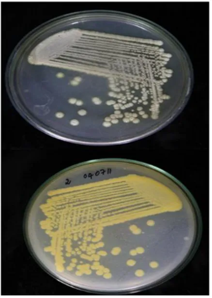
Streptomyces species (Fig. 1).
Figure 1 - Pure culture of pigment producing
Streptomyces griseoaurantiacus JUACT 01 grown on SCNA agar.
The16S ribosomal RNA encoding gene sequence analysis with sequences of representatives of
Streptomycetaceae family confirmed that the
unknown isolate belonged to genus Streptomyces
and the highest 16S rRNA gene sequence
similarity was found with Streptomyces
griseoaurantiacus strain NBRC 15440 (100%).
The sequence was submitted to GenBank database with accession number KJ774106. The organism was identified as Streptomyces griseoaurantiacus
JUACT 01.This was also confirmed by
Phylogenetic analysis performed using Neighbor-Joining method. Based on the phylogenetic tree the strain JUACT 01 showed highest affinity with
Streptomyces griseoaurantiacus by a 100%
bootstrap level and loosely related to others (Fig. 2).
Figure 2 - The evolutionary history was inferred using the
Neighbor-Joining method. The optimal tree with the sum of branch length = 0.06865153 is shown. The percentage of replicate trees in which the associated taxa clustered together in the bootstrap test (1000 replicates) are shown next to the branches. The evolutionary distances were computed using the Maximum Composite Likelihood method and are in the units of the number of base substitutions per site.
The pigment extracted using ethyl acetate as the solvent system was analysed by scanning the absorbance with UV-VIS spectrophotometer and the absorbance peak was observed at 410 nm. In order to select reproducible procedures and stable properties for characterization of Streptomyces
ISP3 and ISP4 media. Neither growth nor pigment production was seen with ISP7 media. Majority of the microbial secondary metabolite extraction procedures use combination of solvents varying in polarity. This method was selected for separation of the fractions conveniently with higher resolution (Seidel et al. 2005). Methanol and hexane in the ratio of 7:3was found to be the best solvent combination for the separation of the pigment by Thin Layer Chromatography (TLC). The yellow pigment was resolved on the silica sheet with Rf value of 0.78. The TLC fraction of the pigment was recovered by preparative TLC and the recovery was found to be 72%. A number of pigmented compounds with bioactivity have been isolated from microorganisms. Pigments from Streptomyces sp particularly have attracted
cancer biologist’s worldwide (Soliev et al. 2011). Chinikomycin A, chinikomycin B and manumycin A compounds isolated from marine Streptomyces
sp. M045are the pigmented antibiotics reported to possess anticancer property (Li et al. 2005). We have found that the yellow pigment which gave positive for the presence of carotenoid from our
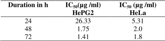
Streptomyces strain JUACT01 exhibited profound
anticancer potentials against HeLa and Hep G2 cells as indicated by their low IC50 concentrations
(Table 1).
Table 1 - IC50 concentration of TLC purified yellow
pigment on HepG2 and HeLa cells at 24, 48 and 72 h of treatment.
Duration in h IC50(µg /ml)
HePG2
IC50 (µg /ml) HeLa
24 48 72
26.33 1.75 1.41
5.31 2.0 1.8
Working on a marine isolate of Streptomyces
griseoaurantiacus strain MK393 Matsumoto et al.
(1998) have identified an antitumor antibiotic called diperamycin. When tested for growth inhibitory potentials on various tumour cell lines, diperamycin had exhibited significant activity. The yellow pigment extract from JUACT 01 when tested for in vitro cytotoxicity on HeLa and Hep G2 cells by using MTT cell viability assay resulted in considerable inhibition of cell growth and proliferation. Conversely the yellow pigment did not show any toxic effect on normal lymphocytes. Microscopic observation revealed that the lymphocytic cell morphology was unaltered and
growth and proliferation were unaffected even after 72 h of treatment (Fig. 3).
Figure 3 - Effect of yellow pigment on cell viability of human lymphocytes tested using MTT assay. Effect after ( ) 24 h, ( ) 48 h and ( ) 72 h.
Despite a number of drawbacks, novel apoptosis triggering drugs/approaches will enhance the development of innovative strategies in cancer therapy. DNA fragmentation is one of the notable biochemical changes occurring due to apoptosis (Huang et al. 1997). Appearance of discrete DNA fragments on conventional agarose gel is a clean evidence for the induction of apoptosis in the pigment treated cells. The DNA isolated from the cells treated with 20 µg /mL of pigment for 24 h, was seen as a smear when compared to intact band of DNA isolated from untreated cells. Along with DNA degradation and increased caspase activity, cells undergoing apoptosis exhibit morphological changes like plasma membrane blebbing, cell body shrinkage and formation of membrane bound apoptotic bodies (Cohen et al. 1992). Microscopic observation of the cells after staining with AO/EtBr stain enabled us to visualise the effect of pigment treatment on the morphology and viability of cancer cells (Telford et al. 1992). Typically viable cells were bright green in colour where as the dead ones were bright orange in colour (Fig. 4).
cycle with the recovered cells after treating with the pigment revealed that, there was a clear difference in the cell cycle stages between the untreated and treated cell populations of HeLa cells. It was seen that untreated cell population was healthy and multiplying with 40.27, 32.31 and 21.01 % of cells in G1, S and G2 phase respectively. The treated cancer cell population showed least number of cells in S phase (9.17%)
and fewer cells in G1 (32.78%) and G2 (12.11%) phase of the cell cycle indicating inhibition of proliferation. These results show the probable effect of pigment to cause apoptosis in the cancer cells. Activity of LDH present in the supernatant obtained by treating the cancer cells with 5.0 µg/mL of purified yellow pigment was higher than the control samples demonstrating the cytotoxic effect of the pigment on cancer cell lines.
Figure 4 - Fluorescence microscopy photos (A) untreated cells (B) pigment treated HeLa cells (C)
pigment treated HepG2 cells. The arrow indicates the fragmented DNA.
The activity obtained by the untreated cells was used to estimate maximum LDH released and the percentage activity was calculated for the control. The percentage cytotoxicity obtained by comparing the LDH activity between the positive, negative controls and the test samples was found to be 91% with HeLa cells and 111% with Hep G2 cells. Caspases are a family of aspertate- specific cysteine proteases that serve as the primary mediators of apoptosis. Also they are the early key indicators of apoptosis (Chang et al. 2000). The caspase activities measured show that there is a drastic increase in the activity of caspases in treated cells compared to the untreated cells with increase in time from t-0 minutes to t- 45minutes (Fig. 5).
Treatment with 5 μg ml-1 concentration of the purified pigment on HeLa cells resulted in 1.6 fold increase in the final caspase activity as compared to the initial caspase activity whereas with Hep G2 cells the increase was found to be 2 folds. The percentage caspase activity values indicate that the treated HeLa and Hep G2 cells show 125.03% and 111.72% more caspase activity than the untreated cells at t-45 minutes respectively. These results infer that the pigment treated cells indeed are undergoing apoptosis.
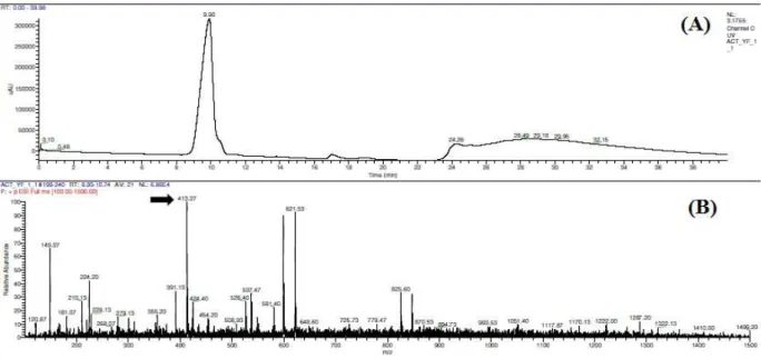
HPLC separation of the TLC fractionated pigment resulted in a major peak with a
retention time of 9.90 minutes. The
chromatograph shows no other major peaks indicating that the separation was ideal. Electro spray Ionisation Mass Spectrometry (ESI-MS) of the sample eluted at 9.90 minutes resulted with a peak showing 100% relative abundance corresponding to a mass to charge ratio (m/z value) of 413.22 (Fig. 6).
Figure 5 - Activity of caspases measured in ( ) Control
Figure 6 - HPLC and ESI-MS (A) HPLC chromatogram of TLC purified yellow fraction showing a major peak eluting at time 9.90 min (B) ESI- MS of HPLC fraction collected at time 9.90 min. Arrow indicates the m/z of the pigment fraction as 413.27 showing 100% relative absorbance.
The IR spectrum for the TLC separated pigment showed characteristic absorption frequencies of hydroxyl group in alcohol or phenol group at 3313 cm-1, carbonyl group at 1640 cm-1, nitro group at 1294 cm -1 and vinyl group at 915 cm-1.The ESI-MS and IR reports suggest that this kind of pigment with anticancer potentials was not reported from this species of Streptomyces and hence appears to be a novel one.
CONCLUSION
With these findings, we conclude that the yellow pigment from Streptomyces gresioaurianticus
JUACT 01 isolated from soil has cytotoxic potential and is nontoxic to human lymphocytes. It inhibits the growth and proliferation of HeLa and HepG2 cells in vitro through induction of apoptosis. These results offer justification for further research to evaluate the structural features of the pigment molecule and its effect on specific cancer cell lines.
ACKNOWLEDGMENTS
The financial support by way of the grant provided by DST (project no. SR/SO/HS-0072/2012) is
greatly acknowledged by the authors. The authors are grateful to the management of Jain Group of Institutions for the infrastructural facilities provided to carry out the work.
REFERENCES
Ajayi IA, Ajibade O, Oderinde RA. Preliminary phytochemical analysis of some plant seeds. Res J Chem Sci. 2011: 1(3); 58-62.
Altschul SF, Gish W, Miller W, Myers EW, Lipman DJ. Basic local alignment search tool. J Mol Biol. 1990: 215(3): 403-410.
Berdy, J. Bioactive microbial metabolites. J Antibiot.
2005: 58(1); 1-26.
Chang HY, Yang X. Proteases for cell suicide: functions and regulation of caspases. Microbiol Mol Biol Rev. 2000: 64(4); 821-846.
Coates J. Interpretation of infrared spectra, a practical approach. In: Meyers RA, editor. Encyclopaedia of Analytical Chemistry. UK: John Wiley and Sons Ltd; 2000. p. 10815–10837.
Cohen GM, Sun XM, Snowden RT, Dinsdale D, Skilleter DN. Key morphological features of apoptosis may occur in the absence of internucleosomal DNA fragmentation. Biochem J. 1992: 286; 331-334.
Ferlay J, Soerjomataram I, Ervik M, Dikshit R, Eser S, Mathers C, et al. Cancer Incidence and Mortality Worldwide: IARC CancerBase No. 11 [Internet]. 2013 [cited 2014 Dec.24] Lyon, France: International Agency for Research on Cancer. Available from: http://globocan.iarc.fr
Gomez A, Gomez A. A statistical procedure for agricultural research. 2. ed. Kwanchi: John Willy and Sons, 1984,188p.
Huang P, Ballal K, Plunkett W. Biochemical characterization of the protein activity responsible for high molecular weight DNA fragmentation during drug-induced apoptosis. Cancer Res. 1997: 57(16); 3407-3414.
Kirchner JG, Miller JM, Keller GJ. Separation and identification of some terpenes by new chromatographic technique. Anal Chem.1951: 23(3); 420-425.
Kirchner JG.Thin-layer chromatographic quantitative analysis. J Chromatogr A. 1973: 82(1); 101-115. Li F, Maskey RP, Qin S, Sattler I, Fiebig HH, Maier A,
et al. Chinikomycins A and B: Isolation, structure elucidation and biological activity of novel antibiotics from a marine Streptomyces sp. Isolate M045. J Nat Prod. 2005: 68 (3); 349-353.
Matsumoto N, Momose I, Umekita M, Kinoshita N, Chino M, Iinuma H et al. (1998). Diperamycin, a new antimicrobial antibiotic produced by Streptomyces
griseoaurantiacus MK393-AF2. I. Taxonomy,
fermentation, isolation, physico-chemical properties and biological activities. J Antibiot. 1998: 51(12); 1087-1092.
Mosmann T. (1983) Rapid colorimetric assay for cellular growth and survival: application to proliferation and cytotoxicity assays. J Immunol Methods. 1983:65 (1); 55-63.
Nadumane VK, Venkat P, Pal A, Dharod H, Shukla M, Prashanthi K. A novel metabolite from Aspergillus ochraceus JGI 25 showing cytotoxicity to HeLa cells.
Indian J Pharm Sci.2013: 75 (5); 507.
Olano C, Méndez C, Salas JA. Antitumor compounds from marine actinomycetes. Mar Drugs. 2009: 7(2); 210-248.
Pozarowski P, Darzynkiewicz Z. Analysis of cell cycle by flow cytometry. In Checkpoint Controls and Cancer. Humana Press; 2004.p.301-311.
Saitou N, Nei M.The neighbour-joining method: a new method for reconstructing phylogenetic trees. Mol Biol Evol . 1987: 4(4); 406-425.
Sambrook J, Fritsch EF, Maniatis T. Molecular cloning.2.ed. New York: Cold spring harbor laboratory press, 1989,149p.
Schwieger F, Tebbe CC. A new approach to utilize PCR–single-strand-conformation polymorphism for 16S rRNA gene-based microbial community analysis.
Appl Environ Microbiol. 1998: 64(12);4870-4876. Seidel V. Initial and bulk extraction. In Natural
products isolation. 2005, Humana Press. p.27-46 Selvameenal L, Radhakrishnan M, Balagurunathan R.
Antibiotic pigment from desert soil actinomycetes; biological activity, purification and chemical screening. Indian J Pharm Sci. 2009: 71(5); 499. Shirling EB, Gottlieb D. Cooperative description of
type cultures of Streptomyces. II. Species descriptions from first study. Int J Syst Bacteriol. 1968:18(2); 69-189.
Snyder LR, Kirkland JJ, Glajch JL. Practical HPLC method development.2012, John Wiley and Sons. Solanki R, Khanna M, Lal R. Bioactive compounds
from marine actinomycetes. Indian J Microbiol.
2008:48(4); 410-431.
Soliev AB, Hosokawa K, Enomoto K. Bioactive pigments from marine bacteria: applications and physiological roles. Evidence-Based Complementary and Altern Med. 2011: 2011(2011); 17.
Tamura K, Peterson D, Peterson N, Stecher G, Nei M, Kumar S. Molecular evolutionary genetics Analysis using maximum likelihood, evolutionary distance, and maximum parsimony methods. Mol Biol Evol. 2011: 28(10); 2731-2739.
Telford WG, King LE, Fraker PJ. Comparative evaluation of several DNA binding dyes in the detection of apoptosis‐associated chromatin degradation by flow cytometry. Cytometry. 1992: 13(2); 137-143.