An
Model of Skeletal Muscle Volume
Regulation
Anna Wibberley1, Caroline A. Staunton1, Claire H. Feetham1, Alexey A. Vereninov2, Richard Barrett-Jolley1*
1Department of Musculoskeletal Biology, Institute of Ageing and Chronic Disease, University of Liverpool, Liverpool, United Kingdom,2Laboratory of Cell Physiology, Institute of Cytology, Russian Academy of Sciences, St-Petersburg, Russia
Abstract
Introduction
Hypertonic media causes cells to shrink due to water loss through aquaporin channels. After acute shrinkage, cells either regulate their volume or, alternatively, undergo a number of metabolic changes which ultimately lead to cell death. In many cell types, hypertonic shrinkage is followed by apoptosis. Due to the complex 3D morphology of skeletal muscle and the difficulty in obtaining isolated human tissue, we have begun skeletal muscle volume regulation studies using the human skeletal muscle cell line TE671RD. In this study we in-vestigated whether hypertonic challenge of the human skeletal muscle cell line TE671RD triggered cell death or evoked a cell volume recovery response.
Methods
The cellular volume of TE671RD cells was calculated from the 2D surface area. Cell death was assessed by both the trypan blue live/dead assay and the TUNEL assay.
Results
Medium osmolality was increased by addition of up to 200mM sucrose. Addition of 200mM sucrose resulted in mean cell shrinkage of 44±1% after 30mins. At later time points (2 and 4 hrs) two separate cell subpopulations with differing mean cell volume became apparent. The first subpopulation (15±2% of the total cell number) continued to shrink whereas the second subpopulation had an increased cell volume. Cell death was observed in a small proportion of cells (approximately 6-8%).
Conclusion
We have established that a substantial proportion of TE671RD cells respond to hypertonic challenge with RVI, but that these cells are resistant to hypertonicity triggered cell death. OPEN ACCESS
Citation:Wibberley A, Staunton CA, Feetham CH, Vereninov AA, Barrett-Jolley R (2015) AnIn Vitro Model of Skeletal Muscle Volume Regulation. PLoS ONE 10(6): e0127889. doi:10.1371/journal. pone.0127889
Academic Editor:Alexander A. Mongin, Albany Medical College, UNITED STATES
Received:March 24, 2015
Accepted:April 20, 2015
Published:June 1, 2015
Copyright:© 2015 Wibberley et al. This is an open access article distributed under the terms of the
Creative Commons Attribution License, which permits unrestricted use, distribution, and reproduction in any medium, provided the original author and source are credited.
Data Availability Statement:All relevant data are within the paper.
Funding:AAV was supported by the Russian Foundation for Basic Research, project no. 15-04-00776-A, and CHF was supported by the BBSRC. The funders had no role in the design, data collection and analysis, decision to publish, or the preparation of the manuscript.
Introduction
Regulatory volume increase (RVI) and apoptotic volume decrease (AVD) are two opposing cel-lular volume-regulatory mechanisms [1–3]. The first of these, RVI, is frequently involved with adaptation to hypertonic media and cell survival, whilst in some cells, but not others AVD leads to cell death [4,5]. Frequently after cells reach a certain critical threshold of shrinkage, cells then undergo RVI or AVD. Hypertonic challenge can lead to apoptosis in a number of cell types [6–10]. In this study we investigate whether hypertonic challenge induces cell death in a human derived skeletal muscle cell line TE671RD.
Overall control of systemic osmolality is affected by a number of periventricular osmosen-sing structures within the brain [11] and involves osmotic response of individual neurones by mechanisms analogous to that of cell volume regulation itself [12]. Older people have an ap-proximately 3% (302.2 compared with 291.2 mOsm/Kg H2O) increased plasma osmolality
compared to healthy younger people [13]. This could be due to changes in kidney function, as a result of hypertension, or due to environmental factors such as diet. However, a loss in os-motic response is also observed in the elderly, suggesting that an issue with osos-motic control could result in increased plasma osmolality [14,15]. Cells are generally able to withstand small (2–3%) changes in tissue osmolality, but beyond this the activation of volume defence mecha-nisms becomes necessary [16]. Such chronic change in plasma osmotic potential of older peo-ple could therefore have a negative impact on skeletal muscle physiology, affecting such parameters as cellular volume. Indeed, a number of genes critical to both cell volume control and apoptosis, including the AQP2 and AQP3 aquaporin channels, are differentially expressed in ageing skeletal muscle [17]. The importance of apoptosis to ageing skeletal muscle physiolo-gy is controversial. It has been argued that apoptosis is not necessarily pathological and is im-portant for the process of remodelling [18], but it is increased subtly during ageing [19]. It has therefore been hypothesised that apoptosis may contribute to sarcopenia in older people [19–
22], potentially resulting from mitochondrial dysregulation [23]. Different skeletal muscle fibre types have been shown to have differing propensity to undergo apoptosis in response to TNFα [24], but apoptosis in response to hypertonic challenge has not previously been investigated. TE671RD cells are potentially an ideal model for cell volume regulation experiments because whilst they derive from human skeletal muscle they readily round-up and facilitate volume measurement. There is no evidence for the presence of t-tubules in TE671RD cells, structures which would naturally confound simple geometric estimation of cellular volume in native skel-etal muscle fibres. The literature shows that TE671RD cells express the skelskel-etal muscle specific form of the nicotinic acetylcholine receptor [25] and TTX-resistant voltage-gated sodium channels characteristic of native skeletal muscle [26]. Another ion channel, however, the KATP
channel is not pharmacologically identical in TE671RD cells [27] to that found in our own rat skeletal muscle studies [28]. It should be noted, however, that most skeletal muscle ion channel studies use rodent or amphibian tissue rather than human and so species is a confounding vari-able. Aside from muscle ion channel expression, a recent study showed the expression of striat-ed muscle developmental micro mRNA mir206 in TE671RD cells [29–31], but there have been few studies of other muscle like properties such as expression of contractile apparatus. There are also very limited data available on their volume regulation.
Methods
Culture
TE671RD cells constitute a human rhabdomyosarcoma cell line showing features of skeletal muscle differentiation [32,33]. These were a kind gift from the group of Prof Vincent (Oxford University, [34]). TE671RD cells were maintained in 300mOsm/Kg H2O DMEM (GIBCO
31885, Life Technologies Ltd, Paisley, UK) medium with 10% fetal calf serum, 1% amphoteri-cin B and 2% penicillin and streptomyamphoteri-cin at 37°C and 5% CO2. All reagents were obtained
from Sigma-Aldrich (UK) unless otherwise stated.
Measurement of cell size
Cells were detached from the flask surface by 0.25% Trypsin-EDTA solution. A pellet was formed by centrifugation of the cell solution (300 x g, 5 min) (MultifugeX1, Thermo Fisher Sci-entific) and supernatant removed. Cells were then re-suspended in culture media with FCS and incubated for 1 hour at 37°C and 5% CO2before the experiment. We measured cell shrinkage
upon exposure to hypertonic media with a wide range of sucrose concentrations (0–200 mM). Osmolality was measured using a freezing point micro-osmometer (3MO, Advanced Instru-ments, Norwood, USA). We compared cell volume in isotonic control and hypertonic samples using light microscopy with an Olympus IX51 equipped with a JVC TK-C921EG digital cam-era. Cell volume was calculated as described previously [35] from the 2D areas with ImageJ software [36].
Cell viability
Cell viability was assessed with the trypan blue exclusion assay. DNA fragmentation character-istic of apoptotic programmed cell death was detected with the terminal deoxynucleotidyl transferase (TdT)-mediated dUTP nick end labelling (TUNEL) assay [37] using the Trevigen apoptotic cell system (TACS) TACS 2 TdT-Fluor Apoptosis Detection Kit (Trevigen, Gaithers-burg, MD, USA). Control and sucrose treated cells were fixed in 10% neutral buffer formalin (NBF), placed onto slides and then processed according to the manufacturer’s instructions. After staining, slides were mounted with DAPI enriched VECTASHIELD Mounting Medium. Statistical analysis was carried out using the non-parametric Mann-Whitney test (p0.05) in MiniTab (Minitab Ltd., Coventry, UK).
Results
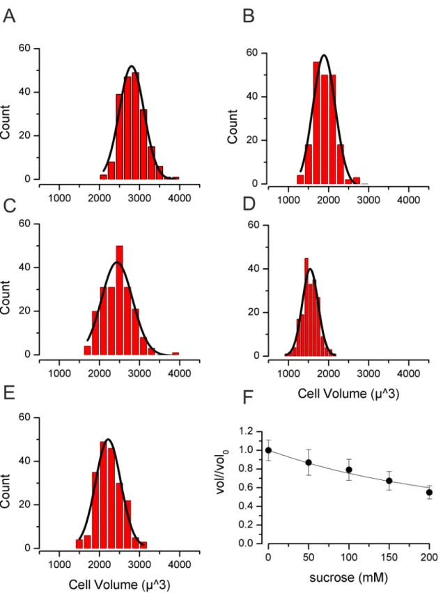
Under iso-osmotic control conditions (300 mOsm/Kg H2O) mean TE671RD cell volume was
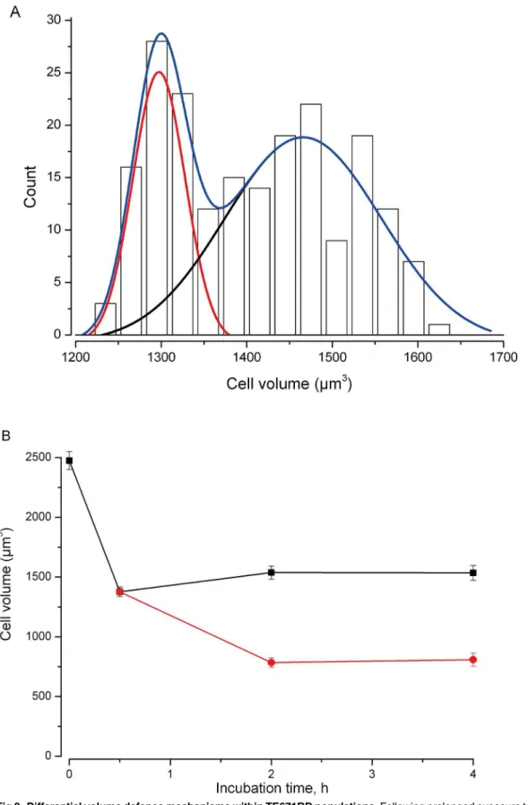
2840±22μm3(n= 200,Fig 1). Following 30 minutes incubation with sucrose (0 to 200mM), volume decreased to close to that predicted from“ideal osmotic behaviour”[38] (Fig 1). We then went on to measure cell volume at a series of time points (0.5, 2 and 4hr) following expo-sure to 200mM sucrose. We meaexpo-sured cell volume in between 100 and 200 cells in each of 13 separate experiments. Initially volume fell to 44±1% (n = 13, t = 0.5hr). By 2hrs cells had clus-tered into two distinct populations (assessed by cellular volume). Those cells identified in the first population reduced in size, whereas cells of the second population significantly increased in cellular volume (Fig 2). At 2hr 18±1% (n = 8) of cells had increased their volume (I.e., exhib-ited RVI) and this rose to 21±1% (n = 6) of cells at 4hr.
Fig 1. Volume of TE671RD following 30 min of increased osmolality. (A)Cells measured in 300mOsm/Kg H2O (no sucrose),(B)with 350 mOsm/Kg H2O (50 mM sucrose),(C)with 400 mOsm/Kg H2O (100mM sucrose)(D)with 450 mOsm/Kg H2O (150mM sucrose),(E)with 500 mOsm/Kg H2O (200mM sucrose). For (A) to (E) smooth lines are Gaussian fits.(F)Normalised volume against sucrose concentration. The smooth line is drawn to that of ideal osmotic behaviour:Vol/Vol0=V0*osm0/osmsucrose. WhereVoloandVolare the initial volume and the volume attained by the cells in the sucrose containing solution (measured at 30mins).osm0is the initial osmolality of the solution (I.e., DMEM alone, 300mOsm/Kg H2O) andosmsucroseis the osmolality once sucrose had been added (i.e., 300mOsm/Kg H2O + [sucrose]).
percentage of dead cells after 2hr and 4hr incubation with 200 mM sucrose was 1.5 and 1.9 fold higher (respectively) than in control samples (Fig 3). These changes were statistically increased from control, but there was no significance between the two time points of 2 and 4 hr. The DNA fragmentation fluorescent microscopy assay showed that after 4hr hypertonic exposure (200mM sucrose) 6±1% (n = 5) of cells had a bright green nucleus with condensed chromatin, consistent with the development of DNA fragmentation.
Discussion
In this report we investigated whether hypertonic challenge triggers cell death and/or regulato-ry volume increase (RVI) in the human skeletal muscle cell line, TE671RD. We found that a significant proportion of cells underwent RVI and there was minimal resultant cell death even with very high osmotic challenge. Initial shrinkage very closely fitted that expected from the
“ideal osmotic shrinkage”described in native skeletal muscle fibres by Blinket al[38] and
Fer-encziet al[39]. Blinket al[38] saw little evidence of RVI and Ferencziet al[39] reported only
a few percent RVI. In contrast, Lindingeret alsaw significant and rapid RVI in mouse soleus
(slow twitch) skeletal muscle [40]. We find that the TE671RD population divides in to subpop-ulations with and without significant RVI capacity in a similar way to that we reported previ-ously for leukaemia U937 cells [2].
Apoptosis is a stereotyped form of programmed cell death which includes a number of mor-phological and biochemical changes including DNA fragmentation [41]. The degree of in-creased osmolality in older people is relatively small (3%) [13], compared with the challenge we applied here of up to 200mOsm/Kg H20, but in our assays only a small proportion of cells
showed evidence of DNA fragmentation. We would therefore tend toexcludeincreased
osmo-lality as a potential trigger for apoptosis and sarcopenia in ageing people. It should be noted, however, that we applied a high osmolality solution for only a few hours, whilst naturally, peo-ple would have their small elevation in plasma osmolality for many years. The osmolalities we used here were quite extreme; (DMEM plus added sucrose) varied between 300 and
500mOsm/Kg H2O. It was shown in rabbit studies that 400mOsm/Kg H2O and above results
in severe clinical symptoms, and most animals died by 450mOsm/Kg H2O [42].
One particular point of note in the present study is the apparently slow rate of shrinkage, with minimum volume being measured at a time of 2hrs following hypertonic challenge. There are several points surrounding this. The first is that since our protocol was to measure at 0.5 hrs and then again at 2 hrs, it is entirely possible that the minimum volume was actually reached well before 2 hrs. The reason for this is that our experimental design was not intended to demonstrate the kinetic properties and follow progression of individual cells (as we have done in some other studies), but to follow populations of cells. Some cells trigger RVI earlier, and some later (some not at all), but we felt that by 2 hrs all cells that were going to undergo RVI would have begun to do so. We caution against kinetic interpretation of our data, however, since the study simply was not designed for that. A more interesting hypothesis is that these cells have very few aquaporin channels and very low water permeability. We will investigate this in the future. From a biological/biochemical perspective it is interesting to note that the rate of attaining a new cellular volume following change of solution osmolality is dependent upon cell water permeability. This can be at least 10 fold different between different types of cells and is greatly affected by the composition of the extracellular solution [43]. In cases where similarly slow rates of cellular shrinkage have been observed [2,43,44], the extracellular
continued to shrink (red circles); the second population of cells (black squares) maintained or increased volume. At 2 hrs 15±2% (n = 9) of cells are included in the smaller population, at 4hrs 17±2% (n = 7) are included in the smaller population.
osmolality was also increased by sucrose (similar to our own study) which seems to decrease cell permeability [43]. One might speculate this could involve block of aquaporin channels by sucrose, but we are not aware if this has been investigated directly. Yurinskayaet al[2] show
comparative shrinkage to be much faster in the presence of elevated NaCl compared to sucrose and the rapid skeletal muscle shrinkage observed by [40] used addition of NaCl. Whilst the aquaporin composition of skeletal muscle varies with age [17], the predominant isoform is AQP4 [45] and further pharmacological analysis of this and other aquaporins would be useful. Finally, it is possible that following passive shrinkage, a degree of active volume shrinkage then takes place; the mechanism of which would be beyond the scope of the current manuscript. This could be some sort of“dying cell volume decrease”(DVD) as seem in many [4,5], but not all cell types [4,46]. Since we did not see a great deal of cell death itself, even at 4 hrs, this seems unlikely. We found that a rather high proportion of cells lacked RVI, but few cells ap-peared to initiate apoptosis and so our data does not support a notion that hypertonic shrink-age and lack of RVI directly correlates to induction of apoptosis. This does not directly shed light on the debate as to whether shrinkage is a necessary step for apoptosis in this cell line, however [4,5], since there was so little evidence of apoptosis itself.
The present study used a human skeletal muscle derived cell line, TE671RD. Previously, TE671RD cells have been used mainly for investigation of the human nicotinic receptor physi-ology [33], but we will continue to investigate its volume regulatory properties in parallel with otherin vitroskeletal muscle systems. Myoblasts formed from freshly isolated rodent tissue
would be an alternative skeletal muscle cell volume model and it would be interesting to com-pare the volume regulatory properties of human TE671RD cells to such cell lines as the rat L6 or mouse C2C12. The attraction to us of TE671RD cells is that they constitute a human skeletal muscle derived cell line and thus express human muscle isoforms of important muscle recep-tors such as the nicotinic acetylcholine receptor [25]. There is unlikely to be a single perfectin vitromodel of skeletal muscle cell volume control. For example, in the introduction to this
manuscript we discuss known similarities and differences between native skeletal muscle and TE671RD cells, but it is also likely that ion channel expression is dissimilar between different skeletal muscle fibre types withinthe samespecies. In terms of volume regulation, TE671RD
cells form round cells and allow for the measurement of cellular volume from 2D surface area in a way difficult to reproduce with freshly dissociated skeletal muscle, but possible with the ro-dent myoblasts. The clear limitation is the differences we have observed (above) between TE671RD cells and previously reported native skeletal muscle. In future studies we will contin-ue to investigate the cellular machinery of skeletal muscle cellular volume control, both in terms of its regulatory volume increases and decreases. Furthermore, we will explore apoptosis in this cell line and investigate responses to inflammatory cytokines known to be important for skeletal muscle senescence. This work may prove useful, not just in the study of ageing, but also in the disease most closely thought to involve skeletal muscle cellular volume control; compartment syndrome.
In conclusion, we found that the human skeletal muscle cell line, TE671RD exhibits greater active cell volume regulation than reported for some native skeletal muscle and the cells are very resistant to hypertonicity triggered cell death. If these effects were reproduced in native human skeletal muscle it would strongly suggest that the relatively modest plasma hypertonici-ty measured in older people is not likely to contribute to cell death or sarcopenia.
microscope. Fluorescent microscopy showed the presence of DNA fragmentation after 4 h of 200 mM sucrose incubation. 6±1% (n = 5) of sucrose-treated cells demonstrated a bright green nucleus with condensed chromatin. The TUNEL apoptosis assay was conducted at 4hrs.
Acknowledgments
We would like to thank Rebecca Lewis for her support during this study, and the Oxford Neu-rosciences Group for the kind gift of the TE671RD cells [33,34]. AAV was supported by the Russian Foundation for Basic Research, projects no. 15-04-00776-A and CHF was supported by the BBSRC. The funders had no role in the design, data collection and analysis, decision to publish, or the preparation of the manuscript.
Author Contributions
Conceived and designed the experiments: AW CAS CHF AAV RBJ. Performed the experi-ments: AW CAS CHF RBJ. Analyzed the data: AW RBJ. Contributed reagents/materials/analy-sis tools: AAV RBJ. Wrote the paper: AW CAS CHF AAV RBJ.
References
1. Maeno E, Takahashi N, Okada Y. Dysfunction of regulatory volume increase is a key component of ap-optosis. FEBS Lett. 2006; 580(27):6513–7. doi:10.1016/j.febslet.2006.10.074PMID:
WOS:000242665700034.
2. Yurinskaya VE, Moshkov AV, Wibberley AV, Lang F, Model MA, Vereninov AA. Dual response of human leukemia U937 cells to hypertonic shrinkage: initial regulatory volume increase (RVI) and de-layed apoptotic volume decrease (AVD). Cellular physiology and biochemistry: international journal of experimental cellular physiology, biochemistry, and pharmacology. 2012; 30(4):964–73. Epub 2012/ 12/12. doi:10.1159/000341473PMID:23221465.
3. Okada Y, Maeno E, Shimizu T, Dezaki K, Wang J, Morishima S. Receptor-mediated control of regulato-ry volume decrease (RVD) and apoptotic volume decrease (AVD). J Physiol. 2001; 532(Pt 1):3–16. Epub 2001/04/03. doi:10.1111/j.1469-7793.2001.0003g.xPMID:11283221; PubMed Central PMCID: PMC2278524.
4. Orlov SN, Model MA, Grygorczyk R. CrossTalk opposing view: the triggering and progression of the cell death machinery can occur without cell volume perturbations. The Journal of Physiology. 2013; 591(24):6123–5. doi:10.1113/jphysiol.2013.258624
5. Lang F, Hoffmann EK. CrossTalk proposal: Cell volume changes are an essential step in the cell death machinery. J Physiol. 2013; 591(Pt 24):6119–21. doi:10.1113/jphysiol.2013.258632PMID:24339145; PubMed Central PMCID: PMC3892460.
6. Bortner CD, Cidlowski JA. Absence of volume regulatory mechanisms contributes to the rapid activa-tion of apoptosis in thymocytes. American journal of physiology. 1996; 271(3 Pt 1):C950–61. PMID: 8843726; PubMed Central PMCID: PMC8843726.
7. Edwards YS, Sutherland LM, Power JH, Nicholas TE, Murray AW. Osmotic stress induces both secre-tion and apoptosis in rat alveolar type II cells. Am J Physiol. 1998; 275(4 Pt 1):L670–8. PMID:9755098. 8. Friis MB, Friborg CR, Schneider L, Nielsen MB, Lambert IH, Christensen ST, et al. Cell shrinkage as a
signal to apoptosis in NIH 3T3 fibroblasts. J Physiol. 2005; 567(Pt 2):427–43. doi:10.1113/jphysiol. 2005.087130PMID:15975986; PubMed Central PMCID: PMC1474190.
9. Ghosh A, Keng PC, Knauf PA. Hypertonicity induced apoptosis in HL-60 cells in the presence of intra-cellular potassium. Apoptosis: an international journal on programmed cell death. 2007; 12(7):1281–8. doi:10.1007/s10495-007-0054-zPMID:17333319.
10. Racz B, Reglodi D, Fodor B, Gasz B, Lubics A, Gallyas F Jr., et al. Hyperosmotic stress-induced apo-ptotic signaling pathways in chondrocytes. Bone. 2007; 40(6):1536–43. doi:10.1016/j.bone.2007.02. 011PMID:17392049.
11. Bourque CW. Central mechanisms of osmosensation and systemic osmoregulation. Nat Rev Neurosci. 2008; 9(7):519–31. Epub 2008/05/30. doi:10.1038/nrn2400PMID:18509340.
12. Feetham CH, Nunn N, Lewis R, Dart C, Barrett-Jolley R. TRPV4 and KCa functionally couple as osmo-sensors in the PVN. Br J Pharmacol. 2014:doi:10.1111/bph.13023doi: 10.1111/bph.13023.
13. McLean KA, O'Neill PA, Davies I, Morris J. Influence of age on plasma osmolality: a community study. Age Ageing. 1992; 21(1):56–60. PMID:1553862.
15. Stachenfeld NS, DiPeitro L, Nadel ER, Mack GW. Mechanism of attenuated thirst in aging: Role of cen-tral volume receptors. American Journal of Physiology-Regulatory Integrative and Comparative Physi-ology. 1997; 272(1):R148–R57. PMID:WOS:A1997WG56500020.
16. Hoffmann EK, Lambert IH, Pedersen SF. Physiology of Cell Volume Regulation in Vertebrates. Physiol Rev. 2009; 89(1):193–277. doi:10.1152/physrev.00037.2007PMID:ISI:000262247600006.
17. Welle S, Brooks AI, Delehanty JM, Needler N, Thornton CA. Gene expression profile of aging in human muscle. Physiol Genomics. 2003; 14(2):149–59. doi:10.1152/physiolgenomics.00049.2003PMID: WOS:000184204600006.
18. Allen DL, Linderman JK, Roy RR, Bigbee AJ, Grindeland RE, Mukku V, et al. Apoptosis: a mechanism contributing to remodeling of skeletal muscle in response to hindlimb unweighting. American Journal of Physiology-Cell Physiology. 1997; 273(2):C579–C87. PMID:WOS:A1997XQ26200024.
19. Dirks A, Leeuwenburgh C. Apoptosis in skeletal muscle with aging. American Journal of Physiology-Regulatory Integrative and Comparative Physiology. 2002; 282(2):R519–R27. PMID:
WOS:000173287100023.
20. Marzetti E, Lees HA, Manini TM, Buford TW, Aranda JM, Calvani R, et al. Skeletal Muscle Apoptotic Signaling Predicts Thigh Muscle Volume and Gait Speed in Community-Dwelling Older Persons: An Exploratory Study. PLoS One. 2012; 7(2). doi:10.1371/journal.pone.0032829PMID:
WOS:000302999600064.
21. Marzetti E, Leeuwenburgh C. Skeletal muscle apoptosis, sarcopenia and frailty at old age. Experimen-tal gerontology. 2006; 41(12):1234–8. doi:10.1016/j.exger.2006.08.011PMID:
WOS:000243219800008.
22. Skulachev VP. What is "phenoptosis" and how to fight it? Biochemistry (Mosc). 2012; 77(7):689–706. Epub 2012/07/24. doi:10.1134/S0006297912070012PMID:22817532.
23. Pollack M, Phaneuf S, Dirks A, Leeuwenburgh C. The role of apoptosis in the normal aging brain, skele-tal muscle, and heart. In: Harman D, editor. Increasing Healthy Life Span: Conventional Measures and Slowing the Innate Aging Process. Annals of the New York Academy of Sciences. 9592002. p. 93– 107. PMID:11976179
24. Phillips T, Leeuwenburgh C. Muscle fiber-specific apoptosis and TNF-alpha signaling in sarcopenia are attenuated by life-long calorie restriction. FASEB J. 2005; 19(1):668–+. doi:10.1096/fj.04-2870fje PMID:WOS:000226576600014.
25. Luther MA, Schoepfer R, Whiting P, Casey B, Blatt Y, Montal MS, et al. A muscle acetylcholine receptor is expressed in the human cerebellar medulloblastoma cell line TE671. J Neurosci. 1989; 9(3):1082– 96. PMID:2564429.
26. Fakler B, Ruppersberg JP, Spittelmeister W, Rudel R. Inactivation of human sodium channels and the effect of tocainide. Pflugers Arch. 1990; 415(6):693–700. PMID:2159618.
27. Miller TR, Taber RD, Molinari EJ, Whiteaker KL, Monteggia LM, Scott VES, et al. Pharmacological and molecular characterization of ATP-sensitive K+ channels in the TE671 human medulloblastoma cell line. Eur J Pharmacol. 1999; 370(2):179–85. doi:http://dx.doi.org/10.1016/S0014-2999(99)00128-4 PMID:10323267
28. Barrett-Jolley R, McPherson GA. Characterization of K-ATP channels in intact mammalian skeletal muscle fibres. Br J Pharmacol. 1998; 123(6):1103–10. PMID:ISI:000072694700012.
29. Goljanek-Whysall K, Pais H, Rathjen T, Sweetman D, Dalmay T, Münsterberg A. Regulation of multiple target genes by miR-1 and miR-206 is pivotal for C2C12 myoblast differentiation. 2012. doi:10.1242/ jcs.101758
30. Williams AH, Valdez G, Moresi V, Qi X, McAnally J, Elliott JL, et al. MicroRNA-206 Delays ALS Progres-sion and Promotes Regeneration of Neuromuscular Synapses in Mice. Science. 2009; 326
(5959):1549–54. doi:10.1126/science.1181046PMID:20007902; PubMed Central PMCID: PMC2796560.
31. Taulli R, Bersani F, Foglizzo V, Linari A, Vigna E, Ladanyi M, et al. The muscle-specific microRNA miR-206 blocks human rhabdomyosarcoma growth in xenotransplanted mice by promoting myogenic differ-entiation. J Clin Invest. 1192009. p. 2366–78.
32. Stratton MR, Darling J, Pilkington GJ, Lantos PL, Reeves BR, Cooper CS. Characterization of the human cell line TE671. Carcinogenesis. 1989; 10(5):899–905. Epub 1989/05/01. PMID:2650908. 33. Barrett-Jolley R, Byrne N, Vincent A, Newsom-Davis J. Plasma from patients with seronegative
myas-thenia gravis inhibit nAChR responses in the TE671/RD cell line. Pflugers Archiv: European journal of physiology. 1994; 428(5–6):492–8. Epub 1994/10/01. PMID:7838671.
35. Lewis R, Asplin KE, Bruce G, Dart C, Mobasheri A, Barrett-Jolley R. The role of the membrane potential in chondrocyte volume regulation. J Cell Physiol. 2011; 226(11):2979–86. Epub 2011/02/18. doi:10. 1002/jcp.22646PMID:21328349; PubMed Central PMCID: PMC3229839.
36. Abramoff MD, Magelhaes PJ, Ram SJ. Image Processing with ImageJ. Biophotonics International. 2004; 11(7):36–42.
37. Gavrieli Y, Sherman Y, Ben-Sasson SA. Identification of programmed cell death in situ via specific la-beling of nuclear DNA fragmentation. J Cell Biol. 1992; 119(3):493–501. PMID:1400587; PubMed Cen-tral PMCID: PMC2289665.
38. Blinks JR. Influence of Osmotic Strength on Cross-Section and Volume of Isolated Single Muscle Fi-bres. J Physiol. 1965; 177:42–57. PMID:14296959; PubMed Central PMCID: PMC1357223.
39. Ferenczi EA, Fraser JA, Chawla S, Skepper JN, Schwiening CJ, Huang CL. Membrane potential stabili-zation in amphibian skeletal muscle fibres in hypertonic solutions. J Physiol. 2004; 555(Pt 2):423–38. doi:10.1113/jphysiol.2003.058545PMID:14694151; PubMed Central PMCID: PMC1664835. 40. Lindinger MI, Leung M, Trajcevski KE, Hawke TJ. Volume regulation in mammalian skeletal muscle:
the role of sodium-potassium-chloride cotransporters during exposure to hypertonic solutions. Journal of Physiology-London. 2011; 589(11):2887–99. doi:10.1113/jphysiol.2011.206730PMID:
WOS:000291256100022.
41. Hengartner MO. The biochemistry of apoptosis. Nature. 2000; 407(6805):770–6. doi:10.1038/ 35037710PMID:11048727
42. Sotos JF, Dodge PR, Meara P, Talbot NB. Studies in Experimental hypertonicity: I. Pathogenesis of the Clinical Syndrome, Biochemical Abnormalities and Cause of Death. Pediatrics. 1960; 26(6):925–38. 43. Troshin AS. CHAPTER II—Do Live Cells Have Osmometric Properties? In: Troshin AS, editor.
Prob-lems of Cell Permeability (Revised and Supplemented Edition). 26: Pergamon; 1966. p. 30–56. 44. Dall’Asta V, Bussolati O, Sala R, Parolari A, Alamanni F, Biglioli P, et al. Amino acids are compatible
osmolytes for volume recovery after hypertonic shrinkage in vascular endothelial cells1999 1999-04-01 00:00:00. C865-C72 p.
45. Frigeri A, Nicchia GP, Balena R, Nico B, Svelto M. Aquaporins in skeletal muscle: reassessment of the functional role of aquaporin-4. The FASEB Journal. 2004; 18(7):905–7. doi:10.1096/fj.03-0987fje PMID:15033928