CECAV-UTAD Publications
Institutional Repository of the
University of Trás-os-Montes and Alto Douro
Found at:
http://repositorio.utad.pt/
This is an author-produced version of a book chapter
published in
Advances in Medicine and Biology. Volume 94
This paper does not include the final publisher
proof-corrections or pagination.
Citation for the published book chapter:
R. Payan-Carreira and M.A. Pires. Ovarian Cysts in Dogs´
Practice. In: Advances in Medicine and Biology. Vol 94, 2016,
Chapter 4, Editor: Berhardt, L.V., Nova Science Publishers Inc:
65 - 88. ISBN: 978-1-63484-185-6
Published in final form at:
Paper back:
https://www.novapublishers.com/catalog/product_info.php?prod
ucts_id=56587
Ebook:
https://www.novapublishers.com/catalog/product_info.php?prod
ucts_id=56588
Copyright: Nova Publishers
Access to the published version may
require subscription.
In: Advances in Medicine and Biology. Vol 94 ISBN: 978-1-63484-185-6
Editor: Berhardt, L.V. © 2016 Nova Science Publishers, Inc.
Chapter XX
OVARIAN CYSTS IN DOGS´ PRACTICE
Rita Payan-Carreira
*and Maria dos Anjos Pires
CECAV - Animal and Veterinary Research Centre, Universidade de Trás-os-Montes e Alto Douro (UTAD), Vila Real, Portugal
Abstract
In dogs, ovarian cysts include the fluid-filled structures of any size present within the ovaries, outside physiological proestrus and estrus; they may be unilateral or bilateral. They are a common finding, particularly in older females and in females under contraceptive treatments. In dogs, cysts can originate from various structures in and around the canine ovary; therefore not all the ovarian cysts are hormonally active. Like in other domestic species, canine ovarian cysts may be grouped into follicle-derived cysts, which are hormonally active, and the non-follicular cysts, which are known to be incapable of steroid production, thereby evolving often silently, in the absence of specific clinical signs, in contrast to follicle-derived cysts that usually interfere with the female normal cyclicity. Yet, most types of cysts may compromise fertility. Recent surveys suggests that the prevalence of one type of the other may vary with the region of the study, possibly representing differences in the reference dog population, in the strategies used to control female dog reproductive status, in methodological approaches to diagnosis or in the routine procedures for the practitioner facing the problem.
The morphology of the canine ovary, together with the tendency for a disturbed growth of the subepithelial structures, makes it prone to the cystic degeneration of the ovarian surface that may physically compromise ovulation and limits the bitch fertility. On another hand, follicular cysts that can arise spontaneously or in consequence of the stimulation of a cycle, can either compromise the ovulation rate of a female and reduce the litter size or predispose to uterine diseases due increased levels of estrogens. Either way, it is important to establish an early diagnose of the condition, and try to restore fertility if the female are intent to be breed. This work intended to provide a comprehensive overview of available information on dog ovarian cysts. Based on our practice, we discuss the trends on prevalence of ovarian cysts in the species, the different types of ovarian cysts and the corresponding clinical conditions and the available diagnostic approaches as well as the existing therapeutic options. The information provided in constitutes a helpful tool in the clinical approach to canine ovarian cysts.
Payan-Carreira and Pires
Introduction
In dogs, cysts arising within or from around the ovaries are relatively common in the clinical practice. However, it is difficult to quantify the prevalence of ovarian cysts from the literature, as most reports describe particular clinical situations affecting a very small number of individuals [1-3] or they focus on particular aspects of the disease [4] or its diagnosis [5]. Clinical studies on the incidence or prevalence of ovarian cysts are rare in dogs, and the existing report cases referred to histopathology services, which per se present an important limitation since they are based on the necropsies findings [6, 7] or depend on the practitioner perception of an existing lesion in an ovariectomy/ovariohysterectomy specimen for which he/she needs a diagnosis and by the owner which accepts to request the histopathological evaluation. An old study refer that non-neoplasic ovarian cysts account to 78% of all canine ovarian diseases [6]. More recently, Pires and Payan-Carreira [8] referred that ovarian cysts (neoplasic and non-neoplasic) represented around 35% of the lesions found in canine ovaries, but only in ca. 54% of the situations the cysts were visible under gross inspection of the ovaries. Therefore, it is possible that prevalence of canine ovarian cysts vary with:
- The geographical location, in particular because of the differences regarding the usual contraceptive practices (whether or not a female is spayed at youngest ages);
- The breeds represented in the region, as often the breeds change with time, modes and even with the sort of veterinary establishment participating in the studies;
- The use of drugs to manipulate the canine estrous cycle, particularly for induction of estrus;
- The procedures used for diagnosing ovarian cysts (such as ultrasonography, endocrine determinations, visual inspection, or histopathology).
Ovarian cysts are broadly defined as fluid-filled structures of any size within the ovary, present outside the physiological follicular phase of the estrous cycle [9]. Although this definition covers both neoplastic and non-neoplastic cavitary lesions, when talking about ovarian cysts the neoplasic cystic lesions (adenomas or carcinomas of the ovary) are usually unconsidered.
It is essential for the clinician as well as for the pathologist to be aware of the structures from which cysts may arise in the canine ovary, either for the implications of its origin on the cyst ability to produce steroid hormones, and thereby to interfere with the female estrous cycle and to induce mammary or uterine diseases, or to hinder ovulation and thus interfering with the female fertility in the absence of other clinical signs. These issues will be detailed in following sections.
The morphology of the canine ovary
In dogs, the ovary is completely enclosed within the ovarian bursa, a structure resulting from the fusion of the distal mesovarium and the mesosalpinx, located close to the caudal pole of the ipsilateral kidney [10, 11]. The ovarian bursa contains a large amount of adipose tissue that hides the ovary from view, except in a small clear area of the mesosalpinx [12]. In general, fat content increases with age, in particular after puberty, so that young female dogs
Ovarian cysts in dogs
may present a more translucent ovarian bursa whereas older or overweighed females have it opaque.
The gonads are long and slightly flattened, and their size may be influenced by breed size. Table 1 summarizes the main available information on the ovary size and weight in dogs. It is generally accepted that differences in size between the right and the left ovary are insignificant [13] provided that no lesions are presented. In dogs the opening (ostium) of the ovarian bursa is very narrow; it is usually made obvious by the predominance of few reddish
tubal fimbriae [11]. Normally, neither the ovary nor any ovarian structure protrudes at the ostium of the ovarian bursa.
Small, minor variations in the ovary size may be perceived when large antral follicles (during final proestrus and estrus until ovulation) or mature corpora lutea (in early and mid diestrus) protrude above the ovarian surface, but it returns to its regular size when these structures sank below the ovarian surface. The changes in size will depend on the number of structures developing in each cycle [14]. The contour of the mature canine ovary is irregular from proestrus to mid-diestrus (i.e., during the follicular and luteal stages of the estrous cycle) because of the presence of follicles and corpora lutea [12]; during anestrus, the surface of the ovary is smooth.
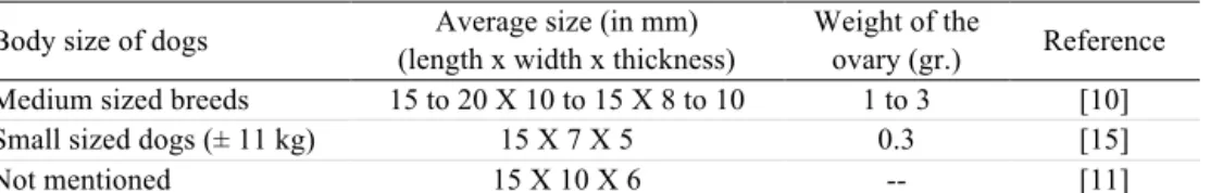
Table 1. Reported size and weight of the canine ovary
Body size of dogs (length x width x thickness) Average size (in mm) Weight of the ovary (gr.) Reference
Medium sized breeds 15 to 20 X 10 to 15 X 8 to 10 1 to 3 [10]
Small sized dogs (± 11 kg) 15 X 7 X 5 0.3 [15]
Not mentioned 15 X 10 X 6 -- [11]
The ovary is outlined by a surface epithelium [10, 16], originated from the coelomic epithelium, which is constituted by a simple layer of low cuboidal to columnar epithelial cells [10, 16]. In older references the surface epithelium of the ovary was named “germinal epithelium” [17]. It is possible to find some small invaginations arising from the surface epithelium, simple or branched tubular, with a variable disposition, that some authors suggested to be at the origin of the sub-surface crypts [12] also named sub-surface epithelial structures (SES) [7, 17, 18]. SES are ingrowths of the peritoneal covering of the ovary that continue to develop throughout life; they are only described in dogs among all other domestic species [12].
The surface epithelium covers a densification of the cortical stroma, inaccurately called ovarian albuginea, disposed as a thin layer of regularly organized, denser connective tissue, poorly delimited on profundity, as it progress into the interstitial stroma of the ovary [10].
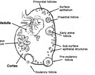
Ovarian cortex
As in other mammalian species, the canine ovary comprises a cortex and a medulla (Figure 1). In dogs, the ovarian cortex is rather thin compared with the medulla. The ovarian cortex is sustained by a dense stroma, rich in reticular and collagen fibres and in a particular type of fusiform cells that resemble smooth muscle cells but are devoid of any striation [10]. Fibroblasts with epithelial-like characteristics are disposed around follicles as follicular theca [19]. A rich network of thin blood vessels covers the cortical area of the ovary and shows a spherical, denser disposition around the follicles, in particular in those on latter developmental stages. In the cortex, a variable number of follicles can also be observed.
Payan-Carreira and Pires
Neoangiogenesis is particularly evident in early developing corpora lutea. Many lymphatic vessels are associated with the external follicular theca layer [19]. It is also possible to observe in the ovarian cortex of female dogs particular structures once named granulosa cell cords or islands [20] also called as interstitial gland. These structures, that possibly arise from atresia of large follicles, form cords of clarifying cytoplasm cells where granulosa cells continued to proliferate [21]; these structures may further differentiate into pseudotesticular tubules [12, 20].
Figure 1. Drawing of the morphology of the canine ovary
The morphology and size of the follicles varies according to their developmental stage and the stage of the cycle. Dogs are a polytocous species, so multiple follicles develop in each cycle [22]. At birth, most oogonia are surrounded by pre-granulosa cells, and follicle differentiation is not completed. Oogonia enter the meiotic prophase shortly after birth, the primordial follicles becoming increasingly abundant by 2 months of age [20, 23]. At 4 months of age, primary follicles are recognized in the canine ovarian cortex, surrounded by a single layer of cuboidal granulosa cells. Secondary follicles show two or more layers of granulosa cells disposed around fully-grown oocytes [24]. Synthesis of follicular fluid marks the transition into tertiary or early antral follicles; these can be found in the canine ovary between 4 and 5 months of age, whereas large, advanced antral follicles can be found in the ovary of female dogs reaching to puberty, close to 6 months of age [20, 24]; from this moment on, the development of large antral follicles coincides the follicular stage of each ovarian cycle.
In dogs, follicular recruitment occurs in the last third of anestrus [25], but it is only in late proestrus that follicles are perceived at the ovarian surface as small vesicular structures. The periovulatory size of canine follicles may vary slightly with the size of dogs. In general, ovarian follicles at ovulation measure 9 to 12 mm in diameter [22]. Fontbonne [26] reports that the average size at ovulation, assessed by ultrasound measurement of the inner follicular diameter, increased with the size of female (4.5±0.5mm 5.3±0.9mm, 5.9±0.4mm and 5.8±1mm, respectively in small, medium, large and giant females), although the differences among groups were devoid of significant, maybe due to the limited number of females. Table 2 provides a summary of available information on the size of antral follicles and corpora lutea
Ovarian cysts in dogs
in dogs. Once the size is an important parameter for the diagnosis of ovarian cysts, it is important that the practitioner and the pathologist to know the size and appearance of normal mature follicles in the species.
After ovulation, the post-ovulated follicles transform into corpora lutea that bulge above the ovarian surface until the 5th week, and gradually decrease in size until they are no longer perceived below the ovarian surface (Table 2). Nevertheless, corpora lutea can still be traced on the canine ovary over the next two estrous cycles.
Ovarian medulla
The medulla is usually in a central position in the ovary, taking a more superficial position at the level of the hilum, where blood vessels and nerves enter and exit from the gonad. The medulla consists of loose collagenous tissue mingled with elastic and reticular fibres [19]. Besides the large blood and lymphatic vessels and the nerves, the medulla of the canine ovary also presents embryonic remnants that form the rete ovarii, formed by intraovarian mesonephric-derived cells. This structure is located adjacent to the hilum [12] and extends into the medulla; its development and disposition may vary among females [7]. The rete ovarii is a cluster of cell cords and tubules, lined with a densely stained cuboid epithelium and containing scant fluid [18]; it frequently forms small papillae, particularly in mature and old females. Andersen and Simpson [20] refer the existence of changes in the size of rete ovarii in diestrus and anestrus, thought Mialot and Parodi [7] could not detect changes in this structure during the stages of the cycle.
In carnivores, the rete system extends, into the periovarian tissue, forming the extra-ovarian rete that ends blindly, derived from mesonephric tubules and cords or the mesonephros [9].
Cyst in and around the canine ovary
There are different types of cyst that develop within and around the ovary (Figure 2; Table 3). Cysts occurring in the ovary may be of two main types [5, 9, 12]:
- The follicular cysts (Figure 2), arising from medium-large antral anovulatory follicles that often originate during a follicular phase and remain in the ovary for longer periods;
- The non-follicular cysts (Figure 2) that include cysts originated from the mesonephric tubules (cysts from the rete ovarii and paraovarian cysts), cysts derived from the surface epithelium (that might evolve to adenoma of the ovary) and cysts coming from the subsurface epithelial structures (SES).
Although in the literature the existence of luteal cysts in dogs is mentioned [4, 6, 27], their existence as a clinical entity is uncertain. Cystic corpora lutea are described in dogs, but in fact antral corpora lutea are always present in the first ~20 days of diestrus; thus, unless a clinical situation encompassing an elongation of the interestrous interval due to prolonged luteal stage (evidenced by higher levels of peripheral progesterone for a period over 60-70 days) accompanies the presence of corpora lutea (CL) larger than the usual for the breed size, the diagnosis of cystic CL should be disregarded. Nonetheless, in dogs, the most frequent cause for interestrous elongation is a prolonged anestrus [28].
Payan-Carreira and Pires
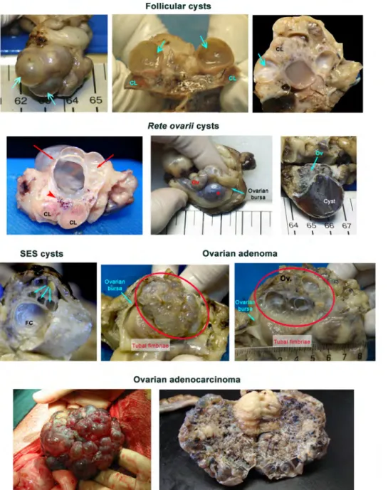
Figure 2. Macroscopic features of canine ovarian cysts. Follicular cysts – non-movable regular cysts
(arrows) delimited by a thick and regular wall, containing a serous fluid, that may escort corpora lutea (CL). On cut sections, the luteinization of the walls is usually limited to the inner mural surface. Rete ovarii cysts (red arrows or *) are cysts located in the hilum (arrowhead) of the ovary. Therefore, ovarian parenchyma (Ov) appears parallel to the cyst origin; the vascular network supplying the ovary is usually found between the cysts and the ovary. SES cysts are usually small sized surface cysts (Cyan arrows) presenting a regular and thin wall, that do not support blood vessels, and containing a serous fluid; they are frequently multiple. In this image, they co-exist with a large follicular cyst (FC). Ovarian adenoma (delimited by the red oval shape) represents a benign proliferation of the surface and SES epithelium, originating multiple small cysts that give the ovary surface a foaming appearance. Usually, the gonad (Ov) is contained within the ovarian bursa and the tubal fimbriae show a normal surface. On the cut surface, the differences in individual cysts sizes are noticeable. In the ovarian adenocarcinoma the size of the cysts at the ovarian surface is rather variable, and its appearance quite heterogeneous. On the cut surface the ovarian parenchyma is usually not visible, and often the entire gonad presents a sponge feature.
Ovarian cysts in dogs
Epidemiological aspects
The majority of ovarian cysts present no signs or symptoms [8]. Thereby, they frequently remain undiagnosed, which makes it difficult to estimate the incidence and prevalence of the condition. As said before, the incidence of the different types of ovarian cysts is difficult to establish in dogs. Moreover, some confusion exits on the classification of cysts, particularly in clinical practice, because the available complementary diagnostic methodologies do not allow to clearly distinguish among the different epithelial ovarian cysts (cystic rete tubules, cysts of SES, paraovarian cysts or adenomas), which impairs to establish the true incidence of the different type of cysts in dogs´ practice. The existence or the absence of pathology in the uterus of the mammary gland may be of additional value for the clinical diagnosis. Although the practitioner must be aware that both the uterine and mammary diseases may co-exists with non-follicular cysts following independent age-related process, and therefore may have independent courses. Aging increases the prevalence of uterine diseases that in dogs are associated with characteristic features of the species reproductive cycle, namely the relatively long recurrent progesterone dominance stages [25].
No clear information exists on the epidemiologic aspects of the canine ovarian cysts. Seldom information based on clinical surveys is reported in the literature. In previous surveys, follicular cysts would be less frequent than those originating from SES or the rete [5, 8, 27] (Table 4). Yet, the number of published case descriptions is higher for the later compared to the former, for which it would surely contribute the fact that follicular cysts or malignant ovarian adenocarcinoma usually evidences exuberant clinical signs, while the cysts of the SES and rete, as well as the surface epithelium adenoma usually progresses in a silent clinical form. Data compiled on Table 4, resulting from the studies of several veterinary histopathology laboratories [8, 27, 29], clearly show that epithelial cysts originated from the surface-SES and rete structures are those presenting a higher prevalence worldwide. In opposition, among the ovarian cystic tumours, adenocarcinoma is rarer than the adenoma [6].
Most reports agree that incidence of cystic disease in dog ovaries increase with age [6, 8, 9], particularly on what concerns epithelial cysts originating from the SES or the rete ovarii [28], which is also true for ovarian adenoma. Age predisposition for follicular cysts is not so clearly defined, in particular because some evidences now exists that deslorelin treatments may induce follicular cysts [2, 30] due to anovulation. Nevertheless, spontaneous follicular cyst have been described more frequently in female dogs above 5 to 6 years of age [2, 31, 32] than in younger females. In contrast, the ovarian adenocarcinoma develops in older females, between 8 to 10 years of age [33, 34] albeit the most exuberant situations have been described in younger females, in general 3-4 years of age [34, 35].
Ov ar ia n cy st s in d og s Ta b le 2 . Si ze a nd a pp ea ra nc e of n or m al an tr al fo ll icl es an d co rp or a lu tea (C L ) in d og s. [1 4, 2 2] De ve lo pme nt al s ta ge St ag e of th e cy cl e Si ze ( m m ) Gr os s appe ar anc e An tr al f ol li cl e La te a ne st ru s 0. 2 – 1. 5 No t v is ib le o n th e ova ri an su rface O nl y vi si bl e in longi tudi na l c ut s of the ova ry Ea rl y pr oe st ru s 1. 5 - 3 Ra nd om ly d is tr ib ut ed i n th e ov ar ia n su rf ac e as c le ar g re yi sh a re as , w ith in dis tin ct bounda ri es . Pr e-ovul at or y fol li cl es La te p ro es tr us an d ear ly est ru s 5 – 8 Fo ll ic le s ap pe ar a s ve si cu la r st ru ct ur es b ul gi ng f ro m t he o va ri an s ur fa ce , sp he ric al in sh ap e Fo ll ic ul ar w al ls a re tr an sl uc en t a nd su pp or t a co nv ol ut ed va sc ul ar ne tw or k Pe ri -ovul at or y fol li cl es On e da y po st LH s ur ge 4 -13 Th e fo ll ic ul ar w al l be com e ri gi d and opa que due t o pr e-ovul at or y lut ei ni za ti on of t he mu ra l g ra nu lo sa la ye r Pr io r to o vu la ti on , t he s ti gm a is v is ib le a s a gr ey s po t i n th e fo ll ic ul ar d om e At o vu la ti on 9 -12 Th e fo ll ic le t en si on d ec re as es , b ut t he f ol li cu la r w al ls d o no t co ll ap se a t ov ul at io n, a nd th e h ae m or rh ag e w ith in th e f ollic le c av ity is n eg lig ib le Co rp or a lu te a Di es tr us 6. 5 – 12 (1 st w eek p os t-ovul at ion) L ut ei ni za ti on of th e po st -ovul at or y fol li cl es i s co m pl et ed i n 2-3 w ee ks . C ha nge s in co lo ur , si ze, pr ot rus ion above t he ova ri an sur fa ce a nd cons is te nc y oc cur dur ing CL lif es pa n. In b rie f, in th e in itia l sta ge s of CL ma tu ra ti on , a sl ig ht ly e cc en tr ic a nt ru m is vi si bl e; t he f ol li cul ar w al ls be com e opa que a nd ar e pi nki sh to ca rm ine i n col our . A ft er tw o w ee ks , th e m atu re CL ar e co m pact , car m in e an d st il l pr ot ru de ab ov e th e su rf ace of th e ov ar y; af te r th e 5 th we ek , t he p ro je ct io n of t he CL at t he ov ar ian su rf ace decr eases, as it d ecr eases in si ze an d beco m e m or e fi rm an d yel lo w ish i n co lo ur . B y we ek 1 2 pos t-ovul at ion CL ar e vi si bl e as sm al l pal e yel lo w st ru ct ur es w it hi n th e ov ar ian co rt ex ; al th ou gh n ot b ul gi ng t hey can b e fo un d by v isu al i ns pe ct ion unt il w ee k 16 pos t-ovul at ion. Co rp or a lu te a co nt in ue t o de cr ea se i n si ze , bu t th ey c an b e tr ac ed o ve r th e ne xt 2 es tr ous c yc le s.
Pa ya n-Ca rr ei ra a nd P ir es Ta b le 3 . Ma in c la ss if ic at io n of o va ri an c ys ts b y or ig in Or ig in Ty pe o f cy st s Br ie f m ac ro sc op ic d es cr ip ti on * Br ie f hi st ol ogi ca l de sc ri pt ion ** C YS T S W IT HI N T HE OVAR Y Fo ll ic le Fo ll ic ul ar c ys ts [G ra af ia n or no n-ovul at or y cys ts ] Va ri ab le in n umb er a nd s iz e No n-mo va bl e cy st s bu lg in g ab ov e th e ov ar ia n su rf ac e In th e ov ary , n orm al ly d ev el op ed C L m ay c o-ex ist Ma y or ig in at e sy nd ro m es of est ro gen im bal an ce Cy st s ar e li ne d by g ra nu lo sa c el ls , ex pr es sin g in hib in al ph a, b ut n eg at iv e fo r desm in o r cy to ker at in s (A E 1/ A E 3) Va ri ab le nu mb er of gr an ul os a ce ll la ye rs , th at ma y pr es ent s igns of a tr es ia Lu te in iz at io n of t he m ur al g ra nu lo sa m ay o r m ay n ot b e pr es ent Th e oo cy te is d eg en er at ed Th ec a la ye r m ay b e r ep la ce d b y f ib ro us tis su e Su b-su rface ep it hel ial st ru ct ur es SES cy st s St ar ti ng of te n as a dy sp la st ic pr ol if er at io n of SES le ad in g to th e fo rm atio n of v es ic le s; co nf lu en t ve sic le s re m ai n se pa ra te d by th in la m el la e of c on ne ct iv e ti ss ue As th ey g ro wt h, S E S c ys ts d ef or m th e ov ar ia n su rf ac e Ra re ly la rg er th an 5 m m Of te n bi la te ra l Mo re of te n in old er an im als an d fe m ale s un de r pr oge st age n tr ea tm ent Li ne d by si m pl e-cu bo id cel ls si m il ar to th os e of th e su rface or S E S ep it hel iu m M ay p resen t v eg et at iv e in gr ow th s St ro ng ex pr es si on of cy to ke ra ti ns (A E 1/ A E 3+ ) an d de sm in. Ma y w ea kl y ex pr es s pl ac ent al al ka li ne phos pha ta se (P L A P ), w hile th e su rf ac e ep it hel iu m sh ow s m od er at e ex pr essi on Re te o va ri i Re te c ys ts Re pr es en ts c ys ti c di la ta ti on s of t he r et e tu bu le s in t he pe ri -hi la r ar ea of the ova ry Mu lt ip le c ys ts o f va ri ab le s iz e Wh en la rg e, th ey m ay c om pr es s th e ov ar ia n co rt ex Fr eq ue nt ly u ni la te ra l Ma in ly in o ld er a ni m al s Cy st s li ne d by a s in gl e la ye r of cu bo id ep it hel iu m ; m ay be c il ia te d Po si ti ve f or S1 00 , d es m in a nd cy to ker at in s (A E 1/ A E 3) C YS T S AR OUND T HE OVAR Y Pa ra ov ar ia n cy st s Wo lf ia n or Mü lle ria n d uc ts Cy st s ar is in g fr om W ol fi an re m na nt s lo ca te in th e me so va ri um, c lo se to th e ov ar y tu bu la r ex tr em it y. Mü ll er ia n du ct c ys ts lo ca te in th e fi m br ia e. Mo va bl e cy st s of th in w al ls on the c ra ni al s ur fa ce of the ova ri es , th at m ay pr otr ud e fr om th e os ti um of the ova ri an bur sa Va ri ab le i n nu mb er , if so li tar y th ey t en d to ach iev e bi g di m ens ions Wo lf ia n cy st s ar e co m po sed o f th in w al l of co nn ect iv e tis su e an d m us cle , lin ed b y a sin gle la ye r of c ub oid al or lo w co lu m na r cel ls w it h cl ear cy to pl asm , co ver in g a ba se m ent m em br ane . S om e ce ll s ar e ci li at ed Mü ll er ia n cy st s pre se nt a n ep it he li um s im il ar to t ha t of th e ute rin e tu be s, m ay o r no t be c ilia te d, an d la ck s a ba se m ent m em br ane
Ov ar ia n cy st s in d og s C YS T S E P IT HE L IAL T UM OUR S OF T HE OVAR Y SES an d Su rf ac e ep it hel iu m Ov ar ia n ad en oma Co ve ri ng th e ov ar ia n su rf ac e En la rg ed a nd ir re gu la r sh ap e of th e ov ar y Sm oo th ed o r no du la r Fr eq ue nt ly b il at er al Mu lt ip le c ys ti c di ff er en tl y si ze d st ru ct ur es Th e en la rg ed o va ry is c on fi ne d w it hi n th e ov ar ia n bu rs a Ma in ly i n m id dl e ag ed o r ol d fe m al es or i n fe m al es unde r cont ra ce pt ive tr ea tm ent [B en ig n le si on ] In fo ld in gs an d pa pi ll ary pro je ct io ns of the ova ri an su rface an d S E S st ru ct ur es Sc an t c on ne ct iv e ti ss ue s tr om a Cy st s ar e li ne d by s m al l cu bo id al o r cy li nd ri ca l ce ll s th at ma y pr es en t c il ia Le ss o ft en , c el ls a re a rr an ge d in a g la nd ul ar p at te rn Ra re m it ot ic f ig ur es Po si ti ve to c yt ok er at in s (A E1 /A E 3) Ov ar ia n ad en oc ar ci no ma Co ve ri ng th e ov ar ia n su rf ac e Irre gu la r sh ap e of th e ov ary M al ig na nt fo rm : Ad en oc ar ci no ma Mu lt ip le c ys ts o f di ff er en t s iz e an d as pe ct Ra pi d gr ow th a nd p ro li fe ra ti on De st ro ys t he o va ri an b ur sa , fr ee in g th e ov ar y in t he ab do m in al cav it y Hi gh ly me ta st at ic à su dd en cl in ical on set acco m pan ied b y asci tes an d dep ressi on De ve lo ps in y ou ng f ema le s [M al ig na nt le si on ] Si m il ar h is to lo gi ca l a pp ea ra nc e to th e ov ar ia n ad en om a In cre as ed m it ot ic a ct iv it y In va si on in to th e ov ari an s tro m a Ex te ns io n in to ti ss ue s ad ja ce nt to th e ov ar y Va sc ul ar in va sio n ma y be o bs er ve d Si ze an d su rf ace per m eat io n ar e im po rta nt cr ite ri a of ma li gn an cy * B as ed o n th e ref er en ces [7 , 9 , 1 2] ** B as ed o n th e ref er en ces [5 , 3 6]
Ovarian cysts in dogs
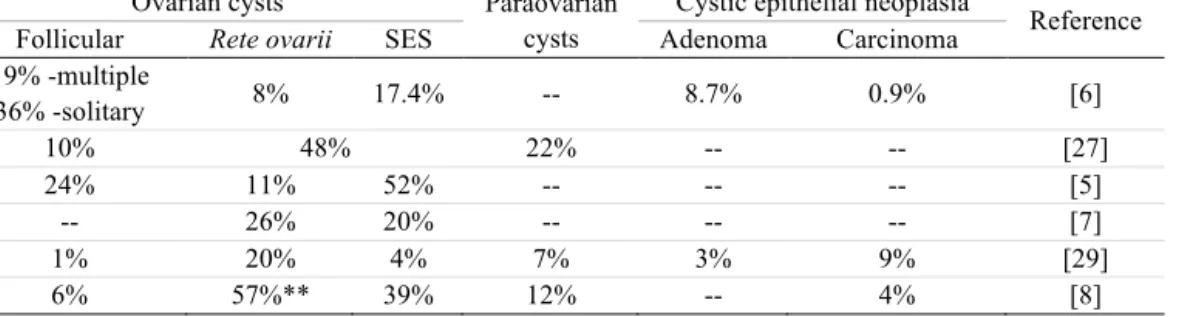
Table 4. Prevalence of canine ovarian cysts (%) according to the type*
Ovarian cysts Paraovarian
cysts
Cystic epithelial neoplasia
Reference
Follicular Rete ovarii SES Adenoma Carcinoma
19% -multiple 36% -solitary 8% 17.4% -- 8.7% 0.9% [6] 10% 48% 22% -- -- [27] 24% 11% 52% -- -- -- [5] -- 26% 20% -- -- -- [7] 1% 20% 4% 7% 3% 9% [29] 6% 57%** 39% 12% -- 4% [8]
* The percentages were estimated from the total number of ovaries with lesions or with cysts, according to references
** this value represents cyst located in the rete ovarii and hilar region, therefore it may include also paraovarian cysts.
Pregnancy and parturition seems not to have a protective role for cystic diseases in the canine ovaries. Independently of the cyst type (epithelial vs. follicular, or neoplasic vs. non-neoplasic cysts), they develop in both the nulliparous and multiparous females. Moreover, in our practice, development of epithelial cysts from SES tends to be anticipated in females under contraceptive treatment.
In dogs, ovarian cysts can evolve as solitary or multiple structures and also can develop on only one ovary or on both ovaries [6]. Nevertheless, epithelial cysts in the ovary are frequently bilateral while paraovarian cysts tend to occur as unilateral structures. In a recent publication, Knauf and colleagues [4] found that multiple follicular cysts were more frequent than the solitary and also that they occurred often in both ovaries, contrasting to our personal experience, corroborated by Dow findings [6] who also observed more often solitary follicular cysts than multiple follicular cysts.
Until now no breed predisposition has been described for the occurrence of ovarian cysts, whether concerning the epithelial or the follicular cysts. Nonetheless, case reports on follicular cysts represented more frequently purebred bitches than mongrels. This can be related to the fact that most mongrels are spayed or under medical contraceptive treatments (depending on the country) to avoid breeding.
Signalement and clinical aspects
The origin of the ovarian cysts clearly influences the development of overt clinical symptoms. Grossly, follicular cysts are often associated with more or less clear signs of hyperestrogenism, particularly when they are multiple, whereas the cysts originating from the ovarian epithelial structures frequently develops for long unnoticed since they do not produce sex steroids. Additionally, ovarian adenocarcinoma is usually accompanied of a severe clinical form distinctive from the other epithelial cysts or cystic tumours of ovarian origin. Therefore, we will distinguish in this section between follicular cysts, silent epithelial ovarian cysts and ovarian adenocarcinoma.
Payan-Carreira and Pires
Follicular cysts
Follicular cysts are hormonally active structures that develop in the female dog ovary. They often represent large antral follicles that failed to ovulate, but did not respond to atresia stimulus. Therefore, under gross examination, follicular cysts resemble Graafian follicles, larger than a normal follicle (see table 2 to evoke the follicular size in dogs).
In the bitch, anovulatory cysts may develop as solitary or multiple structures, and they may also be unilateral or bilateral. As main difference to other carnivore species, the follicular cysts do not luteinise past the existing mural granulosa luteinization existing at the moment of its formation. Therefore, changes from an estrogen to progestagen dominance does not occur in dogs, compared to cats and cows [37].
Spontaneous follicular cysts tend to occur in middle and old age groups. For the canine follicular cysts, based on the literature, Johnston and colleagues [28] describe an age range of 1-19 years and an average age at diagnosis of 8 years old. Recently, evidences exist that they also develop following therapeutic induction of estrus when deslorelin, an GnRH analogue, is used [2, 30]. Authors agree on the need to implant only female in anestrus, after a thoroughly gynaecological exam. However it is important to remember that the interestrous interval may vary between bitches, and this parameter should always be taken into consideration when defining the most suitable moment for insertion of deslorelin implants.
Under gross inspection, ovarian cysts may appear as thin-wall structure(s) containing clear serous fluid (Figure 2), similar in appearance but larger in size than the cohort of existing follicles or corpora lutea bulging above the ovary. In the wall of the cysts, small blood vessels may appear, but the blood network is often less expressive than in normally developing large antral follicles. The size of the cystic follicles varies from 1-1.5 to 10 mm in diameter [9, 12], though sporadic reports on larger [27] or smaller [4] cysts exists.
The pathogenesis of the process remains unclear. It has been proposed that follicular cysts originate from an inability of a large antral follicle to respond to the ovulatory stimulus [37, 38]. This mechanism may originate from a central failure to produce an adequate amount of gonadotropins, in particular of LH, to sustain an adequate follicular growth and ovulation [4], or from at peripheral level due to imbalances in the follicular molecular homeostasis compromising its response to the pre-ovulatory LH surge. The former hypothesis would explain the occurrence of multiple follicular cysts in dogs, while the later would rather apply to the occurrence of solitary follicular cysts.
Follicular cysts are known to be productive, that is they retain the capacity to produce estrogens and/or progesterone, thereby presenting the potential to cause disease [4]. Their reflexes on the bitch´ clinical condition depends on the amount of sex steroids, in particular the estrogens, produced by the cyst(s), which may depend also from the number of the cysts developing in the ovaries. Marino et al. [27] report changes in the ovarian cycle in 53% of the surveyed cases, coexisting with uterine dysfunctions, while the remainder 47% were asymptomatic due to its association with corpora lutea. Moreover, Knauf and colleagues [4] found only a fair correlation between the sex steroid peripheral levels and the estradiol and progesterone in matched follicular cysts, whilst the estradiol or progesterone concentrations in blood were not correlated with the number of ovarian cysts present in a given individual. Moreover, they also refer to the co-existence of follicular cysts and corpora lutea [4], which could be explained by the fact that steroids hormones concentration, particularly those of estrogens, was higher and highly variable in the cyst-fluid than in the peripheral blood.
Ovarian cysts in dogs
In dogs, antral non-ovulatory follicles will suffer atresia during diestrus, sequential degeneration of the oocyte and granulosa cells [6, 9, 27], suggesting that this may be a self-limiting condition, in the absence of co-existing uterine or mammary dysfunction (which in dogs are importing clinical entities requesting treatment by themselves). This can also be the fate for the follicular cysts induced by deslorelin treatment, since as inferred from Borges and collaborators [30] the females recover the ovulation rates in subsequent induced or spontaneous cycles. The role of the progesterone levels in diestrus on the follicular cysts regression has yet to be explored in dogs.
Hyperestrogenism represents a syndrome that may present a variety of clinical signs or symptoms depending on the relative amount of estrogens/progesterone in tissues. The owner could report either persistent estrus [27, 28, 31], which may be lengthened up to 3 months, or shortened interestrous interval [4, 28] compared to the normally exhibited by the female with recurrent heat behaviour. Seldom, if estrogen production by cysts is insufficient to contradict the progesterone dominance, the ovarian cycle may not be disturbed and the female retain her normal cyclicity. In females exhibiting elongated estrus, the visual inspection of the perineum reveals the existence of vulvar oedema; however, if estrogen stimulation persists for more than 30-40 days, the vulva losses the typical turgid swollen aspect displayed during heat, and a flaccid oedema can be noticed; estrogen impregnation may be confirmed in the vaginal cytology, by the presence of typical keratinised cells. Swelling of the mammary glands [9], which can be observed in some cases [39], is not always perceived by the owners.
If the ability to produce estradiol encompasses or overcome that of the progesterone production, changes in the estrogen and progesterone in target tissues for sex steroid would reflect in the onset of disease, particularly in the uterus. Often, the disturbances in the sex steroid ratios will favour the development of mucometra and pyometra [6, 9], which can be favoured by pre-existent cystic endometrial hyperplasia, particularly in older females [37]. Developing mucometra or pyometra may originate main clinical symptoms, particularly the later, which include: anorexia, depression or lethargy, polyuria and polydipsia, vomiting, abdominal distension, abdominal distress or pain, mucous or purulent vaginal discharge and in some cases, sepsis [40]. Less frequently, possibly associated with a chronic exposure to low levels of estrogen, the uterus develops keratinizing squamous metaplasia and chronic endometritis [9], perceived by a chronic purulent vaginal discharge, which is frequently associated with more severe symptoms of uterine endometritis/pyometra.
Skin diseases can be an important symptom that often takes the owner attention [27, 32, 37] in chronic low estrogen impregnation. These include a nonpruritic symmetrical bilateral alopecia, lignification, hyperpigmentation and hyperkeratosis. Alopecia most frequently develops on the caudal aspects of both thighs [32].
Toxic effects associated with chronic exposure to estrogens may also escort hyperestrogenism. The following problems may combine in hyperestrogenism cases: pancytopenia, anaemia, thrombocytopenia and consequent bleeding (represented by epistaxis, haematuria, petechia or haemorrhagic vulvar discharge) or granulocytopenia [9].
Sporadically, the owner complains of disturbed behaviour, in particular pseudopregnancy, but which is seldom clinically confirmed, and no references to it have been found in the literature.
Payan-Carreira and Pires
Ovarian epithelial cysts and ovarian adenomas
Ovarian epithelial cysts (whether they are SES or rete cysts) are non-productive, i.e. they do not produce sex steroid hormones, and therefore they progress asymptomatically [41]. Consequently, they are often found as an incidental finding during routine physical or abdominal ultrasound examinations, during uterine surgery or at necropsy, in the absence of ovarian disorder. As they do not disturb the normal cyclicity of the female dog, they are detected in otherwise normal ovaries [6]. Due to these characteristics, information on the pathology and the clinical course of epithelial cysts of the canine ovary is scarce.
The co-existence of uterine or mammary diseases with ovarian epithelial cysts must not be understood as indicator of an overproduction of sex steroid hormones. As their incidence increase with age, in general, the concomitant present of other age-related diseases should be primarily foresee as a coincidence of independent processes.
SES cysts (Figure 2) develop in the ovarian cortex, contrasting to those of the rete ovarii or the derived from Wolfian ducts, that are found in the ovarian hilus [7, 9] SES and rete cysts may co-exist in the same ovary [12].
Frequently, the SES cysts are small, its size ranging from microscopic cysts, barely visible during visual inspection, until close to 5mm, that give a spumous aspect to the ovarian surface, but they are seldom larger than this [12]; they tend to spread through the ovarian surface, as they are commonly multiple. In general, cysts of the surface/SES extending at the ovarian surface, alike adenomas, usually enlarges the normal ovary size; beyond a given dimension, enlarged ovaries can be palpated in the caudal pole of the kidney, as small, relatively firm ovoid masses, in mature to old females, above 7 years old. Occasionally, during the physical examination, unspecific signs of abdominal distress during transabdominal palpation can be observed. Distinction between SES cysts grossly visible and covering the ovarian surface and adenoma is difficult [9, 38], although the later tend to show thicker, irregular walls; moreover, adenomas increase usually the dimension of the ovary, which appear as large, floating masses caudal to the kidney. Most SES cysts and adenoma are recovered by the surface epithelium of the ovary [7]. It is possible that in more advances situations, the cystic proliferation of the cysts in the ovarian cortex may impose a physical barrier or in some way disturb the ovulation, thus being associated with infertility. Still this hypothesis was never proved.
Contrasting to the SES structures, cystic rete ovarii (Figure 2) are often unilateral, and they develop in the whole or in a segment of the rete tubules. They are easily confounded with paraovarian cysts under macroscopic examination. MacLachlan [38] refers that rete ovarii cysts may induce the compression of the ovarian cortex due to their size; however our practice shows that this is observed when cysts develop as solitary or a small numbered cysts of larger dimensions. According to Mialot and Parodis [7], age seems not to predispose to the development of rete ovarii cysts.
Cysts originating from remnants of the Wolfian ducts and other paraovarian cysts (Figure 2) also develop near the ovarian hilus, and they reach dimensions over the 4 and 5 mm [7, 12]. They are thin-walled, slow growing cysts, which allows them to protrude from the ostium of the ovarian bursa as they became large. Still, the paraovarian cysts are not report to impair ovarian function or compromise the fertility potential for the female.
Ovarian cysts in dogs
Ovarian adenocarcinoma
Ovarian adenocarcinomas are rare malignant tumour of the superficial structures of the ovarian cortex. They are usually multicentric and may be bilateral, which suggests the existence of a genetic background. In the study by Patniak and Greenlee [34], about one third of the ovarian carcinomas were bilateral.
Ovarian carcinomas may vary in size; ovaries are frequently are detected as irregular, multicystic masses above 10 cm, with a typical cauliflower aspect [9] (Figure 2). Nevertheless, small-sized ovarian carcinomas were found in older females [6, 28, 33]; the size of the tumour does not seem to determine the existence of metastatic disease [6]. In most severe situations, they invade and completely destroy the ovarian bursa, thereby exposing the ovary within the abdominal cavity. Our experience suggest that size does not relate with the tumour invasiveness, therefore a more complete histopathological assessment of the tumour (invasion of the ovarian capsules, number of mitosis per high power filed, the presence of necrotic areas, for instance) is needed to provide valuable information concerning the survival expectancy.
Once free of the bursa confinement, the tumour rapidly spread and abdominal implantation quickly occurs [9], and in rare cases giving distant metastasis in the lung or liver, originating exuberant clinical signs. There are evidences that this tumour responds to estrogen stimulation [9], but they are not productive by themselves; thus ovarian carcinoma does not disturb the normal ovarian cycle. The tumours are initially insidious, and initially progresses sub-clinically; clinical symptoms are usually detected when the carcinoma invade and originates metastatic syndromes. Abdominal effusion is commonly associated with carcinomatosis of the peritoneal cavity. Metastasis occurs by invasion and implantation in adjacent tissues but also by lymphatic and vascular permeation [9, 34].
Most reports on ovarian adenocarcinomas describe an acute onset of abdominal distension in a female close to 3 years of age that had recently become lethargic and losing weight [35, 42, 43]. Previous reproductive history is not helpful to the diagnosis of ovarian carcinoma, as they may be diagnosed in nulliparous females, but also in females with a pregnancy and parturition in the previous cycle [9] and the female maintain regular estrous cycles [42]. During physical examination, the palpation of an abdominal mass in the caudal abdomen, as described by Ajadi et al. [35], it is not always possible, depending on the amount of ascites and the abdominal tension. Pleural effusion may be present in more severe situation and with lung metastasis [28], raising difficulties to heart and lung auscultation; in those situations distressful respiratory movements and altered respiratory rate may be observed in the female. Abdominal discomfort may be present, and often the owners report a reduction of activity in particular concerning abdominal stressing exercises. Vulvar discharge is occasionally reported [42]. Dehydration may be detected, which along with diverse degrees of renal insufficiency, may influence the levels of some biochemical parameters in blood analysis [35]. Non-regenerative anaemia and leucocytosis are mentioned in most cases. Additionally, hypercalcemia has been referred due to altered parathyroid function [43].
Diagnosing ovarian cysts
Cystic structures that do involve follicles and thereby do not originate hyperestrogenism may persist silent for long period of time, thus remaining undiagnosed until they have enough size
Payan-Carreira and Pires
to be detected upon abdominal palpation or during an abdominal ultrasound scan. Diagnosis should results from the integration of the information collected by different methods.
An attentive collection of the reproductive anamnesis, along with the physical examination will discard suspicions of hyperestrogenism that accompanies follicular cysts. Although the measurements of peripheral levels of estradiol are considered effective (but not pathognomonic) for its diagnosis, the vaginal cytology may be helpful to confirm the existence of persistently elevated estrogen impregnation if females with history of persistent heat or recurrent short interestrous interval (provided they are in heat). In the physical exam, the vulva is frequently swollen, and a more or less abundant dark sero-haemorrhagic or mucous discharge is present [41, 42]; if estrus symptoms last for more than 28 days, a flaccid oedema of the vulva may be found, and the vulvar discharge may become whitish or mucous-purulent. The existence of a chronic vaginal discharge is also a common complaint. Although the female displayed estrus behaviour, in general she does not allow mounting, and its personality may become altered when heat is prolonged for more than a month. Follicular cysts diagnosis is made based on the history of persistent estrus and/or hyperestrogenism. The main differential diagnosis sought to be made with granulosa cell tumours producing high levels of estrogens, for which the results from the abdominal palpation and the abdominal X-ray and ultrasound are important diagnostic criteria.
On the physical examination, bitches are often alert and do not present discomfort, and the rectal temperature, pulse and respiratory rates were within reference ranges. An exception to this apparent normality is the ovarian adenocarcinoma, which generally is accompanied by complaints of anorexia, discomfort, apathy, pronounced weight loss and ascites [35, 43]. Besides the adenocarcinoma situations, only dogs presenting large ovarian cysts may show abdominal distension. Refusal to climb or descend stairs may, however, be reported by owners when the ovarian masses reach a significant increase in size, which may be associated with stimulation of adrenergic pathways due to distension of the ovarian bursa. Non-pruritic bilateral symmetrical alopecia in the trunk and perineum, as well as lichenification and hyperkeratosis, suggests hormonal imbalance and are associated to excessive estrogen impregnation [35, 37], supporting the diagnosis of productive ovarian cysts.
The transabdominal palpation will allow detecting changes in the ovarian dimensions that often accompanies benign cystic conditions of the ovary, in particular in the case of adenoma. Adenomas increases the size and consistency of the ovary, that can became palpable as a
movable mass in the mid-caudal abdomen, close to kidney, and are often bilateral, while most follicular cysts remains undetected in transabdominal palpation. Ovarian tumours, such as granulosa cell tumours, teratoma or disgerminoma, are the main differential diagnosis for ovary enlargement; in those cases, the abdominal ultrasonography and/or the X-ray are crucial to distinguish between the clinical situations.
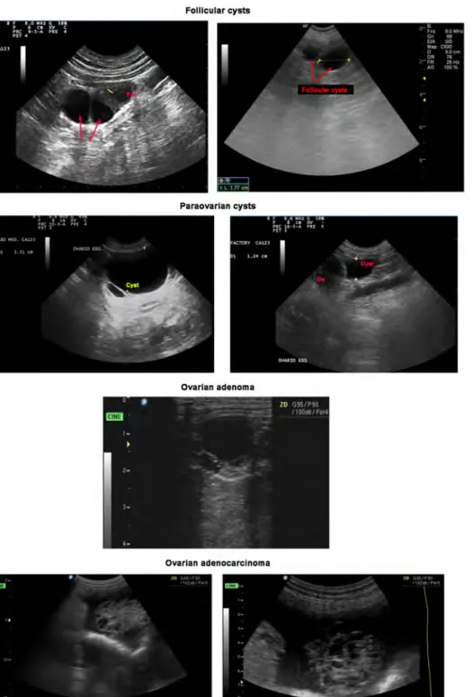
The use of ultrasonography may be helpful to diagnose ovarian cysts. In general, SES and rete cysts not attaining large dimensions may remain unperceived at the ultrasound scanning of the ovaries. Contrasting, follicular cysts are usually visible on ultrasound scans as anechoic structures with thin walls (Figure 3) and acoustic enhancement [44], while the ovary retains its normal ovoid shape; seldom, some of them may evidence signs of granulosa cell luteinisation similar to the detected in normal developing follicles, although thinner. Size is an important parameter when diagnosing follicular cysts as in the majority of the cases they
Ovarian cysts in dogs
will be slightly larger than the coexisting cohort of follicles in the same or the contralateral ovary (Figure 3). Moreover, follicular cysts may be single or multiple. In the later case, larger anechoic structures separated by thin hyperechoic walls are perceived. Frequently, follicular sizes are similar and present normal echoic pattern. If doubts arise, a second scanning could be arranged for 1 to 2 weeks later, to establish comparison to the structures in the ovary. Follicular productive cysts remain unchanged for some weeks, while follicles that ovulated assume the echoic pattern of corpora lutea, with disappearance of the anechoic antrum [45]. Non-follicular cystic structures representing adenomas, rete cysts or paraovarian cysts may incidentally be found during abdominal ultrasonography. Only rete cysts and paraovarian cysts larger than 5mm are detected as anechoic regular structures that a rounder contour to the image of the ovary or are located parallel to the ovary (Figure 3). Distinction from follicular cysts is difficult in US images, particularly when they are not large structures. Adenomas appear as multiple anechoic cavities located within the ovary, with diverse size and echogenicity, and the overall size of the gonad is usually increased. When small cysts compose adenomas, the ovary presents an irregular [44], foamy contour. Acoustic enhancement is sporadic. In the case of adenocarcinoma, the ultrasound image of the ovary is similar in appearance to that evidenced by adenomas. However, the former most frequently appear with more heterogeneous cysts in both size and sonographic pattern [44]; it often co-exists with signs of free abdominal fluid (Figure 3). Then the multicystic ovary seems to float from a pedicle, the representation of the ovarian bursa being inexistent.
Coexistence of uterine disease is possible, but the non-productive cysts seldom cause uterine disease; however, it should be annotated in the clinical file, as it may dictate the available options.
Abdominal radiography is less helpful than ultrasound imaging, and therefore less frequently accounted for the diagnosis of ovarian cysts. Unless it exists an important difference in the ovarian size or in the adnexal tissues, usually associated with the existence of extremely large cysts, no changes are perceived on X-rays. Rather large cysts may be identified as a soft tissue-density mass in the central abdomen, caudal to the kidney. If the cyst is large enough, a
colon filled with faeces, suggesting delayed intestinal transit.
The peripheral measurements of estrogen and progesterone determination are not always helpful in the diagnosis of follicular cysts, because they not always evidence clear changes in the serum levels of estrogen [4]; moreover, they do not correlate with the severity of the process. In other hand, assays for canine estrogen are not always readily available and the normal range values also greatly vary among laboratories. Estrogen impregnation may also be suspected from the results of vaginal cytology. However one should remember that if the follicular cyst does not produce enough amount of estrogen to compromise the progesterone dominance in the luteal stage, the values for peripheral estrogens might be unchanged or inconclusive.
Complete blood count is expectably altered when signs of hyperestrogenism and of estrogen-related toxicity accompany follicular cysts; but the inexistence of changes should not limit the diagnosis, particularly on respect to epithelial ovarian cysts or epithelial tumours of the ovary. In ovarian adenocarcinomas, multiple changes in the blood parameters may escort the clinical condition, derived from the systemic disease (such as azotemia or metabolic acidosis) or hypercalcemia due to paraneoplasic syndrome.
Payan-Carreira and Pires
Figure 3. Ultrasonographic features of canine ovarian cysts. Follicular cysts (arrows) appear as anechoic structures slightly larger than co-existing follicles (Fol), and fail to evidence the regular echoic pattern of granulosa luteinization (yellow bar) typically found in late antral follicles in dogs. Paraovarian cysts are observed as larger anechoic thin-wall structures (Cyst) that often are observed close to the ovary (Ov.). The Adenoma scans show usually multiple anechoic cavities with heterogeneous size and echogenicity. In this image od adenocarcinoma, the gonad presents multiple hypoechoic to anechoic structures with different size and ultrasonographic pattern, surrounded by a collection of free abdominal fluid.
Ov ar ia n cy st s in d og s Fi gu re 4 . D ia gn os tic m ap f or o va ria n c ys ts in d og s
Ovarian cysts in dogs
Therapeutic strategies
Two main issues regarding the available therapeutic approaches to ovarian cysts in dogs concern: the value or interest of the animal for breeding, which in turn also depends on the age, and the type of cystic pathology diagnosed in a particular animal.
As stated by Sontas et al.[3], in veterinary medicine the therapeutic options focus mainly on follicular cysts since other cysts do not cause a clinical disease. Therefore, ovariohysterectomy is often offered as curative and to eliminate the risk of recurrence in non-breeding female dogs or when the cystic condition escorts other genital diseases such as pyometra.
Treatment proposed for follicular cysts is based on the attempt to luteinize the cystic structure so it can regress later, by the end of a diestrus stage. Available options include the administration of GnRH (Gonadotropin-releasing Hormone, 50µg, intramuscular) or hCG (human Chorionic Gonadotropin; 500UI, intramuscular, twice at 48h interval). However the success of these treatments is limited. Data compiled by Johnston and colleagues [28] reveales that the success of GnRH treatment was higher compared with hCG (64% of the cases were resolved by GnRH administration, compared to 43% of resolved cases with hCG). Nevertheless, in 43% and 33% of the situations responding to GnRH and hCG administration, respectively, recurred therefore demanding for a surgical alternative.
Sporadically, follicular cysts have been tentatively treated with tamoxifen citrate or prostaglandin F2α, but application of these drugs ensued recurrence and failure of resolution
respectively (Olson et al. 1989, cited in [28]).
Surgical approaches to ovarian cysts of follicular type also traditionally encompass the ovariohysterectomy [28] whenever the female presents degenerative cystic uterine diseases or pyometra and it is not intent to reproduction or the clinical condition does not respond to medical therapy. Occasionally, the surgical drainage and flushing of the cystic structures has been attempted, and the few reports available mentioned the cure for the situation [46, 47]. As previously mentioned, non-productive benign cysts are usually found as an incidental finding during physical or ultrasound examination, and at OVH. No medical treatment is been available to these cysts, since their etiopathogenesis is unclear. Therefore, surgery is the option remaining. Although the usefulness of OVH may be questionable, since these cysts do not induce clinical disease, nonetheless they will growth with time, and in some situations they may reach such a size that unspecific symptoms of abdominal discomfort may became apparent.
Conversely, the treatment of choice for ovarian adenocarcinomas is surgery, which should be performed according to standard oncologic surgical principles to minimize further tumour seeding into the abdominal cavity. Ideally, the excised genital tract should be sent for histopathological analysis. The characterization of the tumour aggressively and the confirmation of the type of tumour are crucial to establish a prognosis or even to attempt complementary medical treatment. However, rather limited information is available on the
Payan-Carreira and Pires
use of chemotherapy or radiation therapy in the treatment of ovarian tumours in veterinary medicine, which compromises possible recommendations.
Final comments
This work revised the available information on the different types of cysts arising within and around the canine ovary. Although follicular cysts are often referred in the literature, this sort of lesions is less frequent in the canine practice, albeit they may present exuberant hyperestrogenism syndrome that draw the clinician or the owner´s attention. In contrast, cysts arising from the epithelial structures are commonly found in mature and older female dogs, but evolve silently and remain undiagnosed for longer periods. Most infrequently, ovarian adenocarcinoma is diagnosed in females presented at the clinic for a sudden abdominal dilatation, weight loss and depression . The sudden onset of the clinical symptoms and the degradation of the animal condition are often negative prognostic signs.
It was intent to gather in one chapter the scattered information on the ovarian cysts in dogs, including the pathologic aspects of the lesions, while reviewing the symptoms or the clinical findings that will orientate the diagnosis. Definitive diagnosis should be supported by histopathology, as not always the macroscopic assessment of ovarian lesions will allow the practitioner to reach a final diagnosis.
Finally, an attempt was made to summarize the information necessary to establish a tentative diagnosis in a concept map, particularly helpful to students and young professionals.
References
[1] Shille V, Calderwoodmays M, Thatcher M. Infertility in a bitch associated with short interestrous intervals and cystic follicles - A
case-report. Journal of the American Animal Hospital Association. 1984;20:171-6.
[2] Arlt SP, Spankowsky S, Heuwieser W. Follicular cysts and prolonged oestrus in a female dog after administration of a deslorelin implant. N Z Vet J. 2011;59:87-91.
[3] Sontas B, Milani C, Romagnoli S, Bertolini G, Caldin M, Caliari D, et al. A Huge Ovarian Cyst in a Hysterectomized Bitch. Reproduction in Domestic Animals. 2011;46:1107-11.
[4] Knauf Y, Bostedt H, Failing K, Knauf S, Wehrend A. Gross pathology and endocrinology of ovarian cysts in bitches. Reprod Domest Anim. 2014;49:463-8.
[5] Akihara Y, Shimoyama Y, Kawasako K, Komine M, Hirayama K, Kagawa Y, et al. Immunohistochemical evaluation of canine ovarian cysts. Journal of Veterinary Medical Science. 2007;69:1033-7.
[6] DOW C. Ovarian abnormalities in the bitch. J Comp Pathol. 1960;70:59-69.
[7] Mialot M, Parodi A. Particularités histologiques et dysplasies des structures épithéliales de l'ovaire de la chienne. Zentralblatt für Veterinärmedizin Reihe A. 1979;26:800-9.
[8] Pires MA, Payan-Carreira R. Cystic structures in the canine ovary: a prevalence survey. In: Schäfer-Somi SP, A.Niżański, W.Hagman, R., editor. 17th EVSSAR Conference - Reproduction and Pediatrics in dogs, cats and exotic carnivores. Wroclaw, Poland: EVSSAR; 2014. p. 179 [abstr.].
Ovarian cysts in dogs
[9] Schlafer D, Miller R. Pathology of the ovary (non-developmental lesions). In: MG M, editor. Jubb, Kennedy, and Palmer’s Pathology of Domestic Animals. 5th ed. St. Louis, MO: Elsevier Limited; 2007. p. 444-50.
[10] Barone R. Anatomie Comparée des Mammifères Domestiques. Lyon, France: Vigot Frères. [11] Dyce KM, Sack WO, Wensing CJG. The Female Reproductive Organs - The Pelvis and Reproductive Organs of the Dog and Cat. Textbook of Veterinary Anatomy. 4th ed. St. Louis, MI, USA: Saunders, Elsevier; 2010. p. 459-66.
[12] McEntee K. Reproductive Pathology of Domestic Mammals. New York: Academic Press; 1990.
[13] Tanaka K. Morphological Study on the Canine Ovary. Japanese Journal of Veterinary Research: Hokkaido University; 1962. p. 80-1.
[14] Wildt DE, Levinson CJ, Seager SW. Laparoscopic exposure and sequential observation of the ovary of the cycling bitch. Anat Rec. 1977;189:443-9.
[15] Miller ME, Christensen JC, Evans HE. Miller's Anatomy of the Dog. Philadelphia, PA, USA: W. B. Saunders Company,; 1964.
[16] Duke KL. Comparative Aspects of the Mammalian Ovary. In: Motta PM, Hafez ESE, editors. Biology of the Ovary: Springer Netherlands; 1980. p. 16-30.
[17] Barton EP. The cyclic changes of epithelial cords in the dog ovary. J Morphol. 1945;77:317-49.
[18] O'shea J. Histochemical observations on mucin secretion by subsurface epithelial structures in the canine ovary. Journal of Morphology. 1966;120:347-58.
[19] Banks WJ. Female reproductive system. Applied Veterinary Histology. 3rd ed. St. Louis, MI, USA: Mosby; 1993. p. 446-68.
[20] Andersen AC, Simpson ME. The ovary and reproductive cycle of the dog (Beagle). Theriogenology. 1973;1:39-42.
[21] Bacha WJ, L.M. B. Female Reproductive System. Color Atlas of Veterinary Histology Wiley-Blackwell; 2012.
[22] Concannon PW. Endocrinologic control of normal canine ovarian function. Reprod Domest Anim. 2009;44 Suppl 2:3-15.
[23] Reynaud K. Folliculogenesis, ovulation and endocrine control of the oocyte and the embryo in the dog. In: REPRODUCTION ISOCAF, editor. 7th International Symposium on Canine and Feline Reproduction. Whistler, Canada2012. p. pp. 2.
[24] Songsasen N, Wildt DE. Oocyte biology and challenges in developing in vitro maturation systems in the domestic dog. Anim Reprod Sci. 2007;98:2-22.
[25] Concannon PW. Reproductive cycles of the domestic bitch. Anim Reprod Sci. 2011;124:200-10.
[26] Fontbonne A. In vivo ovulation, oocyte maturation and fertilisation in the bitch 2008.
[27] Marino GB, A.Mannarino, C.Di Prima, M.L.Zanghì, A. Le cisti
stromali dell’ovaio di cagna: prevalenza, diagnosi e risvolti clinici. Veterinaria (SCIVAC). 2010;24:9-15.
[28] Johnston S, Root Kustritz M, Olson P. Disorders of the canine ovary. Canine and Feline Theriogenology. Philadelphia: W.B. Saunders Comp; 2001. p. 193-205.
[29] Foster R. Surgical Pathology of the Canine Female Reproductive Tract. http://www.uoguelph.ca/%7Erfoster/repropath/female/dog/female_dog.htm: Ontario Veterinary College, University of Guelph; 2014.
[30] Borges P, Fontaine E, Maenhoudt C, Payan-Carreira R, Santos N, Leblond E, et al. Fertility in adult bitches previously treated with a 4.7mg subcutaneous deslorelin implant. Reproduction in Domestica Animals. 2015;In press:pp.7.
[31] Kim BS, Kim KC, Oh KS. Vaginal prolapse by ovarian follicular cysts in a female Jin-do dog. Korean Journal Veterinary Research. 2008;48:223-5.
[32] Ghaffari MS, Dezfoulian O, Aldavood SJ, Masoudifard M. Estrogen-related alopecia due to polycystic ovaries in a terrier dog. Comparative Clinical Pathology. 2009;18:341-3.
Payan-Carreira and Pires
[33] Sforna M, Brachelente C, Lepri E, Mechelli L. Canine ovarian tumours: a retrospective study of 49 cases. Vet Res Commun. 2003;27 Suppl 1:359-61.
[34] Patnaik AK, Greenlee PG. Canine ovarian neoplasms: a clinicopathologic study of 71 cases, including histology of 12 granulosa cell tumors. Vet Pathol. 1987;24:509-14.
[35] Ajadi AT, Antia ER, Akang EE, AJADI A, ANTIA E, AKANG E. Cystadenocarcinoma Arising from Ovary in a Three Year Old Doberman Bitch. Int J Morphol. 2011;29:988-91.
[36] Kennedy P, Cullen J, Edwards J, Goldschmidt M, Larsen S, Munson L, et al. Histological Classification of the Tumours of the Genital System of Domestic Animals. Washington DC: Armed Forces Institute of Pathology; 1998.
[37] Ortega-Pacheco A, Gutiérrez-Blanco E, Jiménez-Coello M. Common lesions in the female reproductive tract of dogs and cats. Vet Clin North Am Small Anim Pract. 2012;42:547-59, vii. [38] MacLachlan N. Ovarian disorders in domestic animals. Environ Health Perspect. 1987;73:27–33.
[39] Navas JHA, Rodríguez LSD, Quiceno VHA. Reporte de caso. Ovarios poliquísticos en hembra canina. Bucaramanga-Santander, Colombia1. Spei Domus. 2012;8:29-33.
[40] Johnston. Disordres of the canine uterus and uterine tubes (oviducts). In: Johnston SD, Root Kustritz MV, Olson PS, editors. Canine and Feline Theriogenology. 1st ed. Philadelphia: Saunders; 2001. p. 206-24.
[41] Harvey M. Conditions of the non-pregnant female. In: Simpson, G.M., England GCW, Harvey M, editors. Manual of Small Animal Reproduction and Neonatology. Shurdington, Cheltenham, UK: BSAVA Publishing; 1998. p. 46-9.
[42] Yotov S, Simeonov R, Dimitrov F, Vassilev N, Dimitrov M, Georgiev P. Papillary ovarian cystadenocarcinoma in a dog. J S Afr Vet Assoc. 2005;76:43-5.
[43] Hori Y, Uechi M, Kanakubo K, Sano T, Oyamada T. Canine ovarian serous papillary adenocarcinoma with neoplastic hypercalcemia. J Vet Med Sci. 2006;68:979-82.
[44] Diez-Bru N, Garcia-Real I, Martinez EM, Rollan E, Mayenco A, Llorens P. Ultrasonographic appearance of ovarian tumors in 10 dogs. Vet Radiol Ultrasound. 1998;39:226-33.
[45] Hayer P, Günzel-Apel AR, Lüerssen D, Hoppen HO. Ultrasonographic monitoring of follicular development, ovulation and the early luteal phase in the bitch. J Reprod Fertil Suppl. 1993;47:93-100.
[46] Bassu G, Rault D, Besso J, Marseloo N, Fontbonne A. Ultrasound-guided aspiration in management of ovarian fullicular cysts in the bitch. Proceedings of the 4th Congress of the European Veterinary Society for Small Animal Reproduction. Barcelona, Spain2004. p. 269–70. [47] Levy X, Fontaine E, Grellet A, Courtois E, Fontbonne A. Surgical cysts removal: A new technique for the treatment of ovarian cysts in the bitch. 5th Annual Symposium of the European Veterinary Society for Small Animal Reproduction. Estoril, Portugal2007. p. 120.