Irene Maria Teixeira Pais
EFEITO DA ACUPUNCTURA NAS CÉLULAS NK EM DOENTES COM CANCRO
COLORECTAL SUBMETIDOS A QUIMIOTERAPIA- ESTUDO PROSPECTIVO, RANDOMIZADO E CONTROLADO -
Dissertação de Candidatura ao grau de Mestre
em Medicina Tradicional Chinesa submetida ao
Instituto de Ciências Biomédicas de Abel Salazar
da Universidade do Porto
Orientador – Doutor Henry Johannes Greten
Categoria – Professor Associado
Afiliação – Instituto de Ciências Biomédicas Abel
Salazar, Universidade do Porto.
Co-Orientador – Mestre Nuno Correia
Categoria – Especialista de Medicina Interna
Filiação – Centro Hospitalar de S. João do Porto
Co-Orientador – Doutor António Moreira Pinto
Categoria: Director do Serviço de Oncologia
Afiliação: Centro Hospitalar de Vila Nova de
Gaia/ Espinho
Effect of Acupuncture on NK cells in CRC patients undergoing chemotherapy
DEDICATION
I dedicate this study to
my precious friends
that are
always with me.
Effect of Acupuncture on NK cells in CRC patients undergoing chemotherapy
Irene Pais
V
ACKNOWLEDGMENTS
I would like to thank my closest family and friends for all their background support that has enabled me to develop my own education.
To my dears Professors Jorge Machado, Mário Gonçalves and Johannes Greten, for their constant enthusiastic support.
To Drª Esmeralda Neves (Department of Immunology, Hospital Santo António) for her contribution to this project.
To Drª Maria José Teles and Prof. Dr. Tiago Guimarães (Department of Clinical Pathology, Hospital São João); Drª Isabel Pimentel and Drª Margarida Damasceno (Department of Oncology, Hospital São João), for their support and contribution to this project.
To Petra for her support in general management.
To Prof. Dr. Moreira Pinto (Department of Oncology, Centro Hospitalar de V.N.Gaia/Espinho), for the support and contribution for this master thesis.
To Dr. Nuno Correia, for an outstanding colleague and friend; for his support and love by TCM and research, that allowed the execution of this master thesis.
Effect of Acupuncture on NK cells in CRC patients undergoing chemotherapy
Any medicine, to the extent that it justifies the qualification of “rational science”,
does not directly combat disturbances defined empirically, i.e., their symptoms,
their symptomatic abnormalities. Instead, it strives to isolate and define general or
basic postulates producing these disturbances”
Prof. Dr. Manfred Porkert, 1983Traditional Chinese Medicine is a system of sensations and findings
designed to establish a functional vegetative state.
Effect of Acupuncture on NK cells in CRC patients undergoing chemotherapy
Irene Pais
VII
RESUMOIntrodução: As células Natural Killer (NK) parecem constituir a primeira linha de defesa contra as células tumorais. A relevância das células NK é suportada pela associação entre uma diminuição da actividade, ou baixa quantidade de células NK circulantes em pacientes com progressão tumoral. No carcinoma colorectal (CRC), uma diminuição da actividade e do número de células NK parece estar associado a um pior prognóstico. De acordo com dois estudos chineses, a acupunctura e a moxabustão (AcuMoxa) poderão ter um impacto positivo nas células NK no CRC, as quais podem ser distinguidas em dois subtipos: citotóxico (NKdim) e imunoregulador (NKbright). Contudo, o efeito da acupunctura na sua expressão permanece por esclarecer.
Objectivo: Determinar os efeitos da AcuMoxa no sistema imune dos doentes com CRC, principalmente o seu efeito nas células NK e seus subtipos.
Métodos: Doentes com CRC serão recrutados a partir do Departamento de Oncologia do Centro Hospitalar S. João e distribuídos em dois grupos: o grupo experimental (n=9) e o grupo de controlo (lista de espera; n=9). No grupo experimental, os doentes serão submetidos a tratamento de AcuMoxa segundo o Modelo de Heidelberg de Medicina Tradicional Chinesa (8 sessões/mês). Serão efectuadas colheitas de sangue periférico em quatro tempos distintos: a primeira colheita (t=0) será efectuada na semana anterior ao tratamento de quimioterapia e as restantes três colheitas, a cada 8 dias. O tratamento de AcuMoxa consistirá dos pontos LI4, TB5, SP9, Lv3, St36, GB39, PC5 e Lu7.
Parâmetros primários: Hemograma completo; linfócitos T e B, células NK e subtipos NKdim/ NKbright quantificados por citometria de fluxo.
Parâmetros secundários: Avaliação dos níveis de ansiedade e depressão bem como a qualidade de vida (QOL).
Análise estatística: Os resultados serão analisados dentro de cada grupo e entre os grupos (antes e depois da intervenção).
Discussão: Dois estudos abordaram a modulação da actividade dos subtipos de células NK em doentes oncológicos. Baseado nestes dados sobre acupunctura e células NK, pretendemos confirmar uma melhoria do status imune, mais especificamente nas células NK. Adicionalmente, esperamos revelar qual o subtipo NK mais prevalente durante cada fase da QT / intervenção (T0, T1,T2 e T3) incluindo a magnitude do rácio NKdim/ NKbright. Conclusão: O tratamento de AcuMoxa segundo o Modelo de Heidelberg poderá promover a actividade das células NK, especificamente a sua citotoxicidade, de acordo com a literatura. No futuro, este tratamento complementar poderá ter um impacto positivo no prognóstico e na QOL dos doentes com CRC.
Effect of Acupuncture on NK cells in CRC patients undergoing chemotherapy
ABSTRACT
Introduction
NK cells appear to represent a first line of defence against tumour cells. This relevance of NK cells is supported by an association between decreased activity or low numbers of circulating NK cells in patients with progression of cancers. In colorectal cancer (CRC), decreased activity and low number of NK cells appears to be associated with a worse prognosis. According to two Chinese studies, acupuncture and moxibustion may have a positive impact on NK cell, which can be distinguished in two subsets: cytotoxic (NKdim) and immunoregulatory cells (NKbright). However, the effect of acupuncture on its expression remains unclear.
Objectives: To assess the effect of acupuncture and moxibustion on the immune function of colorectal cancer patients.
Methods: CRC patients recruited from the Oncology department of Centro Hospitalar S. João, were randomized in two groups, intervention (n=9) and control/waiting list (n=9). In the intervention group, patients were submitted to acu-moxibustion treatment protocol based on the Heidelberg Model of TCM (8 sessions/ month). Blood samples will be collected at baseline (T0) on the week prior to chemotherapy and the following samples are collected every once a week until next chemotherapy (CT) regimen. The acupuncture/moxa treatment will consist of the points LI4, TB5, SP9, Lv3, St36, GB39, PC5 and Lu7.
Primary Outcome: Complete Blood counts, T and B lymphocytes, NK cells, NKdim/ NKbright counts by flow cytometry.
Secondary Outcomes: Assessment of anxiety and depression levels as well as quality of life.
Statistical analyses: Results will be analysed within each group (before and after intervention) and co- relational comparison between groups
Discussion: two studies have addressed modulation of NK cells subset activity in cancer patients. Based on these data on acupuncture and NK activity, we would like to confirm a improvement in immune status and more specifically in NK cells. In addition, we hope to unveil which NK subset is more prevalent during each phase of the CT / intervention (T0, T1, T2 and T3) regimen, including magnitude of NK dim/ bright ratio.
Conclusion: Acupuncture/moxa treatment according to the Heidelberg Model may promote NK cells activity, especially cytotoxic according to literature. In the future, this complementary treatment might have a positive impact in the prognosis and QOL of CRC patients in the future.
Effect of Acupuncture on NK cells in CRC patients undergoing chemotherapy
CONTENTS
PART ONE –THEORETICAL BACKGROUND... 1
C
HAPTERI
–
I
MMUNOLOGICALA
SPECTS OFC
ANCER ... 31.
Immunoediting Theory ... 1
2. NK cells role in innate and adaptive immune system ... 4
3. NK cells in cancer immunity ... 11
4. Co-stimulatory molecules that regulate NK-cell antitumour immunity and
link innate and acquired immune system ... 12
5. Immune parameters affecting the efficacy of chemotherapy regimens ... 13
6. The immune system and the quality of life (QOL) in cancer patients ... 14
C
HAPTERII
–
C
OLORECTALC
ANCERO
VERVIEW ... 161. Epidemiology and Etiopathophysiology ... 18
2.
Treatment management of CRC ... 27
3. Chemotherapy - common used agents and related side effects ... 32
4. CRC Immunity ... 35
C
HAPTERIII
–
T
RADITIONALC
HINESEM
EDICINE ... 381. Traditional Chinese Medicine - Overview ... 41
2. Heidelberg Model of Chinese Medicine: TCM as System Biology ... 42
3. Acupuncture - overview ... 59
4. Acupuncture and Immunomodulation ... 60
5. The importance of moxibustion in the immune modulation ... 63
PART TWO –STUDY METHODOLOGY ... 66
1. Objectives ... 67
2. Sampling and Recruitment Procedures ... 68
PART THREE –RESULTS AND DISCUSSION ... 71
PART FOUR –CONCLUSIONS AND FUTURE PERSPECTIVES ... 85
REFERENCES ... A
Effect of Acupuncture on NK cells in CRC patients undergoing chemotherapy
Irene Pais
X
INDEX OF FIGURES
Figure 1: Cancer immunoedting theory. ... 2
Figure 2: The biological functions of NK cells. ... 5
Figure 3: NK cells receptors. ... 6
Figure 4: The dynamic regulation of NK cell effector function. ... 7
Figure 5: NK cell tuning. ... 8
Figure 6: NK cells subsets and surface receptors: NKbright (a) and NKdim (b). ...10
Figure 7: Schematic presentation of NK role in immunoediting theory. ...11
Figure 8. Role of CD56bright NK cells and T cell in the interaction between adaptive and innate system. ...13
Figure 9. Colonic features and corresponding barium enema X-rays. ...22
Figure 10. The parallelism between TCM concepts of disease and physiological process at four levels of regulation, according to Heidelberg Model of TCM. ...43
Figure 11. The four components of functional diagnosis. ...45
Figure 12. Schematic model of Algor Laedens Theory. ...47
Figure 13. The six layers of energy. ...49
Figure 14. ALT related orbs and cross-linked orbs. ...53
Figure 15. Most common cancer syndromes, according to TCM. ...54
Figure 16. Hypothetic model of the mechanisms how acupuncture stimulates the immune system. ...62
Figure 17:Study flow chart.. ...69
Figure 18: Effects of AcuMoxa protocol in immune system, according to Heidelberg Model of TCM. ...82
Figure 19. Simplified scheme of acupuncture mechanisms of action on the NK cells tumoricidal activity. ...84
Effect of Acupuncture on NK cells in CRC patients undergoing chemotherapy
Irene Pais
XI
ÍNDEX OF TABLES
Table 1. The 7th edition of AJCC-TNM classification system. ...25
Table 2. The 7th Edition of AJCC-TNM Classification System. ...26
Table 3: Rectal cancer. TNM staging. ...27
Table 4 Common types of chemotherapy agents and their mechanisms. ...33
Table 5. Most common regimens associated side effects in the management of CRCs ...34
Table 6. The six stages of Algor-laedens Theory; Energy layers, mechanisms of action and key symptoms. ...50
Table 7: Socio-demographic and clinical characteristics of patients. ...72
Table 8: Blood analyses at baseline. Comparison between groups. ...73
Table 9. QOL-CR 29 and HADS Scores. Comparison between groups at baseline. ...73
Table 10: WBC and ANC (T0) . ...74
Table 11. Lymphocyte populations: Total lymphocytes, B cells and T cells.. ...75
I
NDEX OFG
RAPHICS Graphic 1: WBC and ANC counts. ...75Graphic 2: Lymphocyte populations. Comparison between groups and acroos time. ...76
Graphic 3: NK cells and subsets. ...77
Graphic 4: Comparison of Anxiety and Depression scores among groups, at begining and at the end of the study. ...77
Graphic 5: QOL assessment through questionnaire CR29 (I). ...78
Effect of Acupuncture on NK cells in CRC patients undergoing chemotherapy
Irene Pais
XII
LIST OF ABBREVIATIONS
5-FU 5- fluorouracil
ACTH adrenocorticotropic hormone ADCC antibody-dependent cell
cytotoxicity
ALT algor laedens theory ANC absolute neutrophil counts APC antigen presenting cell
BRAF serine/threonine-protein kinase CCR7 chemokine receptor 7
CEA carcinoembryonic antigen CRC colorectal cancer
CTL cytolytic T cells DC dendritic cell
DCBE double-contrast barium enema DNA desoxirribonucleic acid
EC ethics committee EGFR epidermal growth factor
FAP familial adenomatous polyposis G-CSF granulocyte colony-stimulating
factor
GM-CSF granulocyte macrophage colony-stimulating factor
H Hepatic
HNPCC hereditary nonpolyposis colorectal cancer
IBD inflammatory bowel disease IFNγ interferon γ
IGF1 insulin-like growth factor-1 IGFBP-3 insulin-like growth factor-binding
protein-3
IGFII insulin-like growth factor II IL Interleukin
KIR killer cell immunoglobulin-like Receptors
KRAS Kirsten rat sarcoma viral oncogene homolog
L Lienal
LAK lymphokine-activated killer
LHA lateral hypothalamic area LV leucovorin
MC microcirculation
MHC major histocompatibility complex NCR natural cytotoxicity receptors NF-Kβ nuclear transcription factor- Kβ NK natural killer NO nitric oxide P pulmonal p53 protein 53 Pb peripheral bllod PC pericardial
PDGF platelet-derived growth factor
PGE2 prostaglandin E2 PMN polymorphonucleocytes PSC primary sclerosing cholangitis PSGL-1 PEN5P-selectin glycoprotein
ligand-1 QOL quality of life
R renal
RNA ribonucleicc acid
RS respondens
ST stomachal
TAMs tumour-associated macrophages TCM tradicional chinese medicine TGF tumor growth factor
Th T helper
TK Tricaloric
TME total mesorectal excision- TNF tumour necrosis factor TNM Tumour nodules metastases VEGF vascular endothelial growth factor WBC white blood cells
HPA hypothalamus-pituitary-adrenal SNS Sympathetic nervous system
Irene Pais
3
CHAPTER I – IMMUNOLOGICAL ASPECTS OF CANCER
Effect of Acupuncture on NK cells in CRC patients undergoing for chemotherapy
Irene Pais
1
1
.Immunoediting TheoryCancer is widely considered to be a cell-autonomous genetic disease that results from alterations in oncogenes, tumour-suppressor genes and genome-stability genes. However, the tumour-cell microenvironment, the stroma and immunity also have a major role in cancer. Indeed, for the development of a neoplasia, cancer cells have to overcome intrinsic (cell autonomous) and extrinsic (immune mediated) barriers to oncogenesis.
Initial studies on cancer immunobiology, proposed the hypothesis of “immunosurveillance”: the immune system is able to recognize and destroy proliferating cancer cells, which express unique antigens at surface that are by-products of the process of malignant transformation and the immune system’s capacity for surveillance can be modified through both medical and lifestyle interventions. Initially, was proposed that an impaired immune system would fail to react even to a highly immunogenic tumour and this impairment was due to genetic factors, radiotherapy or immunosuppressive drugs. Nowadays, it is know that, except for hormone therapy, all conventional treatments for cancer can suppress immune response.1
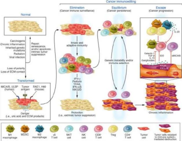
Later studies recognized the existence of a highly complex interrelationship between cancer and the immune system and suggested the replacement of the term “immunosurveillance” by a more comprehensive term, “immunoediting” (figure 1). The cancer immunoediting process engenders three phases: a) elimination phase, in which cancer cells are recognized by the immune system and may be eradicated; b) equilibrium
phase appears if elimination is unsuccessful, and immune system and cancer cells
achieve an equilibrium in which cancer is contained but not eliminated and c) the escape
phase appears as result of constant pressure from the immune system, cancer cells can
undergo genetic alterations becoming resistant to immune attack and, thus, are free to grow even in the presence of an intact immune system.2
Effect of Acupuncture on NK cells in CRC patients undergoing chemotherapy
Irene Pais
2
Figure 1: Cancer immunoedting theory. Diverse immune cells involved in the different phases. Extracted from Swann et al, 2007 3
Elimination phase represents the classical concept of cancer immunosurveillance.
As reviewed by Dunn4, cancer immunosurveillance has two critical effectors functions: IFNγ production and cytotoxicity. Interferon (IFN) γ facilitates development of powerful anti-tumour effector functions mediated by both adaptive and innate immunity: generation of tumour-specific CD4+ Th1 T cells and cytolytic T cells (CTL) and activation of cytocidal activity in macrophages. Moreover, IFNγ has the capacity to upregulate tumour immunogenicity, explaining the effects on tumour detection and elimination in immunocompetent hosts. Perforin was identified as a critical cytolytic molecule in the primary host anti-tumour response.
The molecular definition of tumour antigens provides a firm basis for how the adaptive immune system discriminates between normal and neoplasic cells. The tumour antigens are recognized by CD8+ β T cells in the context of MHC class II and class I proteins, respectively.
Another important component used by both adaptive and innate cells to distinguish cancer cells from normal cells a receptor expressed on the surface of NK cells, the NKG2D-activating receptor. NKG2D binds to the MHC class I chain-related proteins A
Irene Pais
3
an B, to the UL16 binding proteins and to the lymphocyte effector cell toxicity-activating ligand (LETAL).According to the immunoediting hypothesis, tumour’s immunogenic phenotype is continuously shaped by the immunological factors in its environment and, thus, the dynamic interaction between immunity and cancer in the equilibrium phase produces new populations of tumour cells, unstable and rapidly mutating cells. In the equilibrium phase, the tumour has three possible outcomes: eventual elimination by the immune system; permanent maintenance in the equilibrium phase by cellular and molecular controls of immunity, or escape from immune pressure and transit to the final escape phase of the immunoediting process.
In the escape phase, tumour growth proceeds unrestrained by immune pressure. To become clinically detectable in an immunocompetent host, cancer cells must circumvent both innate and adaptive immunologic defences. It has been proposed several mechanisms through which tumour escape from immune control. Tumour escape can be a) a direct consequence of alterations occurring in edited tumour targets themselves, like for instance tumour cells develop lesions in antigen processing and presentation pathways that facilitate evasion from adaptive immune recognition; b) due to the inhibition of the protective functions of the immune system; and c) due to suppression of pro-inflammatory danger signals.4
Tumour microenvironment
Robert Strausberg2 highlights the importance of studying the genetics and phenotypes of the tumour surrounding microenvironment, as well as the cancer cells. In fact, there is compelling evidence that the innate and adaptive immune cells of the tumour microenvironment engage an extensive and dynamic crosstalk with cancer cells, through direct contact or by chemokine and cytokine production5, leading to conclude that tumour related-inflammation and anti-tumour immunity co-exist at different points along tumour progression and that the environmental and micro environmental conditions dictate the balance between the two.
In fact, some studies6,7,8 show that several components of the tumour microenvironment plays an important role in promoting tumour progression. Tumour stroma, containing fibroblasts, inflammatory cells and endothelial cells, plays an important role in promoting tumour progression; where myofibroblast-driven desmoplasic stromal reaction is considered to be a poor prognostic indicator in primary CRC. Several cytokines (TGF-beta, PDGF, IL4 and IGF II), that induce myofibroblastic differentiation, seem to
Effect of Acupuncture on NK cells in CRC patients undergoing chemotherapy
Irene Pais
4
have pro-oncogenic functions.6 In addition the presence of ECM molecules in tumour microenvironment means the existence of an inflammatory process, which predisposes to the acquisition of malignant characteristics for tumour cells.7
The microenvironment of pre-malignant and early tumour lesions is generally composed by immune cells (tumour-infiltrating lymphocytes), such as T lymphocytes, macrophages and dendritic cells (DC) – a component of an inflammatory host response. The most frequent immune cells within the tumour microenvironment are tumour-associated macrophages (TAMs) and T cells. TAMs are tumour-associated with tumour growth and may be pre-requisite for angiogenesis, invasion and metastasis, being correlated with poor prognosis. T Cells have both tumour suppressive and promoting effects. In some cancers, increased T cell numbers were correlated with better survival and, low levels of T cell seem to be associated with more susceptibility to spontaneous or chemical carcinogenesis. However, evidence found in solid tumours has showed that many of T cells subsets are involved in tumour promotion, progression and metastasis.
The NK cells are the unique cells without a pro-tumourigenic role.5 In fact; recent studies revealed that the presence of NK cells in tumour immune infiltration represents a
positive prognostic marker.
2. NK cells role in innate and adaptive immune system
The innate system is composed mainly of NK cells, polymorphonucleocytes (PMN) and macrophages, and is most directly involved in tumour immunology. These cells also participate in the adaptive response and form an important and vital bridge between the
two arms of the immune system. It recognizes non-self molecules according to a specific
pattern. Another feature of the innate immune system is the complement, a group of inactive proteins in the blood which are activated in the presence of pathogens and non-self cells and cause cell lysis.9
As effectors members of the innate immunity, NK cells play a major role in anti-infection activity and tumour surveillance. NK cells can directly kill target cells to which they are capable of adhering within 1 to 4 hours without prior activation, priming or assistance by cytokines. NK cells can be triggered through various receptors depending on specific ligands presented by target cells in a given encounter.
NK cells have been recognized as major producers of cytokines in many physiological and pathological conditions, such as IFNγ, tumour necrosis factor (TNF)
Irene Pais
5
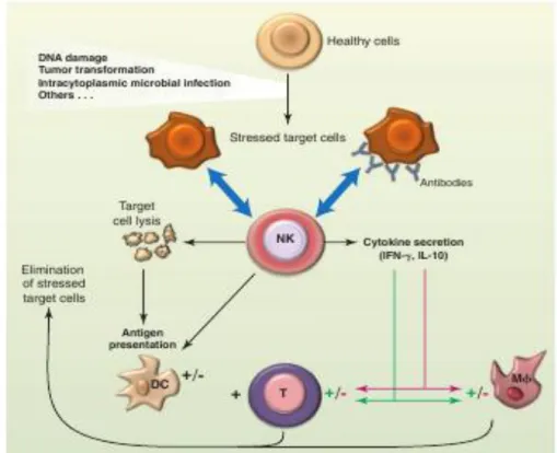
and interleucine (IL)-10, as well as growth factors such as granulocyte macrophage colony-stimulating factor (GM-CSF), granulocyte colony-stimulating factor (G-CSF) and IL3. NK cells also secrete several chemokines, which are vital for their co-localization with other hematopoietic cells such as DC in areas of inflammation. The production of IFNγ is related with the modulation of T cell responses in lymph nodes, possibly by a direct interaction between naïve T cells and NK cells migrating from inflamed peripheral tissues to secondary lymphoid compartments and by indirect effect on DC, promoting the maturation of DC by NK cell-derived IFNγ and TNF and subsequently leading to the enhancement of antigen presentation to T cells.In resume, NK cells modulate emerging B and T cells and are also regulatory cells engaged in reciprocal interactions with DCs, macrophages, T cells and endothelial cells. Thus NK cells are able to limit or exacerbate immune responses (Figure 2).9
Figure 2: The biological functions of NK cells.
DC – dendritic cells. M - Macrophages. Extracted from Vivier et al, 20119
In order to exert effectively their functions, cytolytic and regulatory function of the adaptive immune response, NK cells must be recruited to the site of infection or tumour and must be in closely proximity to antigen-stimulated T and/or B cells, respectively. They require triggering by several factors, such as IL15 presented by DC or macrophages, IL12 or IL18. IL 15 is an important for the maturation and survival of NK cells.
Effect of Acupuncture on NK cells in CRC patients undergoing chemotherapy
Irene Pais
6
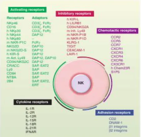
NK cells possess three main groups of receptors: the activating receptors, the inhibitory receptors and the chemotactic receptors (Figure 3). Among the activating receptors groups are included receptors that interact with cytokines (e.g. NKp46, CD16, NKG2D) and receptors that interact with cell surface molecules (e.g. FcRγ). This repertoire of receptors allows NK cells to exert their functions through various means: they can kill a variety of target cells in the absence of antibody; NK cells are able to detect antibody-coated cells through CD16 cell surface receptor and to exert antibody-dependent cell cytotoxicity (ADCC) and cytokine production. The natural cytotoxicity receptors, NKp46/NCR1; NKp44/NCR2 and NKp30/NCR3, play a crucial role in antitumoral responses.
The activating receptor NKG2D is responsible for the NK cells capacity to distinguish self molecules in conditions of cellular stress. This receptor interacts with various ligands that have low levels of expression in most tissues but are over-expressed in situations of cellular distress.
Figure 3: NK cells receptors. Extracted from Vivier et al, 201165
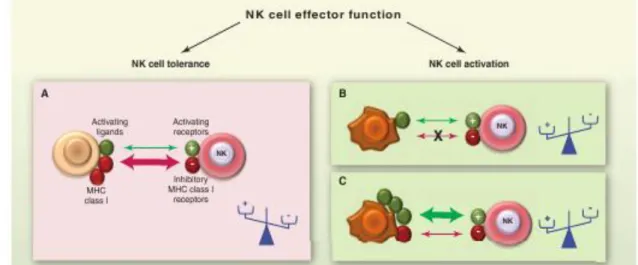
Another function attributed to NK cells is that they are able to detect the lack of major histocompatibility complex (MHC) class I (“missing self”), a situation that occurs during viral infections or cellular transformation (Figure 4). This is the reason why NK cells selectively kill target cells “in distress” and spare healthy cells. This is explained by the
Irene Pais
7
expression on the NK cell surface of several MHC class I-specific inhibitory receptors, such as killer cell immunoglobulin-like receptors (KIRs) and CD94/NKG2D.The array of receptors that individual NK cells express during development is largely random, therefore, some NK cells may express activating receptors for a self-ligand, but they do not express inhibitory receptors for self-MHC molecules. Thus NK cells, like T and B cells, have the potential for auto-reactivity. In order to avoid auto-reactivity, such NK cells acquire a state of hypo-responsiveness to self cells and also to several other stimuli, including MHC class I-deficient tumour cells or cross-linking antibodies specific activating receptors. The findings of several studies leads to the suggestion that in steady-state conditions, NK cell tuning enables NK cells with inhibitory receptors for self-MHC to rapidly eliminate MHC class I-deficient cells that arise in the environment, whereas NK cells with fewer such receptors can be mobilized by inflammatory signals that accompany pathogen infections.
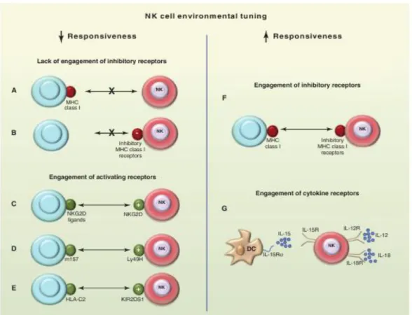
The molecular mechanisms that govern responsiveness are yet not established, although it has been suggested that in the presence of high levels of activating ligands, a negative turning effect may occur (Figure 5). It has been proposed some possible mechanisms for the impaired responsiveness of NK cells: (1) the induction of an anergic state in NK cells that are not inhibited by MHC molecules, as can occur in auto-reactive T and B cells; (2) the failure of these NK cells to undergo functional maturation, which may depend on interactions between MHC and inhibitory receptors on NK cells; (3) the function of inhibitory receptors for non-MHC ligands or (4) the action of suppressor cells.
Figure 4: The dynamic regulation of NK cell effector function. NK cells sense the density of various cell surface molecules expressed at the surface of interacting cells. The integration of these distinct signals dictates the quality and the intensity of the NK cell response. NK cells spare healthy cells that express self-MHC class I molecules and low amounts of stress-induced self molecules (A), whereas they selectively kill target cells “in distress” that down-regulate MHC class I molecules (B) or up-regulate stress-induced self molecules (c). + activating receptors; - inhibitory receptors. Extracted from Vivier et al, 20119
Effect of Acupuncture on NK cells in CRC patients undergoing chemotherapy
Irene Pais
8
Figure 5: NK cell tuning. Schematic experimental conditions in which NK cells show to adapt to their environment. In the absence of detection of MHC class I (lack of MHC class I receptors) (A), or in MHC class I deficient hosts (B), NK cells are hypo-responsive at steady state. NK cells are rendered “anergic” by the chronic engagement of various activating receptors such as NKG2D (C), Ly49H in the mouse (D) or KIR2DS1 in humans (E). NK cells can be educated by MHC class I molecules via their cognate inhibitory receptors (F) and primed by cytokines (G). Extracted from Vivier
et al, 20119
NK cells Subsets
The NK cells are lymphogranular cells having an ability to kill the intruding pathogens and are the most important cells of the system. They are found in the innate and the adaptive system as well. NK cells originate from the lymphoid progenitor and migrate to the secondary lymphoid tissues. When activated, NK cells burst forth from the tonsils, lymph nodes and spleen, and destroy infected and cancerous cells while the immune T and B cells are still mobilizing. They do not require memory from previous encounter and target virus infected cells and tumour cells and are the first line of defence. Activity of the immune cells varies from person to person and may be genetically inherited. The ability to kill cancers cells in less than 24 hours varies from 97% to 2%.10 NK cells have the ability to kill infected target cells and tumour cells and, also, to secrete
Irene Pais
9
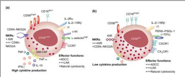
various effectors molecules. The physiological functions of NK cells are tightly regulated by a delicate balance between signals transmitted by activating and inhibitory receptors.Human peripheral blood NK (pbNK) cells can be divided into two subsets based on their cell surface density of CD56 and CD16, the CD56dim and CD56bright subpopulations13. The majority of peripheral blood NK cells, about 90%, is the CD56dim subset, which have low density expression of CD56 and express high levels of CD16 and has been shown to be responsible for the natural cytotoxicity against tumour targets.
CD56bright NK cells, comprising 10% of NK cells, expresses high levels of CD56 and low levels of CD16. CD56bright NK cells secrete higher levels of different cytokines such as IFNγ , TNFβ, GM-CSF, IL-10 and IL-13 in response to monokine stimulation. CD56bright NK cells exhibit low natural cytotoxicity and ADCC, but have potent lymphokine-activated killer (LAK) activity, representing the immunoregulatory subset. CD56bright NK cells are able to regulate adaptive T and B cell responses in secondary lymphoid tissues.11
CD56dim NK cells are associated with low capacity to produce cytokines, are potent mediators of ADCC, LAK activity and natural cytotoxicity and have a more granular morphology than CD56bright NK cells.12 A recent study suggest that CD56dim CD16+NK cells also have their unique abilities in influencing events occurring in their microenvironment.14
Moreover, the two NK cell subsets can be distinguished concerning their receptor expression profiles (Figure 6). The CD56bright NK cell subset expresses high levels of CD94/NKG2A and low levels of killer Ig-like receptors (KIRs), high amounts of chemokine receptor 7 (CCR7) and L-selectin (CD62L). The expression of CCR7 and L-selectin seems to be responsible for their mobilization to secondary lymphoid tissues. The CD56dim NK cells subset show no expression of CCR7 and little or no L-selectin, but high levels of another adhesion molecule – PEN5P-selectin glycoprotein ligand-1(PSGL-1), suggesting that they migrate to peripheral nonlymphoid tissues. Although, after their activation at sites of inflammation in the peripheral tissues, CD56dim NK cells express CCR7, promoting their migration to secondary lymphoid tissues.11,13 CD56dim NK cells also have high level expression of KIRs15
Effect of Acupuncture on NK cells in CRC patients undergoing chemotherapy
Irene Pais
10
Figure 6: NK cells subsets and surface receptors: NKbright (a) and NKdim (b). Extracted from Cooper 200113
As previously described, they exist in both activator and inhibitory forms, and are grouped in three major superfamilies: (1) the killer cell Ig-like receptor (KIR) superfamily, (2) the C-type lectin superfamily (CD94, NKG2 receptors); and (3) natural cytotoxicity receptors (NCRs). Another class of NK’ receptors have been described, the Ig-like transcripts (ILTs). NK receptors (NKRs) are crucial for distinguishing normal cells from infected and/or transformed cells by monitoring the expression levels of MHC molecules.12
Opposite to CD56dim NK cell, CD56bright NK cells have low or no expression of KIRs and ILT-2 (an inhibitory receptor) and high level expression of CD94-NKG2A inhibitory receptors.
Both NK cells subsets, as well as T cells, macrophages and CD8+ T cells, largely express the activating NKG2D receptor, which has the capacity to interact with a diverse family of MHC class I-related ligands that are induced by cellular stress.16
KIRs control the response of NK cells by delivering inhibitory or activating signals upon recognition of MHC class I ligands on the surface of target cells. They provide an alternative means of modulating the immune response to damaged or foreign cells.16
The NCRs are exclusively expressed on NK cells and appear to be the main receptors involved in antitumoral responses.
Relatively to NK maturation, Yu et al17 suggested that the density of CD94 surface expression on CD56dim NK cells identifies a functional and developmental intermediary between CD56bright and CD94lowCD56dim NK cells, and that CD56bright NK cells progress through a continuum of differentiation that ends with CD94lowCD56dim phenotype. 13
Irene Pais
11
3. NK cells in cancer immunitySeveral studies verified that the lack of NK cells or molecules associated with NK cells recognition or effectors function in mice was associated with an increase of tumour incidence. In another studies the loss of immune competence, more precisely the low levels of NK cells, is considered an important cancer risk factor and allows to conclude that NK cells participates in immunosurveillence against certain types of tumours.3
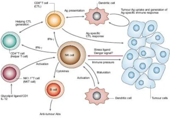
The NK cells act in tumour immunosurveillance (Figure 7), by directly inducing tumour cells death (NK act as cytolytic effectors lymphocytes), even with the absence of surface adhesion molecules and antigenic peptides.4,9 NK cells initially recognize certain ‘stress’ or ‘danger’ signals that are produced by tumours. NK-cell lysis of cancer cells could provide tumour antigens for DCs, which induce them to mature and present antigen to CTLs in lymph nodes. Cytokines, such as IFNγ , produced by activated NK cells, activate CTL and helper T-cell (CD4+) responses. This leads to the proliferation of helper T cells and cytokine production. Activated NKT cells can also induce the antitumour activity of NK cells. Cytokines that are produced by NK cells might also regulate B-cell production of antitumour antibodies. 18
Figure 7: Schematic presentation of NK role in immunoediting theory. Extracted from Smyth et al.18
According to the immunoediting theory cancer cells and immune cells reciprocally modulate each other and the two possible outcomes are: the elimination or escape of
Effect of Acupuncture on NK cells in CRC patients undergoing chemotherapy
Irene Pais
12
tumour cells. NK cells are considered to represent a first line of defence against the
metastatic spread of tumour cells. This idea is supported by the report of an association
between the decreased activity or low numbers of circulating NK cells with progression of cancers; and a correlation between an absolute decrease in the activity of the NK cells and an absolute decrease in the lytic potential of these cells.19 Recent studies of tumour-associated NK cells demonstrated a striking phenotype, supporting the notion that tumour-induced alterations of activating NK cell receptor expression may hamper immune surveillance and promote tumour progression. These tumour-induced alterations includes, in some cases the down-regulation of activating receptors and in others, the
over-expression of inhibitory receptors leading to reduced cytotoxicity.
Studies in metastatic melanoma patients reveal decreased activity and IFN production of NK cells and a redistribution of NK subsets: an increase of non-cytotoxic NK (CD56bright) cells and a decrease of cytotoxic NK cells (CD56dim). Additionally, they found a decreased NKG2D activating receptor and an over-expressed inhibitory NK cell receptor (CD158a), which is correlated to lower NK cell cytotoxicity.20
4. Co-stimulatory molecules that regulate NK-cell antitumour immunity and link innate and acquired immune system
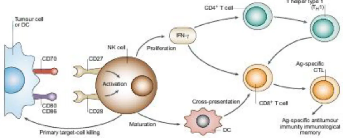
NK cells and DCs have the ability to reciprocally activate one-another, through two possible distinct mechanisms: (1) cell contact involving unknown receptor-ligand pairs and (2) through soluble mediators produced by the two cells. The cytokines, TNF, IL-2, IL-12, IL-18 and IFNγ, have all been implicated in this process. The end result of these interactions is NK cells activated for cytotoxicity, IFNγ production, and proliferation and DC that have matured and are capable of cytokine production and T cell activation (Figure 8).21
NK cells can be activated by tumour-cell- or DC-surface molecules, such as CD70, CD80 or CD86. Activated NK cells proliferate and produce cytokines such as IFNγ, or kill the tumour cell or DC. IFNγ that is produced by activated NK cells can promote (a) the antitumor acquired immune response, inducing the CD8+T cells to become antigen-specific CTLs, which lead to the development of immunological memory against the tumour; and (b) promote differentiation of CD4+T cells towards a Th1-dominant state which promotes CTL differentiation. NK-cell activation following DC-NK-cell interaction can also result in DC maturation. These DCs can facilitate the generation of CD8+T-cell
Irene Pais
13
responses through their ability to internalize and present T cells with tumour-specific antigens, which were derived from NK-cell-mediated tumour-cell lysis. 18Figure 8. Role of CD56bright NK cells and T cell in the interaction between adaptive and innate system. Extracted from Smyth et al.18
Todd et al,22 demonstrate that CD56bright NK cells and T cell are crucial cells in the interaction between adaptive and innate system. They found that endogenous T cell-derived IL-2, acting through the high affinity IL-2 receptor, co-stimulates CD56bright NK cells to secrete IFNγ.
5. Immune parameters affecting the efficacy of chemotherapy regimens
The outcome of chemotherapy can be influenced by the host immune system at multiple levels. Chemotherapy can kill cancer cells by causing them to elicit an immune response or alternatively, by increasing their susceptibility to immune attack. In addition, chemotherapy can stimulate anticancer immune effectors either in a direct fashion or by subverting immunosuppressive mechanisms. Beyond cancer-cell-intrinsic factors that determine the cytotoxic or cytostatic response, as well as the potential immunogenicity of tumour cells, the functional state of the host immune system has a major prognostic and predictive impact on the fate of cancer patients treated with conventional or targeted chemotherapies.23
It was supposed that chemotherapeutic agents elicit immunogenic tumour cell-death and the release of immunogenic signals. These signals are recognized by innate immune effectors (DCs) and trigger an adaptive immune response, which includes CD8+ T cells and IFNγ signalling, and allows the immune system to control residual tumour cells.
Effect of Acupuncture on NK cells in CRC patients undergoing chemotherapy
Irene Pais
14
Assuming this supposition as real, the interruption of this pathway at any level would lead to therapeutic failure: (1) chemotherapeutic agent may be unable to trigger immunogenic cell death; (2) tumour cells may be unable to express the required set of immunogenic signals; (3) the innate immune system may be unable to recognize such immunogenic signals; and (4) local or systemic immunosuppression or tumour-dependent immunosubversion and immunoevasion can prevent the formation, recruitment or action of adaptive immune effectors.23
Studies performed in multiple human carcinomas suggest that DCs, M1 macrophages, cytotoxic CD8+ T cells, Th1 CD4+ T cells, NK cells and Th1 CD4+ T cells present in the tumour bed possibly reduce cancer growth. And by contrast, neutrophils, M2 macrophages, myeloid-derived suppressor cells, Th2 CD4+ T cells, B lymphocytes are suspected to stimulate cancer growth.
6. The immune system and the quality of life (QOL) in cancer patients
Cancer diagnosis is a significant source of anxiety, depression and emotional distress, followed by an extended period of stressful cancer treatment significantly compromise patients’ QOL. About 20-40% of cancer patients exhibit significant levels of depression and anxiety, which was associated with higher frequency and severity of clinical symptoms, such as pain, fatigue, poor appetite, sleep disturbance and poor quality of life.24
Psychological stress may have multiple negative impacts on health outcomes, namely with a) higher incidence of infections, accelerated aging, and greater cardiovascular diseases in several populations; b) in breast cancer patients, with low physical and psychological QOL and significantly shorter disease-free interval; and with (3) a decrease of efficacy or resistance to chemotherapeutics agents, in animal models.
According to psychoneuroimmunology, psychological and emotional stress induces several alterations in diverse biological responses. The activation of the
hypothalamic-pituitary-adrenal axis and the sympathetic nervous system may generate a
change in the immune cell traffics and promotes inflammation via multiple neuroendocrine and immune pathways. Higher stress levels were associated with poorer immune responses (low NK cell activity) and the stress reduction with improvement of immune responses, in women with breast cancer. Furthermore, psychological and emotional stress has negative implications for increased symptom perception and poor quality of life,
Irene Pais
15
and is significantly associated with increased neutrophil percentage but decreased lymphocyte percentage which has been associated with negative cancer outcomes.24A study on early stage breast and prostate cancer found an association between the reduction of stress levels and the depressive pattern with a shift in the balance from a pro-inflammatory to an anti-inflammatory environment.25
WHO defines Quality of Life as individuals’ perception of their position in life in the context of the culture and value systems in which they live and in relation to their goals, expectations, standards and concerns. It is a broad ranging concept affected in a complex way by the person's physical health, psychological state, level of independence, social relationships, personal beliefs and their relationship to salient features of their environment.
The effect that chemotherapy may have on QOL is extremely important. This QOL includes not only the individual's physical well-being, but their mental well-being, role functioning and levels of emotional distress as well.
The inclusion of QOL endpoints in chemotherapy trials with cancer patients started in earliest during the last decade, with the majority of studies assessing the impact of chemotherapeutic agents on symptoms such as pain and fatigue. Since then, the inclusion of QOL endpoints has become a more common outcome in chemotherapy studies, although many continue to neglect the psychological component, focusing instead on the occurrence of symptoms and their impact on physical QOL.
QOL may be considered to be the effect of an illness and its treatment as perceived by patients and is modified by factors such as impairments, functional stress, perceptions and social opportunities.26
The most widely applicable instrument to measure the QOL in cancer patients is the EORTC QLQ-C30.
Alteration of immune cells (Decrease of NK cells)
Hypothalamus-pituitary axis Sympathetic Nervous System Activation
Emotional and Psychological stress
Effect of Acupuncture on NK cells in CRC patients undergoing chemotherapy
Irene Pais
18
1.
Epidemiology and EtiopathophysiologyColorectal cancer (CRC) is the fourth leading cause of cancer deaths worldwide in both men and women.27 Approximately two thirds of the cases are located in the colon.
The disease is often considered as one, irrespective of location, but should probably be regarded as two distinct entities as biology, risk factors, treatment and outcome may differ.28,29 Most CRCs (95%) are epithelial cancers (adenocarcinoma) that originate from glandular tissue. Other, rarer cancers in the colon and rectum are
squamous cell carcinoma and lymphoma.
In Portugal, CRC is the second most common cancer among Portuguese men and women, with an incidence rate of 40.6 and 24.1 cases per 100,000, respectively and with a mortality rate of 19.6 and 16.4 per 100,000, respectively. 30
In respect to CRC mortality in Europe, Portugal is the 8th country with highest mortality indexes, and the male rates higher than female rates; Hungary accounts with the highest mortality rates for both sexes and Greece and Cyprus accounts with the lowest rates.31
The development of CRC has been associated with several important risk factors, which can be aggregated in two main groups: the “environmental” factors group and the genetics factors group.32
The findings from epidemiologic studies that physical inactivity, excess of body weight and a central accumulation of adiposity have influence on CRC risk, as well as the ethnic and racial differences in CRC and studies on migrants suggest that an environmental factor plays an important role in aetiology of this disease. In fact, diet is considered an important factor of increased risk in the development of CRC, playing a significant role in determining the incidence of CRC in the general population. Compelling evidence suggests a strong dose-related association between red meat and fat intake and the development of CRC; and higher cholesterol values correlates significantly with later tumour development. On the other hand, diets rich in vegetables and high fibber grains as well as the consumption of fish and skinless chicken demonstrate a protective effect in the pathophysiology of CRC.
In fact, the modifiable lifestyle factors are thought to contribute substantially to the geographical difference in CRC incidence33.
Mutations in the genes Kras and p53 are highly relevant for CRC development and the same mutations have been linked to reduced physical activity, smoking and diets high in red meat.34
Irene Pais
19
Regular exercise may increase the number and activity of macrophages, natural killer cells, and cytokines with the ability to kill cancer cells and thereby improve anti-tumour immune responses.35 Furthermore, obesity may be a risk factor for CRC development independently of inactivity. Visceral adiposity is associated with low-grade chronic inflammation leading to up-regulation of the nuclear transcription factor-Kβ (NF-Kβ) with transcription of genes that promote tumourigenesis as a consequence.36 Prostaglandin E2 (PGE2) is reduced with high levels of physical activity, possibly through insulin-like growth factor-1 (IGF-1)37 and PGE2 affects both tumour immunity and tumourigenesis.38 The motility of the intestine may affect CRC risk as shortened intestinal transit time reduces the exposure of the mucosa to faecal carcinogens. Exercise is thought to increase the expression of PGF2 and the vagal tone, both stimulating motility. Diets high in fiber, fresh fruits and fresh vegetables may also shorten the intestinal transit time and the formation of fiber may produce mucosa-protecting agents, thereby affecting CRC risk. However, studies regarding diet, transit time and CRC risk are contradictory and firm conclusions cannot be drawn.39,40,41,42Insulin and IGF-1 are up-regulated in the absence of physical activity and in obesity.51 Increased levels of insulin and IFG-1 are correlated with risk of CRC 52 possibly through mechanisms that activate the Wnt pathway and increase the expression of pro-angiogenic proteins such as HIF-11 and vascular endothelial growth factor (VEGF).36 Furthermore, elevated levels of the reciprocally expressed insulin-like growth factor-binding protein-3 (IGFBP-3) have been shown to be associated with reduced risk of CRC.53
Increasing age43 and a family history of CRC44 predispose to the development of CRC, and CRC is slightly more common in men. Furthermore, genetic conditions such as familial adenomatous polyposis (FAP)45, Lynch syndrome (formerly HNPCC; hereditary nonpolyposis colorectal cancer)46 and Gardner’s syndrome (considered a subtype of FAP)47are genetic risk factors. The FAP syndrome accounts for approximately 1% of all CRC cases and patients will most likely present with adenocarcinoma before the age of 40. Therefore, prophylactic colectomy is recommended at an early age for FAP patients.48
Gardner’s syndrome is a rare phenotypic variant of FAP, both caused by mutation
in the APC gene. In addition to colon polyposis, Gardner patients acquire extra-colonic tumours including osteomas, thyroid cancer, epidermoid cysts, fibromas, sebaceous cysts and desmoids tumours 47. The disease is not curable and life expectancy with the condition is 35-45 years.
Effect of Acupuncture on NK cells in CRC patients undergoing chemotherapy
Irene Pais
20
Lynch syndrome is an autossomal dominant trait (as FAP) predisposing for CRC,
the associated mutations impair genes related to DNA mismatch repair. Approximately 1 to 4% of the cases of colon cancer are related to Lynch syndrome.46
Patients with inflammatory bowel disease (IBD; ulcerative colitis and Crohn’s) disease have increased risk of developing CRC and the risk is more likely a result of chronic inflammation rather than genetic predisposition.49 The progression from adenoma to carcinoma that occurs during development of sporadic colorectal tumours appears to be a sequence of inflammation – dysplasia – carcinoma in IBD-associated CRC49. Primary sclerosing cholangitis (PSC) is another chronic inflammatory disease associated with CRC when concomitant with IBD.50 Pro-inflammatory factors of the innate and adaptive immune system contributes to the development and growth of colon neoplasia.
It is too early to establish a causal relationship between smoking and CRC, however most reports point in the direction that smoking is a risk factor.51,54 Furthermore, smoking may be associated with different subtypes with different mutational background than non-smokers (e.g. p53, APC and Kras).55 Heavy alcohol intake (>45g/day) has been associated with increased risk of CRC, while lower levels were not.56 The increased CRC risk has been suggested to be associated with lower intake of folate connected with heavy drinking.57
In conclusion, there is evidence that inactivity, red meat, smoking, alcohol, and obesity are connected to increased risk of CRC, but the quality of the observations varies and in some cases, there are contradictory reports.
1.1. Clinical presentation
Patients with CRC may present in three ways: patients with suspicious symptoms and/or signs; asymptomatic individuals discovered by routine screening; and emergency admission with intestinal obstruction, peritonitis, or rarely, an acute gastrointestinal bleed.
Despite the increasing usage of CRC screening allows the early diagnosis, on an asymptomatic, of CRC, the majority of CRCs is diagnosed after the occurrence of symptoms. The symptomatic presentation of CRC is normally due to growth of the tumour into the lumen or adjacent structures and usually reflects relatively advanced CRC. The symptomatic presentation can be divided in two groups: symptoms that derive from the local tumour and symptoms related to metastasis.58
Irene Pais
21
Symptoms/signs associated with local CRC include hematochezia or melena, abdominal pain, otherwise unexplained iron deficiency anemia, and/or a change in bowel habits. Abdominal distention, and/or nausea and vomiting, which may be indicators of obstruction, are less common presenting symptoms. Obstructive symptoms are more common with cancers that encircle the bowel, producing the so-called "apple-core" (seen on radiologic imaging). Rectal cancer can also cause also cause tenesmus, rectal pain, and diminished caliber of stools.These clinical manifestations differ according on tumour location: a change in bowel habits is generally indicator of left-sided than right-sided CRCs because fecal contents are liquid in the proximal colon and the lumen caliber is larger, and they are therefore less likely to be associated with obstructive symptoms; Hematochezia is more often caused by rectosigmoid than right-sided colon cancer; Iron deficiency anemia is more common with right-sided CRCs. Cecal and ascending colon tumors have a fourfold higher mean daily blood loss (approximately 9 mL/day) than tumors at other colonic sites.
Abdominal pain can occur with tumors arising at all sites; it can be caused by a partial obstruction, peritoneal dissemination, or intestinal perforation leading to generalized peritonitis.
The symptoms/signs of metastatic disease include the presence of right upper quadrant pain, abdominal distention, early satiety, supraclavicular adenopathy, or periumbilical nodules. These categories of symptoms are explained by de fact that the most common metastatic sites are regional lymph nodes, liver, lungs and peritoneum. Since the venous drainage of the intestinal tract is via the portal system, the first site of hematogenous dissemination is generally the liver, followed by the lungs, bone, and many other sites, including the brain. However, tumors arising in the distal rectum may metastasize initially to the lungs because the inferior rectal vein drains into the inferior vena cava rather than into the portal venous system.
There are also a diversity of atypical presentations of CRC, including: a) local invasion or a contained perforation causing malignant fistula formation into adjacent organs, such as bladder (resulting in pneumaturia) or small bowel; b) Fever of unknown origin, the existence of abscesses (intra abdominal, retroperitoneal, abdominal wall or intrahepatic) due to a localized perforated colon cancer; c) Streptococcus bovis
bacteremia and Clostridium septicum sepsis are associated with underlying colonic
malignancies in about 10% to 25% of patients. Seldom, other extra-abdominal infections caused by colonic anaerobic organisms (eg, Bacteroides fragilis) may be associated with CRC.
Effect of Acupuncture on NK cells in CRC patients undergoing chemotherapy
Irene Pais
22
The presence of symptoms and their particular type provide some prognostic importance: symptomatic patients at time of diagnosis have a more advanced disease and a worse prognosis; however, asymptomatic patients tend to have a more favorable pathological stage and consequently better prognosis.
1.2 Diagnosis
CRC may be suspected from one or more of the symptoms and signs described above or may be asymptomatic and discovered by routine screening of average and high-risk subjects. Once a CRC is suspected, the next test can be a colonoscopy, barium enema, or CT colonography. However, examination of tissue is required to establish the diagnosis; this is usually accomplished by colonoscopy. In fact, colonoscopy is the most accurate and resourceful diagnostic test for CRC, since it can localize and biopsy lesions throughout the large bowel, detect synchronous neoplasm, and remove polyps. Synchronous CRCs, refers to the diagnosis, within six months of an initial CRC, of two or more distinct primary tumors, separated by normal bowel, and not due to direct extension or metastasis.
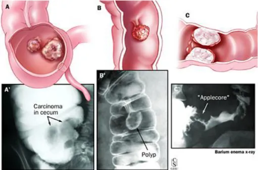
The vast majority of colon and rectal cancers are endoluminal masses (exophytic or polypoid) that arise from the mucosa and protrude into the lumen. Bleeding may be seen with lesions that are friable, necrotic, or ulcerated. Circumferential or near-circumferential involvement of the bowel wall correlates with the so-called "apple-core" description seen on radiologic imaging.
Figure 9. Colonic features and corresponding barium enema X-rays. A: carcinoma in the cecum; B: pedunculated polyp; C: apple core lesion.
Irene Pais
23
A minority of neoplastic lesions in the gastrointestinal tract is nonpolypoid and relatively flat or depressed, which are more difficult to detect by colonoscopy.If a malignant obstruction precludes a full colonoscopy preoperatively, the entire residual colon should be examined soon after resection.
Flexible sigmoidoscopy is generally not considered to be an adequate diagnostic study for a patient suspected of having a CRC, unless a palpable mass is felt in the rectum. In such cases, a full colonoscopy will still be needed to evaluate the remainder of the colon for synchronous polyps and cancers.
Barium enema is widely available and may be used to investigate patients with symptoms suggesting of CRC. However, the diagnostic yield of both double-contrast barium enema (DCBE) alone and the combination of DCBE plus flexible sigmoidoscopy is less than that of colonoscopy or CT colonography for the evaluation of lower tract symptoms. After detection by barium enema of a polyp or a mass, colonoscopy is recommended to establish the histology, remove the polyp, and search for synchronous lesions.
CT colonography (virtual colonoscopy or CT colography) provides a computer-simulated endoluminal perspective of the air-filled distended colon. CT colonography requires a mechanical bowel preparation that is similar to that needed for barium enema, since stool can simulate polyps.
Reasons for incompleteness include the inability of the colonoscope to reach the tumour or to visualize the mucosa proximal to the tumour for technical reasons (eg, partially or completely obstructing cancer, tortuous colon, and poor preparation) and patient intolerance of the examination. CT colonography should be restricted to patients who are able to pass flatus and capable of tolerating the oral preparation. For clinically obstructed patients, a gastrointestinal (GI) CT scan is a good alternative to CT colonography.
Resuming, the CT colonography provides a similarly sensitive, less invasive alternative to colonoscopy in patients presenting with symptoms suggestive of CRC. However, since colonoscopy permits removal/biopsy of the lesion and the detection of any synchronous cancers or polyps, colonoscopy seems to constitute the gold standard for investigation of symptoms suggestive of CRC.
In respect to laboratory tests, although CRC is often associated with iron deficiency anemia, its absence does not reliably exclude the disease. There is no diagnostic role for other routine laboratory test, including liver function tests, which lack sensitivity for detection of liver metastases.
Effect of Acupuncture on NK cells in CRC patients undergoing chemotherapy
Irene Pais
24
A variety of serum markers have been associated with CRC, particularly carcinoembryonic antigen (CEA). However, all these markers have a low diagnostic ability to detect primary CRC due low sensitivity for early stage disease. And on other hand, non-cancer-related causes can elevate CEA levels, including gastritis, peptic ulcer disease, diverticulitis, liver disease, chronic obstructive pulmonary disease, diabetes, and any acute or chronic inflammatory state. The American Society of Clinical Oncology (ASCO) and the European Group on Tumour Markers recommended that neither serum CEA nor CA 19.9 levels should be used as a screening or diagnostic test for colorectal cancer.
However, CEA levels do have value in the follow-up of patients with diagnosed CRC as well as on preoperative scenarios with the objective to aid in surgical treatment planning, post-treatment follow up, and in the assessment of prognosis: CEA >5 ng/mL have a worse prognosis, stage for stage, than those with lower levels; and elevated preoperative CEA levels that do not normalize following surgical resection imply the presence of persistent disease and the need for further evaluation.
Furthermore it’s recommended to perform a serial assay of postoperative CEA levels for five years for patients with stage II and III disease candidate for surgery or chemotherapy (metastatic disease). A rising CEA level after surgical resection implies recurrent disease and should prompt follow-up radiologic imaging.58
1.3 CRC Staging
Two different staging systems are used to provide prognostic information and determine treatment strategy in CRC. The Duke classification system from 1932 59 has been replaced by the more detailed TNM staging system developed and maintained by the American Joint Committee on Cancer (AJCC). Duke staging is no longer recommended for use in clinical practice and is not further discussed here. TNM staging (tumour, node and metastasis) is used on many solid cancers, adapted for CRC and is now in the 7th edition as of 2010. As Table I shows, the tumour is first scored with respect to the TNM variables then assigned to stage I-IV in the CRC specific (Table II). However, TNM does not adapt to recent advances in metastatic treatment. For example, the survival of a patient with resectable solitary liver metastasis is better than that of a patient with stage III disease.60 Current observations regarding the clinical course of the patients with CRLM support emerging arguments for a new staging system in CRC. 60,61,62
Irene Pais
25
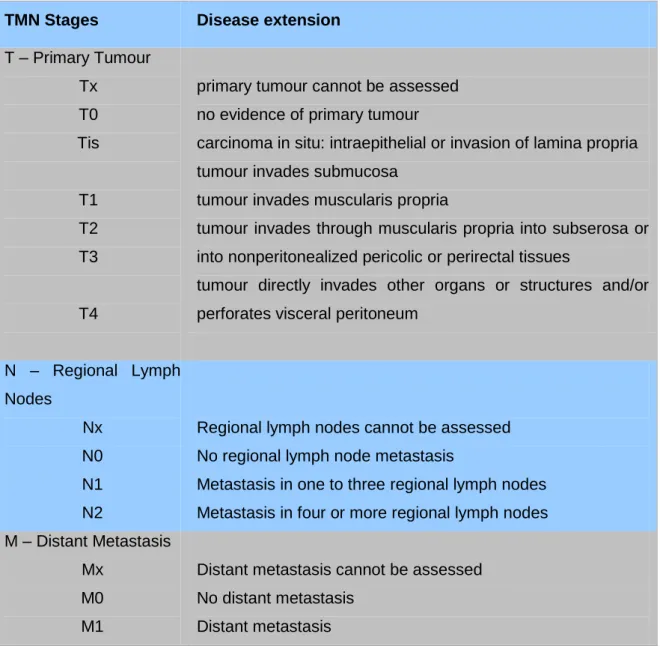
TMN Stages Disease extension
T – Primary Tumour Tx T0 Tis T1 T2 T3 T4
primary tumour cannot be assessed no evidence of primary tumour
carcinoma in situ: intraepithelial or invasion of lamina propria tumour invades submucosa
tumour invades muscularis propria
tumour invades through muscularis propria into subserosa or into nonperitonealized pericolic or perirectal tissues
tumour directly invades other organs or structures and/or perforates visceral peritoneum
N – Regional Lymph Nodes Nx N0 N1 N2
Regional lymph nodes cannot be assessed No regional lymph node metastasis
Metastasis in one to three regional lymph nodes Metastasis in four or more regional lymph nodes M – Distant Metastasis
Mx M0 M1
Distant metastasis cannot be assessed No distant metastasis
Distant metastasis
Effect of Acupuncture on NK cells in CRC patients undergoing chemotherapy Irene Pais
26
Stages T N M 0 Tis N0 M0 I T1 N0 M0 T2 N0 M0 II T3 N0 M0 T4 N0 M0 III T1, T2 N1 or N2 M0 T3, T4 N1 or N2 M0 IV Any T Any N M1Table 2. The 7th Edition of AJCC-TNM Classification System.
In respect to rectal cancer, the TNM staging should be used, being recommended the version 5 from 1997 over TNM version 6 (2002) and 7 (2010), due to marked interobserver variation in defining stage II and Stage II, on the version 7. And also, there is the need for further subclassification particularly of cT3 (Table 3).63
T1 tumours refer to cancers that are in a stalked adenoma and according to the submucosa (sm)-system in a sessile adenoma. The level of infiltration into the submucosa predicts the risk of lymph node metastases and thus the type of surgery.