47
ABSTRACT
Introduction. Fabry disease is a rare metabolic
disorder caused by the genetic deficiency of the lysosomal hydrolase alpha-galactosidase A, located on chromosome X. Females with the defective gene are more than carriers and can develop a wide range of symptoms. Nevertheless, disease symp-toms generally occur later and are less severe in women than in men. The enzyme deficiency mani-fests as a glycosphingolipidosis with progressive accumulation of glycosphingolipids and deposit of inclusion bodies in lysosomes giving a myelin-like appearance.
Patients and Methods. Records of renal biopsies
performed on adults from 1st January 2008 to 31st
August 2011, were retrospectively examined at the Renal Pathology Laboratory. We retrieved biopsies diagnosed with Fabry disease and reviewed clinical and laboratory data and pathology findings.
Results. Four female patients with a mean age of
49.3±4.5 (44-55) years were identified. The mean proteinuria was 0.75±0.3 g/24h (0.4-1.2) and esti-mated glomerular filtration rate (CKD EPI equation)
was 71±15.7 ml/min/1.73m2 (48-83). Three patients
experienced extra-renal organ involvement (cerebro-vascular, cardiac, dermatologic, ophthalmologic and thyroid) with distinct severity degrees. Leukocyte α-GAL A activity was below normal range in the four cases but plasma and urinary enzymatic activity was normal.
Light microscopy showed predominant vacuolisa-tion of the podocyte cytoplasm and darkly staining granular inclusions on paraffin and plastic-embedded semi-thin sections. Electron microscopy showed in three patients the characteristic myelin-like inclusions in the podocyte cytoplasm and also focal podocyte foot process effacement. In one case the inclusions were also present in parietal glomerular cells, endothe-lial cells of peritubular capillary and arterioles.
Conclusion. Clinical signs and symptoms are varied
and can be severe among heterozygous females with Fabry disease. Intracellular accumulation of gly-cosphingolipids is a characteristic histologic finding of Fabry nephropathy. Since this disease is a poten-tially treatable condition, its early identification is imperative. We should consider it in the differential diagnosis of any patient presenting with proteinuria and/or chronic kidney disease, especially if there is a family history of kidney disease.
Key-Words:
Fabry disease; glomerulopathy; renal biopsy.
INTRODUCTION
Fabry disease (FD) is a progressive, X-linked inher-ited disorder of glycosphingolipid metabolism due to deficient or absent lysosomal α-galactosidase A (α-GAL A) activity. This results in progressive accu-mulation of globotriaosylceramide (Gb3 or GL-3) and
Clinical and pathological findings
in women with Fabry disease
Ana Natário1,2, Helena Viana2, Maria João Galvão2, Fernanda Carvalho2
1 Nephrology Department, Centro Hospitalar de Setúbal. Setúbal, Portugal.
2 Renal Pathology Laboratory, Nephrology Department, Hospital Curry Cabral. Lisbon, Portugal.
Received for publication: 29/12/2011
Accepted in revised form: 16/02/2012
Nefro - 26-1 - MIOLO.indd 47
48 Port J Nephrol Hypert 2012; 26(1): 47-54
related glycosphingolipids within lysosomes in a variety of cell types, including capillary endothelial
cells, renal, cardiac and nerve cells1.
The incidence of Fabry disease has been estimated
to be 1:117,000 overall births2. The age of symptom
onset and the age of diagnosis tend to be
approxi-mately 10 years later in females than males3. There
is often significant delay in the diagnosis4.
Phenotype differences are due in part to random X-chromosome inactivation, resulting in considerable variability in α-GAL A activity among carriers and within one carrier individual among various tissues
or regions of a single tissue4.
The classical phenotype presents with early der-matologic, ophthalmologic, and peripheral nervous system involvement. Renal, cardiac and cerebrovas-cular complications are major causes of morbidity
and mortality in adults1.
Renal impairment often begins with microalbu-minuria and proteinuria in the second to third decade of life. Isosthenuria accompanied by altera-tions in tubular reabsorption, secretion and
excre-tion also develops1. By adulthood, renal failure
frequently becomes a major complication of Fabry disease, with more than half of males eventually developing advanced renal failure or end-stage renal
disease (ESRD)5.
Heterozygous female individuals can develop a wide range of symptoms, ranging from asymptomatic
to overt disease6. The majority of females have slowly
progressive kidney disease, but a smaller subset seem to be more seriously affected with progression
to ESRD at the same median age as men7.
Classically, renal lesions result from Gb3 deposi-tion in the lysosomes of podocytes, glomerular endothelial, parietal epithelial, mesangial and inter-stitial cells that may lead to progressive microvas-cular dysfunction, occlusion and ischaemia, with subsequent development of segmental and global glomerular sclerosis, tubular atrophy and interstitial
fibrosis8-10. Podocyte injury might correlate directly
with proteinuria and play a pivotal role in the
devel-opment and progression of Fabry nephropathy11.
Glycosphingolipid storage also occurs in the epithe-lium of the loop of Henle and the distal tubules,
and in the endothelial and smooth muscle cells of
the renal arterioles1.
Kidney biopsy may be useful as a baseline assess-ment and in patients with atypical presentations, includ-ing a repeat kidney biopsy when the disease is
progress-ing despite therapy1. In some cases, females can develop
the same kidney histopathology as males8,12,13.
The aim of this study is to review the clinico-pathological findings of females with Fabry disease, whose renal biopsies were analysed in a single neph-ropathology laboratory.
PATIENTS AND METHODS
Four renal biopsies performed on four adult
females and analysed at our institution from 1st
Janu-ary 2008 to 31st August 2011 were retrospectively
examined.
We recorded the following data from each patient: age, gender, race, indication for renal biopsy (clinical syndrome), clinical manifestations in other organs,
GLA gene mutation, plasma and leukocyte α-GAL A
levels and urinary activity measurements.
Renal biopsy specimens were stained and analysed by light microscopy (LM), immunofluorescence (IMF) and electron microscopy (EM). Routine LM examina-tion was performed on haematoxylin and eosin (HE), periodic acid-Schiff (PAS), Jones methenamine silver (SM), and trichrome stains (TR). Histological findings were scored by LM, with respect to glomerular and interstitial changes as 0, none; 1, mild (<25%); 2,
moderate (25-50%); and 3, severe change (>50%)8.
Vascular changes were analysed for intimal thicken-ing and hyalinosis usthicken-ing the same scorthicken-ing system.
We reviewed the semi-thin toluidine blue-stain sections that were prepared as survey sections for electron microscopy.
All specimens were studied by IMF using anti-immunoglobulin (A, G and M), anti-C3, C4, C1q of complement and anti-albumin antibodies.
Electron microscopy images were reviewed in all cases.
Nefro - 26-1 - MIOLO.indd 48
Port J Nephrol Hypert 2012; 26(1): 47-54 49
RESULTS
Clinical findings
We retrospectively identified four female patients with Fabry disease in our renal biopsy files, and their clinical and laboratory data are summarised in Table I.
All patients were Caucasian, ranging in age from 44 to 55 years (mean 49.3±4.5). Estimated glom-erular filtration rate (CKD EPI equation) was 71±15.7
ml/min/1.73m2 (48-83). Three patients with a mean
age of 39.3±6.0 (33-45) years were diagnosed with hypertension and, in these cases, 24-hour urine total protein (mean 0.75±0.34 g/24h) was measured while on treatment with angiotensin converting enzyme inhibitors and/or angiotensin receptor blockers.
Two patients were sisters (patients 1 and 2) and all had at least one case of first-degree family history of renal disease.
The indications for renal biopsy were non-nephrotic proteinuria in three patients and chronic renal failure with proteinuria in the other.
In three cases the diagnosis was established by biochemical and genetic studies prior to renal biopsy. Skin biopsy of angiokeratomas performed in our nephropathology centre (Fig. 1) allowed the diagnosis in one case (patient 3).
All the females had leukocyte α-GAL A activity below normal range but plasma and urinary enzymatic activity was normal.
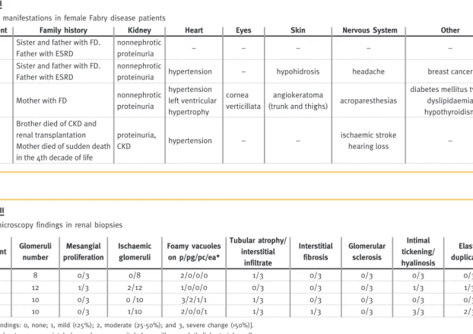
Clinical manifestations were provided by the patients' physicians and are shown in Table II. Car-diovascular involvement was exhibited as hyperten-sion in three patients and also left ventricular hyper-trophy in other case. Cryptogenic ischaemic stroke occurred in one patient at the age of 38 years. Ophthalmologic, cutaneous lesions, thyroid dysfunc-tion and peripheral neuropathic pain were present in one patient. Finally, in one case the only mani-festation was non-nephrotic proteinuria.
Light microscopy
On average, 10±1.6 (range 8-12) glomeruli were present for light microscopic assessment, and 2.8±1.3
Figure 1
Electron microscopy of skin biopsy (patient 3) of a small superficial angioma showing lysosomal inclusion with myelin-like figures within endothelial cells and capillary lumen).
Table I
Clinical and laboratory data at the time of renal biopsy
Patient Age (years) Serum creatinine (mg/dL) eGFR (CKD EPI equation) (ml/min/1.73m2) Urine protein (g/24h) Leukoyte α-galactosidase (Nmol/h/mg protein) Plasma α-galactosidase (Nmol/h/mL plasma) Urinary Gb3 (ug/mmol creat)
GLA gene muta-tion
1 49 0.9 75 0.4 20 10 8 Arg112His
2 55 0.8 83 0.6 2 5 No data Arg112His
3 44 0.9 78 0.8 1.9 6 No data Pro265Arg
4 49 1.3 48 1.2 27 6 2.9 ΔGlu358
* Reference range: 36-80 nmol/h/mg protein ** Reference range: 6-19 nmol/h/mL plasma *** Reference range: <25 μmmol creat
Nefro - 26-1 - MIOLO.indd 49
50 Port J Nephrol Hypert 2012; 26(1): 47-54
Table II
Clinical manifestations in female Fabry disease patients
Patient Family history Kidney Heart Eyes Skin Nervous System Other
1 Sister and father with FD. Father with ESRD
nonnephrotic
proteinuria – – – – –
2 Sister and father with FD. Father with ESRD
nonnephrotic
proteinuria hypertension – hypohidrosis headache breast cancer
3 Mother with FD nonnephrotic proteinuria hypertension left ventricular hypertrophy cornea verticillata angiokeratoma
(trunk and thighs) acroparesthesias
diabetes mellitus type 2 dyslipidaemia hypothyroidism
4
Brother died of CKD and renal transplantation Mother died of sudden death in the 4th decade of life
proteinuria,
CKD hypertension – –
ischaemic stroke
hearing loss –
Table III
Light microscopy findings in renal biopsies
Patient Glomeruli number Mesangial proliferation Ischaemic glomeruli Foamy vacuoles on p/pg/pc/ea* Tubular atrophy/ interstitial infiltrate Interstitial fibrosis Glomerular sclerosis Intimal tickening/ hyalinosis Elastic duplication 1 8 0/3 0/8 2/0/0/0 1/3 0/3 0/3 0/3 0/3 2 12 1/3 2/12 1/0/0/0 0/3 0/3 0/3 1/3 1/3 3 10 0/3 0 /10 3/2/1/1 1/3 0/3 0/3 0/3 0/3 4 10 0/3 1/10 2/0/0/1 1/3 1/3 0/3 3/3 2/3
[Score findings: 0, none; 1, mild (<25%); 2, moderate (25-50%); and 3, severe change (>50%)]. * p – podocytes, pg – parietal glomerular, pc – peritubular capillary, endothelial arteriolar cells
A B
Figure 2
Light microscopy in Fabry disease
(A) Renal biopsy of patient 4 with a glomerulus showing hypertrophic glomerular podocytes distended with foamy appearing vacuoles (PAS stain; mag-nification, x400)
(B) Renal biopsy of patient 3 with a glomerulus showing extensive inclusion bodies of glycolipid in podocytes and parietal cells (arrows; toluidine blue stain; magnification, x 400)
Nefro - 26-1 - MIOLO.indd 50
Port J Nephrol Hypert 2012; 26(1): 47-54 51
(range 1-4) glomeruli were examined on toluidine blue-stained semi-thin sections.
Renal pathology from light microscopy is described in Table III.
LM showed in all patients varying degrees of hypertrophic glomerular visceral epithelial cells dis-tended with foamy appearing vacuoles (Fig. 2A), corresponding to extracted Gb3 deposits. There were also vacuoles in parietal glomerular, endothelial cells of peritubular capillary and arterioles in patient 3.
In one biopsy there was mild mesangial widening. In the tubulo-interstitial compartment, two patients had mild interstitial fibrosis, a chronic inflammatory infiltrate and tubular atrophy (less than 5%). Glomerular sclerosis (segmental or global) was not observed.
Vascular involvement was observed in two patients: arteriolar thickening, hyalinosis and elastic duplica-tion, which led to the initial histological diagnosis of nephroangiosclerosis in one of the cases. The female patient with diabetes mellitus type 2 showed no lesions of diabetic nephropathy.
Semi-thin sections with toluidine blue showed fine but darkly staining granular inclusions (Fig. 2B) in
varying degrees in the cytoplasm of podocytes in all patients. The most prominent location of the inclu-sions was in podocytes but in two cases (patients 3 and 4) it was present in peritubular capillary, glomerular endothelial and vascular intimal cells.
On frozen sections under polarised light, the gly-cosphingolipids showed birefringence and autofluo-rescence (Fig. 3).
Immunofluorescence was negative in all cases (including albumin and Ig G in the diabetic female).
Electron microscopy
All the biopsies were analysed by electron microscopy.
In three specimens, podocyte inclusions that varied from granular to concentric lamellated were demon-strated (Fig. 4) and in one biopsy it was also found on peritubular capillaries, parietal glomerular cells and arterial endothelial cells. Inclusions were surrounded by a single unit membrane, indicating their location in lysosomes. Areas of focal fusion of podocyte foot processes were present in three biopsies. No lesions were found in dense membrane of the glomeruli.
Unfortunately in one patient there was no sample for electron microscopy. The diagnosis of Fabry
Figure 3
Light microscopy with polarised light (patient 3) showing glycosphingolip-ids within a glomeruli that are birefrigent and exhibit autofluorescence (magnification, x 400)
Figure 4
Electron microscopy in renal biopsy of patient 1. Accumulation of storage material, with the appearance of lamellated membrane structures is dem-onstrated in the secondary lysosomes of podocytes. There is focal foot process effacement (arrow).
Nefro - 26-1 - MIOLO.indd 51
52 Port J Nephrol Hypert 2012; 26(1): 47-54
disease was made afterwards with identification of
GLA mutation.
DISCUSSION
In this case series of heterozygous females, clinical signs and symptoms varied widely. This phenotypic heterogeneity is thought to be partly due to lyonisa-tion, a process whereby one copy of the X-chromo-some is randomly inactivated in all cells of the female
embryo1. Therefore, heterozygous females are
essen-tially a mosaic of normal and mutant cells in varying
proportions21.
The degree of symptoms depends on the residual enzymatic activity of α-GAL A: females can have anywhere from near-normal levels to no active
enzyme1. Of the 1077 females enrolled in the Fabry
Registry, 69.4% had symptoms and signs of FD and twenty percent experienced major cerebrovascular, cardiac, or renal events at a median age of 46
years5.
One female of our case series suffered an ischae-mic stroke at the age of 38 years and on light microscopy, vascular abnormalities of severe hyper-tension superimposed on the changes of FD were observed. There is a high prevalence of hypertension, cardiac disease and renal disease in patients who
have had a stroke in the context of FD22. Data from
the Fabry Registry22 and the Fabry Outcome Survey23
have shown that the majority of strokes in FD are due to small vessel events. In a large cohort of young Portuguese patients the prevalence of GLA missense mutations was 3.8% in patients with cryptogenic stroke, and these mutations were associated with residual α-galactosidase activity levels much higher than those observed in classically affected Fabry
patients14.
Incidental findings of corneal opacity by ophthal-mologists can be another sign of FD. Corneal changes (cornea verticillata), rarely disturbing visual acuity,
are frequently encountered in case series1 and was
detected in one of our patients. Hearing loss has
also been reported1,15.
In a small study, subclinical hypothyroidism was
found in 36.4% of the patients16. Hypothyroidism
was detected in one of our four cases. Faggiano et
al. consider that endocrine work-up should be
rec-ommended in all patients suffering from FD17.
Proteinuria and CKD are important manifestations
of Fabry disease7. All the female patients had
pro-teinuria and only one had moderate renal function impairment.
According to some authors, proteinuria and/or a reduced glomerular filtration rate may be found in
40% of adult females7,18. Proteinuria was recently
shown to be an important risk factor for progression
of kidney dysfunction in Fabry disease19,20 with a
much greater predictive value for men than women20.
Deegan et al. found that proteinuria was not
associ-ated with a more rapid decline in GFR in females18.
An international effort has been mounted to create a standardised scoring system of the extent and severity of renal involvement, supporting the role of kidney biopsy in the baseline evaluation of Fabry nephropathy. This International Study Group consid-ered that optimal slides for scoring should ideally include >10 glomeruli for light microscopic assess-ment, and at least 3 glomeruli for semi-thin section
scoring8. Renal specimens of our study contained a
mean glomeruli number of 10±1.6 (range 8-12) pres-ent for light microscopy and 2.8±1.3 (range 1-4) on toluidine blue-stained semi-thin sections.
Vacuolisation of podocytes and epithelial cells is
a characteristic histological finding8. Glomerular
pari-etal epithelial cells are particularly involved in females
with severe GLA gene mutations13 and this was found
in patient 3 that had serious extra-renal manifesta-tions of FD.
Fogo et al. found that arteriolar hyalinosis was simi-lar in both genders, but females had significantly more arterial hyalinosis which was probably related to their
significantly higher age8. We detected mild and severe
arteriolar hyalinosis, respectively, in two biopsies. Although mild interstitial fibrosis and tubular atro-phy was found in only half of our patients, it should be emphasised that chronic glomerular and interstitial damage develops early in the course of Fabry disease, and the absence of typical clinical signs of CKD does
not rule out Fabry nephropathy8. Our results are in
Nefro - 26-1 - MIOLO.indd 52
Port J Nephrol Hypert 2012; 26(1): 47-54 53
agreement with previous series of Fabry patients, showing histological changes before renal function is decreased. Renal biopsy in female patients with FD may therefore be important for establishing the diagnosis and characterisation of renal lesions.
During the study, it became clear to us that his-tological findings suggestive of Fabry nephropathy (e.g. vacuolisation) may be missed on routine his-tological sections unless there is a strong clinical suspicion. However, semi-thin sections are best for
characterising podocyte Gb3 inclusions8.
Cellular inclusions of Gb3 within lysosomes have been described by various names, including zebra bodies and myelin figures, according to different morphologies of their pattern of lamellation. Podo-cytes and distal tubular epithelial cells have been described as containing the highest concentrations
of Gb324. In some cell types, inclusions appear as
small, dark, dense-beaded granules; in others, they
appear as larger complex laminated bodies24, as
seen in this case series.
Noteworthy is that none of our patients had an history of taking medications known to cause Fabry-like morphologic changes, such as chloroquine,
amio-darone, or aminoglycosides25-27.
Immunofluorescence was negative in all cases. According to literature, routine immunofluorescence microscopy is usually negative but in glomeruli with advanced lesions, immunoglobulin M, A, and comple-ment components (C3 and C1q) may be detectable in capillary walls and mesangial regions showing a
segmental distribution and granular pattern24.
In symptomatic individuals, the diagnosis of FD can be established on the basis of low activity of α-GAL A in plasma, leukocytes or cultured skin
fibro-blasts28. There is significant overlap in the α-GAL A
activity levels of carrier females and the general population; thus, α-GAL A activity may not reliably distinguish the affected (carrier) from unaffected (non-carrier) women. As shown in this series, females had a normal amount of plasma α-GAL A enzyme activity and still carried the defective gene.
For females with normal to low-normal α-GAL A,
a genetic test is needed for accurate diagnosis15.
Mutation subtypes of FD may have impact on
disease progression9. The mutation Arg112His
dem-onstrated in patients 1 and 2 is predominantly asso-ciated with residual enzyme activity and a mild variant
phenotype of FD29 which is consistent with our
clini-cal and histologiclini-cal findings.
There are several limitations when analysing our data, especially in rare diseases where the limited num-ber of cases may not be representative of the Fabry female population. The prevalence of CKD and ESRD in the general population is much higher than in Fabry disease and, particularly in patients carrying GLA muta-tions associated with significant residual enzyme activ-ity, it should be noted that the cause of CKD may not always be a consequence of Fabry disease.
The diagnosis of Fabry disease among patients without a positive family history has been a chal-lenge to nephrologists and since Fabry disease is a potentially treatable condition it is imperative to consider it in the differential diagnosis of any patient presenting with proteinuria and/or chronic kidney disease, especially if there is a family history of
kidney disease30.
CONCLUSION
Clinical signs and symptoms are varied and can be severe among heterozygous females with Fabry disease. Histological renal findings are notorious even in women with little clinical involvement – myelin-like structures were found within lysosomes in many renal cells. Glomerular sclerosis and tubulointerstitial fibrosis are minimal in females with no clinical expres-sion of renal disease.
Fabry disease nephropathy should be considered for diagnosis of any patient presenting with CKD, especially in those with proteinuria and family history of renal disease.
This study should raise the awareness of this rare disease among nephrologists and other specialists that are likely to encounter unrecognised Fabry female patients to improve early diagnosis.
Conflict of interest statement. None declared.
Nefro - 26-1 - MIOLO.indd 53
54 Port J Nephrol Hypert 2012; 26(1): 47-54
Acknowledgements
The authors gratefully acknowledge the physicians of Fabry disease patients: Dr Carlos Barreto (Setúbal), Dr Jesus Garrido and Dr Joana Vidinha (Viseu), Dr Isabel Pataca (Lisboa) for their contribu-tion to clinical data colleccontribu-tion.
References
1. Germain DP. Fabry disease. Orphanet J Rare Dis 2010;5:30
2. Meikle PJ, Hopwood JJ, Clague AE, Carey WF. Prevalence of lysosomal storage disorders.
JAMA 1999;281:249-254
3. MacDermot KD, Holmes A, Miners AH. Anderson-Fabry disease: Clinical manifestations
and impact of disease in a cohort of 60 obligate carrier females. J Med Genet 2001;38:769-775
4. Anderson-Fabry disease: a nephrology perspective. J Am Soc Nephrol 2002;(Ssuppl
2);13:S125-S153
5. Wilcox WR, Oliveira JP, Hopkin RJ, et al. Females with Fabry disease frequently have
major organ involvement: lessons from the Fabry Registry. Mol Genet Metab 2008;93:112-128
6. Warnock DG, Daina E, Remuzzi G, West M. Enzyme replacement therapy and Fabry
nephropathy. Clin J Am Soc Nephrol 2010;5:371-378
7. Ortiz A, Oliveira JP, Waldek S, Warnock DG, Cianciaruso B, Wanner C. Nephropathy in
males and females with Fabry disease: cross-sectional description of patients before treatment with enzyme replacement therapy. Nephrol Dial Transplant 2008;23:1600-1607
8. Fogo AB, Bostad L, Svarstad E, et al. Scoring system for renal pathology in Fabry
disease: report of the International Study Group of Fabry Nephropathy (ISGFN). Neph-rol Dial Transplant 2010;25:2168-2177
9. Branton MH, Schiffmann R, Sabnis SG, et al. Natural history of Fabry renal disease:
Influence of alpha-galactosidase A activity and genetic mutations on clinical course. Medicine (Baltimore) 2002; 81:122-138
10. Alroy J, Sabnis S, Kopp JB. Renal pathology in Fabry disease. J Am Soc Nephrol
2002;13:S134-S138
11. Najafian B, Svarstad E, Bostad L, et al. Progressive podocyte injury and
globotriao-sylceramide (GL-3) accumulation in young patients with Fabry disease. Kidney Int 2011;79: 663-670
12. Gubler MC, Lenoir G, Grunfeld JP, Ulmann A, Droz D, Habib R. Early changes in
hem-izygous and heterozygous patients with Fabry’s disease. Kidney Int 1978;13:223-235
13. Valbuena C, Carvalho E, Burstorff M, et al. Kidney biopsy findings in heterozygous
Fabry disease females with early nephropathy. Virchow Arch 2008;453:329-338
14. Baptista MV, Ferreira S, Pinho-e-Melo T, et al. Mutations of the GLA gene in young
patients with stroke: the POTYSTROKE Study. Stroke 2010; 41:431-436
15. Keilmann A, Hajioff D, Ramaswami U. Ear symptoms in children with Fabry disease:
data from the Fabry Outcome Survey. J Inherit Metab Dis 2009;32:739-744
16. Hauser AC, Gessl A, Lorenz M, Voigtlander T, Fodinger M, Sunder-Plassmann G. High
prevalence of subclinical hypothyroidism in patients with Anderson-Fabry disease. J Inherit Metab Dis 2005;28:715-722
17. Faggiano A, Pisani A, Milone F, et al. Endocrine dysfunction in patients with Fabry
disease. J Clin Endocrinol Metab 2006; 91:4319-4325.
18. Deegan PB, Baehner AF, Barba Romero MA, et al. Natural history of Fabry disease in
females in the Fabry Outcome Survey. J Med Genet 2006;43:347-352
19. Banikazemi M, Bultas J, Waldek S, et al. Agalsidase-beta therapy for advanced Fabry
disease: a randomized trial. Ann Intern Med 2007;146:77-86
20. Wanner C, Oliveira JP, Ortiz A, et al. Prognostic indicators of renal disease progression
in adults with Fabry disease: natural history data from the Fabry Registry. Clin J Am Soc Nephrol 2010;5:2220-2228
21. Germain DP. Genetics of Fabry disease: diagnostic and therapeutic implications. Presse
Med 2007;36:S14-S19
22. Sims K, Politei J, Banikazemi M, Lee P. Stroke in Fabry disease frequently occurs before
diagnosis and in the absence of other clinical events: natural history data from the Fabry Registry. Stroke 2009;40:788-794
23. Fellgiebel A, Muller MJ, Ginsberg L. CNS manifestations of Fabry’s disease. Lancet
Neurol 2006;5:791-795
24. Mehta A, Beck M, Sunder-Plassmann G. Fabry disease: perspectives from 5 years of
FOS. Oxford PharmaGenesis 2006;Chapter 21.
25. Albay D, Adler SG, Philipose J, Calesdbetta CC, Romansky SG, Cohen AH.
Chloroquine-indeuced lipidosis mimicking Fabry disease. Mod Pathol 2005;18:733-738
26. Liu FLW, Cohen RD, Downar E, et al. Amiodarone pulmonary toxicity: functional and
ultrastructural evaluation. Thorax 1986;41:100-105
27. Coulon G, Saint-Hillier Y, Carbillet JP et al. Urinary myelin bodies and the
nephrotoxic-ity of aminoglycosides. Nephrologie 1984;5:107-114.
28. Chamoles NA, Blanco M, Gaggioli D. Fabry disease: enzymatic diagnosis in dried blood
spots on filter paper. Clin Chim Acta 2001;308:195-196
29. Eng CM, Niehaus DJ, Enriquez AL, Burgert TS, Ludman MD, Desnick RJ. Fabry disease:
twenty-three mutations including sense and antisense CpG alterations and identifica-tion of a deleidentifica-tional hot-spot in the alpha-galactosidase A gene. Hum Mol Genet 1994;3:1795-1799
30. Fervenza FC, Torra R, Warnock DG. Safety and efficacy of enzyme replacement
thera-py in the nephropathy of Fabry disease. Biologics 2008;2:823-843
Correspondence to:
Dr Ana Natário
Department of Nephrology, Centro Hospitalar Setúbal Rua Camilo Castelo Branco
2910-446 Setúbal, Portugal e-mail: ananatario@hotmail.com
Nefro - 26-1 - MIOLO.indd 54