Universidade de Aveiro 2011
Departamento de Biologia
Sónia Marina Pinto
Nunes da Silva
TOXICIDADE DO ALUMÍNIO EM TRIGO E CENTEIO
Sónia Marina Pinto
Nunes da Silva
TOXICIDADE DO ALUMÍNIO EM TRIGO E CENTEIO
ALUMINIUM TOXICITY IN WHEAT AND RYE
Tese apresentada à Universidade de Aveiro para cumprimento dos requisitos necessários à obtenção do grau de Doutor em Biologia, realizada sob a orientação científica da Professora Doutora Conceição Santos, Professora Associada com Agregação do Departamento de Biologia da Universidade de Aveiro e co-orientação científica da Professora Doutora Olinda Pinto Carnide, Professora Catedrática do Departamento de Genética e Biotecnologia da Universidade de Trás-os-Montes e Alto Douro.
Apoio financeiro da FCT e do POPH/FSE.
o júri
presidente Prof. Doutor Anibal Guimarães da Costa
Professor Catedrático do Departamento de Engenharia Civil da Universidade de Aveiro
Prof. Doutor Olinda da Conceição Pinto Carnide
Professora Catedrática do Departamento de Genética e Biotecnologia da Universidade de Trás-os-Montes e Alto Douro
(co-orientadora)
Prof. Doutor Amadeu Mortágua Velho da Maia Soares
Professor Catedrático do Departamento de Biologia da Universidade de Aveiro
Prof. Doutor António Carlos Matias Correia
Professor Catedrático do Departamento de Biologia da Universidade de Aveiro
Prof. Doutor Anabela Maria Lopes Romano
Professora Associada com Agregação da Faculdade de Ciências e Tecnologia da Universidade do Algarve
Prof. Doutor Maria da Conceição Lopes Vieira dos Santos
Professora Associada com Agregação do Departamento de Biologia da Universidade de Aveiro (orientadora)
Prof. Doutor Jorge Manuel Pataca Leal Canhoto
Professor Auxiliar com Agregação da Faculdade de Ciências e Tecnologia da Universidade de Coimbra
Prof. Doutor Maria Manuela Outeiro Correia de Matos
Professora Auxiliar do Departamento de Genética e Biotecnologia da Universidade de Trás-os-Montes e Alto Douro
agradecimentos/
acknowledgements
This PhD Thesis is in part the result of a FCT Project
(FCT/POCI/AGR/58174/04) between the Institute of Genetics and
Biotechnology/Centre of Genetics and Biotechnology-UTAD and the Laboratory of Biotechnology and Cytomics (Department of Biology)/CESAM.
I would like to express my gratitude to my major professors, Conceição Santos (University of Aveiro/CESAM) and Olinda Carnide (University of Trás-os-Montes and Alto Douro) for giving me the necessary guidance, support and encouragement.
I wish to express my gratefulness to all those friends that have been by my side giving me encouragement through all the years, special the colleagues from the Laboratory of Biotechnology and Cytomics.
In special I would like to express my deepest gratitude to Glória Pinto, which was always available to help, hear and advise me. Thanks for the friendship and for always been present.
My gratitude goes also to Armando Costa, for his technical support and help in the laboratory, but especially for his patience.
The foundation for Science and technology (FCT) is thanked for supporting this work providing the PhD fellowship no. SFRH/BD/32257/2006.
palavras-chave Alumínio (Al), Al-tolerância,Al-sensibilidade,calose, centeio, ciclo celular, exposição,fotossíntese,histologia,nutrientes,raiz, Secale cereale, stress oxidativo,trigo, Triticum aestivum.
resumo Com o presente trabalho pretendeu-se determinar e compreender melhor
quais os alvos do Alumínio (Al) nas plantas, e contribuir para um melhor entendimento dos mecanismos de tolerância presentes em genótipos com elevado grau de tolerância ao Al.
O Al é um dos maiores constituintes do solo e torna-se biodisponível em solos com baixo pH. Nesses casos, a exposição ao Al afecta negativamente o crescimento das plantas conduzindo a uma diminuição da produção. Estes factos são especialmente visíveis nos cereais, sendo a exposição ao Al uma das principais causas das quebras de produção nestas espécies.
O Capítulo I consiste numa revisão geral sobre a toxicidade do Al nas plantas, apontando os seus principais alvos. Apresenta também os mecanismos de resistência, que inclui Al-destoxificação externa e interna, em diferentes espécies.
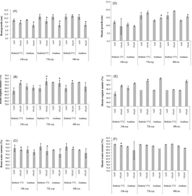
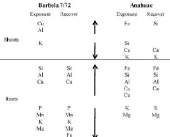
O Capítulo II aborda os estudos sobre a exposição de curto prazo ao Al em duas espécies de cereais: Triticum aestivum L. e Secale cereale L., tendo-se sempre utilizado um genótipo Al-tolerante e um Al-sensível para cada espécie. Este capítulo está dividido em três estudos: no Capítulo II.1 realça-se o efeito da exposição a 185 µM de Al no equilíbrio nutricional em trigo. Verificou-se que em ambos os genótipos (sensível e tolerante) o perfil de macro e micro
nutrientes se alterou, tendo uma interferência negativa, sobretudo no nível de P, Mg e K. Além disso, registaram-se diferenças na diferenciação da
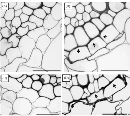
endoderme consoante o grau de tolerância/sensibilidade do genótipo. No Capítulo II.2 apresenta-se uma visão mais abrangente dos efeitos da exposição a 185 µM de Al em trigo, incluindo parâmetros fisiológicos,
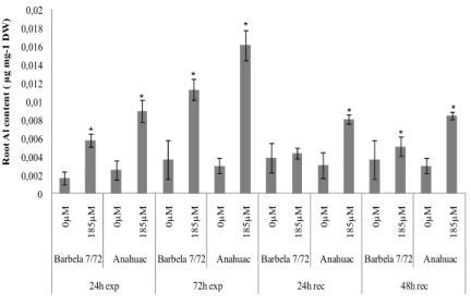
estruturais, citológicos e genotóxicos. Demonstra-se, pela primeira vez, que a progressão do ciclo celular é diferentemente regulada, dependendo da tolerância/sensibilidade do genótipo e que, mesmo em zonas já diferenciadas da raiz a exposição ao Al leva à deposição de calose. O Capítulo II.3 aborda os efeitos da exposição de 1.1 mM de Al em centeio, numa perspectiva bastante alargada. Apresenta-se o desequilíbrio nutricional, sobretudo no genótipo sensível, assim como a translocação de Al para a parte aérea nesse mesmo genótipo. Analisa-se também o comportamento de ambos os genótipos no que se refere ao ciclo celular, diferenciação da endoderme, crescimento radicular, reservas de hidratos de carbono, entre outros. Os resultados apontam para estratégias bem definidas adoptadas pelo genótipo tolerante de forma a minimizar a acção do Al no sistema radicular.
resumo (cont.) O Capítulo III compreende a exposição longa ao Al. Dois genótipos de centeio com diferentes graus de tolerância ao Al foram expostos a 1.11 mM e 1.85 mM de Al durante 21 dias, tendo sido usados dois pontos de amostragem (15 e 21 dias). Este capítulo está dividido em dois estudos: No Capítulo III. 1 analisam-se os mecanismos antioxidantes (folhas e raízes) como resposta à exposição ao Al, dando-se especial atenção ao ciclo do ascorbato-glutationas. A
exposição ao Al levou a stress oxidativo e a alterações na actividade de enzimas antioxidantes e no conteúdo de antioxidantes não-enzimáticos. Demonstra-se que os dois órgãos apresentam respostas diferentes à
exposição ao Al e que a capacidade de sobreviver em ambientes ricos em Al depende da eficácia da resposta antioxidante. Para além disso, a resposta do ciclo ascorbato-glutationas parece estar dependente do tipo de órgão, grau de tolerância e do tempo de exposição ao Al. No Capítulo III. 2 analisam-se os efeitos da exposição ao Al na fotossíntese. Verificou-se que o Al afecta negativamente a taxa fotossintética em ambos os genótipos, embora as alterações que o Al provoca nas trocas gasosas e no Ciclo de Calvin sejam dependentes do genótipo. Verificou-se também que os danos no genótipo sensível surgem mais cedo do que no genótipo tolerante, mas que ambos apresentam susceptibilidade ao Al após exposição de longo termo. Por fim, no Capítulo IV são apresentadas as conclusões da Tese de Doutoramento.
Keywords Aluminium (Al), Al-tolerance, Al-sensitivity,cell cycle, exposure, histology, nutrients, oxidative stress, photosynthesis, root, rye, Secale cereale,Triticum aestivum , wheat.
abstract The present work aims to better understand which are the targets of aluminium (Al) in plants, and contribute to a better understanding of the tolerance
mechanisms present in genotypes with high Al tolerance.
Al is a major constituent of the soil and become bioavailable in soils with low pH. In such cases, exposure to Al adversely affects plant growth and leads to a decrease in production. These facts are especially evident in crops and
exposure to Al is a major reason for reduction in crops production.
Chapter I present a general review about Al toxicity in plants, pointing out their main targets. In addition, also presents the mechanisms of resistance, including external and internal Al-detoxification, shown in different species.
Chapter II focuses on short term Al exposure in two species of cereals: Triticum aestivum L. and Secale cereal L., having always used one Al-tolerant and one Al-sensitive genotype for each species. This chapter is divided into three studies: In Chapter II.1 is enhanced the effect of exposure to 185μM of Al in the nutrient balance in wheat. It was found that in both genotypes (sensitive and tolerant) the profile of macro and micronutrients changed, with a negative interference on the level of P, Mg and K. Also, presents differences in endoderm differentiation depending on the degree of tolerance / sensitivity of the genotype. In Chapter II.2 a more comprehensive view of the effects of exposure to 185 μM Al in wheat is presented. This section includes
physiological, structural, cytological and genotoxic effects. For the first time is demonstrated that the cell cycle progression is differently regulated depending on the tolerance / sensitivity of the genotype, and that even in differentiated root zones Al exposure leads to callose deposition. Chapter II.3 focuses on the effects of 1.1mm Al exposure in rye, in a very broad perspective. It is presented nutritional imbalances observed mainly in sensitive genotype and the Al
translocation to shoots in the same genotype. It is also referred the different behaviour of both genotypes in relation to cell cycle, differentiation of
endodermis, root growth, carbohydrate reserves, among others. All that points to well-defined strategies taken up by the tolerant genotype, in order to minimize the action of Al in roots.
abstract (cont.) Chapter III focuses in long-term Al exposure. Two rye genotypes with different Al tolerance were exposed to 1.11 mM and 1.85 mM Al during 21 days, having been used two sampling points (15 and 21 days).This chapter is divided into two studies: In Chapter III. 1, are analyse the antioxidant mechanisms (leaves and roots) in response to Al exposure, with particular focus on the ascorbate-glutathione cycle. Al exposure leaded to oxidative stress and to changes in the activity of antioxidant enzymes and in the content of non-enzymatic
antioxidants. Is demonstrated that the two organs have different responses to Al exposure and that the ability to survive in environments rich in Al depends on the effectiveness of the antioxidant response. Furthermore, the response of the ascorbate-glutathione cycle seems to be dependent on organ type, Al-tolerance and time of Al-exposure. Chapter III. 2 analyze the effects of Al-exposure in photosynthesis. It is presented that Al negatively affects the photosynthetic rate in both genotypes, however the Al-induced alterations on gas exchange parameters and in Calvin cycle are genotype dependent. In this chapter, is demonstrated that Al induce earlier damages in the sensitive genotype, but both genotypes showed long-term susceptibility to Al.
Finally, in Chapter IV the general conclusions of the present PhD Thesis are presented.
Abbreviations:
1
O2: Singlet oxygen A: Net photosynthetic rate
Al: Aluminium
ALA: 5-aminolevulinic acid AlCl3: Aluminium chloride
AlCl3·6H2O: Aluminium chloride hexahydride ALMT1: Aluminium-activated malate transporter
APX: Ascorbate peroxidase
AS6: Rye Al sensitive of Barbela line AsA: Ascorbate
AT6: Rye Al tolerant of Barbela line
AtALMT1: Arabidopsis homolog of the ALMT1
AtBCB: Arabidopsis blue-copper-binding protein gene
B: Borum
BSA: Bovine serum albumin Ca: Calcium
CAT: Catalase Chl a: Chlorophyll a Chl b: Chlorophyll b
CHM: Nonspecific protein-synthesis inhibitor
Ci/Ca: Intercellular to atmospheric CO2 concentration ratio
CS: Citrate synthase Cu: Copper
CV: Coefficient of variation DHA: Dehydroascorbate
DHAR: Dehydroascorbate reductase DNA: Deoxyribonucleic acid
DTNB: 5,5’-dithiobis-(2-nitrobenzoic acid) DTT: Dithiothreitol
DTZ: Distal transition zone DW: Dry weight
ETR: Electron transport rate EZ: Elongation zone
F0: Minimal fluorescence
Fe: Iron
FeCl3: Iron chloride
FCM: Flow cytometry Fm: Maximal fluorescence
FPCV: Full peak coefficient of variation Fv: Variable fluorescence
FW: Fresh weight
G-POX: Guaiacol peroxidase GR: Gluthatione reductase gs: stomatal conductance GSH: Gluthatione GSHt: Total gluthatione GSSG: Oxized glutathione GST: Glutathione-S-transferase H+: Hydrogen HCL: Chloride acid H2O2: Hydrogen peroxide H2SO4: Sulfuric acid
HZ: Hairy root zone
IRGA: Infra-red gas analyser K+: Potassium
KCl: Potassium chloride
MATE: Mmultidrug and toxic compound exudation
MDA: malondialdehyde MDH: Malate dehydrogenase
MDHAR: Monodehydroascorbate reductase Mg2+: Magnesium
Mn: Manganese
MZ: Meristematic zone N2: Liquid nitrogen
NAD(P)H: Nicotinamide adenine (phosphate) dinucleotide NsHCO3: Sodium bicarbonate
NBT: Nitro-blue tetrazolium chloride NH4+: Ammonium
NtGDI1: Tobacco GDP-dissociation inhibitor gene NtPox: Tobacco peroxidase gene
O2-: Superoxide radical .
OH: Hydroxyl radical P: Phosphorus
parB: Tobacco glutathione S-transferase gene
PAS: Periodic acid-Schiff reaction PEPC: Phosphoenolpyruvate carboxylase PGI: Phosphoglucose isomerase
PI: Propidium iodide
PIPES: Piperazine-N,N’-bis-2-ethanesulfonic qp: Photochemical quenching
qN: Non-photochemical quenching
RO.: Alkoxyl radical
ROS: Reactive oxygen species
RuBisCo: Ribulose-1,5-bisphosphate carboxylase/oxigenase RWC: Relative water content
SD: Standard deviation SE: Standard error
sFBPase: Stromal fructose-1,6-bisphosphatase Si: Silicon
SOD: Superoxide dismutase TCA: Trichloroacetic acid Zn: Zinc
Acknowledgments Resumo
Abstract Abbreviations
CHAPTER I
ALUMINIUM TOXICITY IN PLANTS: A GENERAL APPROACH
I.1 Aluminium Chemistry in the Soil 3
I.2 Aluminium Toxicity 4
I.2.1 Root Growth 4
I.2.2 Oxidative Stress 6
I.2.3 Cell Wall, Plasma Membrane and Nutrient Unbalances 9
I.2.4 Cytoplasmatic Ca2+ 11
I.2.5 Callose 12
I.2.6 Others 12
I.3 Aluminium Resistance 13
I.3.1 External Detoxification 13
I.3.1.1 Organic Acids 13
I.3.1.2 Phenolic Compounds 16
I.3.1.3 Root Mucilage 17
I.3.2 Internal Detoxification 18
I.4 Model Species 19
I.5 Aims 21
CHAPTER II
SHORT-TERM ALUMINIUM EXPOSURE
II.1: Differential aluminium changes on nutrient accumulation and root differentiation in Al sensitive vs tolerant wheat
Abstract 39
Introduction 41
Material and Methods 42
Results 44
Discussion and Conclusions 50
References 54
II.2: Different structural and functional responses of sensitive vs tolerant wheat roots during Al exposure and recovery
Abstract 61
Introduction 63
Material and Methods 65
Results 68
Discussion and Conclusions 78
References 83
II.3: Al toxicity mechanisms in tolerant and sensitive rye genotypes: a comprehensive physiological and histological study
Abstract 91
Introduction 93
Material and Methods 95
Results 98
Discussion and Conclusions 108
III.1: Antioxidant mechanisms in response to Al of tolerant and sensitive rye genotypes
Abstract 121
Introduction 123
Material and Methods 125
Results 129
Discussion and Conclusions 138
References 142
III.2: Aluminium long-term stress differently affects photosynthesis in rye genotypes
Abstract 149
Introduction 151
Material and Methods 153
Results 156
Discussion and Conclusions 163
References 167
CHAPTER IV
CONCLUDING REMARKS
Short-term Al Exposure 173
Long-term Al Exposure 175
Aluminium toxicity in wheat and rye 1
CHAPTER I
Aluminium toxicity in wheat and rye 3
I.1 Aluminium Chemistry in the Soil
Aluminium (Al) ranks third in abundance among the Earth’s crust elements, after oxygen and silicon, and is the most abundant metallic element. A large amount of Al is incorporated into aluminosilicate soil minerals and very small quantities appear in the soluble form, capable of influencing biological systems (May and Nordstrom, 1991). Al bioavailability and, in consequence toxicity, is mainly restricted to acid environments. Acid soils (with a pH of 5.5 or lower) are among the most important limitations to agricultural production. The production of staple food crops, in particular grain crops, is negatively influenced by acid soils (Kochian et al., 2005). It has been estimated that 15% of the world´s soil is acidic (http://www.fao.org/ag/agl/agll/terrastat/#terrastatlinks) and that over 50% of the world’s potentially arable lands are acidic (von Uexküll and Mutert, 1995; Panda et al., 2009). In Portugal, 80% of soils have pH lower than 6.5 and more than 40% lower than 5.5 (Almeida, 1955) and are located mostly in the Northern regions (Fig. 1). Furthermore, 60% of acid soils in the world are located in the humid tropics and subtropics. Thus, in many developing countries, acid soils limit crop production. In developed countries, some agricultural practices, as removal of products from the farm or paddock, leaching of nitrogen below the plant root zone, inappropriate use of nitrogenous fertilizers and build up in organic matter are causing further acidification of agricultural soils (http://www.dpi.nsw.gov.au/).
When pH drops below 5.5, aluminosilicate clays and aluminium hydroxide minerals begin to dissolve, releasing aluminium-hydroxy cations and Al(H2O)63+ (Al3+), that then
exchange with other cations. The chemistry of Al3+ in soil solution is complicated by the fact that soluble inorganic (such sulfate and fluoride) and organic ligands form complexes with Al3+. Whether a ligand increases or decrease aluminium solubility depends on the particular aluminium-ligand complex and its tendency to remain in solution or precipitate (McBride, 1994; Panda and Matsumoto, 2007). On that conditions, Al3+ also forms the mononuclear species AlOH2+, Al(OH)2+, Al(OH)3 and Al(OH)4 (Panda and Matsumoto,
Al13 (AlO4Al12(OH)24(H2O)127+) (Massor-Pietraszewska, 2001). The mononuclear Al3+
species and Al13 are considered as the most toxic forms (Kinraide, 1991; Kochian, 1995).
I.2 Aluminium Toxicity
I.2.1 Root Growth
A major consequence of Al toxicity is the inhibition of root growth, and this outcome has been reported during the last century (e.g. Eisenmenger, 1935) for innumerous species (Jan, 1991; Ryan et al., 1993; Shen et al., 1993; Crowford and Wilkens, 1998; Ahn et al., 2002; Ma et al., 2002; Kikui et al., 2005; Abdel-Basset et al., 2010). Consequently, root growth inhibition has been widely used to assess Al toxicity.
Root growth is the combination of cell division and elongation. Only during the last decade, researchers started to look at the cell cycle (de)regulation induced by Al, with some works focusing unbalances on mitosis phase, and very few on other interphase phases (e.g. Silva et al., 2000). Decrease of mitotic activity was reported as a consequence of Al exposure in root tips of several species as: wheat (Frantzios et al., 2001; Li et al., 2008), maize (Doncheva et al., 2005), barley (Budikova and Durcekova, 2004), and oat (Marienfeld et al., 1995). Some authors defended that inhibition of cell elongation was the primary mechanism leading to root growth inhibition (Čiamporová, 2002; Zheng and
Fig. 1. Distribution of acid soils in Portugal
Aluminium toxicity in wheat and rye 5
Yang, 2005). The reason for that is that root growth inhibition could occur within a short time period - 30 min in Al-sensitive maize (Llugany et al., 1995) - and that cell division is a slow process (cell cycle takes usually several hours to be completed). However, Doncheva et al (2005) reported inhibition of cell division (decrease of S-phase cells) in the proximal meristem after 5 min Al exposure and inhibition of root cell division in the apical meristem within 10 or 30 minutes. Furthermore, Al can accumulate in the nuclei of cells in the meristematic region of the root tip within 30 minutes (Silva et al., 2000). So, although root elongation inhibition is a primary target of Al toxicity, it does not preclude fast Al effects on root cell division (Doncheva et al., 2005). Therefore, whereas inhibition of cell elongation or cell division is the primary mechanism leading to root growth inhibition is still unclear. Recently, Yi et al (2009) reported that Al exposure leaded to abnormal progress through mitosis and induced micronuclei formation in Vicia faba roots, which is in agreement with Al-induced chromosome aberrations found in wheat roots (Bulanova et al., 2001) and Al-induced chromosome stickness and breacks in Oryza sativa (Mohanty et al., 2004). From the literature review, it is evident that Al leads to cell cycle unbalances, but many questions still remain to clarify. For example it still remains unclear how and where, Al exerts its influence throughout the cell cycle, if these changes are species and region dependent (most studies are performed in root apices), how the putative changes are exerted through time and/or if they may be reversible after Al removal.
The root growth inhibition and increase in root diameter observed in roots exposed to Al (Zobel et al., 2007) suggested that plant cytoskeleton could be a cellular target of Al phytotoxicity (Blancaflor et al., 1998). Blancaflor et al (1998) and Horst et al (1999) studied Al-induced effects on microtubules and actin microfilaments and showed that microtubules and microfilaments are altered, in their stability, organization and polymerization, when exposed to Al. Also, in Triticum turgidum Al treatment leaded to disorganization of actin filaments and formation of actin deposits (Frantzios, 2005). Recently, Zhang et al (2007a) showed that Al inhibited actin and profilin genes. Profilin, as an actin-binding protein, provides cells with the ability to remodel the cytoskeleton (Krishnan and Moens, 2009). In Arabidopsis thaliana a decrease in profilin expression resulted in an elongation defect (Ramachandran et al., 2000). Furthermore, Sivaguru et al (1999) and Čiamporová (2002) showed that organization of cytoskeleton is most sensitive in the distal transition zone of the root apex, providing evidence that this zone represents a potential target with respect to Al toxicity.
The most sensitive root zone to Al toxicity is under great attention. Earlier, it was hypothesized that root cap played a major role in the mechanism of Al toxicity/protection (Bennet and Breen, 1991). However, Ryan et al (1993) demonstrated that the removal of the root cap had no effect on the Al-induced inhibition of root growth in maize. Furthermore, the same authors also suggested that the meristem is the primary site of Al-toxicity. Later, Sivaguru and Horst (1998), applying Al to 1mm root segments, reported that Al accumulation in the distal transition zone (DTZ: 1-2mm) led to a rapid inhibition of the root elongation and suggested that this root zone is the primary target of Al in an Al-sensitive maize cultivar.
I.2.2 Oxidative Stress
Al-induced oxidative stress and changes in cell wall properties have been suggested as the two major factors leading to Al toxicity (Yamamoto et al., 2003; Zheng and Yang, 2005). Oxidative stress occurs when any condition disrupt the cellular redox homeostasis. Reactive oxygen species (ROS) include the superoxide radical (O2-), the hydroxyl radical
(.OH), the alkoxyl radical (RO.), hydrogen peroxide (H2O2), and singlet oxygen (1O2).
These ROS have the capacity to oxidize cellular components such as lipids, proteins, enzymes and nucleic acids, leading to cell death. Metals are known to act as catalysts in ROS production and to induce oxidative damage in plants. Al itself is not a transition metal and cannot catalyze redox reactions, however, Al exposure leads to oxidative stress (Yamamoto et al., 2001, 2003; Boscolo et al., 2003; Kuo and Kao, 2003; Jones et al., 2006; Liu et al., 2008a, b). Because aluminium ions form electrostatic bonds preferentially with oxygen donor ligands (e.g. carboxylate or phosphate groups), cell wall pectin and the outer surface of the plasma membrane seem to be major targets of aluminium (Yamamoto et al., 2001). Al binding to biomembranes lead to rigidification (Jones et al., 2006), which seems to facilitate the radical chain reactions by iron (Fe) ions and enhance the peroxidation of lipids (Yamamoto et al., 2003).
Al induction lipid peroxidation has been reported for some species, including barley (Guo et al., 2004), sorghum (Peixoto et al., 1999), triticale (Liu et al., 2008a), rice (Kuo and Kao, 2003), greengram (Panda et al., 2003) and wheat (Hossain et al., 2005). Yamamoto et al (2001) found that, for Pisum sativum seedlings treated with Al in a simple Ca solution, Al accumulation, lipid peroxidation and callose production had a similar distribution on the
Aluminium toxicity in wheat and rye 7
root apex surface and was accompanied by root growth inhibition. However, the loss of membrane integrity was only detected at the periphery of the cracks on the surface of the root apex. Furthermore, Yamamoto et al (2003) concluded that the Al enhancement of lipid peroxidation is an early symptom of Al accumulation and appears to cause partly callose production, but not root growth inhibition. Later, however, in maize, Al treatment didn´t induced lipid peroxidation, indicating that lipids are not the primary cellular target of oxidative stress in maize (Boscolo et al., 2003). So, it seems that cellular target of oxidative stress depend on plant species.
Plant cells are equipped with a defensive system composed by enzymatic antioxidants (catalase (CAT), ascorbate peroxidase (APX), guaiacol peroxidase (G-POX), superoxide dismutase (SOD), monodehydroascorbate reductase (MDHAR), dehydroascorbate reductase (DHAR), glutathione-S-transferase (GST), and gluthatione reductase (GR) and nonenzymatic antioxidants (ascorbate (AsA), glutathione (GSH), α-tocopherol, carotenoids) that help to detoxify the ROS. Some works reported ROS production and alterations in the antioxidant system as a consequence of Al exposure. In pea seedlings, ROS production is detected in root apex after two hours of Al exposure and increase with time exposure (Yamamoto et al., 2003). Furthermore, Yamamoto et al (2002) found, in pea, that ATP content, respiration and root elongation decreased as consequence of Al treatment. In maize roots, Al-treatment also lead to increase in ROS production rate in all epidermal cells, only within 10 min of Al exposure and continued to increase during Al exposure (Jones et al., 2006). APX and SOD activity increased in roots of both Al-resistant and Al-sensitive triticale cultivars (with higher magnitude in the sensitive one), but changes were detected first in the sensitive cultivar (6h) and then in the resistant (12h) (Liu et al., 2008a). Boscolo et al (2003) reported for maize root tips an increase of SOD and APX activities, mostly in the sensitive line, and suggested that Al exposure lead to formation of O2- in a greater quantity than the pre-existent SOD can remove and therefore
induce the cells to initiate SOD synthesis. Furthermore, these authors found that SOD and APX activity are inversely proportional to root growth rate and, therefore, suggested that the increase of O2- and H2O2 production are related to Al toxicity. An increase in SOD,
APX and GR activities was reported for greengram seedlings, whereas a decrease in CAT activity and glutathione and ascorbate contents was also found at higher Al concentrations (Panda et al., 2003). These authors justified the decrease in CAT activity due to the fact that this enzyme is photosensitive and, therefore, needs constant synthesis and suggested
that glutathione and ascorbate may be enable to detoxify the ROS directly (Panda et al., 2003). Devi et al (2003) found an increase in manganese superoxide dismutase (MnSOD) activity in both sensitive and tolerant cell lines of tobacco, and in AsA and GSH contents, mostly in the tolerant line. These data indicated that AsA and GSH seem to be in part responsible for the tolerance mechanisms of the tolerant line to Al. Activities of SOD, CAT and APX also increased in roots of tea plants and in cultured tea cells exposed to Al (Ghanati et al., 2005). That increase seemed to result in increased membrane integrity, since lipid peroxidation reduced with Al exposure (Ghanati et al., 2005).
These findings reporting increase of antioxidants (enzymatic and nonenzymatic), are accompanied with others that prove gene regulation associated with oxidative stress. A large range of genes induced by Al stress have been isolated from a variety of plant species, and some of them are related to oxidative stress. For example, Ezaki et al (2000) expressed nine genes derived from Arabidopsis, tobacco, wheat, and yeast in Arabidopsis ecotype Landsberg. An Arabidopsis blue-copper-binding protein gene (AtBCB), a tobacco glutathione-S-transferase gene (parB), a tobacco peroxidase gene (NtPox), and a tobacco GDP-dissociation inhibitor gene (NtGDI1) conferred a degree of resistance to Al: significative differences in relative root growth, and decrease in Al content and oxidative damages. They also showed that overexpression of three Al-induced genes in plants conferred oxidative stress resistance. Furthermore, overexpression of the parB gene simultaneously conferred resistance to both Al and oxidative stresses. Therefore, Ezaki and coworkers concluded that some of the genes induced during Al-exposure and oxidative stresses play protective roles against both stresses. Cancado et al (2005) identified a maize Al-inducible cDNA encoding a glutathione-S-transferase (GST). Expression of that gene (GST27.2) was up-regulated in response to various Al concentrations in both Al-tolerant and Al-sensitive maize lines. Recently, using Al-sensitive Medicago truncatula cultivar Jemalong genotype A17, 324 genes were up-regulated and 267 genes were down-regulated after Al exposure (Chandran et al., 2008). Up-regulated genes were enriched in transcripts involved in cell-wall modification and abiotic and biotic stress responses, while down-regulated genes were enriched in transcripts involved in primary metabolism, secondary metabolism, protein synthesis and processing, and the cell cycle. Known markers of Al-induced gene expression including genes associated with oxidative stress and cell wall stiffening, were differentially regulated in that study (Chandran et al., 2008). For maize plants, Al exposure leaded to alteration in gene expression, mostly in the Al sensitive
Aluminium toxicity in wheat and rye 9
genotype. Although in Al-sensitive genotype was altered the expression of more genes, several Al regulated genes exhibited higher expression in the tolerant genotype (Marom et al., 2008). So, it is clear that expression of some genes confers Al resistance and contributes to reduce oxidative stress.
I.2.3 Cell Wall, Plasma Membrane and Nutrient Unbalances
Al accumulation is primarily and predominantly in the root apoplast (30-90% of the total absorved Al (e.g. Rengel and Reid, 1997; Liu et al., 2008a) of peripherical cells, and is only very slowly translocated to more central tissues (Marienfeld and Stelzer, 1993; Marienfeld et al., 2000; Schmohl and Horst, 2000). The primary binding of Al3+ in the apoplast is probably the pectin matrix, with its negatively charged carboxylic groups (Chang et al., 1999; Schmohl and Horst, 2000).
Several works reported increases of pectin levels in Al-sensitive genotypes (Van et al., 1994; Chang et al., 1999; Horst et al., 1999; Schmohl and Horst, 2000; Hossain et al., 2006; Liu et al., 2008b) and, some, also detected increase in Al contents in the same sensitive genotypes (Horst et al., 1999; Schmohl and Horst, 2000; Hossain at al, 2006). These findings indicate that pectin plays a major role in the binding of Al, and suggest that some of the additional Al accumulation in sensitive genotypes, bound in the newly formed cell wall pectin (Chang et al., 1999; Schmohl and Horst, 2000; Liu et al., 2008b). Binding of Al to the pectin matrix and other cell wall constituents could alter cell wall characteristics and functions such as extensibility (Tabuchi and Matsumoto, 2001), porosity and enzyme activities thus leading to inhibition of root growth (Schmohl and Horst, 2000). Another mechanism for Al toxicity targeted to the apoplast invokes a rapid and irreversible displacement of Ca2+ from cell wall components by Al ions (Tabuchi and Matsumoto, 2001; Zheng and Yang, 2005).
Accumulation of Al occurs predominantly in the root apoplast. Nevertheless, Al accumulates also in the symplast and with a fast rate (Marienfeld et al., 2000). Recently, Xia et al (2010) reported a transporter, Nrat1 (Nramp aluminium transporter 1), specific for Al3+ localized at the plasma membrane of all rice root tips cells, except epidermal cells. Those authors referred that the elimination of the Nrat1 enhanced Al-sensitivity, decreased Al uptake, increased Al binding to cell wall and concluded that this transporter is required for prior step of final Al detoxification through sequestration of Al into vacuoles.
Furthermore, given its physicochemical properties, Al can interact strongly with the negatively charged plasma membrane. Plasma membrane usually possesses two types of membrane potential. One reflects the unbalance in ions concentration inside the cell and the other is caused by the net concentration of fixe ions on the membrane surface (Zheng and Yang, 2005). Al seems to be able to affect both types of potentials (Ahn et al., 2001; 2002). For instance, Al can displace other cations (e.g. Ca2+) that may form bridges between the phospholipid head groups of the membrane bilayer (Akeson and Munns, 1989). Furthermore, Al interaction with plasma membrane could lead to depolarization of the transmembranar potential (e.g. Kinraide et al., 1992) and/or reduction of H+-ATPase (e.g. Ahn et al., 2001; 2002) which, in turn, can alter the activities of ions near the plasma membrane surface and impede the formation and maintenance of the trans-membrane H+ gradient (Kochian et al., 2005). Moreover, Al changes in plasma membrane can modify the uptake of several cations (e.g. Ca2+, Mg2+, K+, NH4+) (Jan, 1991; Nichol et al., 1993;
Poschenrieder et al., 1995; Mariano and Keltjens, 2005). These changes are related to direct Al3+ interactions with plasma membrane ion channels (Piñeros and Kochian, 2001) and changes in membrane potential.
Nutrional unbalances induced by Al exposure were reported for several plant species. Eleven families of pteridophytes presented different nutritional unbalances (mostly in Ca, Mg, P, K) depending on Al accumulation (Olivares et al., 2009) and in maize, Al had negative effects on the uptake of macro and micronutrients, being Ca and Mg the macro- and Mn and Zn the micronutrients more affected (Mariano and Keltjens, 2005). Also, the maize Al-tolerant genotypes accumulated higher concentration of Ca, Mg (Mariano and Keltjens, 2005) and K (Giannakoula et al., 2008) than the sensitive genotypes. However, others studies reported different results in Al-induced nutritional imbalances in maize: Lidon et al (2000) referred that all elements in roots, except K, Mn and Zn, increased in Al-treated roots and that in shoots Ca and Mg had little variation. Poschenrieder et al. (1995) reported that only the specific absorption rate of B was correlated to the Al-induced root growth inhibition. Al exposure leaded to decrease in K, Mg, Ca and P contents and uptake in rice plants and, as observed in maize, the tolerant cultivar presented less negative effects in nutrient content than the sensitive one (de Mendonça et al., 2003). In tomato cultivars, Al exposure decreased the content of Ca, K, Mg, Mn, Fe and Zn in roots, stems and leaves (Simon et al., 1994). Zobel et al (2007) related changes in fine root diameter with changes in concentration of some nutrients, as N, P and Al. It seems that the
Aluminium toxicity in wheat and rye 11
differential tolerance to Al may be due to their differences in uptake, ability to keep adequate concentrations and to use the nutrients efficiently. Differences in nutrient uptake, accumulation and translocation are evident between plant species and within each species. Furthermore, since each author utilized different Al concentrations, variated nutritive solutions and time exposures is difficult to make a general and accurate model of Al-induced nutritional imbalances.
I.2.4 Cytoplasmic Ca2+
Disturbance of cytoplasmic Ca2+ homeostasis is believed to be the primary target of Al toxicity (Rengel and Zhang, 2003) and may be involved in the inhibition of the cell division or root elongation by causing potentially disruptions of Ca2+-dependent biochemical and physiological processes (Sivaguru et al., 1999; Rengel and Zhang, 2003). In wheat root apices, Jones and Kochian (1995; 1997) found that Al inhibit Ca2+- dependent phospholipase C, which acts on the lipid substrate phosphatidylinositol-4,5-biphosphate. The authors hypothesized that phosphoinosite signilling pathway might be the initial target of Al. In accordance, Zhang et al (2007a) found Al-induced inhibition of genes related to phosphoinosite signilling pathway and hypothesized that the gene inhibition could result in disruption of this pathway. Also, it was reported that components of the actin-based cytoskeleton interact directly with phospholipase C in oat (Huang and Crain, 2009).
Most works reported an increase in cytoplasmic Ca2+ when plants were exposed to Al (Zhang and Rengel, 1999; Ma et al., 2002; Bhuja et al., 2004). However, Jones et al (1998) reported a decrease in cytoplasmic Ca2+ in tobacco cell cultures in the presence of Al. Furthermore, Zhang and Rengel (1999) reported an increase in cytoplasmic Ca2+ in two lines with different tolerance to Al and correlated it with the inhibition of root growth in both lines. As well, Ma et al (2002) correlated cytoplasmic Ca2+ to root growth response. Moreover, alteration in cytoplasmic Ca2+ homeostasis can occur within few minutes (20-30 minutes) in root hair tips of Arabidopsis thaliana (Jones et al., 1998).
It is certain that Al exposure influences cytoplasmic Ca2+ homeostasis, but it is still unclear if it is a primary cause of Al-induced inhibition of root growth or a secondary effect. The source of Ca2+ for the increase if cytosolic Ca2+ activity could be extracellular and/or
intracellular, but is still insufficiently documented, as well the effects on increased cytosolic Ca2+ (for review see Rengel and Zhang, 2003).
I.2.5 Callose
The induction of callose (1,3-β-D-glucan) formation in Al exposed roots has been reported in many plant species (e.g. Jorns et al., 1991; Schereiner et al., 1994; Poschenrieder et al., 1995; Budikova and Durcekova, 2004; Eticha et al., 2005; Tahara et al., 2005; Jones et al., 2006 ). Al-induced callose formation in root tips is recognized as an excellent indicator of Al sensibility (Horst et al., 1997; Massot et al., 1999; Meriga et al., 2003; Bhuja et al., 2004; Hirano et al., 2004; Tahara et al., 2005) and some works negatively correlated root elongation with callose formation during Al exposure (e.g. Nagy et al., 2004; Tahara et al., 2005). In maize roots, Jones et al (2006) found a close spatial and temporal coordination between Al accumulation and callose production in roots. Still, Tahara et al (2005) reported that, in some Myrtaceae species, induction of callose formation was not accompanied by root growth inhibition and suggested that callose formation is a more sensitive indicator to Al than root elongation.
Since Al induces a transient rise of cytosolic Ca2+, an increase of callose accumulation under Al stress is not unexpected. Cytosolic Ca2+ is one of the prerequisites for the induction of callose synthesis, but not the only factor modulating increases in callose synthesis and deposition (Bhuja et al., 2004). Callose formation, as response to Al, is described in sensitive and, to a lesser extent, in tolerant roots (Horst et al., 1997; Eticha et al., 2005). In a less extent, callose deposition has been considered as a mechanism to prevent Al from penetrating into the apoplast. Also, this accumulation is reported to inhibit the symplastic transport and cell communication by blocking plasmadesmata, avoiding Al induced lesions in the symplast (Sivaguru et al., 2000). However, callose deposition in sensitive roots has also been shown to lead to uncontrolled rigidity of cell walls (Jones et al., 2006) leading ultimately to protoplast degradation.
I.2.6 Others
Al-induced effects/damages are first detected in the root system (Barceló and Poschenrieder, 2002; Doncheva et al., 2005). Changes in the root system, may affect
Aluminium toxicity in wheat and rye 13
nutrient uptake, which can lead to nutritional deficiencies in shoots and leaves (Moustakas et al., 1996). Except for Al-accumulator plants, Al accumulates more in roots than in leaves (Konarska, 2010). In some species, Al-induced alterations in leaves were considered indirect, since Al accumulation was not detected in leaves (Moustakas et al., 1996). Nevertheless, alterations in leaves induced by Al-exposure were reported for many species. Several works reported leaves biomass reduction (Azmat and Hasan, 2008), thickness (Konarska, 2010), lipid peroxidation (Zhang et al., 2007b), nutritional imbalances (Guo et al., 2007), changes in the photosynthetic performance (Jiang et al., 2008), changes in chlorophyll contents (Chen et al., 2005; Zhang et al., 2007; Azmat and Hasan, 2008; Jiang et al., 2008), among others. Reductions in carbon dioxide (CO2) assimilation rate due to Al
toxicity are reported for several species (Moustakas et al., 1996; Oleksyn et al., 1996; Lidon et al., 1999; Pereira et al., 2000; Peixoto et al., 2002; Chen et al., 2005; Jiang et al., 2008) and some works indicated that Al-exposure induced damage of the photosystem II (Zhang et al., 2007b; Reyes-Dias et al., 2010). Very few works focused on the consequence of Al-treatment in the carbohydrate metabolism. The effects of Al exposure on Ribulose-1,5-bisphosphate carboxylase/oxygenase (RuBisCo) content and activity is still unclear and the few reports available were performed in citrus (Chen et al., 2005; Jiang et al., 2008) and in wild rice (Cao et al., 2010).
I.3 Aluminium Resistance
Aluminium resistance is the property which allows a plant to grow with little or no injury with elevates Al supplies. Resistance strategies can be separated into: a) exclusion of Al from the root apex; b) Al-tolerance (Al binding and uptake without Al injury) (Panda et al., 2009; for review see Horst et al., 2010).
I.3.1 External Detoxification
I.3.1.1 Organic Acids
There has been considerable evidence in the literature for an Al-exclusion mechanism based on carboxylate exudation from the root apex (for review see Barceló and
Poschenrieder, 2002; Kochian et al., 2005; Horst et al., 2010). The ability of organic acids to chelate and render Al nonphytotoxic is well established. However, the exudation rate and the form of organic acid anions exuded depend on plants species and Al-resistance. Al induced exudation of several organic acids has been described from roots of several species, as malate in wheat (Ryan et al., 1995; Zheng et al., 1998; Li et al., 2000), citrate in maize (Pellet at al, 1995), citrate and malate in rye (Li et al., 2002), citrate in soybean (Yang et al., 2001) and oxalate in buckwheat (Zheng et al., 1998).
In Al-resistant genotypes, the efflux of organic acids may present two patterns (Ma et al., 2001): I) immediately upon Al exposure (e.g. malate and/or citrate efflux in wheat, bean, soybean, maize, shrub bushclover); II) requires a lag-time of several hours (e.g. malate exudation in rye, triticale, Cassia sp.). For pattern I, it seems that Al induce the opening of anion efflux channels facilitating the organic acid efflux (Delhaize and Ryan, 1995; Ryan et al., 1995), while it seems that there is some gene activation in the pattern II, with protein synthesis that may be involved in organic acid metabolism or in the transport of organic acid anions.
Since organic acids in the cytoplasm are largely deprotonated and exist as anions, the equilibrium potencial of organic acid anions is much more positive than that of the resting membrane potencial. So, activation of membrane anion channels will result in an anion (as organic acids) efflux down the outward electrochemical gradient. Although many types of organic acids are found in root cells, only one or two are secreted in response to Al treatment for a given species. This suggests the existence of specific transport systems for the organic acid anions. In protoplasts from wheat roots apex, Ryan et al (1997) reported that Al activated an ion channel that allows anion efflux. Later, in maize protoplasts derived from cortical cells of the DTZ (Kollmeier et al., 2001) and in protoplasts from the maize hybrid South American 3 (Piñeros et al., 2001), was found that Al activated anion channel permeable to malate and citrate anions and to citrate, respectively. Also in maize, Piñeros et al (2002) found Al-induced root citrate release (as far as 5 cm beyond root cap) within 30 min, where the rate of exudation was positively correlated with the level of Al in the nutrient solution, and Al-induced plasma membrane anion channel in protoplasts isolated from stellar or cortical tissues. Later, Yang et al (2006) used a nonspecific protein-synthesis inhibitor (CHM) to investigate the relationship between Al stress and organic acid secretion in buckwheat, which presents a typically pattern I behavior. They demonstrated that the induction of oxalate secretion was rapid, and CHM did not affect the
Aluminium toxicity in wheat and rye 15
process, showing that all the necessary metabolic “machinery” was constitutively expressed in buckwheat roots, and that oxalate secretion was simply triggered by the Al stimulus, no induction of a novel protein is required.
The Al response in some species, mostly those that present a lag-time in organic acids exudation, is accompanied by enhancements of key enzymes in organic acids synthesis, as malate dehydrogenase (MDH), citrate synthase (CS) (Li et al., 2000; Yang et al., 2001), and phosphoenolpyruvate carboxylase (PEPC) (Gaume et al., 2001). Furthermore, some works have shown, that genetic transformation of plants to overexpress enzymes involved in organic acid synthesis, lead to increased efflux of organic acid and Al resistance (de la Fuente et al., 1997; Tesfaye et al., 2001; Anoop et al., 2003). However, several studies reported that the activity of enzymes related to organic acid synthesis, was not enhanced under Al exposure, even when organic acids efflux occurred. For example, in barley, Zhao et al (2003) reported an increase in citrate secretion, which was positively correlated with Al resistance and negatively correlated with Al root apices content. However, the activity of CS was unaffected by Al in both Al-resistant and Al-sensitive barley varieties. Also, in triticale were not detected differences in the activity of CS, MDH and PEPC in both tolerant and sensitive lines (Hayes and Ma, 2003). Furthermore, those authors concluded that the release of organic acids does not appear to be regulated by their levels in the roots or by the capacity of the root tissues to synthesize them. Yang et al (2006) used Cassia tora L., which belongs to pattern II, and observed that CHM inhibited the citrate efflux, de novo protein synthesis and the CS activity, but Al treatment alone induced significant exudation of citrate and was not able to affect CS activity. So, these authors suggested that the enhancement of citrate efflux was mainly caused by the de novo synthesis of anion channels.
Among the organic acid anions, citrate forms the most stable complexes with Al. The Al-citrate 1:1 complex is not phytotoxic and its transport through the plasmalemma seems to be very slow (Kochian, 1995). At a 1:1 ratio the Al-oxalate complex also had little toxic effects in Al-sensitive wheat and the complex prevented Al root accumulation (Ma et al., 2001). Malate efflux is specifically triggered by Al3+, whereas it is apparently the less effective mechanism in preventing Al uptake.
The major Al resistance gene in wheat (ALMT1-aluminium-activated malate transporter) encodes an Al activated malate transporter protein located in the plasma membrane of root cells, which contains six transmembrane domains with the amino and carboxyl termini
located on the extracellular side of the plasma membrane (Motoda et al., 2007). ALMT1 confers an Al activated efflux of malate from root apices (Raman et al., 2005). Expression of ALMT1 of in vitro tobacco cells (Sasaki et al., 2004), Xenopus oocytes (Sasaki et al., 2004), rice (Sasaki et al., 2004), and transgenic barley (Delhaize et al., 2004), conferred an Al-activated efflux of malate and in some of them increased resistance to Al. Recently, transgenic wheat lines with over-expression of the TaALMT1 gene showed increases in malate efflux and in Al resistance (Pereira et al., 2010). Also, Hoekenga et al (2006) identified in Arabidopsis a homolog of the ALMT1 (AtALMT1) as an essential factor for Al resistance. Beyond ALMT1 genes, Al-resistance in some crop species such sorghum (SbMATE) and barley (HvAACT1), was related to the expression of some genes of the MATE (multidrug and toxic compound exudation) family based on Al-activated citrate exudation (Magalhaes, 2010). Based on the homology to these genes (ALMT1 and MATE), others resistance genes were isolated in others species, such as rye (Collins et al, 2008; Yokosho et al, 2010), maize (Maron et al, 2010) and common bean (Eticha et al, 2010). It is clear that Al-resistance mechanisms are controlled by numerous genes, but their physiology and molecular biology is not well understood (Ryan et al, 2010).
I.3.1.2 Phenolic Compounds
Some studies did not find a correlation between Al resistance and the amount of organic acid efflux (Piñeros et al., 2005). This indicates that exudation of organic acids may not be the only mechanism of Al exclusion. Other ligands and mechanisms may also play a role in this process (Barceló and Poschenrieder, 2002). According to stability constants of metal-ligand complexes, citrate, oxalate, and malate are more suited for binding Al in acid soil solutions than simple phenolics, because of the high competition of protons for binding. However, in flavonoid-type phenolic, the competitive effect of protons is substantially lower (Barceló and Poschenrieder, 2002). Under the less acidic conditions in the apoplast or inside plant cells, Al-binding by phenolics may even be more relevant (Tolrà et al., 2005). Furthermore, the phenolic compounds in presence of carboxylic groups from organic acids can, by a deprotonation reaction, strengthen the interaction between Al3+ and the organic acid ligand (Barceló and Poschenrieder, 2002).
Enhancements of phenolic compounds contents were already reported under certain conditions. It was also detected, by fluorescence spectroscopy, the complexation of Al by
Aluminium toxicity in wheat and rye 17
phelonics released by the roots of Norway spruce (Heim et al., 1999). In maize, Si induced amelioration of Al toxicity enhanced root exudation of flavonoid-type phenolics (Kidd et al., 2001; Barceló and Poschenrieder, 2002). Also, in Rumex acetosa L., Tolrà et al (2005) reported that Al induced high levels of anthraquinone in roots and of catechol, catechin and rutin in shoots and concluded that phenolics, rather than organic acids, are involved in detoxification of Al in leaves. Recently, in Matricaria chamomilla plants phenolics increased in the shoots of plants exposed to Al (Kovacik et al., 2010).
I.3.1.3 Root Mucilage
Rhizodepositions in the form of mucilage has also been implied in Al resistance mechanisms (Horst, 1995). Root mucilage consists mainly of polysaccharides, and is exuded from the outer layers of the root cap. It was proposed that one of the roles of mucilage is the detoxification of toxic metal cations, as Al3+ (Horst et al., 1982). That ability is due to the uronic acids, in its structural polysaccharides, which adsorb metal cations. Archambault et al (1996) found that Al bound to the mucilage accounted approximately 25-35% of nonexchangeable Al, representing a significant pool of apoplastic Al in wheat. In maize, Li et al (2000) reported that mucilage strongly binds to Al, and Al bound to mucilage is not phytotoxic. Higher mucilage production was found in the Al tolerant wheat cultivar (Atlas 66) than in the sensitive (Puthota et al., 1991). However, the mucilage contribution to Al tolerance is not still understood and is under debate. Li et al (2000) reported that inhibition of root elongation by Al, was independent of the presence or absence of mucilage prior to the Al treatment, and concluded that although mucilage affects the accumulation of Al by roots, it does not confer Al resistance to Z. mays root apices. Furthermore, in Melastoma malabathricum, an Al accumulator, the removal of root mucilage reduced the Al concentration in shoots (Watanabe et al., 2008). Those authors also reported that binding strength between mucilage and Al was very weak, and that the characteristics of mucilage in adsorption of cations differ between M. malabathricum and Z. mays. M. malabathricum mucilage has higher adsorption affinities for trivalent cations, whereas Z. mays mucilage has a higher affinity for divalent cations (Watanabe et al., 2008). All this together, shows that the rule of root mucilage in Al detoxification is still unknown.
I.3.2 Internal Detoxification
Some species have the ability to accumulate high Al concentrations without showing symptoms of Al toxicity. Al accumulator plants have been defined as those that have ≥1000 mg kg-1 DM Al in aerial tissues, as more recently suggested by Jansen et al (2002) in agreement with Chenery (1948). However, other authors proposed, in addition to the refered criterion, the use of Al/Ca ratio to define Al accumulators, principly when Al concentration is higher than 3000 mg kg-1 DM (Masunaga et al., 1998). Accumulation of Al in leaves may occur in cell walls or in the leaf vacuoles (Poschenrieder et al., 2008). One of Al accumulator species is M. malabathricum which growth is even stimulated in the presence of Al (Watanabe et al., 2008). Also, tea and hydrangea plants are well known Al accumulators. The concentration of free Al3+ at the pH (app. 7.4) of the symplasm, is <10–11M because of the pH-dependent hydrolysis of Al in solution and the formation of insoluble Al(OH)3 (Martin, 1986). Despite low concentration, Al can still be phytotoxic because of the strong affinity for oxygen ligands (Martin, 1986). This suggests that Al accumulating plants must have Al3+ internal detoxification.
A number of low-molecular-weight di- and tri-carboxylic acids form strong complexes with Al3+, which are less phytotoxic than free Al3+ions. Among the ligands that form stable complexes with Al, organic acids, phenolics and silicon may be implied in Al internal detoxification (Wenzl et al., 2002; Tolrà et al., 2005; Kovacik et al., 2010; for review see Ma et al., 2001 and Barceló and Poschenrieder, 2002). For example, considerable fraction of the total Al accumulation in roots of both Brachiaria decumbens and Brachiaria ruziziensis was complexed by soluble low-molecular-weight ligands (Wenzl et al., 2002). In B. decumbens, 90% of organic acids were retained and accumulated within root apices, whereas in mature root zones were secreted into the growth medium (Wenzl et al., 2002). This pattern shows that Brachiaria species employ organic acids (citrate and trans-aconitate) for Al internal detoxification. In buckwheat, Al, once taken up, is chelated internally by oxalate (forming 1:3 complex) in root cells and when is loaded to the xylem, ligand exchange from oxalate to citrate (Ma and Hiradate, 2000). When Al is unloaded from the xylem to the cells of the leaf, citrate ligand exchange to form again Al-oxalate complex and is stored in the vacuole (Ma et al., 2001).
Aluminium toxicity in wheat and rye 19
I.4 Model species
Although some crops (e.g. pineapple, tea) are considered tolerant to high levels of exchangeable of Al, for most crops it is a serious constraint. For most crops, fertilization and attempts of soil correction (e.g. liming) may not be enough per se to reduce Al toxicity (e.g. as the soil reaction remains strongly acid), and in most target countries these strategies may also be jeopardized by economical constrains (Marschner et al., 1995). Therefore, is imperative to fully understand the mechanisms that are used by the Al-tolerant species to cope Al toxicity, as well which genotypes, within the most resistant/tolerant cereal species, are more suitable to grow in acidic soils in order to increase world cereal production. Furthermore, the development of new cultivars (or the reinvestment in ancient genotypes from Al rich regions) with increased Al-tolerance is fundamental and economic solution to increase world food productionnn. It is widely known that species and genotypes within species greatly differ in their tolerance to Al. Among the cereal species, rye (Secale cereale L.) is one of the most Al-tolerant and wheat (Triticum aestivum L.) is less tolerant (Aniol and Gustafson, 1984; Pinto-Carnide and Guedes-Pinto, 2000; Kim et al., 2001). Within rye genotypes, the Portuguese regional populations showed higher Al-tolerance than others European cultivars/populations, since they show a great intraspecific variability and a higher Al-tolerance than others European cultivars (Pinto-Carnide and Guedes-Pinto, 2000). One of them is the Montalegre population from Trás-os-Montes regions, which is considered an Al-tolerant genotype (Pinto-Carnide and Guedes-Pinto, 2000). Furthermore, within this population, two lines were selected based on their Al-tolerance during six generations, at 30 ppm Al concentration: AS6 (sensitive: without root regrowth) and AT6 (tolerante: with root regrowth). Other rye genotype is ‘Riodeva’, which is a sensitive inbred line at 150 µM Al (4 ppm) (Gallego and Benito, 1997). ‘Dankowski Zlote’, a rye Polish cultivar has being used as tolerant tester (Pinto-Carnide and Guedes-Pinto, 1999; Kim et al., 2001). Relatively to wheat genotypes, again Portuguese landraces are very tolerant to Al. ‘Barbela’, a very old Portuguese landrace, revealed high Al-tolerance (Pinto-Carnide and Guedes-Pinto, 1999; Martins-Lopes et al., 2009) and a unique repository of genetic variability (Guedes-Pinto et al., 1998). Line 7/72, selected from ‘Barbela’ landrace, showed particular tolerance to Al (Martins-Lopes et al., 2009). On the other hand,
‘Anahuac’, a Polish cultivar (IHAR-Radzików, Poland) has being used as non-tolerant tester without root regrowth at 5 ppm Al (Pinto-Carnide and Guedes-Pinto, 1999).
Triticum aestivum L. (common wheat) and Secale cereale (rye), grain cereals, belong to the Triticeae tribe. Bread wheat is the most widely grown food crop in the world and it primary use is for bread manufacture. Furthermore, wheat is also used for flour, animal feed, conversion of wheat starch to ethanol, for wheat beer, biofuel, among others. World wheat production is currently forecast to reach near 650 million tones (2010), having been produced 611 and 685 million tones in 2007/08 and in 2008/09, respectively (http://www.fao.org/docrep/012/ak354e/ak354e00.pdf). According to the Food and Agriculture Organization (FAO), Europe produced in 2009 more than 228 million tones of rye, which more than 61 million tones were produced in the Russian Federation. In Portugal, the production was about 110200 tones. The area cultivated with wheat in 2009 in the world was about 225.5 millions ha, which more than 61 millions ha are located in Europe (see details in http://faostat.fao.org).
Rey grain is used for flour, bread, beer, some whiskies, vodkas and animal fodder. According to the same international organization FAO, World rye production was in 2009 approximately 18 million tones, which near 16.5 million tones were produced in Europe. In Europe, rye production is mainly located in the Eastern region with about 10.5 million tones produced in 2009. Russian Federation is on the top of the table with about 4.3 million tones produced. In Portugal, rye production in 2009 was 18300 tones. The world area cultivated with rye in 2009 was about 6.5 millions ha and in Europe about 5.8 million ha (see http://faostat.fao.org).
As conclusion, many regions where wheat is grown have acidic problems. This implies that wheat production has to be improved or alternative crops should be searched. On other hand, although rye production diminished during the past decades and is not a major crop, it continues a good food source. Furthermore, rye can resist in extreme conditions, as high Al external levels, representing an important crop for those critical regions. Here we highlight the need and urgency to study the Al-tolerance physiology of these related crops and contribute to their improvement and incorporation in plant breeding strategies.
Most of the studies on Al tolerance using cereal focus rice, barley, maize and wheat species (e.g. Samac and Tesfaye, 2003). However, most studies are performed with different media composition (as simple CaCl2 solution (Liu et al., 2008a; Panda et al.,
Aluminium toxicity in wheat and rye 21
variations of Hoaglands (Darkó et al., 2004; Guo et al., 2007) among others (Stass et al., 2008)), Al concentration and period of exposure. This battery of non harmonized experimental data needs caution during interpretation, mostly concerning generalizations of functional models. A particular critical issue is the period of Al-exposure, which vary between some minutes to several weeks. In this thesis, was considered as a short-term exposure the experiments that last less than one week period and as long-term exposure the experiments that last more than one week. Comparing, for the same species, short and longer time exposure would represent an important insight to harmonize some of the previous data and also clarify different mechanisms involved in the different periods.
I.5 Aims
Considering the above mentioned and the importance of both rye and wheat species, as well as the unique patrimony of some regional genotypes (e.g. Montalegre rye and ‘Barbela’ wheat), and also considering the high levels of Al present in the most part of the soils, namely in Portugal, the main aim of this thesis was to study functional aspects
related with Al toxicity (e.g. root growth related with cell cycle unbalances, nutrient impairment, oxidative stress and carbon metabolism) in tolerant vs sensitive genotypes of wheat and rye, using some regional genotypes among others.
Most functional aspects studied in this thesis were selected based on the lack of information in literature (e.g. carbon metabolism), complexity and ambiguity of data available (nutrient imbalances, cell cycle regulation and oxidative stress).
To achieve this main goal, several restrict hypothesis were designed, considering short and/or long Al exposures:
a) Al toxicity induces damages in wheat and rye genotypes after short and long-term exposure.
b) Al differently affects sensitive and tolerant genotypes.
c) Short-term Al exposure leads to nutritional imbalances, cell cycle alterations, root structure modifications and/or callose deposition.
d) The tolerant genotypes are able to revert more efficiently the damages induced by Al short-term exposure after Al removal.
References
Abdel-Basset R, Ozuka S, Demiral T, Furuichi T, Sawatani I, Baskin TI, Matsumoto H, Yamamoto Y. 2010. Aluminium reduces sugar uptake in tobacco cell cultures: a potential cause of inhibited elongation but not of toxicity. Journal of Experimental Botany 61, 1597-1610. Akeson MA, Munns DN, Burau RG. 1989. Adsoption of Al3+ to phosphatidylcholine vesicles.
Biochimica et Biophysica Acta 986, 200-206.
Ahn SJ, Sivaguru M, Chung GC, Rengel Z, Matsumoto H. 2002. Aluminium-induced growth inhibition is associated with impaired efflux and influx of H+ across the plasma membrane in root apices of squash (Cucurbita pepo). Journal of Experimental Botany 53, 1959-1966. Ahn SJ, Sivaguru M, Osawa H, Chung GC, Matsumoto H. 2001. Aluminum Inhibits the H-ATPase
Activity by Permanently Altering the Plasma Membrane Surface Potentials in Squash Roots. Plant Physiology 126, 1831-1390.
Almeida LAV. 1955. A matéria orgânica e a calagem na fertilizaçao da terra. Boletim Ordem Engenheiros 4, 1–16.
Aniol A, Gustafson JP. 1984. Chromosome location of genes controlling aluminum tolerance in wheat, rye and triticale. Canadian Journal of Genetic and Cytology 26, 701-705.
Anoop VM, Basu U, McCammom MT, McAlister-Henn L, Taylor GJ. 2003. Modulation of Citrate Metabolism Alters Aluminum Tolerance in Yeast and Transgenic Canola Overexpressing a Mitochondrial Citrate Synthase. Plant Physiology 132, 2205-2217.
Archambault DJ, Zhang GC, Taylor GJ. 1996. Accumulation of Al in root mucilage of an Al-resistant and an Al-sensitive cultivar of wheat. Plant Physiology 112, 1471-1478.
Azmat R, Hasan S. 2008. Photochemistry of light harvesting pigments and some biochemical changes under aluminium stress. Pakistan Journal of Botany 40, 779-784.
Barcelo J, Poschenrieder C. 2002. Fast root growth responses, root exudates, and internal detoxification as clues to the mechanisms of aluminium toxicity and resistance: a review. Environmental and Experimental Botany 48, 75-92.
Bennet RJ, Breen CM. 1991. The aluminium signal: New dimensions to mechanisms of aluminium tolerance. Plant and Soil 134, 153-166.
Bhuja P, McLachlan K, Stephens J, Taylor G. 2004. Accumulation of 1,3-beta-D-glucans, in response to aluminum and cytosolic calcium in Triticum aestivum. Plant and Cell Physiology 45, 543-549.
Blacaflor EB, Jones DL, Gilroy S. 1998. Alterations in the Cytoskeleton accompany aluminum-induced growth inhibition and morphological changes in primary roots of maize. Plant Physiology 118, 159-172.