O caos é uma ordem por decifrar.
José Saramago
(SFRH/BD/80831/2011), under the POPH – QREN – Type 4.1 – Advanced Training, funded by the European Social Fund and by National funds from the “Ministério da Educação e Ciência”, and FEDER/COMPETE (Grants UID/QUI/00081/2015,
Acknowledgements
It is hard for me to conceive how four and so years of my life can be condensed in such a short amount of words. The fact is that it isn’t, but as my parents taught me, always try your best. However, in my tangled state of mind, I might overlook some of you. But, freight not! I got you covered: If you are reading this, thank you, you are indeed really special to me! Let’s do this:
I’d like to thank Foundation for Science and Technology for the PhD grant SFRH/BD/80831/2011 and FEDER/COMPETE (Grants UID/QUI/00081/2015, POCI-01-0145-FEDER-006980) for their financial support.
First things first, Prof. Fernanda Borges, thank you! Since 2009, you have been behind me and my work, and I know that everything that I accomplish professionally will be always thanks to you. Just for that you would have a special place in my heart, but you always take the extra mile and I can confidently say that I’m also a better person because of you.
Prof. Eugenio Uriarte, I really don’t know how you manage to always stay in excellent mood and see the good in everyone and everything. It was a pleasure to work these years with you and I also could not thank you enough. I will definitely try to take your approach in life whenever possible.
Prof. Lourdes Santana, thank you for all your help, my first years would not be the same without all the bibliographic data you so generously gave me. Also, nobody made me care for lab safety and proper conduct than you. I grew as a chemist also under your wing, thank you.
Alexandra, your wit and quick comebacks always astonish me. My first contact with a proper lab setting was under your scrupulous rule and as you can see I didn’t turn out so bad, did I? Work is important, but life is more. More than a NMR consultant, you are a true friend. Thank you!
Maria, this work has so much of you in it, how can I ever repay you? Thank you for showing what it takes to be a good chemist and for all the knowledge you conveyed. Our friendship, started by the remarkable coumarin scaffold, was blessed with all Galician coffee liquor possible and will undoubtedly never end.
To all my colleagues and friends of the Santiago and Porto labs, all my gratitude. It was not easy to put up with all my jokes and questions, work related or not, so really really
thank you: Javi, Elias, Dolo, Maria, Jacobo, Niccolo, Francesco, Giulio, Andrea, Xosé, from across the border, and closer to home: Carlos, Sofia and Sofia, Joana, Tiago, Catarina, Fernando, Daniel “the father” and Daniel.
A broad acknowledgement is nice and all but Carlos, you were the uber before Uber. Thank you and Sofia for all the dinner parties and for your true friendship! Tiago, wow guys, who knew you would turn out to be such an important piece in this written testament of my work? Thank you for your insight. Joana, my go-to-girl in biological assays, thank you for all the nice moments, without you Ara Batúr would not have such a powerful meaning. Daniel “the father”, ten years have gone by and always you challenge me and open my mind to something new, I’ll always be grateful for that.
Pedro e Hugo, Santiago would not have been the same without some Portuguese guys trying to convince folks that Portugal is more than towels and cod fish. Thank you for everything.
To my beer veterans, Manu, Carlos, Fon, Gabry, Carbón, Victor and Suizo, all the moments we shared were really special to me. More than handball magicians, you were, and still are, awesome friends. Thank you all from the bottom of my heart for making Santiago incredible.
To Anahí, thank you for your support throughout the beginning of my work. You, your friends and your family have forever a place in my heart. I am grateful for the time we spent together and for making me feel at home in a foreign place.
Handball was always a great part of my life, and I am in debt to all the people in F.C. Infesta, my team for twenty years and until my dying days. Thanks to this institution I have made many friends: Marinho, my best friend since god knows when, Ferreira, o captain my captain, Farelo, because what happens in Resende stays in Resende, Cesário, the beach handball master and president… And all the people I shared a locker room with, since, truth be told, I probably spent more time with you guys playing than at home. Paulo, my coach and friend since I was fifteen, thank you for all the advice in and out of the court.
To my all-time friends Ricardo, Calvão, Fábio and Daniel, because even though time passes without us being together, it’s like it never does.
To all my family who accompanied me throughout this entire time, you guys are the greatest. Without you and your laughs, love and support I could never got where I am today. When I grow up, I wish I could be just like you.
To my parents, I owe you everything. Everything I am and will be it’s because of you. Thank you for showing me what unconditional love is like. You must know that if all people were like you, the world would be a better place.
To Saleta, my love, how did it come to this? This last year and a half was undoubtedly the best of my entire life. You and your family opened your hearts and your house to me and that is something that I will never be able to fully repay. There was not a moment with you that I didn’t feel so overwhelmed by how much I love you. I am looking forward for whatever life reserved for us. My genuine hope is that what we share together keeps growing. All I know is, Home is wherever you are. Thank you for showing me the world and believing in me.
Life is a road not meant for walking in a straight line. Detours and bumps along the way will always happen and the people around you will determine how difficult the curves are and how strong the bumps hit. Thank you all for making my journey softer!
“Yes, there are two paths you can go by, but in the long run there’s still time to change the road you’re on.”
Abstract
According to epidemiological studies, Parkinson’s and Alzheimer’s diseases are the most prevalent neurodegenerative disorders, representing a heavy burden on the patient, family, caregivers and society. Unfortunately, the available therapies are only palliative, which makes the design and development of new disease-modifying drugs an unmet clinical need for millions of patients worldwide.
Coumarins and chromones have been validated as privileged structures and are remarkable frameworks for the design and development of monoamine oxidase B and acetylcholinesterase inhibitors, and adenosine receptors ligands. Accordingly, benzopyrone libraries were designed to interact with multiple targets linked to neurodegeneration. The work developed in this project enabled the establishment of structure-activity relationships and hit/lead identification for the aforementioned targets. The results gathered from in vitro assays showed that the most potent and selective monoamine oxidase B inhibitors were compound 29 (IC50 = 4.66 nM), and compound
125 (IC50 = 3.94 nM). These two derivatives were non-competitive and reversible monoamine oxidase B inhibitors. Additionally, screening of 3-arylcoumarins showed affinity towards adenosine receptors, especially for the A3 subtype. The derivatization of the C-6 position of the coumarin scaffold was of particular importance to modulate adenosine receptor selectivity and monoamine oxidase B inhibition. Compound 44 (IC50 MAO-B = 228 nM, Ki A1AR = 41.0 µM, Ki A3AR = 22.0 µM) was then identified as a monoamine oxidase B inhibitor/adenosine receptors ligand hit. Furthermore, preliminary acetylcholinesterase in vitro assays revealed low efficacy of the tested compounds (IC50 ≥ 186 µM). However, compound 24 was identified as a monoamine oxidase B/acetylcholinesterase inhibitor hit (IC50 MAO-B = 11.4 nM, IC50 AChE = 359 µM). Additionally, docking studies were performed to assess the interaction of the most promising compounds with monoamine oxidase B, acetylcholinesterase and adenosine receptors. These studies’ results confirmed the strong monoamine oxidase B inhibitory activity of compound 30, and highlighted the fact that coumarin derivatives seem to be able to interact with the enzyme active center in two equally valid poses. Moreover, chromone derivatives may establish an additional hydrogen bond with Cys172. This interaction is of particular significance, since it leads to decreased plasticity of chromones over their coumarin counterparts, without compromising bioactivity.
Similarly, docking studies with adenosine receptors and acetylcholinesterase provided significant information regarding the ligand/receptor or ligand/enzyme complexes, namely potency and selectivity differences between derivatives. For instance, a glutamine residue existent only in the A3 adenosine receptor has a key role in modulating selectivity towards this target, as shown by the docking poses of compounds 5 and 11. Moreover, the drug-like properties of all the synthesized compounds were also calculated. The theoretical parameters considered (molecular weight, lipophilicity, number of hydrogen bond donors and acceptors, topological polar surface area and blood-brain partitioning multiparameter score) showed that the majority of derivatives might be able to cross the blood-brain barrier and have good oral bioavailability.
Altogether, the benzopyrone scaffold was further validated as a relevant framework for the development of target directed drugs for neurodegenerative diseases. The gathered data provided a solid understanding of the bioactivity of the developed benzopyrone libraries. Additionally, it also provided a base for further exploration of this scaffold towards other targets associated with neurodegeneration.
Keywords: Neurodegenerative diseases, Parkinson’s disease, Alzheimer’s disease,
Monoamine oxidase, Acetylcholinesterase, Adenosine receptors, Benzopyrone, Coumarins, Chromones, in vitro assays, Docking studies, Drug-like properties.
Resumo
De acordo com estudos epidemiológicos, as doenças de Parkinson e Alzheimer são as doenças neurodegenerativas mais prevalentes, representando um pesado encargo para o paciente, família, cuidadores e a sociedade. No entanto, as terapias disponíveis são apenas paliativas, o que torna urgente o desenvolvimento de novos fármacos que alterem a progressão da doença.
Os núcleos das cumarinas e cromonas foram previamente validados como estruturas privilegiadas, sendo considerados úteis para o desenvolvimento de inibidores da monoamino oxidase B e acetilcolinesterase, bem como ligandos de recetores da adenosina. Deste modo, foram delineadas e sintetizadas diferentes séries de derivados da benzopirona como potenciais compostos dirigidos a múltiplos alvos intrinsecamente conectados com neurodegeneração. O trabalho desenvolvido neste projeto permitiu a determinação de relações estrutura-atividade e a identificação de hits e leads para os alvos previamente mencionados.
Os resultados in vitro mostraram que o composto 29 (IC50 = 4.66 nM) e o composto 125 (IC50 = 3.94 nM) foram os mais potentes e seletivos inibidores da monoamino oxidase B, apresentando um mecanismo de inibição não-competitivo e reversível. Adicionalmente, a série de 3-arilcumarinas exibiu afinidade para os recetores da adenosina, especialmente para o subtipo A3. A modificação da posição 6 do esqueleto cumarínico é de particular importância para modular a seletividade dos recetores da adenosina e inibição da monoamino oxidase B. Foi ainda possível identificar o composto
44 (IC50 MAO-B = 228 nM, Ki A1AR = 41.0 µM, Ki A3AR = 22.0 µM) como um hit inibidor da monoamino oxidase B/ligando de recetores da adenosina. Além disso, ensaios preliminares in vitro de determinação de inibição da acetilcolinesterase revelaram que os compostos testados apresentam baixa eficácia (IC50 ≥ 186 µM). No entanto, o composto 24 foi identificado como um hit para o desenvolvimento de inibidores monoamino oxidase B/acetilcolinesterase (IC50 MAO-B = 11.4 nM, IC50 AChE = 359 µM). Finalmente, foram efetuados estudos de docking dos compostos mais promissores por forma a analisar a sua interação com os alvos terapêuticos pretendidos. Estes estudos corroboraram a atividade do composto 30 em relação à monoamino oxidase B. Para além disso, as cumarinas estudadas podem interagir com o centro ativo adotando duas conformações, ambas igualmente válidas e sem detrimento da atividade farmacológica. Por outro lado, as cromonas estudadas estabelecem uma ligação de hidrogénio
adicional com o resíduo de cisteína 172, conferindo a estes derivados uma conformação mais rígida dentro do centro ativo, igualmente sem comprometer a sua atividade. Os estudos computacionais efetuados com os recetores da adenosina e a acetilcolinesterase geraram parâmetros e dados fundamentais para o futuro desenvolvimento de novos compostos mais potentes e seletivos. Com estes estudos foi possível identificar interações conducentes à modulação de afinidade e/ou seletividade em alguns compostos. Por exemplo, um resíduo de glutamina existente apenas no recetor A3 de adenosina tem um papel fundamental na determinação da seletividade, conforme observado para os compostos 5 e 11.
De forma a estabelecer uma caracterização mais detalhada dos compostos sintetizados e de avaliar a sua potencial utilização como fármacos, foram calculadas algumas propriedades físico-químicas de interesse para fármacos com ação central. Os parâmetros teóricos considerados (peso molecular, lipofilia, número de dadores ou aceitadores de pontes de hidrogénio e área topológica polar superficial), juntamente com o cálculo teórico de um parâmetro que descreve a probabilidade de passagem da barreira hemato-encefálica, permitem concluir que a maioria dos derivados sintetizados se enquadram nos requisitos gerais previamente estabelecidos na literatura, e podem efetivamente alcançar o sistema nervoso central.
Em suma, os dados obtidos neste trabalho sustentam a validação do núcleo da benzopirona como uma estrutura relevante para o desenvolvimento de novas entidades químicas dirigidas a alvos diretamente conectados com a neurodegeneração. Da mesma forma, estes resultados cimentaram a base para o futuro desenvolvimento de novos derivados baseados nos hits encontrados neste trabalho.
Palavras-chave: Doenças neurodegenerativas; Doença de Parkinson; Doença de
Alzheimer; monoamino oxidase; acetilcolinesterase; recetores da adenosina; Benzopirona; Cumarinas; Cromonas; ensaios in vitro; Estudos de docking;
Thesis summary
This thesis is organized in four chapters:
Chapter 1. Introduction. Chapter 1 includes a literature review on epidemiological, pathophysiological and pharmacological aspects of neurodegenerative diseases, namely Parkinson’s disease and Alzheimer’s disease. It also encloses a brief review of the synthesis, biological activity of the benzopyrone scaffold, focusing on neurodegenerative diseases. Finally, the main goals of this thesis are also outlined in this chapter.
Chapter 2. Experimental section. Chapter 2 comprises the scientific articles prepared within the scope of the thesis.
Chapter 3. Discussion, conclusions and future perspectives. Chapter 3 encloses the general discussion and conclusions of the experimental results obtained. The most relevant findings and future undertakings are also addressed in this section.
Chapter 4. References. Chapter 4 lists all the bibliographic references used in the previous chapters.
Annexes. Additional unpublished information and results of this thesis are included in this section.
Table of Contents
Acknowledgements ... v
Abstract ... ix
Resumo ... xi
Thesis summary ... xiii
Table of Contents ... xv
Figure Index ... xix
Table Index ... xxiii
Abbreviations Index ... xxv
CHAPTER 1 ... 1
1.1. Introduction ... 3
1.2. Neurodegenerative diseases ... 4
1.2.1. Parkinson’s disease ... 5
1.2.1.1. Dopaminergic therapy of Parkinson’s disease ... 6
1.2.1.1.1. Dopamine ... 6
1.2.1.1.2. Dopamine receptor agonists ... 7
1.2.1.1.3. Catechol-O-methyltransferase inhibitors ... 7
1.2.1.1.4. Monoamine oxidase inhibitors ... 10
1.2.1.2. Non-dopaminergic approaches in PD therapy... 11
1.2.1.2.1. Adenosine and adenosine receptors ... 11
1.2.2. Alzheimer’s disease ... 14
1.2.2.1. Acetylcholinesterase inhibitors ... 15
1.2.2.2. N-Methyl-D-aspartate receptor antagonists ... 17
1.2.2.3. Other targets in Alzheimer’s disease ... 18
1.3. One target versus multi target drugs ... 22
1.3.1. Multi target drugs for Parkinson’s disease ... 23
1.3.2. Multi target drugs for Alzheimer’s disease ... 24
1.4.1. Coumarins ... 27
1.4.1.1. Biological activities ... 27
1.4.1.2. Coumarins in neurodegenerative diseases... 30
1.4.1.3. Synthesis of coumarins ... 31
1.4.2. Chromones ... 34
1.4.2.1. Biological activities ... 35
1.4.2.2. Chromones in neurodegenerative diseases ... 37
1.4.2.3. Synthesis of chromones ... 38 1.5. Objectives ... 43 CHAPTER 2 ... 45 Manuscript I ... 47 Manuscript II ... 59 Manuscript III ... 73 Manuscript IV ... 85 Manuscript V ... 125 Manuscript VI ... 133 Manuscript VII ... 155 Manuscript VIII ... 167 CHAPTER 3 ... 193 3.1. Discussion ... 195
3.1.1. Design and development of benzopyrone-based libraries ... 195
3.1.1.1. Synthesis of 3-carboxamidocoumarin library ... 196
3.1.1.2. Synthesis of 3-arylcoumarin based library ... 202
3.1.1.3. Synthesis of nitrooxy-coumarin based library ... 205
3.1.1.4. Synthesis of 3-carboxamidochromone-based library ... 206
3.1.2. Structural characterization of the benzopyrone derivatives ... 208
3.1.3. Looking for target directed drugs for neurodegenerative diseases based on the benzopyrone scaffold ... 211
3.1.3.2. Benzopyrones as monoamine oxidase B inhibitors ... 213
3.1.3.3. Benzopyrones as adenosine receptors ligands ... 216
3.1.3.4. Benzopyrones as AChE inhibitors ... 219
3.2. Conclusions and future perspectives ... 222
CHAPTER 4 ... 225
4.1. References ... 227
ANNEXES ... 243
Annex I ... 245
Figure Index
Figure 1 – Global life expectancy for both genders since 1950. From the United Nations Population Division and Human Mortality Database.4 ... 4 Figure 2 – Dopamine metabolism. ... 6 Figure 3 – Structures of carbidopa and benserazide. ... 7 Figure 4 – Structures of ergoline, and ergot and non-ergot dopamine receptor agonists. ... 8 Figure 5 – L-DOPA, dopamine and norepinephrine metabolism by COMT. ... 9 Figure 6 – Structure of second generation COMT inhibitors. ... 9 Figure 7 – MAOs catalysed reactions. ... 10 Figure 8 – Structures of selegiline, rasagiline and safinamide. ... 11 Figure 9 – Adenosine receptors. ... 12 Figure 10 – Structure of A2A AR antagonists. ... 13 Figure 11 – AChE catalysis of ACh. ... 15 Figure 12 – Active site of AChE. ... 16 Figure 13 – Structures of tacrine, in red, and rivastigmine, donepezil and galanthamine. ... 17 Figure 14 – Structure of memantine. ... 18 Figure 15 – Representation of the transmembrane protein APP and its degradation by different secretases... 19 Figure 16 – Structure of BACE inhibitors presently on clinical trials. ... 20 Figure 17 – Caspase activation by β-amyloid fragments. ... 20 Figure 18 – Structures of caspase inhibitors. ... 21 Figure 19 – Structures of ladostigil and its major metabolite. ... 23 Figure 20 – Structures of MAO-B inhibitors and A2AAR antagonists. ... 24 Figure 21 – Structures of memoquin, lipocrine and carbacrine. ... 25 Figure 22 – Structures of recognised privileged scaffolds. ... 26 Figure 23 – Biosynthesis of coumarin and related hydroxylated derivatives. ... 27 Figure 24 – Structures of: dicoumarol, warfarin, acenocoumarol and carbochromen. . 28 Figure 25 – Structure of novobiocin, clorobiocin and coumermycin A1. ... 28 Figure 26 – Structure of suksdorfin. ... 29 Figure 27 – Structures of geiparvarin (A) and novobiocin-inspired compounds (B). .... 29 Figure 28 – Structures of dual-target inhibitors... 30 Figure 29 – Condensation reactions used for coumarin synthesis. ... 31 Figure 30 – Perkin reaction. ... 32
Figure 31 – Pechmann reaction. ... 33 Figure 32 – Wittig (at the top) and Reformatsky reactions (at the bottom). ... 33 Figure 33 – Knoevenagel condensation. ... 34 Figure 34 – Structures of khellin, cromoglycate and pranlukast. ... 36 Figure 35 – Structure of stellatin. ... 36 Figure 36 – Structure of chromone derivatives with anti-microbial activities. ... 37 Figure 37 – Structures of 3-phenylcarboxamidochromones with picomolar MAO-B activity. ... 38 Figure 38 – Structures of chromone-based AR ligands. ... 38 Figure 39 – Simonis (left) and Ruhemann reactions (right). ... 39 Figure 40 – Synthesis of coumarins and chromones depending on MA structure. ... 39 Figure 41 – Chromones synthesised using salicylic acids as building blocks. ... 40 Figure 42 – Tautomerism between 2-hydroxychromones and 4-hydroxycoumarins .... 40 Figure 43 – Synthesis of 2-ethylchromone from 4-hydroxy-3-propionylcoumarin. ... 41 Figure 44 – Proposed conversion mechanism of 4-hydroxycoumarin (A) into
2,3-disubstituted chromones (B). ... 41 Figure 45 – Claisen condensation and lactonization. ... 42 Figure 46 – Chromone synthesis via Baker-Venkataraman rearrangement. ... 43 Figure 47 – Synthesis of 3-substituted chromones. ... 43 Figure 48 – Benzopyrone-based libraries developed in the thesis. ... 195 Figure 49 – Methodologies followed for the synthesis of 3-carboxamidocoumarin-based libraries. ... 196 Figure 50 – Methodology followed for the synthesis of 3-arylcoumarin-based library. 202 Figure 51 – Methodology followed for the synthesis of compounds 90-102. ... 202 Figure 52 – Methodology followed for the synthesis of compounds 103-108. ... 205 Figure 53 – Methodology followed for the synthesis of compounds 109-115. ... 205 Figure 54 – Methodologies followed for the synthesis of 3-carboxamidochromone-based library. ... 207 Figure 55 – HMBC spectrum of compound 22. ... 209 Figure 56 – X-ray structure of compound 33. ... 209 Figure 57 – X-ray structure of compound 58. ... 210 Figure 58 – X-ray structure of compound 27. ... 210 Figure 59 – MAO-B activity of compounds 29 and 33. ... 213 Figure 60 – Kinetic and reversibility profiles of compound 29. ... 214 Figure 61 – MAO-B activity of coumarin and chromone derivatives. ... 215 Figure 62 - Kinetic and reversibility profiles of compounds 30 and 118. ... 215 Figure 63 – MAO-B docking poses of compounds 30 and 118. ... 216
Figure 64 – AR affinity of compounds 96, 100 and 101. ... 217 Figure 65 – AR selectivity and affinity of ocmpounds 5, 22 and 39... 217 Figure 66 – AR affinity and MAO-B activity of compounds 11 and 44. ... 218 Figure 67 - A3AR docking poses of compounds 5 and 11. ... 218 Figure 68 - A3AR and A2AAR docking poses of compound 39. ... 219 Figure 69 – Structures and AChE and MAO-B inhibitory activities of compounds 12, 14 and 24. ... 220 Figure 70 – Rational design of coumarin-quinoline hybrids. ... 220 Figure 71 – Docking pose of compound 81, in yellow, in AChE active site. ... 221 Figure 72 – Lead compounds for MAO-B inhibitors. ... 223 Figure 73 – Multi target hits: compound 24 and 44. ... 224
Table Index
Table 1 – Benzopyrone library based on aryl-3-carboxamidocoumarins. ... 197 Table 2 - Benzopyrone library based on quinolinyl-3-carboxamidocoumarins. ... 199 Table 3 - Benzopyrone library based on 3-arylcoumarins derivatives. ... 203 Table 4 - Benzopyrone library based on nitrooxy coumarin derivatives. ... 206 Table 5 - Benzopyrone library based on 3-carboxamidochromone... 208 Table 6 – Drug-like properties of several compounds of the benzopyrone library. ... 212
Abbreviations Index
ACh – Acetylcholine
AChE – Acetylcholinesterase AD – Alzheimer’s disease. AIBN – Azobisisobutyronitrile AMP – Adenosine monophosphate AR – Adenosine receptors
APP – Amyloid protein precursor BACE – β-secretase
BBB – Blood-brain barrier CAS - Catalytic anionic site CNS – Central nervous system
COMT – Catechol O-methyl transferase COSY – Correlation spectroscopy DA – Dopamine
DCC – N,N’-diciclohexylcarbodiimide DCM – Dichloromethane
DEPT – Distortionless enhancement by polarization transfer DMAP – 4-Dimethylaminopyridine
DMF – Dimethylformamide DMSO – Dimethyl sulfoxide
EDC – 1-Ethyl-3-(3-dimethylaminopropyl)carbodiimide EI/MS – Electronic impact mass spectrometry
EOAD – Early onset Alzheimer’s disease ES – Esteratic site
FAD – Flavin adenine dinucleotide FDA – Food and Drug Administration GABA – γ-aminobutyric acid
HBA – Hydrogen bond acceptors HBD – Hydrogen bond donors
HIV – Human immunodeficiency virus
HSQC - Heteronuclear single quantum coherence HTS - High throughput screening
IUPAC – Internation Union of Pure and Applied Chemistry L-DOPA – Levodopa
LOAD – Late onset Alzheimer’s disease MA – Meldrum’s acids
MAO – Monoamine oxidase
MRSA - Methicillin-resistant Staphylococcus aureus MW – Molecular weight
NBS – N-bromosuccinimide NCE – New chemical entities. ND – Neurodegenerative diseases NFT – Neurofibrillary tangles NMDA – N-methyl-D-aspartate NMR – Nuclear magnetic resonance
Nrotb – Number of rotatable bonds
PAS – Peripheral anionic site PD – Parkinson’s disease. ROS – Reactive oxygen species SAR – Structure-activity relationship SI – Selectivity index
SNP – Senile neuritic plaques
TBMDSCL – tert-butyldimethylsilyl chloride TFA – Trifluoroacetic acid
1.1. Introduction
Chemistry has been defined and redefined throughout the years, most recently by Dr. Raymond Chang, who gave the broadest definition of all stating that “Chemistry is the
study of matter and the changes it undergoes”.1 Indeed, a broad definition is probably the best, given that so many fields of study are intrinsically connected to Chemistry, even the study of life itself, Biology. From the smallest to the largest, every living organism is dependent on organic molecules and the changes, or reactions, they undergo. In this sense, it is not surprising that many areas of study encompass these two subjects and, in this symbiosis, the study of Medicinal Chemistry grew.
This discipline deals with the design and discovery of new compounds and their development into useful medicines.2 Even though its definition is fairly recent, for several thousands of years mankind has been practising Medicinal Chemistry by searching cures for ailments using diverse natural sources, like plants, roots, and berries. Initially, the search and use of medicines based on natural sources was based on empiric knowledge, and neither the chemical nature of the pharmacologically active compounds nor their exact mechanism of action was understood. Since then, our understanding of the chemistry and biology of the human organism grew exponentially. The development of new chemical entities (NCE) inspired in natural products drove drug discovery and development into its golden age.3
Nevertheless, there is still a long road to travel when it comes to neurodegenerative diseases (ND), since the pace of drug development has not accompanied the shift in demographics in industrialized countries. According to the latest data from the United Nations development report,4 the majority of developed countries faces a demographic shift towards an elderly age group (Fig. 1). This implies that the number of patients with age-associated illnesses, like Alzheimer’s disease (AD) and Parkinson’s disease (PD), is increasing. In fact, in a 2015 report the Alzheimer’s Disease International estimated that over 46 million people worldwide had AD and forecasted that number to increase up to 131 million by 2050.5 Aside from the obvious health concerns and the progressive loss of cognitive function for the affected population, this also represents an important strain on the global economy, since the cost of AD is expected to reach 1 trillion US dollars by 2018.5
The same applies to PD, with an estimated prevalence of 1% of the worldwide population over 60 years old, potentially reaching 4% in oldest demographics.6 Unfortunately, no shift in this paradigm is foreseeable, as it is projected that the population affected with
PD will double by 2030.7 The economic burden of PD is also significant;8 for instance, it estimated that in Europe, the yearly PD-associated costs will sum up to approximately 14 billion euros.8
Figure 1 – Global life expectancy for both genders since 1950. From the United Nations Population Division and Human Mortality Database.4
Although these matters spark great interest and effort from pharmaceutical companies, no new and effective drugs have been released in the market. As it stands today, drug discovery is a fairly complex and time consuming endeavour. Moreover, the classic «one drug-one target» strategy, albeit successful in many cases, is slowly being replaced by a multi target approach, especially in multifactorial diseases like AD and PD.9–11 Nevertheless, there is still a pressing and unmet clinical demand for NCE that can effectively overcome the challenges posed by these diseases.
1.2. Neurodegenerative diseases
Adding to their heterogeneous clinical expression, ND are broadly characterized by progressive degeneration of the structure and function of neuronal networks. According to epidemiological studies, AD and PD are the most prevalent ND, representing a heavy burden on the patient, family, caregivers and society.5 Still, there is no cure available and, in spite of several different hypothesis being explored and studied in detail, no exact causes have yet been identified. However, it is consensual that these diseases are multifactorial and that symptoms arise as a result of impaired neuronal networks. Moreover, the available therapies are only palliative and are unable to halt or reverse
disease progression. As such, the design and development of new disease modifying drugs is a priority and an unmet clinical need for millions of patients worldwide.
1.2.1.
Parkinson’s disease
The english surgeon James Parkinson, first described PD as an “Involuntary tremulous
motion, with lessened muscular power, in parts not in action and even when supported; with a propensity to bend the trunk forwards, and to pass from a walking to a running pace: the senses and intellects being uninjured.”12 Nowadays, PD affects up to 10 million people worldwide, which makes it the second most prevalent ND.6 Its main symptoms are tremors, hypokinesia, postural instability and mobility problems in the later stage, sometimes accompanied with psychological disorders such as anxiety and depression.13 The pathophysiology of PD is mainly characterized by the loss of dopaminergic neurons in the substantia nigra pars compacta region of the midbrain, which leads to widespread dopamine (DA) depletion.14
In spite of being a high impact and extensively studied disorder, PD is still incurable and its aetiology remains unknown. Nevertheless, there are clues that point out that the loss of the dopaminergic neurons is associated with several different pathological mechanisms, such as impaired calcium homeostasis, inflammation, protein aggregation and inefficient metabolism.15,16 In addition, several studies have shown that oxidative stress is a key factor in neuronal death, since mitochondrial dysfunction was observed in patients with PD.17,18 Other risk factors have also been associated with PD, like genetics and environmental toxins.19–21
The motor symptoms of PD may derive mainly from DA reduction in the basal ganglia.22 Several pathological features have been identified in PD, such as functional and anatomical alterations in the basal ganglia, thalamus and cortex, as well as DA depletion in these areas.22 Moreover, the motor impairments and psychiatric manifestations are associated not only with DA deficiency in the brain, but also with its role in the modulation of other neurotransmitters, such as serotonin, acetylcholine (ACh), γ-aminobutyric acid (GABA), glutamate or norepinephrine.14
1.2.1.1.
Dopaminergic therapy of Parkinson’s disease
1.2.1.1.1. Dopamine
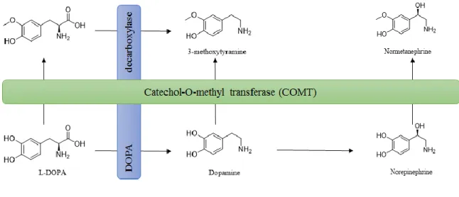
Given the establishment of dopaminergic loss as a pathological hallmark of PD, the therapeutic strategies until now have been focused on boosting the levels of DA in the brain. Since the results published by Yahr et al.23 in the late 60’s, levodopa (L-DOPA) is the gold standard drug for the clinical management of PD.24 L-DOPA is a biosynthetic DA precursor which is decarboxylated by DOPA decarboxylase to yield DA in the brain (Fig. 2). The administration of L-DOPA is a therapeutic strategy to indirectly enhance DA levels within the central nervous system (CNS).
Figure 2 – Dopamine metabolism.
However, long-term treatment with L-DOPA often induces serious adverse effects like nausea, vomiting, psychosis and diskinesias.25 Moreover, its biological half-life is very reduced (60-90 min), which requires multiple daily administrations.26 Furthermore, L-DOPA sustained treatment in PD patients induces “on-off” phenomenon.27 The “on-off” phenomenon comprises profound diurnal fluctuations in the psychomotor state, where phases of dyskinesia and incapacity associated with depression alternate with jubilant periods.27 In order to improve L-DOPA bioavailability, the use of enzyme inhibitors that prevent L-DOPA peripheral catabolism (e.g. carbidopa or benserazide) has been introduced (Fig. 3).
Figure 3 – Structures of carbidopa and benserazide.
Notwithstanding, L-DOPA remains the most effective treatment for the archetypal motor symptoms of PD. In addition to stimulating DA receptors, L-DOPA-derived DA might also activate adrenoceptors (a class of G protein-coupled proteins), DA transporters and trace amine receptors, all of which might contribute to the superior effect of L-DOPA in PD.28
1.2.1.1.2. Dopamine receptor agonists
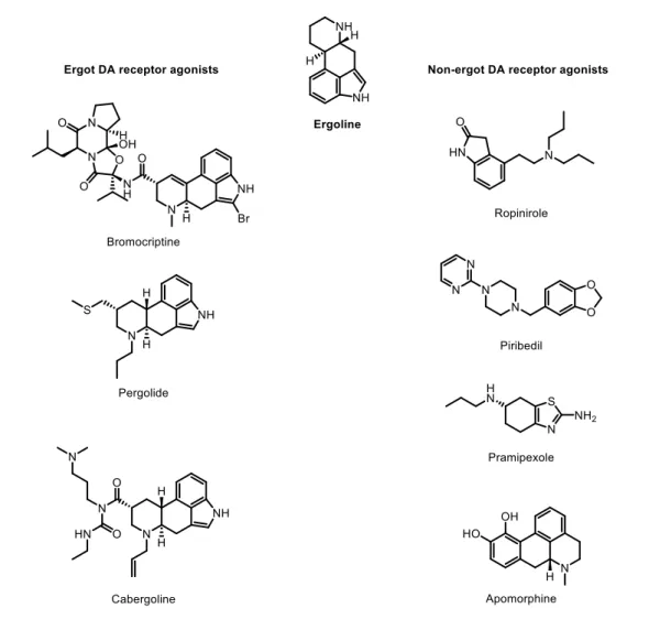
In general, DA receptor agonists are divided into two main classes: ergot or non-ergot agonists. These two classes are defined according to a particular structural parameter, which is the presence or absence of an ergoline-like structure (Fig. 4). As an example, ergot agonists include bromocriptine, pergolide, and cabergoline, while non-ergot agonists include pramipexole, ropinirole, apomorphine and piribedil (Fig. 4).29
Generally, DA receptor agonists are effective in reducing the incidence of motor complications arising from long-term L-DOPA therapy.29–31 There is increasing evidence that DA receptor agonists are not only beneficial to postpone L-DOPA therapy in early PD, but also have a neuroprotective effect.32 The mechanism of DA receptor agonist-induced neuroprotection is mainly associated with their radical scavenging properties.32 Also, DA receptor agonists were reported to have an important role in the CNS as neurogenesis promotors.33
However, many side effects to DA receptor agonists have been reported, mainly impulse control disorders such as hyper sexuality or binge eating. In order to diminish these effects, this therapy is initiated with low dosages and is slowly increased throughout an extended period of time to improve tolerance.34
1.2.1.1.3. Catechol-O-methyltransferase inhibitors
Catechol-O-methyltransferase (COMT) was first discovered by biochemist Julius Axelrod in 1957 and since then its function has been extensively studied.35,36 Due to its role in DA metabolism (Fig. 5), COMT is a pharmacological target for the treatment of PD.
Figure 4 – Structures of ergoline, and ergot and non-ergot dopamine receptor agonists.
Peripherally, COMT can be found in large quantities in the liver and kidneys, where it promotes the meta oriented methylation of catecholamines.36 Specifically, L-DOPA is extensively inactivated by peripheral COMT.37 The pharmacological blockade of the degradation of L-DOPA by COMT inhibitors prevents its peripheral degradation and increases its plasma half-life, allowing orally administered L-DOPA to cross the blood-brain barrier (BBB) into the blood-brain, where it is locally decarboxylated to DA.38,39 This pharmacological approach indirectly compensates the extensive dopaminergic loss observed in PD patients.
Early catechol-based competitive COMT inhibitors lacked in vivo efficacy and had low plasmatic half-lives, showing poor selectivity and toxicological constrains.36
Figure 5 – L-DOPA, dopamine and norepinephrine metabolism by COMT.
Later on, second generation inhibitors were developed and, due to their increased efficacy over first generation inhibitors,40 were introduced in the market for PD adjuvant therapy.41 These selective and orally active COMT inhibitors bear a nitrocatechol scaffold, which is the pharmacophore for second generation tight-binding COMT inhibitors (Fig. 6).
Figure 6 – Structure of second generation COMT inhibitors.
Tolcapone and entacapone (Fig. 6) are highly selective and potent COMT inhibitors. The main difference between these two drugs relies on pharmacokinetics, particularly in brain bioavailability. Entacapone is a peripheral COMT inhibitor, while tolcapone is able to cross the BBB into the CNS, where it can locally prevent L-DOPA degradation catalysed by COMT.42 Nevertheless, tolcapone has a high risk of hepatotoxicity,43 and is currently used under very strict conditions and controlled medical supervision. Although entacapone has not been linked to hepatotoxicity risk, it has been associated with adverse effects, including nausea, vomiting and dyskinesia.44 Accordingly, a third
generation inhibitors was designed and tested for adjunctive therapy for PD. A new drug called opicapone (Fig. 6) successfully passed clinical trials, with 9 out of 600 patients experiencing severe adverse effects in phase III trials.45 Also, it reduced off-times in PD patients by 2 hours compared to entacapone.45 Under the brand name Ongentys®, opicapone was recently introduced in the United Kingdom and it is expected to be launched in the rest of European market in 2017.46
1.2.1.1.4. Monoamine oxidase inhibitors
Monoamine oxidases (MAOs) are flavin adenine dinucleotide (FAD)-containing enzymes mainly found in the outer mitochondrial membrane and are responsible for the metabolism of endogenous amines.47 MAOs have two known isoforms, MAO-A and MAO-B, which catalyse the oxidation of different biogenic amines. The main MAO substrates are neurotransmitters such as adrenaline, noradrenaline, DA, serotonin and
β-phenylethylamine. Under physiological conditions, noradrenaline and serotonin are
substrates of A, while DA and β-phenylethylamine have a greater affinity for MAO-B.48 MAOs metabolise neurotransmitters by oxidizing the amine function to an aldehyde, in a process where the FAD cofactor is reduced (Fig. 7). The FAD cofactor is then oxidized again by molecular oxygen, in a reaction where hydrogen peroxide (H2O2) is generated (Fig. 7).
Figure 7 – MAOs catalysed reactions.
Although glutathione peroxidases can reduce hydrogen peroxide into water, the highly reactive hydroxyl radical can still be generated by a Fenton and Haber-Weiss reaction.49,50 In this sense, MAO-B is a pharmacological target for the treatment of PD, since its inhibition enhances DA levels. Selective MAO-B inhibitors are generally a valid asset in early PD therapy in combination with L-DOPA. In fact, MAO-B inhibitors are administered in patients with mild motor symptoms before other pharmacological alternatives.47 Moreover, MAO-B inhibitors may reduce the rate of motor fluctuations
compared to initial L-DOPA therapy, with fewer adverse effects than DA receptor agonists.51
The first selective and irreversible MAO-B inhibitors introduced in the market were selegiline (L-deprenyl) and rasagiline (Fig. 8). However, some authors suggest that selegiline metabolism can lead to amphetamine-like metabolites, which can be neurotoxic.52 On the other hand, the metabolism of rasagiline does not produce toxic metabolites.53 In fact (R)-1-aminoindan, the major metabolite of rasagiline, exerts neuroprotective effects in PD animal models.54 Similarly to selegiline, rasagiline improves the symptoms of PD patients, but it is more efficient if administered on the early stages of the disease.55
Recently, safinamide (Fig. 8), commercialized under the name of Xadago® was approved in Europe and United States.56 Contrarily to selegiline and rasagiline, safinamide is a reversible MAO-B inhibitor.57 In Europe, safinamide is approved for the treatment of mid- to late-stage fluctuating PD as add-on therapy to a stable dose of levodopa alone or in combination with other PD medications.56 In 24-week, placebo-controlled clinical trials, safinamide increased daily on-time without dyskinesia in patients with mid- to late-stage PD with motor fluctuations.58
Figure 8 – Structures of selegiline, rasagiline and safinamide.
1.2.1.2. Non-dopaminergic approaches in PD therapy
1.2.1.2.1. Adenosine and adenosine receptors
Current therapies for PD are mainly focused on targets of the dopaminergic system. However, it was found that this approach is not innocuous and could lead to severe complications, as previously discussed in item 1.2.1.1..57,59 The drawbacks of dopaminergic therapies provided a driving force for the study of non-dopaminergic targets, such as adenosine receptors (AR). Adenosine is a purine nucleoside which regulates a plethora of CNS processes, such as learning and memory.60 The responses
triggered by adenosine are mediated by AR, which are G protein-coupled receptors embedded in cell membranes. Four different subtypes of AR are known, namely A1, A2A, A2B and A3.61 Mechanistically, they differ on their action towards adenylate cyclase; A1AR and A3AR inhibit downstream adenylate cyclase, while the A2AAR and A2BAR subtypes have the opposite outcome (Fig. 9).62
Figure 9 – Adenosine receptors.
While adenosine signalling is important in several CNS functions,60 the most important within the context of PD are neuroprotection, locomotor effects and the control of DA levels.63 The crosstalk between adenosine and DA has been extensively studied.64 Besides DA, adenosine modulates the production and transmission of other neurotransmitters such as glutamate and, more importantly, GABA.60,65 Stimulation of A2AAR resulted in an increased release of GABA, while the activation of A1AR inhibits its production.60
The A2AAR is currently considered an important target for PD, due to its location and physical association with DA receptors.66 In spite of their physical proximity and a tight interplay, they may have antagonic effects on specific signaling mechanisms, for instance in cyclic adenosine monophosphate (cAMP) production.67,68 Moreover, it is known that A2AAR activation reduces affinity of DA receptors in the striatum for its agonists and that its blockade increases DA receptors activity.65 Aside from reducing the effects of DA depletion in the brain, A2AAR antagonists showed potential neuroprotection
and anti-inflammatory activity.69 Preclinical studies in animal models of PD have shown that A2AAR antagonists in monotherapy can alleviate PD symptoms.70,71 Moreover, the combination of A2AAR antagonists with L-DOPA can successfully reduce the effective dosage of the latter and, consequently, its side effects.72,73
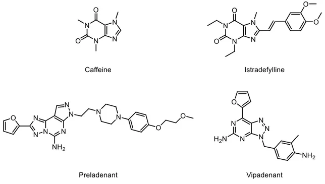
Chemically, A2AAR antagonists can be classified in two categories: xanthine derivatives (e.g.: caffeine), and non-xanthine derivatives (e.g.: pyrimidines) (Fig. 10). Several compounds were found to act as A2AAR antagonists (Fig. 10), but clinical data regarding their effects in PD is scarce. Albeit several A2AAR antagonists have been described, only istradefylline, preladenant and vipadenant (Fig. 10) were studied in detail in preclinical and clinical settings for PD.
Figure 10 – Structure of A2A AR antagonists.
Istradefylline (Fig. 10) was well tolerated in phase I clinical trials,74 and offered a clinically meaningful reduction in off-time without increased troublesome dyskinesia.75 However, the Food and Drug Administration (FDA) issued a non-approvable letter in 2008 demanding more follow-up data as part of phase IV evaluation commitments.74 Thereafter, istradefylline has been approved for the treatment of PD in Japan.74 Preladenant had positive results in phase II clinical trials in humans, although it did not prove to be more effective than placebo in phase III studies and, as a result, was discontinued in May 2013.76,77 Finally, vipadenant studies were suspended due to safety concerns.78,79 Nevertheless, vipadenant served as a scaffold for new generation compounds, one of which is in phase I clinical trials, still with undisclosed results.80
1.2.2.
Alzheimer’s disease
AD was first described in 1906, when a German neuropathologist named Alois Alzheimer gave a lecture on a case of a 51 year old woman suffering of severe dementia. At that time, her case was a curiosity, but it is now evident that she was suffering from a rare early onset form of AD. Nowadays, this progressive and degenerative disease is the most common cause of senile dementia.81
Chronologically, AD can be divided into three stages. In the initial stage, symptoms usually start with manifestations of cognitive deficit, such as the inability to produce new memories and skills.82 In the intermediate stage, patients begin to show problems with speech and not being able to handle simple everyday tasks, such as dressing themselves and attending to their personal hygiene.83 In the most advanced phase, AD is characterised by a state of full incapacity, confusion and disorientation.84 In this phase, patients have major difficulties in mobility and frequently suffer from hallucinations and deliriums.85 The most common form of this disease is the late-onset AD (LOAD) form, which accounts for approximately 95% of AD cases.86 The specific cause of AD is still unknown, although some risk factors have been pointed out, such as advanced age or family history.87 On the contrary, early onset AD (EOAD) is inherited by an autosomal dominant disorder, with four genes associated with disease onset and progression: the amyloid protein precursor (APP), preselinin 1 and 2, and the ε4 allele of the apolipoprotein E gene.88
The classic features in AD patients brains are: a) neuronal loss in regions linked with memory and cognition, mainly in the cholinergic neurons; b) low neurotransmitter levels, mainly acetylcholine (ACh) and c) synaptic dysfunction.86,89 The histopathology of the disease is well studied, and AD brains often present abnormal protein deposits like senile neuritic plaques (SNP) and neurofibrillary tangles (NFT). Physiological β-amyloid protein has short beta sheets in solution in the cell, but when misfolded into tertiary structures, it can form SNP and abnormal extracellular deposits.90 On the other hand, NFT are formed intracellularly, and are a product of an abnormal protein aggregation. Although tau protein is normally responsible for stabilizing the cytoskeleton, in AD it is aberrantly phosphorylated, which leads to protein aggregation and, ultimately, to an insoluble mass inside the cell.91
The formation of SNP and/or NFT leads to atrophy in the affected areas, usually the temporal and parietal lobes and in certain parts of the frontal cortex,92 leading to reduced ACh levels. These effects may contribute to more death of cholinergic neurons resulting
in a positive feedback loop.93 Along this process, the levels of serotonin and norepinephrine levels are low while glutamate levels are usually high.94,95
Even though the histological hallmarks of the disease are well known, the primary cause of AD remains unknown. There are several hypothesis pointed out by different research groups in order to explain the disease onset and progression. The first hypothesis to be suggested was the cholinergic hypothesis,96–98 but other have been proposed such as the amyloid hypothesis,99–101 the glutamatergic hypothesis,98,102,103 the oxidative stress hypothesis,104–106 metal hypothesis,107–109 and the inflammatory hypothesis.87,110,111 Current AD therapies are based on the cholinergic and glutamatergic hypothesis, and include acetylcholinesterase (AChE) inhibitors and N-methyl-D-aspartate (NMDA) receptor antagonists.112 Nonetheless, none of the currently approved drugs is able to slow down disease progression, and provide only symptomatic relief. Thus, there is a pressing and unmet clinical need for new and potent anti-Alzheimer drugs.
1.2.2.1. Acetylcholinesterase inhibitors
The contractions of smooth muscle in several organ systems, including gastrointestinal and urinary tract, and eye movement is mediated by ACh.113 This neurotransmitter is also responsible for decreasing heart rate and vasodilation.114 Moreover, the cholinergic transmission has a vital role in the modulation of cerebral blood flow, memory and cognition.115 According to the cholinergic hypothesis, the cholinergic system is the most affected in the early AD development.93 As a consequence, there is a loss of enzymatic function for ACh synthesis, leading to memory loss and deterioration of cognitive and non-cognitive functions which sometimes culminates in psychiatric symptoms.116 The levels of ACh in the synaptic cleft are tightly regulated by AChE (Fig. 11), which can also be found in neuromuscular junctions and plasma.87
Figure 11 – AChE catalysis of ACh.
According to Sussman et al.117, the active site of AChE is located at the bottom of a narrow gap, in which the catalytic anionic site (CAS) encloses four subsites: a) the anionic site, where the positively charged ACh interacts, b) the esteratic site (ES), which
contains the three residues of the catalytic triad, c) the oxyanion hole and d) the acyl pocket, which is responsible for substrate selectivity. Another different sub-unit in the enzyme’s active site is known as the peripheral anionic site (PAS) and it is located roughly 15 Å above the CAS (Fig. 12).
Figure 12 – Active site of AChE.
It is generally accepted that AChE acts not only as a key regulator of cholinergic transmission, but can also display a non-cholinergic function, attributed to the active site’s PAS, namely promoting β-amyloid deposition.118 This interaction promotes conformational changes in β-amyloid fibrils and induces the formation of SNP.119 All these findings deepen the overly simplistic cholinergic hypothesis, and validate the use of AChE inhibitors as a valid approach to manage AD.87
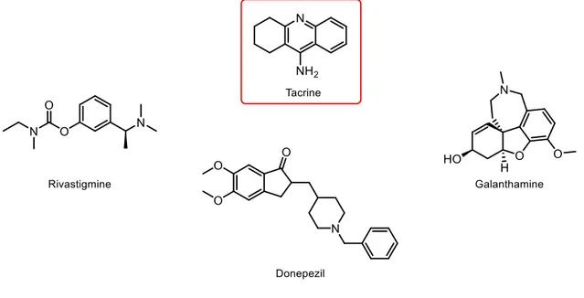
The first AChE inhibitor approved as a drug was tacrine (Fig. 13), but its use has been discontinued due to its severe hepatotoxicity.120 In spite of not being used anymore, tacrine is still used as scaffold for the design and development of novel AChE inhibitors.87 Currently, only three AChE inhibitors (Fig. 13) were approved by the FDA for the clinical management of AD, namely donepezil, rivastigmine and galantamine.121 All compounds are known for their clinical benefits to patients’ cognitive functions, leading to an improvement of the day-to-day activities and global functions.122,123 However, donepezil is the only compound approved in the USA for patients with severe AD.124 Although this type of inhibitors proved to delay cognitive decline for periods of up to 12 months, they
exhibit several adverse effects, such as nausea, vomiting and diarrhoea.122 Moreover, their use is also associated with higher rates of bradycardia and syncope, outlining a feeble risk-benefit relationship that requires careful consideration on later disease stages.125
Figure 13 – Structures of tacrine, in red, and rivastigmine, donepezil and galanthamine.
Even though all these compounds enabled a noteworthy advance in the clinical management of AD, the research and development of new potent AChE inhibitors having disease-modifying properties is still an ongoing effort.126
1.2.2.2. N-Methyl-D-aspartate receptor antagonists
Glutamate is the most abundant neurotransmitter and it has an important role in neuronal differentiation, migration and survival in the developing brain.127 It is also of particular interest due to its putative participation in the neurodegenerative processes.128,129 Glutamate, endogenous and exogenous, can initiate cell death processes in AD, by causing excitotoxicity.130 Excitotoxicity is a condition where neuronal damage is promoted by glutamatergic overstimulation, especially through an agonist effect on N-methyl-D-aspartate (NMDA) receptors, ultimately leading to calcium overload.131,132 The role of NMDA in the neurodegenerative cascade is still elusive, but the subsequent selective neuronal death appears to be dependent on NMDA receptor activation.130 To this effect, NMDA receptor antagonists are an important therapeutic option for moderate to severe AD. Currently, only one NMDA receptor antagonist is marketed for
the clinical management of AD.131 Memantine (Fig. 14) is a NMDA receptor antagonist that protects neurons from excitotoxicity. McShane et al.133 conducted a study in patients with moderate to severe AD showed a significant improvement in cognition, activities of daily living and behaviour following a 6 months treatment with memantine. Another study confirmed the previous findings and added that, under the treatment with memantine, patients reduced psychological symptoms of dementia.134
Figure 14 – Structure of memantine.
Nevertheless, patients undergoing memantine therapy frequently report side effects such as dizziness, headaches, confusion and agitation.135 The combined therapy with memantine and donepezil (Fig. 13) in patients with severe AD showed a significant improvement in cognitive function, language and daily life activities when compared to memantine monotherapy.136–138 The same outcome was not observed in patients with mild AD, therefore memantine is preferably administered in later stages of the disease, when patients have been already treated with donepezil.139
1.2.2.3.
Other targets in Alzheimer’s disease
The neurodegenerative cascade in AD has a wide range of pathologic features, which unveiled a large set of putative therapeutic targets. These targets are mostly enzymes involved in key physiological processes connected to neurodegeneration, such as MAO-B, secretases and caspases, among others.87,140
MAO-B inhibitors are more often used in PD therapy, as described in item 1.2.1.1.4. However, their neuroprotective effects may be useful in therapy of other ND including AD.47,141 The metabolism of MAO-B leads to the formation of ROS and to an amplification of neuronal oxidative stress, as depicted in Fig. 7. Neurons are particularly susceptible to oxidative stress as a consequence of: a) their low pool in endogenous antioxidants, such as glutathione, b) high content on polyunsaturated fatty acids c) the great oxygen brain consumption and also d) a high content in iron.142–144
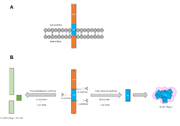
Secretases are enzymes that modulate the cleavage of the transmembrane APP (Fig. 15A) and are expressed in three isoforms: α-, β- and γ-secretase.145 Briefly, the
sequential cleavage by β-secretase (BACE) and γ-secretase leads to the β-amyloid fragment, which aggregates into SNP in AD patients (Fig. 15B). Contrarily, the processing of APP by α-secretase is non-amyloidogenic and yields a protective outcome, preventing the production of SNP (Fig. 15B).145 Thus, owing to its amyloidogenic role, BACE is now considered a therapeutic target for AD.145
First generation BACE inhibitors were based on peptides, similar to the BACE cleavage site,146 but several high affinity inhibitors with non-peptide backbones have thenceforth been developed.147,148
Figure 15 – Representation of the transmembrane protein APP and its degradation by different secretases.
Currently, the pharmaceutical company CoMentis has completed the first phase I clinical trial for a BACE inhibitor. However, the structure of the compound and trial results are undisclosed.145,149 Additionally, two other inhibitors (AZD3293 and MK-8931, Fig. 16) finished Phase I clinical trials and are expected to end Phase II/III studies in 2019. Although the development of BACE inhibitors is in early stages, and information regarding their safety and toxicity is scarce, these preliminary findings of new BACE inhibitors are encouraging and may have a favourable outcome in LOAD.149
Figure 16 – Structure of BACE inhibitors presently on clinical trials.
Caspases (cysteine-aspartic proteases) are a family of proteases are linked in cell regulatory networks, controlling inflammation and cell death.150 Deregulated apoptosis caused by caspases have been linked to neurodegeneration processes and promotion of the biological cascade leading to AD.151 Since the first caspase was identified by Horvitz et al.152 several others have been discovered and categorised into two sub-families based on their biological function. Briefly, they were classified either as inflammatory caspases 1, -4, -5, -11 and -12) or apoptopic caspases (caspase-2. -3, -6, -7, -8, -9 and -10).153,154
Rohn et al.151 hypothesized that β-amyloid fragments trigger the activation of caspases, leading to the proteolysis of tau and consequent NFT formation (Fig. 17). Along the biological cascade the over activation of caspases can lead to cytoskeletal disruption and tau protein hyperphosphorilation, ultimately ending in neuronal death (Fig. 17). Other studies have confirmed the hypothesis supporting the link between deregulated caspases and NFT production,91,155,156 mainly caspase-2.157
Although caspase inhibitors could be beneficial in AD therapy, no drug has been researched and developed with that goal. Nevertheless, there are caspase inhibitors that could in a near future serve as inspiration for new drugs focusing on AD therapy. In Fig. 18 examples of caspase inhibitors are shown. Pralnacasan, first developed to treat ischemia, is a selective and reversible caspase-1 inhibitor, withdrawn from clinical trials due to hepatotoxicity.158 VX765 is also a reversible capase-1 inhibitor currently in Phase II trials for the treatment of inflammatory diseases.159 Also developed as an anti-inflammatory drug, emricasan was withdrawn from clinical studies for undisclosed reasons in spite of promising results.160 NCX1000 is a steroid-based, nitric oxide releasing and non-selective caspase inhibitor developed to treat portal hypertension.161 It was deemed safe however the study was terminated in Phase II for unmet efficacy requirements.
1.3. One target versus multi target drugs
Since the introduction of the “magic bullet” concept by Ehrlich, drug discovery efforts have been focusing on identifying single target drugs that interact with a specific target with a defined disease mechanism.162 Generally, this approach has been successful, however it was unable to effectively tackle multifactorial diseases with high socioeconomic impact. Additionally, the single target approach is oblivious to processes connected by complex networks in biological systems. In these complex systems, cells may display a redundant effect since they have several mechanisms that yield the same outcome, such as gene expression, receptor response or protein degradation.163 Therefore, this redundancy nulls the expected effect of single target drugs in a primary mechanism. Indeed, a clinical effect of a drug is sometimes owed to the interaction with multiple targets.164
In this context, the development of multi target drugs has gathered increased interest from academia and industry.165 Nevertheless, the rational design of multi target drugs is still in its infancy and further development is critical. From a medicinal chemistry standpoint the rational design and multi target hit identification is very challenging. Moreover, lead optimization and comprehensive SAR studies for several different targets is extremely complex. To this end, different approaches were used, such as fragment-based design, molecular hybridization of active scaffolds or combination of compounds with known bioactivity.166 Moreover, the analysis of approved drugs and recurrent bioactive compounds showed multi target binding affinity.167
The multi target approach is specifically relevant for ND, by definition multifactorial diseases with a complex network of pathological events. As mentioned in items 1.2.1. and 1.2.2., currently available single target treatments for PD and AD are only palliative and unable to alter disease progression. As such, the development of multi target directed drugs for different pharmacological ND targets is increasingly enticing attention.168
A significant advance in the progress of multi target directed drugs for ND was the development ladostigil, a drug designed to act as an AChE/MAO-B inhibitor.169 Ladostigil (Fig. 19A) bears the propargyl moiety of MAO-B inhibitor rasagiline (Fig. 8) and the carbamate pharmacophore of AChE inhibitor rivastigmine (Fig. 13). As expected, the resulting molecule is a dual AChE/MAO-B inhibitor, developed for AD therapy and currently enrolled in phase II clinical trials.169 Moreover, ladostigil major metabolite
exhibits potent AChE inhibitory activity in vivo, in addition of neuroprotective effects (Fig. 19B).170,171
Figure 19 – Structures of ladostigil and its major metabolite.
1.3.1.
Multi target drugs for Parkinson’s disease
The majority of PD multi target directed drugs developed so far combine MAO-B inhibition with other relevant targets. As discussed in item 1.2.1.1.4., MAO-B catalysis is one of the main sources of H2O2, a byproduct of selective oxidative monoamine deamination. This H2O2 may react with iron and copper and generate harmful ROS, namely ·OH. Thus, iron chelation combined with MAO-B inhibition is considered a valid multi target approach. Zheng et al.172 combined the iron chelating hydroxyquinoline scaffold with the propargylamine moiety and developed non-selective MAO-B inhibitors that exhibited potent inhibition of iron-dependent lipoperoxidation in rat brain homogenates. Moreover, due to the non-selectivity profile of these derivatives, they also showed potential adjunctive antidepressant activity.173
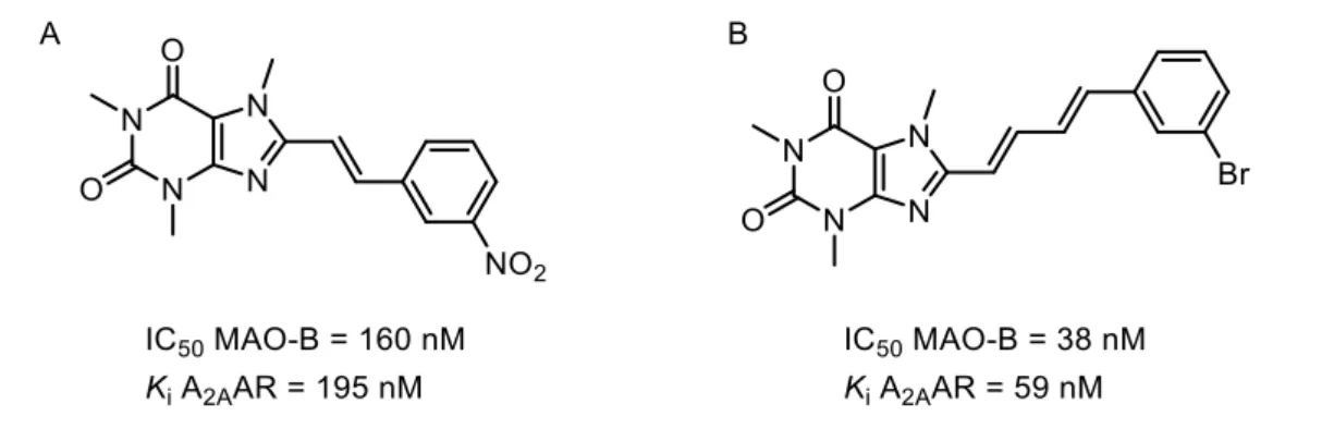
As previously discussed in item 1.2.1.2.1., A2AAR antagonists effectively protected against neurodegeneration in different disease models, and were hypothesized to provide symptomatic relief of PD motor symptoms.70 Petzer et al.174 developed selective and potent nanomolar MAO-B inhibitors, which also exerted neuroprotective effects by means of A2AAR antagonism (Fig. 20A). More recently, Pretorius et al.175 found that 8-(4-phenylbutadien-1-yl)caffeine analogues were potent reversible MAO-B inhibitors with remarkable nanomolar A2AAR affinity and antagonism (Fig. 20B).
Figure 20 – Structures of MAO-B inhibitors and A2AAR antagonists.
1.3.2. Multi target drugs for Alzheimer’s disease
The discovery of AChE non-cholinergic actions, mainly their implication in the development of SNP, provided the driving force for the development of multi target drugs for AD. This strategy represents a new area of research based on both cholinergic and amyloid hypothesis. Furthermore, crystallographic studies of AChE provided additional insight of the enzyme’s structure, and triggered the development of dual binding AChE inhibitors, which interact with the catalytic site and the PAS.176 Adding to ACh degradation inhibition, these dual-binding inhibitors also target the PAS and decrease the aggregation rate of β-amyloid.
Early dual-binding AChE inhibitors were based on the structural elucidation provided by Sussman et al.117 and the reported X-ray structure of the donepezil-AChE complex.177 Since then, a study by Tumiatti et al.178 unraveled the potential of less flexible or rigid moieties (e. g. dipiperidines), as PAS-binding scaffolds. Moreover, the authors found that the synthesized compounds were able to inhibit self- and AChE-induced β-amyloid aggregation within the low micromolar range. In fact, a dual binding AChE inhibitor reached Phase II clinical trials for AD, however no structure is available and studies results are undisclosed.176
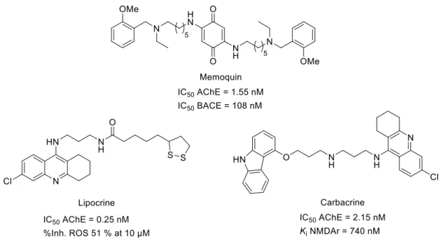
The development of dual-binding AChE inhibitors evolved to incorporate other bioactivities, such BACE inhibition. Since both enzymes are involved in protein aggregation, this constitutes a valid strategy for the development of new AD therapies. Within this class of compounds, memoquin (Fig. 21) is a potent AChE and BACE inhibitor, while retaining micromolar inhibition of β-amyloid aggregation.179 Furthermore, memoquine was shown to improve cognitive impairment, and short and long-term memory in scopolamine-induced amnesia models.180
On the other hand, AChE inhibitors with antioxidant activity were also developed as a therapeutic strategy to tackle both cognitive deficit and the harmful effects associated with neuronal redox deregulation and ROS-induced damage observed in AD. To this effect, lipocrine (Fig. 21), a tacrine/lipoic acid heterodimer was developed as a potent AChE inhibitor able to interact with the enzyme’s PAS and inhibit AChE-induced β-amyloid aggregation.181 Moreover, lipocrine decreased by half the production of ROS at 10 µM and demonstrated improved neuroprotection against oxidative stress.168,181 Glutamate-related excitotocicity contributes to AD and occurs in part because of NMDA overactivation.130 Moreover, oxidative stress and increased intracellular Ca2+ have been reported to enhance glutamate mediated neurotoxicity in vitro.182 In this context, carbacrine (Fig. 21) was successfully developed as: a) AChE inhibitor within the nanomolar range, b) β-amyloid self- and AChE-induced aggregation inhibitor, c) NMDA antagonist, and d) antioxidant agent.183