0103 - 5053 $6.00+0.00
Article
*e-mail: [email protected]
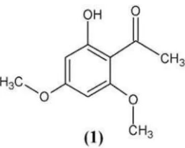
2-Hydroxy-4,6-dimethoxyacetophenone from Leaves of Peperomia glabella
Marisi G. Soares,a Ana P. V. de Felippe,a Elsie F. Guimarães,a Massuo J. Kato,a Javier Ellenab and Antonio C. Doriguetto*,c
a
Instituto de Química, Universidade de São Paulo, Av. Prof. Lineu Prestes, 748, Cidade Universitária, 05508-900 São Paulo-SP, Brazil
b
Instituto de Física de São Carlos, Universidade de São Paulo, CP 369, 13560-970 São Carlos-SP, Brazil
c
Departamento de Ciências Exatas, Universidade Federal de Alfenas, 37130-000 Alfenas-MG, Brazil
A 2-hidroxi-4,6-dimetoxiacetofenona foi isolada de folhas de Peperomia glabella (família
Piperaceae). A substância cristaliza no grupo espacial P—1 com duas moléculas na unidade assimétrica. A análise estrutural revela uma rede bidimensional infinita no plano (011), estabilizada por ligações de hidrogênio inter-moleculares ao longo da direção [100] e interações de van der Waals ao longo da direção [011]. As moléculas apresentam fortes ligações de hidrogênio intra-moleculares [O1-HO1...O4 = 1,53(2) Å e O5-HO5...O8 = 1,38(2) Å]. A conformação molecular foi analisada usando o MOGUL, uma base de dados de geometria molecular derivada do CSD (Cambridge Structural Database).
The 2-hydroxy-4,6-dimethoxyacetophenone was isolated from the leaves of the Peperomia glabella (Piperaceae family). The molecule crystallizes in the space group P—1 with two
independent molecules in the asymmetric unit. The structural analysis reveals an infinite two-dimensional network in the (011) plane, stabilized by intermolecular hydrogen bonds along [100] and van der Waals interactions along [011]. The molecules present strong intra-molecular hydrogen bonds [O1-HO1...O4 = 1.53(2) Å and O5-HO5...O8 = 1.38(2) Å]. The molecular conformation was analyzed using the MOGUL, a knowledge base of molecular geometry derived from the CSD (Cambridge Structural Database).
Keywords:Peperomia glabella, 2-hydroxy-4,6-dimethoxyacetophenone, X-ray diffraction
Introduction
Peperomia glabella (Piperaceae) is an epiphyte used in
Venezuelan folk medicine as an antiasthmatic.1 Piperaceae
family comprises 14 genera and 1950 species.2 Among these,
Piper and Peperomia are the most abundant with
approximately 700 and 600 species, respectively.3
Phytochemical profile from genus Piper is characterized by
occurrence of lignans and neolignans,4,5 chromenes,6-8
amides,9,10 alkaloids,11 Phenylpropanoids,12 and
cyclopente-nedione derivatives.13 Comparative study showed divergence
of secondary metabolism in cell suspension cultures and
differentiated plants of P. cernuum and P.crassinervium.14
Compared with the genus Piper, few chemical investigations
reported so far for Peperomia species have shown their
common constituents as phenylpropanoids, benzopyrans,
chromenes, and prenylated hydroquinones.15-17 Additionally
there are several nor/seco-compounds, e.g., the cyclobutane
compound from Peperomia pellucida that seems to be
produced by dimerisation of styryl phenol (a
nor-phenylpropanoid).18P. glabella has been shown to contain
one secolignan of butenolide skeleton.19 In this work we wish
to report the structure of the
2-hydroxy-4,6-dimethoxy-acetophenone, (1), extracted from leaves of Peperomia
glabella. Acetophenone derivatives have shown many
interesting biological properties such as antinflammatory,20,21
cytotoxic,22 and choleretic23 activities. This compound was
previously isolated from Artemisia maritima,24Artemisia
gypsaceae,25 and Plagiochila fasciculata showing antifungal
activities against Trichophyton mentagrophytes26 from strain
of Trichoderma pseudokoningii Rifai.27It is important to
emphasize that (1) can be also obtained by synthetic route,
from different starting compound, such as
1-hydroxy-3,5-dimethoxybenzene,28, 29 2,4,6-trihydroxiacetophenone,30 and
J. Braz. Chem. Soc.
The aim of the present work is to determine
unam-biguously the molecular structure of (1) by X-ray diffraction
(XRD) and its intra and inter-molecular geometry as well. To the best of our knowledge, this is the first occurrence of
this class of compound in Peperomia species.
Experimental
General Considerations
The compound was isolated from leaves of samples of Peperomia glabella. All chemicals used during the
extraction, purification and crystallization were of analytical or chromatography grade.
1H and 13C NMR spectra (4.9 T) were recorded on a
Bruker AC-200 spectrometer operating at room temperature, using tetramethylsilane as an internal reference.
Plant material, extraction and isolation of 2-hydroxy-4,6-dimethoxyacetophenone
Peperomia glabella samples were collected at Lençóis
(12o 45' 19.5" S 41o 30' 34.1" W) Bahia, Brazil. The leaves
(8.20 g) were extracted four times with methanol (300 mL each time) at room temperature. This extract (2.07 g) was submitted to normal-phase silica gel column chromatography
using increasing proportions of hexane:CH2Cl2.
2-hydroxy-4,6-dimethoxyacetophenone was obtained as solid white, 172
mg, crystallized from CH3OH:CH2Cl2.
Identification of 2-hydroxy-4,6-dimethoxyacetophenone
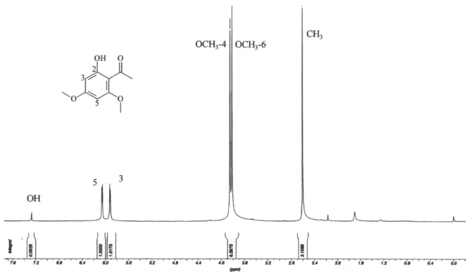
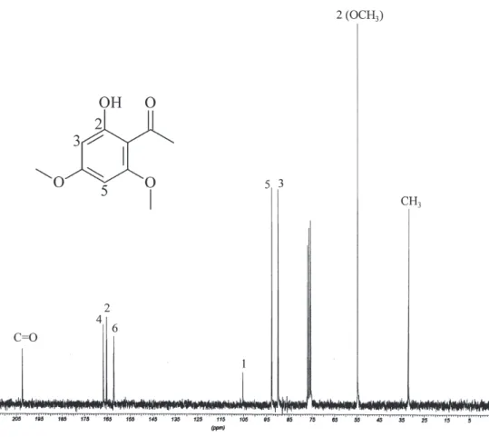
The 1H NMR spectrum of (1) showed two aromatic
meta hydrogens at δ 6.05 and 5.93 (d, J 2.2 Hz). In
addition, the acetophenone group was inferred from the
methyl group at δ 2.60 and confirmed by 13C NMR
spectrum at δ 32.7 and by acetophenone carbonyl at δ
203.5. The additional substituents of the aromatic ring
were defined as one quelated hydroxyl at δ 11.20 and
two methoxyls observed at δ 3.85 (δ 55.4) and δ 3.81
(δ 55.4). The 13C NMR spectrum (4.9 T) showed signals
corresponding to four aromatic carbon quaternary atoms
(δ 105.8; 167.5; 166.0; and 162.7); and two aromatic
methyne (δ 90.6 and 93.0). Thus, compound (1) was
identified as 2-hydroxy-4,6-dimethoxyacetophenone.24
X-ray Structure analysis of 2-hydroxy-4,6-dimethoxyaceto-phenone
A well-shaped single crystal of (1) was selected for the
XRD experiments.The intensity data were collected at
150 K on an Enraf-Nonius Kappa-CCD diffractometer (95 mm CCD camera on k-goniostat) with graphite
monochromated Mo Kα (λ = 0.71073 Å) radiation. The
temperature was controlled using an Oxford Cryosystem
low temperature device. Data collection (ϕ scans and ω
scans with κ offsets) was made using the COLLECT;32
integration and scaling of the reflections were performed
with the HKL Denzo-Scalepack system of programs.33 The
final unit cell parameters were based on all reflections using
HKL Scalepack.33 The structure was solved using Direct
Methods with SHELXS-97.34 The model was refined by
full-matrix least-squares procedures on F2 using
SHELXL-97.34 H atoms of the phenyl and methyl groups were
positioned stereochemically and were refined with fixed
individual displacement parameters [Uiso(H) =
1.2Ueq(Caromatic) or 1.5Ueq(Cmethoxy)] using a riding model, with aromatic C-H distances of 0.95 Å and methyl C-H distances of 0.98 Å. The two hydroxyl H atoms were located by difference Fourier synthesis and were set as isotropic.
Data collections and experimental details for (1) are
summarized in Table 1. The programs SHELXL-97,
SHELXS-97, and ORTEP-335 were used within WinGX36
to prepare materials for publication. The programs
Mercury37 and ORTEP-3 were used to prepare the
molecular graphics.
Results and Discussion
Figure 1 shows an ORTEP-335 view of (1) with the
atom numbering scheme. The structure crystallizes in
P—1 with two independent molecules in the asymmetric
unit (labelled as A and B in Figure 1). Comparison of
these molecules by the method of Kabsch38 showed them
examined using PLATON,39 and it was concluded that
P—1 is in fact the correct space group.
The main geometric parameters are given in Table 2. The molecular conformation was analyzed using the
MOGUL,40 a knowledge base of molecular geometry
derived from the Cambridge Structural Database (CSD)41
that provides rapid access to information on the preferred values of bond lengths, valence angles and acyclic torsion angles. The Figure 2 shows an example of the graphical result of the query molecule studied here (molecule A). The resulting histogram is the C1-O1 bond length comparing with the C-O bond lengths in CSD entries containing similar structures. As emphasized by Bruno et
al.,40 comparison of the dimensions of a newly determined
small-molecule crystal structure with bond lengths and angles of similar structures in the CSD is useful as a check against refinement errors and to highlight unusual geometrical features. This study showed that all bond lengths and bond angles are in agreement with the expected values for a good X-ray diffraction structure refinement. However, the MOGUL analysis has pointed out two interesting geometrical features due to resonance involving the moiety O3-C5-C6-C7-O4 (see Figure 1 and Table 2). It was observed that C5-O3 and C6-C7 is shorter than the average query values whereas the C5-C6 and C7-O4 is longer than the expected ones. It is important to emphasize that the same behaviour (not shown in Table 2) was observed to the molecule B present in the asymmetric unit.
Both molecules in the asymmetric unit are almost flat. The largest deviations from the least squares plane through the aromatic ring system A are -0.176(4) and 0.112(4) Å for C8 and C9 atoms, respectively. Similar results were observed to the molecule B: the largest
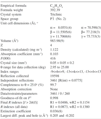
Table 1. Crystal data, data collection details and structure refinement re-sults of 1
Empirical formula C10H12O4 Formula weight 392.39 Crystal system Triclinic Space group P—1 (No. 2) Unit cell dimensions (Å), o
α = 8.0551(4) α = 70.590(3)
β = 11.5505(6) β= 77.218(3)
γ = 11.7068(6) γ = 75.733(3) Volume (Å3) 983.98(9)
Z 4
Density (calculated) (mg m-3) 1.122
Absorption coefficient (mm-1) 0.103
F(000) 416
Crystal size (mm3) 0.05 × 0.05 × 0.2
θ range for data collection (deg) 2.95 to 25.00
Index ranges -9<=h<=9, -13<=k<=13, -13<=l<=13 Reflection collected 19598
Independent reflections 3461 [R(int) = 0.0775] Completeness to θ = 25.0o (%) 99.7
Absorption correction None Data/restraints/parameters 3461 / 0 / 260 Goodness-of-fit on F2 0.998
Final R indexes [I > 2σ(I)] R1 = 0.0486, wR2 = 0.1154 R indexes (all data) R1 = 0.0873, wR2 = 0.1380 Extinction coefficient 0.036(8)
Largest diff. peak and hole (e.Å-3) 0.205 and -0.202
J. Braz. Chem. Soc.
deviations were observed to the C18 (-0.173(4) Å) and C19 (0.120(4) Å) atoms. The least squares planes of the
A and B molecules form an angle of 0.8(1)o. This result
shows that the two moieties are in the same plane, which is also illustrated in Figures 3 and 4.
The molecules A and B exhibited strong intra-molecular hydrogen bonds involving O1-HO1...O4 and O5-HO5...O8, respectively (Table 3 and Figure 1). The weak inter-molecular hydrogen bond between the aromatic hydrogen linked to C4 (molecule A) and the adjacent carboxyl oxygen O1 at x+1, y, z stabilizes the packing of the molecule A and gives rise to an infinite one dimensional chain parallel to the [100] direction (Table 3, Figures 1 and 3). The same interaction occur to the molecule B involving the aromatic hydrogen linked to C14 (molecule B) and the adjacent carboxyl oxygen O5 at x+1, y, z. The chains form a planar structure, connected by van der Waals interactions along the [011] direction. Therefore, the chains are linked
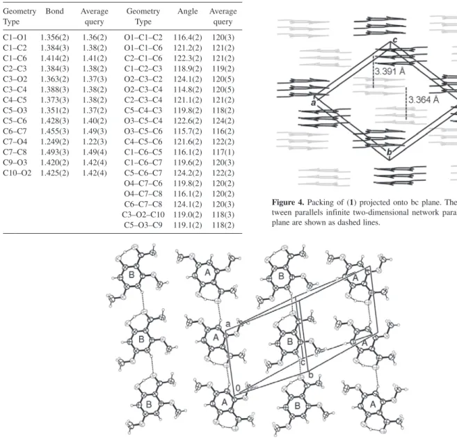
Table 2. Selected Bond lengths (Å) and angles (°) for compound (1) and MOGUL bond analysis
Geometry Bond Average Geometry Angle Average
Type query Type query
C1–O1 1.356(2) 1.36(2) O1–C1–C2 116.4(2) 120(3) C1–C2 1.384(3) 1.38(2) O1–C1–C6 121.2(2) 121(2) C1–C6 1.414(2) 1.41(2) C2–C1–C6 122.3(2) 121(2) C2–C3 1.384(3) 1.38(2) C1–C2–C3 118.9(2) 119(2) C3–O2 1.363(2) 1.37(3) O2–C3–C2 124.1(2) 120(5) C3–C4 1.388(3) 1.38(2) O2–C3–C4 114.8(2) 120(5) C4–C5 1.373(3) 1.38(2) C2–C3–C4 121.1(2) 121(2) C5–O3 1.351(2) 1.37(2) C5–C4–C3 119.8(2) 118(2) C5–C6 1.428(3) 1.40(2) O3–C5–C4 122.6(2) 124(2) C6–C7 1.455(3) 1.49(3) O3–C5–C6 115.7(2) 116(2) C7–O4 1.249(2) 1.22(3) C4–C5–C6 121.6(2) 122(2) C7–C8 1.493(3) 1.49(4) C1–C6–C5 116.1(2) 117(1) C9–O3 1.420(2) 1.42(4) C1–C6–C7 119.6(2) 120(3) C10–O2 1.425(2) 1.42(4) C5–C6–C7 124.2(2) 122(2) O4–C7–C6 119.8(2) 120(2) O4–C7–C8 116.1(2) 120(2) C6–C7–C8 124.1(2) 120(3) C3–O2–C10 119.0(2) 118(3) C5–O3–C9 119.1(2) 118(2) Figure 2. Histogram comparing the C1–O1 bond length with the C–O bond lengths in CSD entries containing structures similar to 2-hydroxy-4,6-dimethoxyacetophenone.
Figure 4. Packing of (1) projected onto bc plane. The separations be-tween parallels infinite two-dimensional network parallel to the (011) plane are shown as dashed lines.
themselves, forming an infinite two-dimensional network parallel to the (011) plane. The stacking of these two-dimensional networks is shown in the Figure 4. It is also observed that double chains, which are related by the inversion symmetry, are formed along [100] direction either to molecule A or B. The distance between parallels two-dimensional networks is about 3.4 Å.
Acknowledgments
The authors are grateful to CNPq and FAPESP for financial support. One of us, MGS, thanks FAPESP for the postdoctoral fellowship.
Supplementary Information
1H and 13C NMR data for 1 are avaliable free of charge
at http://jbcs.sbq.org.br, as PDF file.
Supplementary crystallographic data sets for (1) is
available through the Cambridge Structural Data Base, deposition number CCDC 296321. Copies of this information may be obtained free of charge from The Director, CCDC, 12 Union Road, Cambridge, CB2 1EZ, UK (fax: +44123-336-033; e-mail: [email protected] or http:www.ccdc.ac.uk
References
1. Salas, F. V. In Plantas Medicinales de Venezuela; Velez Boza, F., ed.; INAGRO: Caracas, 1982.
2. Mabberley, D. J.; The Plant-book. A Portable Dictionary of the Higher Plants, Cambridge University Press: New York, USA, 1997. 3. Joly, A. B.; Introdução a Taxonomia Vegetal. Editora Nacional:
São Paulo, Brazil, 1985.
4. Parmar, V. S.; Jain, S. C.; Bisht, K. S.; Jain, R.; Taneja, P.; Jha, A.; Tyagi, O. D.; Prasad, A. K.; Wengel, J.; Olsen, C. E.; Bol, P.M.; Phytochemistry 1997, 46, 597.
5. Martins, R. C. C.; Lago, J. H. G.; Albuquerque, S.; Kato, M. J.; Phytochemistry 2003, 64, 667.
6. Lago, J. H. G.; Ramos, C. S.; Casanova, C. C. D.; Morandim, A. A.; Bergamo, C.D.; Cavalheiro, A. J.; Bolzani, V. S.; Furlan, M.; Guimarães, E. F.; Young, M. M. C.; Kato, M.J.; J. Nat. Prod. 2004, 67, 1783.
7. Moreira, D. L.; Guimarães, E. F.; Kaplan, M. A.; Phytochemistry 1998, 49, 1339.
8. Baldoqui, D. C.; Kato, M. J.; Bolzani, V. S.; Young, M. C. M.; Furlan, M.; Phytochemistry 1999, 51, 889.
9. Silva, R. V.; Navickiene, H. M. D.; Kato, M. J.; Bolzani, V. S.; Méda, C. I.; Young, M. C. M.; Phytochemistry 2002, 59, 521. 10. Alécio, A. C.; Bolzani, V. S.; Young, M. C. M., Furlan, M.; J.
Nat. Prod, 1998, 61, 637.
11. Dodson, C. D.; Dyer, L. A.; Searcy, J.; Wright, Z.; Letourneau, D. K.; Phytochemistry 2002, 53, 51.
12. Orjala, J.; Erdelmeier, C. A. J.; Wright, A. D.; Rali, T.; Sticher, O.; Planta Med. 1993, 59, 813.
13. Facundo, V. A.; Sa, A. L.; Silva, S. A. F.; Morais, S. M.; Matos, C. R. R.; Braz, R.; J. Braz. Chem. Soc. 2004, 15, 140. 14. Danelutte, A. P.; Costantin, M. B.; Delgado, G. E.; Braz, R.;
Kato, M. J.; J. Braz. Chem. Soc. 2005 , 6B,1425.
15. Salazar, K. J. M.; Paredes, G. E. D.; Lluncor, L. R.; Young, M. C. M.; Kato, M. J.; Phytochemistry 2005, 66, 573. 16. Tanaka, T.; Asai, F.; Iinuma, M.; Phytochemistry 1998, 49, 229. 17. Cheng, M. J.; Lee, S. J.; Chang, Y. Y.; Wu, S. H.; Tsai, I. L.; Jayaprakasam, B.; Chen, I. S.; Phytochemistry 2003, 63, 603. 18. Bayma, J. C.; Arruda, M. S. P; Müller, A. H.; Arruda, A. C.;
Canto, W. C. C.; Phytochemistry 2000, 55, 779.
19. Monache, F. D.; Compagnone, R. S.; Phytochemistry 1996, 43, 1097.
20. Sala, A.; Recio, M. C.; Giner, R. M.; Màñez, S.; Ríos, J. L.; J. Nat. Prod. 2001, 64, 1360.
21. Favier, L.; Tonn, C.; Guerreiro, E.; Rotelli, A.; Pelzer, L.; Planta Med. 1998, 64, 657.
22. Huang, P.; Won, S.; Day, S.; Lin, C.; Helv. Chim. Acta 1999, 82, 1716.
23. Suksamrarn, A.; Eiamong, S.; Piyachaturawat, P.; Byrnes L.T.; Phytochemistry 1997, 45, 103.
24. Saxena, S.; Jain, D. C.; J. Ind. Chem. Soc. 2002, 79, 970. 25. Rustaiyan, A.; Zare, K.; Gangi, M. T.; Sadri, H. A.;
Phytochemistry 1989, 28, 1535.
26. Lorimer, S.D.; Perry, N.B; Planta Med. 1994, 60, 386. 27. Astudillo, L.; Schmeda-Hirschmann, G.; Soto, R.; Sandoval,
C.; Afonso, C.; Gonzales, M. J.; Kijjoa, A.; World J. Microbiol. Biotechnol. 2000, 16, 585.
28. Goldoni, L.; Cravorro, G.; Penoni, A.; Tollari, S.; Palmsano, G.; Synlett 2005, 6 927.
29. Cunningham, B. D. M.; Lowe, P. R.; Threadgill, M. D.; J. Chem. Soc. 1989, 9, 1275.
30. Kawadias, D.; Sand, P.; Youndim, K. A.; Qaiser, M. Z.; Rice-Evans, C.; Baur, R.; Sigel, E.; Rausch, W. D.; Riederer, P.; Schreier, P.; Br. J. Pharmacol. 2004, 142, 811.
31. Matieva, N. N.; Rao, N.; Redda, K. K.; J. Heterocycl. Chem. 2002, 39, 1251.
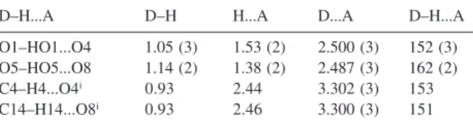
32. Nonius, Collect Software, Delft, The Netherlands, 1998. 33. Otwinowski, Z.; Minor, W; Methods Enzymol. 1997, 276, 307. Table 3. Hydrogen-bonding geometry (Å, o)
D–H...A D–H H...A D...A D–H...A
O1–HO1...O4 1.05 (3) 1.53 (2) 2.500 (3) 152 (3) O5–HO5...O8 1.14 (2) 1.38 (2) 2.487 (3) 162 (2) C4–H4...O4i 0.93 2.44 3.302 (3) 153
J. Braz. Chem. Soc.
34. Sheldrick, G. M.; SHELXS-97 and SHELXL-97; Program for Crystal Structure Refinement, University of Göttingen: Germany, 1997.
35. Farrugia, L. J.; J. Appl. Crystallogr. 1997, 30, 565. 36. Farrugia, L.J.; J. Appl. Crystallogr. 1999, 32, 837.
37. Bruno, I. J.; Cole, J. C.; Edgington, P. R.; Kessler, M. K.; Macrae, C. F.; McCabe, P.; Pearson, J.; Taylor, R.; Acta Crystallogr., Sect. B: Struct. Sci. 2002, 58, 389.
38. Kabsch, W.; Acta Crystallogr., Sect. A: Found. Crystallogr. 1976, 32, 922.
39. Spek A.L.; PLATON, A Multipurpose Crystallographic Tool, Utrecht University: Utrecht, The Netherlands, 2005. 40. Bruno, I. J.; Cole, J. C.; Kessler, M.; Luo, J.; Motherwell, W. D. S.;
Purkis, L. H.; Smith, B. R.; Taylor, R.; Cooper, R. I.; Harris S. E.; Orpen, A. G.; J. Chem. Inf. Comput. Sci. 2004, 44, 2133. 41. Allen, F. H.; Acta Crystallogr., Sect. B: Struct. Sci. 2002, 58, 380.
Received: January 31, 2006
Published on the web: July 20, 2006
0103 - 5053 $6.00+0.00
Supplementary Information
*e-mail: [email protected]
2-Hydroxy-4,6-dimethoxyacetophenone from Leaves of Peperomia glabella
Marisi G. Soares,a Ana P. V. de Felippe,a Elsie F. Guimarães,a Massuo J. Kato,a Javier Ellenab and Antonio C. Doriguetto*,c
a
Instituto de Química, Universidade de São Paulo, Av. Prof. Lineu Prestes, 748, Cidade Universitária, 05508-900 São Paulo-SP, Brazil
b
Instituto de Física de São Carlos, Universidade de São Paulo, CP 369, 13560-970 São Carlos-SP, Brazil
c
Departamento de Ciências Exatas, Universidade Federal de Alfenas, 37130-000 Alfenas-MG, Brazil
J. Braz. Chem. Soc.