HAEMODYNAMIC PARAMETERS OBTAINED
BY TRANSTHORACIC
ECHOCARDIOGRAPHY AND SWAN-GANZ
CATHETER
A Comparative Study in Liver Transplant Patients
PAULO MARCELINO, NUNO GERMANO, SUSAN MARUM, ANA PAULA FERNANDES, PALMEIRO RIBEIRO, MÁRIO G LOPES
Unidade de Cuidados Intensivos. Hospital de Curry Cabral. Centro de Cardiologia da Universidade de Lisboa (CCUL). Lisbon.
Objective: To compare the haemodynamic measurements of cardiac output (CO), central venous pressure (CVP), pulmonary capillary wedge pressure (Pw) and pulmonary artery systolic pressure (PASP), obtained by Swan-Ganz catheter and transthoracic echocardiography.
Material and methods: Prospective study in a Medical/Surgical Intensive Care Unit (ICU). A total of 41 post liver transplant patients were enrolled. CO, CVP, Pw and PASP, were simultaneously determined by two independent observers, utilizing a Swan-Ganz catheter and transthoracic echocardiography, using equations described in the literature. A linear correlation and a Bland-Altman analysis were performed.
Results: A good correlation between invasive and non- invasive measurements for CO (r=0.97) and CVP (r=0.88) was found, but determinations of Pw (r=0.41) and PASP (r=0.18) did not correlate well. Bias and 95% confidence interval for CO were negligible namely when a CO<6 l/min was considered. Pulsed-wave Doppler-echocardiography underestimates the CO when compared with thermodilution, but the 2 techniques agree on average and can be used interchangeably.
Conclusions: The non-invasive determination of CO in critical care post liver transplant patients correlates well with the invasive determinations. Transthoracic echocardiography was not appropriate for calculating filling parameters studied. Although the data was obtained in post liver transplant patients, they could be useful in defining the role of echocardiography in the ICU.
Key-words: Echocardiography, Intensive Care, Haemodynamic
INTRODUCTION
The hemodynamic characterization and classification of ICU patients usually depends upon variables obtained using invasive techniques, namely the catheterization of the pulmonary artery.
Echocardiography has been used over the last two decades as a non-invasive haemodynamic evaluation tech-nique, and this has as a rule, involved attempts to sepa-rately reproduce and quantify parameters traditionally obtained by catheterism of the pulmonary artery.
Pulmonary artery systolic pressure (PASP) was the first non-invasive parameter calculated1,2, with the first
practical application of Bernoulli’s modified formula3.
The echocardiographic evaluation of central venous pressure (CVP) is assessed by determining the size of the inferior vena cava and its index4,5, and is the result of
ground-breaking studies that have recently led to the de-fining of equations for the calculation of CVP6,7.
Echocardiographic assessment of cardiac output (CO) can be done in several ways. The integral flow/velocity,
which is done by analysing transvalvular aortic pulsed-wave Doppler, shows a good correlation with the ther-modilution technique7.
In the 1990s several attempts were made to reproduce a quantified value of the pulmonary capillary wedge pres-sure(Pw)8-15, but the methods and the equations found
are difficult to reproduce in clinical practice. On the other hand, the results are highly influenced by the patients’ characteristics, such as the presence of systolic or diastolic dysfunction13.
The patients on general Critical Care, (ventilated and non-cardiologic) were poorly studied.
In order to help to define the potential role for echocardiography in the Critical Care, we evaluated the possibility to determine these four parameters non-invasively using transthoracic echocardiography, and then comparing these results with those obtained by the cath-eterization of the pulmonary artery. If successfully tested in this way, TTE could possibly be considered as a non-invasive Swan-Ganz catheter at bedside.
R E S U M O
PARÂMETROS HEMODINÂMICOS OBTIDOS POR ECOCARDIOGRAFIA TRANSTORÁCICA E CATETERISMO DA ARTÉRIA PULMONAR
Estudo comparativo em doentes submetidos a transplante hepático
Objectivo: estudo comparativo simultâneo de medições invasivas utilizando o cateterismo da artéria pulmonar e não invasivas utilizando a ecocardiografia transtorácica (ETT) de 4 parâmetros hemodinâmicos: débito cardíaco (DC), pressão de encravamento da artéria pulmonar (PCP), pressão venosa central (PVC), e pressão sistólica da artéria pulmonar (PSAP).
Material e Métodos: estudo prospectivo numa Unidade de Cuidados Intensivos (UCI) médico-cirurgica. Foram estudados 41 doentes em pós-operatório de transplante hepático, nos quais o DC, a PCP, a PVC e a PSAP foram obtidos em simultâneo por 2 observadores independentes, utilizando a ETT e o catetrismo invasivo da artéria pulmonar. Para a quantificação por ETT dos parâmetros foram utilizadas fórmulas descritas na literatura. As medições invasivas e não invasivas foram comparadas através de uma análise de correlação linear e de Bland-Altman.
Resultados: Verificou-se uma boa correlação nas medições invasivas e não invasivas do DC (r=0,97) e PVC (r=0,88). As correlações entre as medições invasivas e não invasivas da PCP e da PSAP foram fracas (r=0,41 e r= 0,118 respectivamente). O intervalo de confiança de 95% e bias para o DC foi negligenciável, em especial para valores de DC abaixo dos 6l/minuto. A ETT subestima em regra o DC, mas as duas técnicas mostraram uma correlação significativa entre si.
Conclusões: a ETT pode estimar de forma fidedigna o DC em doentes submetidos a transplante hepático. A determinação não invasiva das restantes variáveis hemodinâmicas por ETT pode estar sujeita a uma variabilidade grande relacionada com as características dos doentes. Apesar dos dados terem sido obtidos num grupo específico de doentes, podem ajudar a definir uma aplicação futura da ecocardiografia em Cuidados Intensivos.
Palavras chave: Ecocardiografia, Cuidados Intensivos, Hemodinâmica PAULO MARCELINO et al
haemodynamic monitoring (pulmonary artery catheter), and compared the data with those obtained by transtho-racic echocardiography.
MATERIAL AND METHODS
The study was of an observational nature, and ap-proval by the hospital’s Ethics Board was previously ob-tained.
The patients selected were in a general intensive care unit, and invasive haemodynamic monitoring was present exclusively in early post liver transplant patients.
Patients with valvular heart disease and those in whom it was impossible to obtain a good quality Doppler record-ing of the mitral inflow or left ventricular outflow were excluded. The enrolment of patients was carried out over a period of 18 months, and during this time 66 post-liver transplant patients were admitted to the ICU. The proto-col required the presence of 2 observers for the proto-collection of data, and therefore only 49 patients were studied, and of these 41 were included in the final analysis.
The main characteristics of the patients included are described in Table I. The non-invasive echocardiographic evaluation of CVP and PSAP was possible in 26.
was an average based upon 3 readings. When one read-ing was substantially different from the other 2, this result was discarded, and new readings were performed. In this case an average based upon 5 readings was considered. CVP, Pw and PSAP were measured with the same catheter and the same haemodynamic monitor. Arterial blood pres-sure was obtained invasively in all patients. The prespres-sure transducers were zero-referenced at mid-chest.
For transthoracic echocardiography an ALOKA – SSD 2200 echocardiograph was used.
The dimensions of cavities were registered and left ventricular systolic function was determined by the short-ening fraction, obtained in M mode utilizing the paraster-nal long axis view. A shortening fraction higher than 30% was considered normal.
The parameters determined by pulsed-wave Doppler were: acceleration and deceleration time of the E-wave, the E/A ratio, duration of the A wave and the isovolumetric relaxation time of the left ventricle, the deceleration time of the E-wave and the E/A ratio of the transvalvular tricus-pid flow. When present, tricustricus-pid regurgitation was meas-ured utilizing continuous-wave Doppler.
M-colour mode technique was used to determine the early mitral transvalvular flow propagation velocity, utiliz-ing the technique described by Brun et al16.
Cardiac output was calculated by using the aortic flow/ velocity integral (FVI). The cross-sectional area (CSA) of the aortic valvular annulus was obtained in parasternal long-axis view.
The inferior vena cava (IVC) was evaluated at a dis-tance of 2 cm from the right atrium and the maximum and minimum diameters were measured. IVC index was calcu-lated with the equation: (maximum diameter-minimum di-ameter) x 100/maximum diameter. In all ventilated patients the IVC index was obtained with a maximum positive end-expiratory pressure (PEEP) of 4 cm H2O.
Central Venous Pressure was calculated using a re-cently described equation: CVP = (tricuspid deceleration E) x 0.136 + (tricuspid transvalvular gradient x 0.16) – (vari-ation of the inferior vena cava)6. This equation is a result
of a study by the authors aimed at quantifying the CVP by transthoracic echocardiography.
Pulmonary capillary wedge pressure was measured using the equation described by Gonçalez-Vilchez10:(1000/
2 x IVRT + FVP) x 4.5 - 9, where IVRT is the isovolumetric relaxation time (measured as the time between the end of
Parameter Value SD
Patients (n) 41 Male gender (n) 25
Age
(mean and limits – years)) 44 (29-61) +/-17,6
Total ventilated 26
ICU stay
(mean and limits, in days) 3,1 (2,2-6,7) +/-1,9 Ventilation time
(mean and limits, in days) 1,1 (0,8-3,9) +/-0,7 APACHE II
(mean and limits) 19,7 (14-32) +/-6,9 Systolic arterial pressure (mean and limits,
mmHg) 129 (104-164) +/-22,3 Diastolic arterial pressure (mean and limits,
mmHg 71 (68-90) +/-10,8 Heart rate (mean and limits; heart beats per
minute) 95 (66-108) +/-15,2
Table I - Population characteristics
CO, Pw, CVP and PASP were measured utilizing the pulmonary artery catheter, and these data were compared with the same data obtained by transthoracic echocardiography. These measurements were performed simultaneously by two independent observers.
Invasive haemodynamic measurements were made using a pulmonary artery catheter with 110cm, 5-lumen, 7.5 – Fr (Arrow, AH – 05050G), connected to an AGILENT
the aortic flow and the beginning of the mitral E-wave), and FPV is the flow propagation velocity, obtained as scribed by the authors. This method is similar to that de-scribed above. PASP was determined using two different methods. First the authors used the maximum value found in the pressure gradient between the right ventricle and atrium, using the modified Bernoulli equation, to which the estimated value of CVP was added. This latter value was previously obtained as described by Yock et al2: 10
mmHg if the IVC was not dilated and with an index of more than 50%, 14 mmHg if the IVC was dilated and with an index of less than 50%. In the second method, the authors added the value of the CVP obtained by invasive means. All measurements were obtained at the end of expira-tion and the data used was an average based upon 3 con-secutive measurements.
The patients were divided into two groups, one con-sisting of ventilated patients (n = 26) and another of non-ventilated patients (n = 15).
A Pearson´s correlation was performed to determine the correlation between invasive and non invasive haemo-dynamic measurements of parameters evaluated (CO, CVP, and Pw) and to compare invasive parameters and TTE variables.
The Bland-Altman analysis was also carried out in or-der to determine the mean differences between the two techniques (bias) and the standard deviation of the differ-ences (precision) together with 95% confidence interval (limits of agreement).
A correlation coefficient (r) higher than 0.8 (concord-ance of values of over 80%) for a value of p < 0.05 was considered clinically significant.
A comparative statistical analysis was also carried out (invasive versus non-invasive), with the following inter-vals: CO higher than 6 l/min, between 4.01 and 6 l/min and lower than 4 l/min; Pw values higher and lower than 12 mmHg; CVP values higher and lower than 10 mmHg (above and below normal reference values for each variable).
RESULTS
Three patients had a shortening fraction < 30%, and 5 patients had a CO < 4 l/min. The results for Pearson´s correlation and Bland-Altman analysis for bias, precision and 95% confidence interval comparing the parameters are listed in the table II.
The detailed analysis of the haemodynamic parameters revealed the following aspects.
• • • •
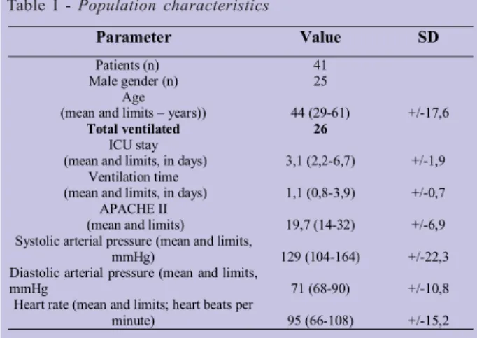
• Cardiac output (CO): the average CO calculated by
Doppler (COc) was 6.88 l/min +/- 2,08, less than that obtained by thermodilution (COi), for which the aver-age was 7.46 +/- 2,35 l/min. There was a good linear correlation between COi and COc (r=0.97) (Figure 1a), with this ratio being higher in ventilated patients. The BlandAltman analysis (Figure 1b) revealed a bias of -0.6 with a 95% confidence interval of -1.8 l/min– -0.6 l/ min. For a CO lower than 6 l/min the bias was - 0.4, and the 95% confidence interval varied between -1 l/min and 0.1 l/min.
Parameter n r Bias
Precision CI 95%
COc vs COi (l/min) 41 0,970 -0.6 0.6 -1.8 – 0.6
CVPc vs CVPi (mmHg) 35 0,881 0.1 1.7 -3.3 – 3.4
PW vs PWi (mmHg) 41 0,491 0.2 4.2 -8.1 – 8.4
PASPc1 vs PASPi (mmHg) 26 -0,118 -0.1 9.8 -19.4 – 19.2 PASPc2 vs PASPi (mmHg) 26 0,093 -0.7 9.5 -19.4 – 18.0 Legend: r=correlation ; Bias=mean of all errors;Precision=standard deviation of errors; CI, confidence interval; CO, cardiac output measured by echocardiography (COc) and thermodilution COi); CVP, central venous pressure measured by echocardiography (CVPc) and invasively (CVPi); Pw, capillary pulmonary wedge pressure measured by echocardiography (Pwc) and invasively (Pwi); PASP, pulmonary artery measured by echocardiography (PASPc1 And PASPc2, see text for details) and invasively (PASPi)
Table II - Correlation and Bland-Altman statistics
Fig. 1A - Regression (A) and Bland-Altman (B) scatterplot
for CO, ecocardiography vs pulmonary artery catheter
Legend: COi – invasive cardiac output; COc – non invasive
CO. Units: litres per minute
y = 0,859x + 0.470 r=0.970 0 2 4 6 8 10 12 14 0 5 10 15 C O i, l/m in A) • • • •
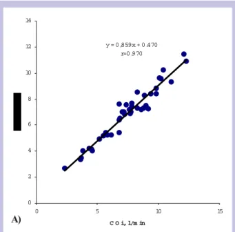
• Central venous pressure (CVP): The correlation
co-efficient was r=0.88 (p<0.001, Figure 2a). The corre-lation between invasive CVP (with an average value of 10.8 mmHg) and that calculated by echocardiography (with an average value of 11.2 PAULO MARCELINO et al
mmHg) was better in the ventilated patient (r = 0.9). A significant correlation of CVP with the tricuspid E-wave deceleration time (r = 0.51) was also found. This correlation has a higher significance for el-evated CVP values - higher than 10 mmHg (r=0.598). The authors also found a good correlation between the value of the tricuspid E/A ratio and tricuspid E-wave deceleration time (the higher the E/A value, the longer the E wave deceleration time). The Bland-Altman analysis (figure 2b) showed a bias of 0.1 and a 95% confidence interval varying between -3.3 mmHg and 3.4 mmHg.
• • • •
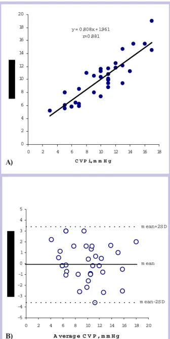
• Pulmonary Capillary Wedge Pressure: the authors
found a correlation coefficient of r=0.49 (Figure 3a) when comparing the data between the invasive val-ues (average value, 9.6 mmHg) and those obtained by the Gonzallez-Vilchez formula (average of 10.6 mmHg). In the analysis of the intervals considered (higher or lower than 12 mmHg) non significant con-cordance of values was found. Bivariate analysis showed a statistically significant correlation (p = 0.002) between the deceleration time of the mitral E-wave and pulmonary capillary wedge pressure for values less than 12mmHg (the shorter the decelera-tion time the greater the wedge pressure), and be-tween the duration of the A wave and pulmonary
Fig. 1B - Regression (A) and Bland-Altman (B) scatterplot for
CO, ecocardiography vs pulmonary artery catheter
Legend: COi – invasive cardiac output; COc – non invasive
CO. Units: litres per minute
-3 -2,5 -2 -1,5 -1 -0,5 0 0, 5 1 1, 5 2 2, 5 3 0 5 1 0 15 Average CO, l/min mean+2S D mean-2SD mea n B)
Fig. 2 - Regression (A) and Bland-Altman (B) scatterplot for
CVP, ecocardiography vs. pulmonary artery catheter Legend: CVPi - invasive central venous pressure; CVPc – non-invasive central venous pressure
y = 0.808x + 1.961 r=0.881 0 2 4 6 8 10 12 14 16 18 20 0 2 4 6 8 10 12 14 16 18 C V P i, m m H g -5 -4 -3 -2 -1 0 1 2 3 4 5 0 2 4 6 8 10 12 14 16 18 20 A verag e C V P , m m H g m ean+2S D m ean-2S D m ean B) A)
capillary wedge pressure for values higher than 12 mmHg (p = 0.007). The Bland-Altman analysis (fig-ure 3b) showed a bias of 0.2. The 95% limits of agree-ment were between -8.1 mmHg and 8.4 mmHg.
• • • •
• Pulmonary artery systolic pressure (PASP): two
equa-tions were used in the study of this parameter conducted on 26 patients (average value of the invasive measure-ment 39 mmHg). The first (average value of 32 mmHg), showed a deficient correlation between the calculated
and the invasive pulmonary artery systolic pressure, with a correlation coefficient of r=0.118 (figure 4a). No significant difference between ventilated and non-ven-tilated patients was found. The concordance between the values of invasive pulmonary artery systolic pres-sure and that calculated by echocardiography in the intervals studied (< 30 mmHg and >30 mmHg) was r=0.53, clinically not significant. The Bland Altman analysis (figure 4b) revealed a 95% confidence interval between -19.4 mmHg and 19.2 mmHg. The second method
(aver-age value of 33.6 mmHg) showed a correlation of r=0,093, statistically not significant. By the Bland-Altman analy-sis a bias of -0.7 was obtained and the 95% limits of agreement were between -19.4 mmHg and 18 mmHg.
Fig. 3 - Regression (A) and Bland-Altman (B) scatterplot for
PW, ecocardiography vs. pulmonary artery catheter. Legend: Pwi – invasive pulmonary capillary wedge pressure; Pwc – non invasive pulmonary capillary wedge pressure
y = 0.363x + 7.637 r=0.491 0 5 10 15 20 25 0 5 10 15 20 25 P W i, m m H g -15 -13 -11 -9 -7 -5 -3 -1 1 3 5 7 9 11 13 15 0 5 10 15 20 25 A verag e P W , m m H g m ean+2S D m ean-2S D m ean
Fig. 4 - Regression (A) and Bland-Altman (B) scatterplot for
PASP1, ecocardiography vs. pulmonary artery catheter.
Legend: PASPi – invasive pulmonary artery systolic pressure;
PASPc1: non invasive pulmonary artery systolic pressure, calculated by the Yock et al equation (see text for details)
y = -0.114x + 34.400 r=-0.118 0 5 10 15 20 25 30 35 40 45 50 0 10 20 30 40 50 P A S P i, m m H g -25 -20 -15 -10 -5 0 5 10 15 20 0 10 20 30 40 50 A verag e P A S P , m m H g m ean+2S D m ean-2S D m ean DISCUSSION
In post liver transplant patients, the authors were able to use a non-invasive TTE approach to reliably reproduce invasive pulmonary artery catheter haemodynamic data, PAULO MARCELINO et al
with respect to CO and CVP. Bias for these 2 variables is negligible, namely for CO < 6 l/min, and the confidence interval is acceptable for clinical purposes. However this was not the case for PSAP and Pw. The main issue ad-dressed on this study was the possible use of echocardiography as a non invasive Swan-Ganz catheter at bedside in the ICU. As we could only study liver trans-plant patients, mentioned hypothesis was not confirmed in this particular group.
Patients in a general ICU are heterogeneous. Liver trans-plant patients represented over 15 % of total admitted at the ICU in study period, and most of them had a normal echocardiographic examination.
Most equations for non invasive determination of de-scribed variables require a good quality imaging in order to reliably obtain Doppler derived parameters. This prob-lem was faced by the authors, and a considerable number of patients could not be studied.
CO obtained using TTE correlated well with thermodi-lution. This finding is in agreement with other descrip-tions in the literature. Unlike other authors, who used trans-oesophageal approach17,18, we relied on transthoracic
echocardiography and pulsed wave Doppler analysis of aortic flow for the determination of the CO. Darmon et al18,
using continuous wave Doppler and trans-oesophageal echocardiography, found that this method may underesti-mate the CO when compared to the thermodilution tech-nique. Our findings confirm this fact; however, this did not influence the final correlation.
Pw could not be reliably determined using TTE. Al-though the authors used a formula partially obtained in ventilated patients with no cardiac pathology, with Pw values between 5mmHg and 30 mmHg (limits of present study: 6 mmHg – 22 mmHg), they were unable to obtain a statistically significant correlation. The difficulty in ap-plying techniques for the non-invasive determination of pulmonary capillary wedge pressure by echocardiography to different groups of patients was described when at-tempts were made to apply equations obtained in patients with ischemic heart disease to patients with hypertrophic cardiomyopathy13 or atrial fibrillation11.
The relationship between mitral E-wave deceleration time and pulmonary capillary wedge pressure has been widely described in the literature8-10,19. In this study,
car-ried out in patients with good global systolic function and non dilated heart cavities, the data could only be repro-duced in patients with normal values of pulmonary capil-lary wedge pressure.
To obtain the equation for CVP, the authors studied 45 patients in the Intensive Care Unit, 32 of them connected
to a ventilator, 32 in sinus rhythm and 13 with atrial fibril-lation. Patients underwent simultaneous CVP measure-ments and transthoracic Doppler echocardiography and a bivariate correlation and analysis of variance was per-formed. Several parameters showed a good correlation with the measured CVP, namely the tricuspide E wave decelera-tion time (r=0.67), the tricuspide transvalvular pressure gradient (r = 0.40) and the absolute inspiratory variation (maximal dimension – minimal dimension in millimetres) of the inferior vena cava (r = 0.67). Standardized coefficients were applied to these parameters and the equation for the calculation of the CVP was obtained.
Regarding non-invasive assessment of CVP, the au-thors faced a number of difficulties. The first difficulty encountered was the evaluation of the inferior vena cava in patients with abdominal dressings, this being the case in most of those studied - mostly post-operative patients following liver transplantation. Second, a tricuspid regurgitant jet could not be determined in all patients. This fact was noted in normal volunteers, without pulmonary hypertension20.
Non-invasive calculation of CVP by echocardiography correlated significantly with the invasive technique. This success can be attributed to the use of an equation ob-tained in similar patients and by the same authors (opera-tors). This fact may raise the question on the operator dependence of echocardiography. The problem may be more serious if a particular formula is dependent on the determination of several parameters. We can possibly con-clude that the equation tested for non invasive assess-ment of CVP was validated in liver transplant patients, but it can not be stated that it can be used in a general ICU population.
The bivariate relationship between the echocardio-graphic parameters is similar to those described. The au-thors were not able to achieve any significant relationship between CVP and inferior vena cava indexes. This fact could be attributed to the difficulty in scanning the infe-rior vena cava, although the main reason may be related to the fact that only eight patients had a CVP higher than 10 mmHg and that the lowest CVP was 8 mmHg, which pre-vented the authors from testing this parameter in extreme situations.
In the cohort of patients studied, PASP could not be reliably determined by TTE. This may be due to the fact that no patients with pulmonary hypertension were evalu-ated, as opposed to the patients in whom the technique was described. On the other hand, it is also possible that, in the absence of significant tricuspid regurgitation, the highest value of regurgitation was not obtained. The fact
that tricuspid regurgitation was only detectable in 26 (63.4%) of the 41 patients studied could be significant. In a cohort of healthy volunteers, studied in an echocardio-graphy laboratory, tricuspid regurgitation was found in 73.5% of those studied20; the use of intra venous (IV)
contrast may significantly increase the level of detection of these cases21. But a recent report on the TTE
determi-nation on patients waiting pulmonary transplant and with pulmonary hypertension, found that the technique could not reliably evaluate PSAP22.
The main results of the present study questioned the possibility to use echocardiography as a mean to non invasively obtain cardiac filling pressures in an heteroge-neous ICU population. To apply equations described in the literature aimed to determine these parameters, the original conditions of the patients should be respected, namely the basic clinical and cardiac condition.
STUDY LIMITATIONS
The most important limitation of this study is the pa-tients’ characteristics, with all of them being post liver transplants, and most of them presenting a normal haemo-dynamic profile - only 5 patients presented a decreased CO, and 3 patients presented a shortening fraction under 30%. This fact may also influence the results for CO; many patients presented a CO > 6 l/min. The authors’ could not find a description of the echocardiographic or hemodynamic characteristics of patients in the general ICU. For this reason it is not possible to know how similar the patients studied are to those admitted to a Medical/Surgi-cal ICU.
CONCLUSIONS
The authors were able to use transthoracic echocar-diography to evaluate cardiac output in post liver trans-plant patients, with statistically significant correlation with the data obtained by invasive haemodynamic tech-niques. Transthoracic echocardiography was not ap-propriate for calculating the filling parameters studied even though the equations used were those found in the literature. It is important to respect the conditions defined when the original equations were described, particularly in the case of pulmonary capillary wedge pressure and possibly for CVP. In the case of pulmo-nary artery systolic pressure, obtaining this parameter by echocardiography in patients without pulmonary hypertension is questionable.
Although this study was performed in post liver trans-plant patients, the data could be useful in defining the role of echocardiography in the ICU.
ACKNOWLEDGEMENTS
We thank Antoine Vieillard-Baron for revising and comment the manuscript
REFERENCES
1. CURRIE PJ, SEWARD JB, K-L CHAN et al: Continuous wave Doppler determination of right ventricular pressure: a simultaneous Doppler-catheterization study. J Am Col Cardiol 1985; 6:750-756
2. YOCK PG, POPP RL: Noninvasive estimation of right ven-tricular systolic pressure by Doppler ultrasound in patients with tricuspid regurgitation. Circulation 1984; 4:657-662 3. SKJAERPE T, HATLE L: Diagnosis and assessment of tri-cuspid regurgitation with Doppler ultrasound. In: Risterborgh H, editor. Echocardiology. The Hage, Martinus Nijoff 1981: 299.
4. KIRCHER B, HIMELMAN RB, SCHILLER NB: Noninvasive estimation of right atrial pressure from the inspiratory colapse of the inferior vena cava. Am J Cardiol 1990; 66:493-496 5. MORENO F, HAGAN AD, HOLMEN JR, PRYOR TA, STRICKLAND RD, CASTLE H: Evaluation of Size and Dy-namics of Inferior Vena Cava as an Index of Right Sided Car-diac Function. Am J Cardiol 1984; 53:579-585
6. MARCELINO P, MARUM S, FERNANDES AP, RIBEIRO JP: Non invasive evaluation of Central Venous Pressure by echocardiography. Rev Port Cardiol 2002; 21:125-133 7. HARVEY FEIGENBAUM: Echocardiography. 5th ed. Mal-vern, Pennsylvania: Lea & Febiger, 1994.
8. APPLETON CP, HATLE LK, POP RL: Demonstration of restrictive ventricular physiology by Doppler echocardiography. J Am Col Cardiol 1988; 11:757-768. 9. GIANNUZZI P, IMPARATO A, TEMPORELLI PL et al: Doppler defived mitral decelaration time of early filling as a strong predictor of pulmonary capillary wedge pressure in post-infaction patients with left ventricular dysfunction. J Am Col Cardiol 1994; 23:1630-1637.
10. GONZALEZ-VILCHEZ F, ARES M, AYUELA J, ALONSO L: Combined use of pulsed and color M-mode Doppler echocardiography for the estimation of pulmonary capillary wedge pressure: an empirical approach based on an analytical relation. J Am Col Cardiol 1999; 34:515-523
11. NAGUEH SF, KOPELEN HN, QUINONES MA: Assess-ment of left ventricular filling pressures by Doppler echocardiography in the presence of atrial fibrillation. Circu-lation 1996; 94:2138-2145
12. NAGUEH SF: Noninvasive evaluation of hemodynamics by Doppler echocardiography. Curr Opin Cardiol 1999; 3:217-2 3 7
13. NISHIMURA RA, APPLETON CP, REDFIELD MM, ILSTRUP DM, HOLMES DR, TAJIK AJ: Noninvasive Dop-pler echocardiographic evaluation of left ventricular filling pressures in patients with cardimyopathies: a simultaneous Doppler echocardiographic and cardiac catheterization study. J Am Col Cardiol 1996; 28:1226-1233
14. TENENBAUM A, MOTRO M, HOD H, KAPLINSKY E, VERED Z: Shortened Doppler-derived A wave decelaration time: an important predictor of elevated left ventricular fill-ing pressure. J Am Col Cardiol 1996; 27:700-705
15. THOMAS JD, WEYMAN AE:. Echocardiographic Dop-pler evaluation of left ventricular diastolic function. Circula-tion 1991; 84:977-1002
16. BRUN P, TRIBOUILLY C, DUVAL A-M et al: Left ven-tricular flow propagation during early filling is related to wall relaxation: a color M-mode Doppler analysis. J Am Col Cardiol 1992; 20:420-432
17. GOLA A, POZZOLI M, CAPOMOLLA S et al: Comparision of Doppler echocardiography with thermodilution for assess-ing cardiac output in advanced congestive heart failure. Am J Cardiol 1996; 78:708-713
18. DARMON PL, HILLEL Z, MOGTADER A et al: Cardiac output by transesophageal echocardiographic using continu-ous-wave Doppler across the aortic valve. Anesthesiol 1994; 80:796-805
19. THOMAS JD, WEYMAN AE: Echocardiographic Dop-pler evaluation of left ventricular diastolic function: physics and physiology. C 1991; 84:977-990
20. BORGESON DD, SEWARD JB, MILLER FA et al: Fre-quency of Doppler measurable pulmonary artery pressures. J Am Soc Echocardiogr 1996; 9:832-837
21. TOKUSHIMA T, UTSUNOMIA T, YOSHIDA K et al: Estimation of the systolic pulmonary arterial pressure using contrast-enhanced continuous-wave Doppler in patients with trivial tricuspid regurgitation. Jpn Heart J 1999; 40:311-320. 22. HOMMA A, ANZUETO A, PETERS J et al: Pulmonary artery systolic pressures estimated by echocardiogram vs car-diac catheterizationin patients awaiting lung transplantation. J Heart Lung Transplant 2001; 20:833-839