Rev Bras Anestesiol CLINICAL INFORMATION 2011; 61: 5: 610-618
610 Revista Brasileira de Anestesiologia
Vol. 61, No 5, September-October, 2011 Received from Hospital das Clínicas da Faculdade de Medicina da USP – São Paulo, Brazil.
1. Medical Degree from FMUSP; Supervisor of Anestesia Obstétrica HC-FMUSP 2. Professor of FMUSP; Professor of the Disciplina de Anestesiologia of FMUSP 3. Anesthesiologist; Centro Obstétrico – HCFMUSP
4. Master’s and Medical Degree from Faculdade de Medicina da USP; Technical Director of Serviço de Pré-Natal da Clínica Obstétrica do HC FMUSP; responsible for the sector of Cardiopatia e Gravidez. Member of the sectors of Hipertensão e Trombose na Gravidez 5. Medical Degree from FMUSP; Professor of the Disciplina de Obstetrícia of FMUSP 6. Professor of FMUSP; Disciplina de Obstetrícia of FMUSP
Submitted on October 27, 2010. Approved on January 31, 2011.
Correspondence to: Dr. Fernando Bliacheriene
Divisão de Anestesia dos Hospital das Clínicas - FMUSP Av. Dr. Enéas de Carvalho Aguiar, 155, 8o andar, Bloco 3 Cerqueira Cesar
05403-000 – São Paulo, SP, Brazil E-mail: [email protected]
CLINICAL INFORMATION
Use of a Minimally Invasive Uncalibrated Cardiac Output
Monitor in Patients Undergoing Cesarean Section under
Spinal Anesthesia: Report of Four Cases
Fernando Bliacheriene, TSA
1, Maria José Carvalho Carmona, TSA
2, Cristina de Freitas Madeira Barretti
3,
Cristiane Maria Federicci Haddad
3, Elaine Soubhi Mouchalwat
3, Maria Rita de Figueiredo Lemos Bortolotto
4,
Rossana Pulcineli Vieira Francisco
5, Marcelo Zugaib
6Summary: Bliacheriene F, Carmona MJC, Barretti CFM, Haddad CMF, Mouchalwat ES, Bortolotto MRFL, Francisco RPV, Zugaib M– Use of a Minimally Invasive Uncalibrated Cardiac Output Monitor in Patients Undergoing Cesarean Section under Spinal Anesthesia: Report of Four Cases.
Background and Objectives: Hemodynamic changes are observed during cesarean section under spinal anesthesia. Non-invasive blood pres-sure (BP) and heart rate (HR) meapres-surements are performed to diagnose these changes, but they are delayed and inaccurate. Other monitors such as filling pressure and cardiac output (CO) catheters with external calibration are very invasive or inaccurate. The objective of the present study was to report the cardiac output measurements obtained with a minimally invasive uncalibrated monitor (LiDCO rapid) in patients under-going cesarean section under spinal anesthesia.
Case report: After approval by the Ethics Commission, four patients agreed to participate in this study. They underwent cesarean section under spinal anesthesia while at the same time being connected to the LiDCO rapid by a radial artery line. Cardiac output, HR, and BP were recorded at baseline, after spinal anesthesia, after fetal and placental extraction, and after the infusion of oxytocin and metaraminol. We observed a fall in BP with an increase of HR and CO after spinal anesthesia and oxytocin infusion; and an increase in BP with a fall in HR and CO after bolus of the vasopressor.
Conclusions: Although this monitor had not been calibrated, it showed a tendency for consistent hemodynamic data in obstetric patients and it may be used as a therapeutic guide or experimental tool.
Keywords: Anesthesia, Spinal; Monitoring, Intraoperative; Hemodynamics; Cardiac Output; Hypotension; Cesarean Section.
©2011 Elsevier Editora Ltda. All rights reserved.
INTRODUCTION
Spinal anesthesia has been consolidating as the technique of choice for cesarean section worldwide 1 due to the
quali-ty of the blockade and safequali-ty of the technique, using small anesthetic mass and not requiring ventilatory assistance and tracheal intubation.
However, it produces a non-selective blockade: the synap-ses in sensorial, motor, and autonomic fibers are temporarily interrupted. The autonomous blockade is regional, but inten-se enough to lead to inten-severe hemodynamic changes, such as hypotension. The sympathetic blockade is not the only cause of hypotension; aortocaval compression by the fetus 2 and
in-fusion of oxytocin 3 also contribute.
The diagnosis of hemodynamic compromise during cesa-rean section under spinal anesthesia is usually non-invasive, such as heart rate and non-invasive blood pressure monitors, used as substitute markers of maternal cardiac output 4.
However, the delay and the lack of accuracy of these para-meters in reflecting the fall in uterine-placental blood flow are significant 5. Cardiac filling pressure catheters, besides being
very invasive, also may be unreliable. Cardiac output (CO) is considered one of the most accurate parameters to detect the-se changes 6 and has better correlation with uterine-placental
blood flow 7. With the advent of minimally invasive equipment
USE OF A MINIMALLY INVASIVE UNCALIBRATED CARDIAC OUTPUT MONITOR IN PATIENTS UNDERGOING CESAREAN SECTION UNDER SPINAL ANESTHESIA: REPORT OF FOUR CASES
Revista Brasileira de Anestesiologia 611
Vol. 61, No 5, September-October, 2011
behavior of CO measured by a minimally invasive uncalibra-ted monitor (LiDCO rapid) in patients undergoing cesarean section under spinal anesthesia.
METHODS
After approval by the Ethics Committee of the institution, pa-tients referred to the pre-delivery unit of the Obstetric Center were questioned whether they wanted to participate in the study. Detailed information on the protocol and procedures emphasizing the voluntariness of participation without any repercussions on treatment in case they did not wish to parti-cipate was provided. Four patients agreed to partiparti-cipate and they signed an informed consent. Exclusion criteria, besides refusal to give consent, were not established.
The patient was transferred to the operating room where venous cannulation was performed with an 18G catheter. This was followed by puncture of the radial artery with a 22G venous catheter for invasive blood pressure monitoring, which was connected to a minimally invasive uncalibrated cardiac output (CO) monitor (LiDCO rapid – Pulse CO system, LiDCO Ltda, Cambridge, GB). This equipment uses the algorithm of pulse power analysis with correction for anthropometric cha-racteristics for individual arterial complacency, and external calibration was not performed. Thus, it converts changes in blood pressure over time on an estimate of the systolic volu-me and CO (nominal value), which can be used in the analy-sis of tendencies 8.
The first measures of CO, BP, and heart rate (HR) were recorded with the patient in partial left lateral decubitus over a 15 degree wedge under the right gluteal region, considered the baseline measurements. The patient was, then, anestheti-zed with the standardianestheti-zed spinal anesthesia for cesarean sec-tion of the institusec-tion consisting of 15 mg of 0.5% hyperbaric bupivacaine associated with 80 µg of 0.02% morphine through a 27G Whitacre needle. The patient was once more placed on a 15 degree wedge on the right gluteal region resulting in uterine dislocation to the left 9 until after the surgical fields
were placed, at which time the wedge was removed. Crystal-loids were not administered before the spinal anesthesia 10, but
immediately after it, resulting in co-hydration with 10 mL.kg-1
of Ringer’s lactate 11. Blood pressure was constantly
monito-red and any fall was immediately corrected to guarantee fetal well-being 12 with a bolus of 200 g of metaraminol IV 13, a
predominantly alpha-agonist vasopressor 14,15, and the
mea-surements were recorded at this moment as “araminol”. When incision of the skin was performed, hemodynamic parameters were recorded as “anesthesia”, and we expected to be re-cording the peak of the sympathetic blockade. As soon as the fetus was extracted, followed by delivery of the afterbir-th, new measurements were recorded as “birth”. According to the protocol of the institution, 3 IU IV 16 of oxytocin over at
least 60 seconds were infused, followed by recording of the measurements as “oxytocin”. The infusion was repeated ac-cording to the evaluation of the uterine tonus by the obstetri-cian, up to a limit of 9 IU 17. No patient required more oxytocin
or other classes of uterotonics, such as ergot derivatives or prostaglandins. The “final” measurement was recorded when closing of the aponeurosis. The arterial catheter was removed and pressure was applied to the puncture site for five minutes, and the patient was followed-up for eight hours for detection of any vascular complications. Data was stored and tabulated on the memory card of the LiDCO rapid monitor and treated by its specific software, the LiDCO view.
CASE 1
This is a 39-year old, 86 kg, 165 cm, gesta 6 (2 cesarean sections and 3 abortions) with a diagnosis of a 34-week twin pregnancy, and acute fetal distress, which were the indica-tions for cesarean section. Her physical status was classified as ASA II, since she had gestational diabetes on insulin, and hypertension controlled with alpha-methyldopa (1.5 g.day-1).
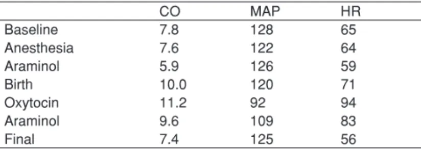
The case developed according to that described in “methods”, and the measurements are shown in Table I.
Table I – Case 1 Measurements
CO MAP HR
Baseline 7.8 128 65
Anesthesia 7.6 122 64
Araminol 5.9 126 59
Birth 10.0 120 71
Oxytocin 11.2 92 94
Araminol 9.6 109 83
Final 7.4 125 56
CO: Cardiac Output in L.min-1; MAP: Mean Arterial Pressure in mmHg; HR:
Heart Rate in beats.min-1.
CASE 2
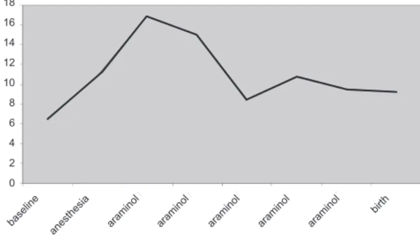
This is a 27-year old female, 89 kg, 162 cm, gesta 1, with diagnosis of a single fetus pregnancy on pelvic presentation, in labor, with indication of cesarean section. She was clas-sified as physical status (ASA) II, since she had gestational Figure 1 – CO in L.min-1 in Case 1.
anesthesia
baseline araminol araminol araminol araminol araminol
18
16
14
12
10
8
6
4
2
BLIACHERIENE, CARMONA, BARRETTI ET AL.
612 Revista Brasileira de Anestesiologia
Vol. 61, No 5, September-October, 2011 diabetes controlled with diet. The case developed according
to that described in “methods”, and her measurements are shown in Table II.
Table II – Case 2 Measurements
CO MAP HR
Baseline 6.5 97 67
Anesthesia 10.8 106 91
Araminol 16.9 79 111
Araminol 15.0 76 107
Araminol 8.5 78 97
Araminol 10.8 70 94
Araminol 9.5 77 88
Birth 9.2 100 87
Oxytocin 9.3 95 81
Final 6.5 90 82
CO: Cardiac Output in L.min-1; MAP: Mean Arterial Pressure in mmHg; HR:
Heart Rate in beats.min-1.
CASE 4
This is a 32-year old female, 63 kg, 149 cm, physical status ASA I, gesta 2, with a history of cesarean section, with a diag-nosis of twin pregnancy in which one fetus was on pelvic pre-sentation, with indication for cesarean section. Measurements are shown in Table IV.
Table IV – Case 4 Measurements
HR MAP HR
Baseline 6.7 101 75
Anesthesia 6.4 103 88
Araminol 7.0 95 94
Araminol 5.3 93 70
Araminol 5.5 92 71
Araminol 4.1 91 65
Araminol 6.4 100 70
Birth 6.4 84 86
Oxytocin 7.8 79 100
Araminol 3.1 97 43
Final 5.7 93 64
CO: Cardiac Output in L.min-1; MAP: Mean Arterial Pressure in mmHg; HR:
Heart Rate in beats.min-1. CASE 3
This is a 31-year old female, 106 kg, 179 cm, gesta 2, with a history of cesarean section, classified as ASA II, with a diag-nosis of gestational diabetes controlled with diet, controlled systemic lupus erythematosus, fetal macrosomia, and iterati-veness, and cesarean section was indicated. Measurements obtained after the onset of the surgery are shown in Table III.
Table III – Case 3 Measurements
CO MAP HR
Baseline 8.4 114 73
Anesthesia 8.8 95 90
Araminol 7.4 92 61
Araminol 7.9 105 71
Birth 8.4 87 73
Oxytocin 8.7 70 79
Araminol 5.4 102 73
Final 7.4 104 70
CO: Cardiac Output in L.min-1; MAP: Mean Arterial Pressure in mmHg; HR:
Heart Rate in beats.min-1.
Figure 2 – CO in L.min-1 in Case 2.
Figure 3 – CO in L.min-1 in Case 3.
Figure 4 – CO in L.min-1 in Case 4.
18
16
14
12
10
8
6
4
2
0
baseline
anesthesia araminol araminol araminol araminol araminol birth
oxytocin final
18
16
14
12
10
8
6
4
2
0
baseline
anesthesia araminol araminol araminol araminol araminol birth
18
16
14
12
10
8
6
4
2
0
baseline
anesthesia araminol araminol araminol araminol araminol birth
oxytocin
USE OF A MINIMALLY INVASIVE UNCALIBRATED CARDIAC OUTPUT MONITOR IN PATIENTS UNDERGOING CESAREAN SECTION UNDER SPINAL ANESTHESIA: REPORT OF FOUR CASES
Revista Brasileira de Anestesiologia 613
Vol. 61, No 5, September-October, 2011 DISCUSSION
In general, the behavior of hemodynamic variables observed during interventions in patients undergoing cesarean sections under spinal anesthesia was:
1. After installation of the blockade, a fall in MAP with oc-casional elevation of HR and CO is observed; 2. When using the vasopressor metaraminol, a
predomi-nantly alpha-agonist drug, an increase in MAP with fall in HR and CO is observed;
3. When the fetus is extracted, the behavior is not uni-form, with occasional elevation of CO and fall in MAP; 4. After infusion of oxytocin, marked changes are obser-ved, such as fall in MAP besides an increase in HR and CO.
In case 1, after the blockade, measurements are close to baseline possibly because the sympathetic blockade did not achieve its peak or the effectiveness of conservative measu-res like dislocation of the uterus to the left and co-hydration. Unlike cases 2 and 4, who presented initially a compensatory increase of CO secondary to an increase in HR without com-promising the BP, but which later manifested with a signifi-cant fall in BP. Since the beginning, case 3 showed signifisignifi-cant hemodynamic changes with fall in MAP and increase in HR and CO. In response to the fall in MAP, boluses of the alpha-agonist vasopressor metaraminol were used, resulting in a typical response 18, with an increase in MAP and fall in HR
and CO, most likely due to an increase in afterload, except in case 2 in which only after fetal extraction a recovery in MAP was observed. Regarding delivery, in all cases, only after fetal extraction a similar response was observed with an increase in CO, which can be explained by self-transfusion resulting from uterine involution and contraction, as well as reduction in aortocaval compression, which increased preload. Oxyto-cin has vasodilator actions, with fall in MAP and increase in HR and CO, which were observed in all cases except in case 2. At the end, parameters returned to baseline levels most likely due to all interventions with less intense actions. The behavior of MAP demonstrated that its variations, most likely
due to changes in systemic vascular resistance (SVR), are secondary to vasodilator (spinal anesthesia and oxytocin) and vasoconstrictor (metaraminol) actions of interventions perfor-med during cesarean sections that affect arterial tonus and not due to bleeding. Regarding the absence of SVR measure-ments as a limiting factor of this study, we decide not to use it, since it is an invasive measurement because it requires a right atrium catheter to measure pressure, which is required to its calculation.
Therefore, physiologic situations are observed, such as aortocaval decompression after birth and self-transfusion of uterine blood, that impose a great overload on the cardiovas-cular system with an increase of venous return, which is ac-commodated through the cardiac valves and pulmonary blood vessels. However, this may be a problem in patients with low cardiovascular reserve or with cardiopathies, such as mitral and aortic stenosis, besides pulmonary hypertension, resul-ting in pulmonary edema or cardiac failure. The same can be seen in situations that are induced like spinal anesthesia and oxytocin infusion in which normal patients respond with an increase in CO and fall in SVR and venous return, but in cardiopathies, these responses should be controlled or even avoided.
This cardiac output monitor produced data compatible with those of prior publications 3,6,8,18,19, and in this scenery,
exter-nal calibration with data obtained by thermodilution or lithium dilution was dispensable, especially when analysis is made based on tendencies and nominal values and not absolute values. Measurements produced can orient regarding a pos-sible cause of hemodynamic changes and indicate the most appropriate treatment for the case, besides signaling reckless conducts, such as rapid infusion of oxytocin. They can also be useful in patients with limited cardiovascular reserve, since they cannot tolerate significant changes in SVR and they can-not compensate for changes in CO.