Universidade de Lisboa
Faculdade de Ciências
Departamento de Biologia Animal
Optimization of the differentiation process of umbilical cord
matrix mesenchymal stem cells into hepatocyte-‐like cells
Alexandra Martins de Medeiros Vieira da Silva
Dissertação
Mestrado em Biologia Evolutiva e do Desenvolvimento
2013
Universidade de Lisboa
Faculdade de Ciências
Departamento de Biologia Animal
Optimization of the differentiation process of umbilical cord
matrix mesenchymal stem cells into hepatocyte-‐like cells
Alexandra Martins de Medeiros Vieira da Silva
Dissertação
Mestrado em Biologia Evolutiva e do Desenvolvimento
Dissertação orientada por:
Doutora Joana Miranda -‐ iMed-‐FFUL
Professora Doutora Gabriela Rodrigues -‐ FCUL
2013
Acknowledgements
First of all I would like to thank the team CBT-‐iMED for receiving me and for making this thesis project possible.
To my supervisor at CBT-‐iMED, Dra. Joana Miranda, I would like to express my sincere gratitude for her patience in guiding me during all the process of research and writing. To Professora Gabriela Rodrigues, my FCUL supervisor, for being always available for receiving me and help.
I would also like to thank to my collegues at CBT-‐iMED laboratory, in no particular order: Elysse Filipe, Patrícia Guerreiro, Pedro Pinheiro, Madalena Cipriano, Sérgio Camões, João Costa, Ana Matos, Sandrina Gonçalves and Nuno Torres. Thank you for your help, with your sense of humour and positivity you always made my day better.
I also thank Prof. Nuno Oliveira, Prof. Fátima Cabral, Prof. Judite Costa, and Prof. Ana Francisca for their advice and suggestions.
To my friends: Fábio, Daniela, Filipe, Catarina, Clara, Maié, Mário, Sara Mendes, Sara Costa, Ana Quendera, Ana Marques, Gonçalo, António, Cláudia and Renato for their support.
To my parents, my sister, my family in Azores and Jorge who are always by my side.
Abstract
The few literature described protocols of hepatic differentiation of human umbilical cord matrix mesenchymal stem cells (ucmMSC) lead to populations of cells with less than satisfactory differentiation rates.
In this study a hepatic differentiation protocol was firstly optimized in conventional monolayer (2D) cultures and further implemented in a 3D culture method, in order to achieve a homogenous population of functional hepatocyte-‐like cells (HLCs), as part of the creation of an alternative model for in vitro toxicology studies.
UCX®, ucmMSC isolated by a proprietary method developed by ECBio S.A., were subjected to 4 differentiation protocols in 2D culture conditions being the most promising procedure applied to 3D culture method. The characterization of differentiated UCX® included the analysis of the expression of hepatic markers by quantitative real time reverse-‐transcription polymerase chain reaction (qRT-‐PCR) and immunofluorescence assays. Metabolic activity was assessed through ECOD and UGT activity assay, and also by glycogen accumulation and urea production. Undifferentiated UCX®, HepG2 and primary rat hepatocytes were used as controls.
The HLCs that resulted from the optimized protocol showed hepatocyte-‐like morphology, expression of hepatic lineage markers, including ALB, CK18 and HNF4α as well as the underexpression of CK19. These also exhibited biotransformation activity (ECOD and UGT activities), and the ability to store glycogen and to produce urea.
When transposed into 3D cultures, the optimized method induced the expression of hepatic markers CK18, HNF4α and ALB and higher urea production.
In summary a more homogenous and functional population of HLCs, with hepatocyte expression pattern and metabolic activity at a superior level than HepG2 and in some aspects at the same activity level of rat primary hepatocytes, was successfully obtained, opening doors to the construction of a humanly-‐close metabolism and toxicology model for drug testing.
Keywords
Differentiation, Hepatocytes, Mesenchymal stem cells (MSCs), Umbilical Cord matrix, UCX®, toxicology model.
Resumo
O fígado é o maior órgão interno do corpo humano, sendo este constituído por células parenquimatosas e não parenquimatosas. No grupo das células parenquimatosas, incluem-‐se os hepatócitos, que constituem 60% a 70% do número total de células do fígado. As células endoteliais, células de Kupffer, células de ito e células estaminais (células ovais) são as células não parenquimatosas do fígado [1-‐2].
O fígado desempenha funções muito importantes no organismo, nomeadamente, o armazenamento e processamento dos nutrientes, produção de proteínas plasmáticas e destoxificação do organismo através da alteração da estrutura de moléculas ou através da excreção pela bílis [2]. O ciclo da ureia é o principal processo de destoxificação do sangue, responsável pela conversão de amónia em ureia, sendo esta depois filtrada e excretada pelos rins [2].
O fígado é também o principal órgão responsável pela biotransformação. Os hepatócitos expressam enzimas de biotransformação de fase I e fase II: os citocromos P450, enzimas de fase I responsáveis pelas vias de oxidação, redução e hidrólise que adicionam ou expõem um grupo funcional, como hidroxilo, tiol, ou amina ao substrato. As reações de fase II conduzem à formação de conjugados desses mesmos grupos funcionais com ácido glucurónico, sulfato e glutationa, que levam à eliminação directa (no caso dos dois primeiros) e destoxificação (no caso da conjugação com glutationa) das moléculas metabolizadas [3].
Sendo o fígado o principal órgão de metabolização de xenobióticos, este é mais exposto aos efeitos potencialmente tóxicos dos fármacos e seus metabolitos. Para detectar precocemente os potenciais riscos toxicológicos é essencial ter bons modelos in vitro de teste de fármacos antes de estes entrarem em ensaios clínicos [4].
Vários modelos in vitro são usados em estudos de toxicologia, por exemplo: bactérias ou vírus modificados geneticamente para expressarem isoformas de enzimas de fase I e fase II [5-‐6], fígados isolados [1, 7], culturas primárias de hepatócitos [1, 8] e linhas celulares [9-‐10].
Modelos baseados na perfusão do fígado, tanto in vivo com ex vivo, são muito utilizados para testar o metabolismo hepático e farmacocinética. Estes modelos têm a capacidade de manter a estrutura hepática intacta, todavia a complexidade do mesmo dificulta a compreensão dos processos que ocorrem a nível intracelular [1, 7].
Os modelos celulares mais utilizados baseiam-‐se em linhas celulares de carcinoma hepatocelular, como HepG2 e HepRG [9-‐10]. HepG2 é uma linha celular humana acessível, fácil de manter em cultura. Todavia, não apresenta algumas funções características dos hepatócitos, como por exemplo, apresenta um perfil de expressão de enzimas de biotransformação diferente [10]. Recentemente, foi isolada e caracterizada a linha celular-‐HepRG. Estas células encontram-‐se num estado bipotente semelhante ao de hepatoblasto e a sua maturação pode ser induzida através da adição de 2% dimetil sulfóxido (DMSO) e 50 μM hidrocortisona. Quando maturadas, estas células expressam 85% dos genes expressos em hepatócitos humanos. Contudo, muitos marcadores importantes não são expressos, como é o caso dos factores de transcrição C/EBPα, C/EBPβ e enzimas de fase II [9].
Os hepatócitos primários, são um modelo de excelência para a compreensão de processos metabólicos e efeitos hepatotóxicos de fármacos [1, 11], dado que durante um período de tempo após o seu isolamento, preservam muitas das funções hepáticas, como a capacidade de biotransformação, metabolismo de glucose e destoxificação da amónia. Apesar disso, a dificuldade em obter material biológico traduz-‐se na difícil implementação deste sistema de forma contínua e prolongada [1].
O uso de hepatócitos primários de rato é mais acessível, porém existem diferenças interespécies que não permitem a total correlação entre este modelo e o fígado
humano in vivo. Além de que, a perda de funcionalidade persiste, problema inerente a todos os hepatócitos primários [12-‐13].
Tendo em consideração os problemas éticos e diferenças interespecies mencionados, um novo método foi proposto como alternativa para substituir as culturas de hepatócitos primários nos ensaios de toxicologia, que se baseia na utilização de hepatócitos obtidos por diferenciação de células estaminais humanas [1, 4, 14].
Os protocolos de diferenciação em hepatócitos são alicerçados nos 4 passos do desenvolvimento do fígado: indução da formação da endoderme, indução da competência hepática, formação de hepatoblastos e finalmente maturação em hepatócitos [15].
Como ponto de partida para esta diferenciação já foram testados diferentes tipos de células: células estaminais embrionárias [16-‐19], células mesenquimais da medula óssea [20-‐21], adipócitos [22], e células estaminais pluripotentes induzidas [23-‐24]. Todavia os estudos que utilizam células estaminais mesenquimais do cordão umbilical para esta finalidade, são escassos [25]. O cordão umbilical é uma fonte muito vantajosa de células estaminais mesenquimais visto que permite um isolamento inicial rápido de um grande número de células multipotentes e a sua utilização levanta muito menos problemas éticos [26-‐27].
Os sistemas de cultura em monocamada (2D) são muito utilizados para a cultura in vitro de hepatócitos. Este tipo de cultura obriga os hepatócitos a adquirirem uma morfologia achatada que não corresponde à que possuem no ambiente in vivo [1-‐2]. Recentemente, têm-‐se vindo a abordar modelos de cultura em 3D. São vários os métodos que se podem aplicar, entre os quais se destaca o método de formação de agregados [28]. Esta técnica assenta sobre a capacidade inata que algumas células têm em se agregar, permitindo a formação de matriz extracelular natural, de extrema importância na manutenção das funções dos hepatócitos [1].
Neste sentido, o objectivo deste trabalho foi desenvolver um protocolo para diferenciação de células mesenquimais estaminais da matriz do cordão umbilical (UCX®) em hepatócitos maduros com a finalidade de se criarem modelos de toxicologia.
Com o intuito de se obter uma população homogénea e funcional de hepatócitos, algo não alcançado em estudos anteriores, novos protocolos foram testados e implementados em modelos 3D que permitem reproduzir melhor o ambiente in vivo, logo, tendo um grande potencial para aumentar o sucesso da diferenciação em hepatócitos.
Nas UCX® diferenciadas foi analisada a expressão de marcadores hepáticos CK18, ALB e HNF4α por qRT-‐PCR e imunofluorescência. A atividade de biotransformação foi determinada através dos ensaios de atividade ECOD e UGT. Também foi avaliada a capacidade de acumulação do glicogénio através do método PAS e a capacidade de destoxificação da amónia, foi determinada a partir da quantificação da ureia produzida. HepG2 e hepatócitos primários de rato foram usados como controlo.
Primeiramente, foi implementado o protocolo descrito por Campard et al. (2008), uma vez que este foi aplicado a MSCs da matriz do cordão, para o nosso conhecimento o único. Utilizando este protocolo como base, modificações foram feitas nos 3 passos que o constituem. O protocolo optimizado permitiu a obtenção de hepatócitos com expressão dos marcadores hepáticos CK18, HNF4α e ALB com níveis de função superiores a HepG2 e em alguns ensaios ao mesmo nível que os hepatócitos primários de rato.
O protocolo optimizado em 2D foi testado em 3D, na medida em que este método permite que as células adquiram uma estrutura mais semelhante ao que acontece in vivo e os hepatócitos diferenciados foram caracterizados no que respeita a morfologia, através de coloração com hematoxilina eosina, detecção de marcadores hepáticos CK18, HNF4α e ALB através de imunofluorescência, acumulação de glicogénio através de coloração PAS e produção de ureia. Os resultados obtidos demonstram uma clara melhoria em termos de presença dos marcadores hepáticos (CK18, HNF4α e ALB) relativamente às células diferenciadas em 2D com o mesmo protocolo. Em relação à
atividade metabólica, as células diferenciadas em cultura 3D, apresentam maior competência em termos de metabolismo de glucose (PAS) e destoxificação de amónia, o que confirma o potencial das culturas em 3D no que respeita à diferenciação e obtenção de hepatócitos funcionais.
Palavras-‐chave
Diferenciação, Hepatócitos, Células mesenquimais estaminais, Matriz do cordão umbilical, UCX®, modelos toxicológicos.
Index
Acknowledgments i Abstract ii Keywords iii Resumo iii Palavras-‐chave viiFigure index ix
Glossary x
1. Introduction 1.1. Liver 1.2. In vitro toxicology models
1.2.1. 3D models 1.3. Alternative models 1.4. Objectives
2. Materials and methods 2.1. Cell culture reagents
2.2. Collagen extraction and plate coating 2.3. Cell culture
2.4. BCA protein quantification
2.5. Hepatocyte differentiation protocols
2.6. Quantitative real time polymerase chain reaction (qRT-‐PCR) 2.7. Immunofluorescence
2.8. Periodic acid Schiff’s staining (PAS) 2.9. Hematoxylin-‐eosin staining (H&E) 2.10. Urea production
2.11. 7-‐ethoxycoumarin-‐O-‐deethylase (ECOD) activity
2.12. Uridine 5’-‐diphosphate glucuronosyltransferase (UGT) activity 2.13. Statistical analysis
3. Results
3.1. UCX® differentiation into hepatocyte-‐like cells under 2D conditions 3.1.1. Optimization of coating, seeding density and FBS concentration 3.1.2. Induction of hepatic competence
3.1.3. Characterization of differentiation Pattern of UCX® into hepatocyte-‐like cells:
Reference Protocol, Protocol A, Protocol B and Protocol C
3.2. UCX® differentiation into Hepatocyte-‐like cells under 3D conditions
3.2.1. Characterization of the Differentiation Pattern of UCX® into hepatocyte-‐like Cells (HLCs): Reference Protocol versus Protocol C
4. Discussion 5. Bibliography Annex 1 Annex 2 i ii iii iii vii ix x 1 1 2 4 6 13 14 14 14 15 15 16 17 18 19 19 20 20 21 21 22 22 23 23 24 25 35 40 45
Figure Index
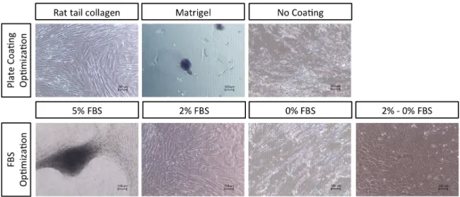
Figure 1 Figure 2 Figure 3 Figure 4 Figure 5 Figure 6 Figure 7 Figure 8 Figure 9 Figure 10 Figure 11 Figure 12 Figure 13 Figure 14 Figure 15 Figure 16 Figure 17Morphology of differentiated UCX® resultant from Reference Protocol with different plate coatings and FBS concentrations.
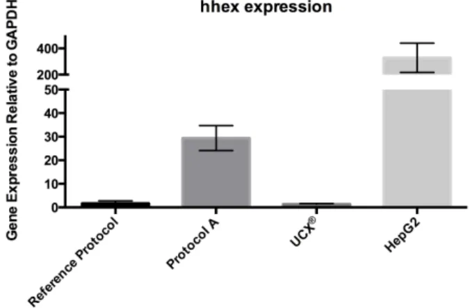
Relative hhex gene expression of differentiated UCX® from Reference Protocol and Protocol A, at day 2, of undifferentiated UCX® and HepG2, determined by qRT-‐PCR.
Morphology of differentiated UCX® at days 15, 21 and 24 and of undifferentiated UCX®, HepG2, and ratPHep in 2D culture method.
Relative ck19 gene expression of differentiated UCX® resultant from Reference Protocol, Protocol B and C at day 24 and of undifferentiated UCX® and HepG2, determined by qRT-‐PCR.
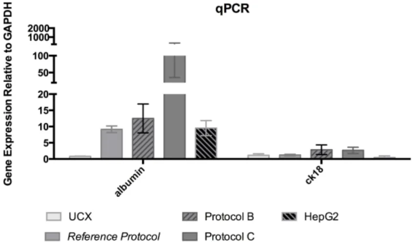
Relative alb and ck18 gene expression of differentiated UCX® resultant from Reference Protocol, Protocol B and C at day 24 and of undifferentiated UCX® and HepG2, determined by qRT-‐PCR.
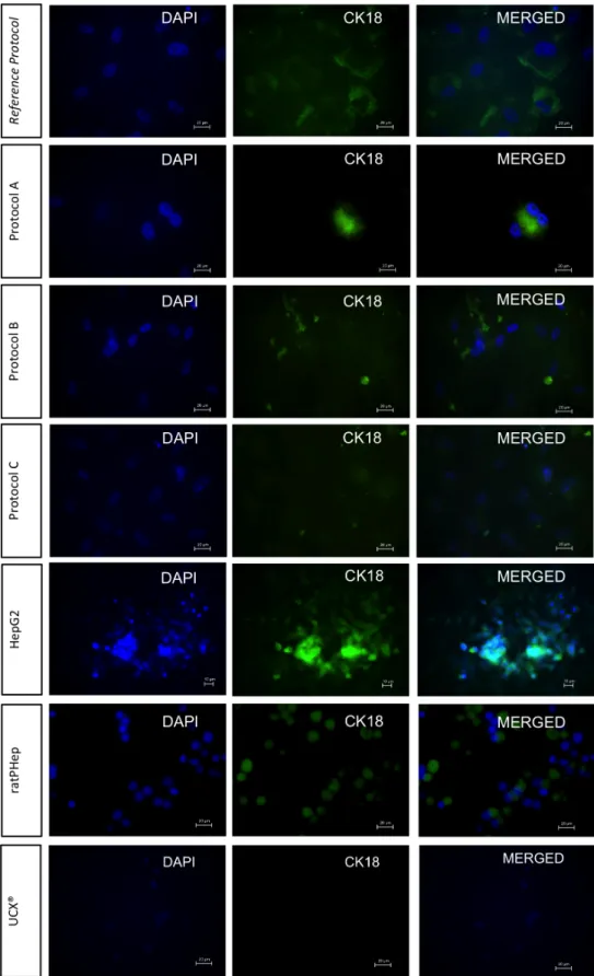
Presence of CK18 in the differentiated UCX® at day 24, in undifferentiated UCX®, ratPHep and HepG2 in 2D culture method, detected by immunofluorescence staining.
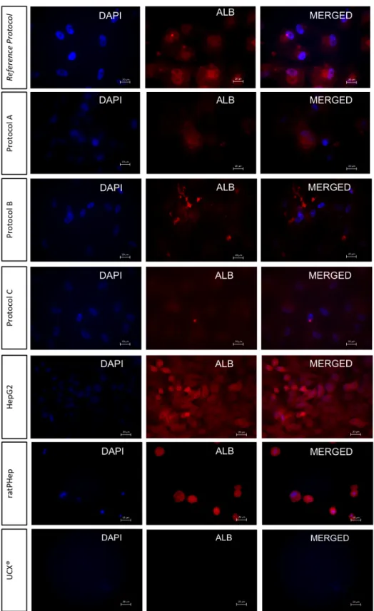
Presence of ALB in differentiated UCX® at day 24, in undifferentiated UCX®, ratPHep and HepG2 in 2D culture method, detected by immunofluorescence staining.
Presence of HNF4α in the differentiated UCX® at day 24, in undifferentiated UCX®, ratPHep and HepG2 in 2D culture method, detected by immunofluorescence staining.
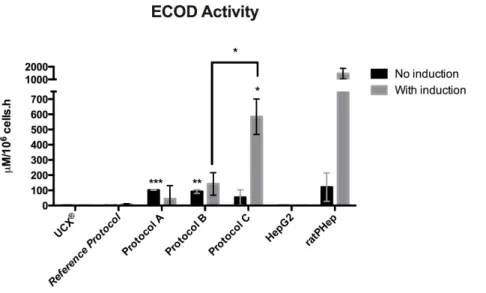
ECOD activity of differentiated UCX® of differentiated UCX® at day 24 and of undifferentiated UCX®, HepG2, and ratPHep in 2D culture method.
UGT activity of differentiated UCX® at day 24 and of undifferentiated UCX®, HepG2 and ratPHep in 2D culture method.
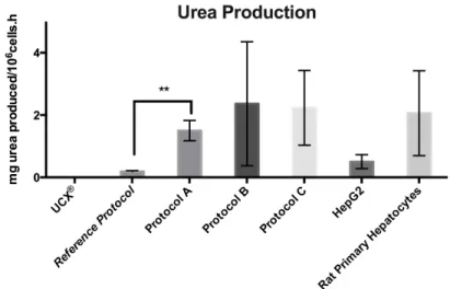
Urea production of differentiated UCX® at day 24 and of undifferentiated UCX®, and in HepG2 and ratPHep in 2D culture method.
Glycogen accumulation in differentiated UCX® at day 24, undifferentiated UCX®, HepG2 and ratPHep in 2D culture method, revealed by PAS staining.
Undifferentiated UCX® spheroids at day 2 and differentiated UCX® spheroids of Reference
Protocol and Protocol C at day 24.
Hematoxylin-‐eosin staining of differentiated UCX® spheroids resultant from Reference Protocol
Presence of HNF4α, ALB and CK18 in the differentiated UCX® at day 24 and in undifferentiated UCX® in 3D culture method, detected by immunofluorescence staining.
Glycogen accumulation in differentiated UCX® resultant from Reference Protocol and Protocol C and undifferentiated UCX® in 3D culture method, determined by PAS staining
Urea production of differentiated UCX® resultant from Reference Protocol and Protocol C in 2D and 3D cell culture method at day 24.
24 25 26 27 28 29 30 31 32 33 33 34 36 37 37 38 39
Glossary
3-‐MC 4-‐MU AFP ALB ASC BCA bmMSC BMP BSA C/EBPα C/EBPβ cDNA CK18 CK19 CYP DAPI DMSO ECM ECOD EDTA EGF ESC FBS FGF Foxa1-‐3 GAPDH GATA 4-‐6 Gp130 HGF HHEX HLA HLC HNF4α HNF1α H&E IFN-‐γ IMDM iPSC ISCT ITS JAK/Stat3 KLF4 MAPK MSC OCT 3 OCT 4 OSM 3-‐Methylcholanthrene 4-‐ Methylumbeliferone α-‐ fetoprotein Albumin Adult stem cells Bicinchoninic acid
Bone marrow mesenchymal stem cells Bone morphogenic protein
Bovine serum albumin
CCAAT-‐enhancer-‐binding proteins α CCAAT-‐enhancer-‐binding proteins β Complementary deoxyribonucleic acid Cytokeratin 18 Cytokeratin 19 Cytochrome 4',6-‐diamidino-‐2-‐phenylindole Dimethyl sulfoxide Extracellular matrix 7-‐ethoxycoumarin-‐O-‐deethylase Ethylenediaminetetraacetic acid Epidermal growth factor Embryonic stem cells Fetal bovine serum Fibroblast growth factor Forkhead box a1 to 3
Glyceraldehyde 3-‐phosphate dehydrogenase GATA binding factor 4 to 6
Glycoprotein 130
Hepatocyte growth Factor
Hematopoietically-‐expressed homeobox protein Human leukocyte antigen
Hepatocyte-‐like cells
Hepatocyte nuclear factor 4α Hepatocyte nuclear factor 1α Hematoxylin-‐Eosin
Interferon γ
Iscove modified dulbeco medium Induced pluripotent stem cells
International Society for Celular Therapy Insulin–transferrin–sodium selenite
Janus Kinase/Signal Transducer and Activator of Transcription 3 Kruppel-‐like factor 4
Mitogen-‐activated protein kinases Mesenchymal stem cells
Octomer-‐binding transcription factor 3 Octomer-‐binding transcription factor 4 Oncostatin M
PCR P/S/A PAS staining PBS Pdx 1 PFA Prox1 qRT-‐PCR ratPHep RGD RNA RT SD Sox17 STM TGF-‐β Tm ucmMSC UGT α-‐MEM
Polymerase chain reaction
Peninsilin/streptomicin/ amphotericin B Periodic acid Schiffs staining
Phosphate Buffered Saline
Pancreatic and duodenal homeobox 1 Paraformaldehyde
Prospero homeobox protein 1
Quantitative real-‐time polymerase chain reaction Rat Primary Hepatocyte
Arginin-‐glycin-‐aspartic acid Ribonucleic acid
Room temperature Standard deviation Sry-‐related HMG box 17
Septum transverse mesenchyme Transforming growth factor β Melting temperature
Umbilical cord matrix mesenchymal stem cells Uridine 5’-‐diphosphate glucuronosyltransferase Minimum essential medium Eagle alfa modification
1. Introduction
1.1. Liver
The liver is the largest intern organ of the human body. It is populated by parenchymal and non-‐parenchymal cells. The hepatocytes, which are parenchymal cells, represent 60-‐70% of the total liver cells; whereas, the non-‐parenchymal cells include, hepatic endothelial cells, Kupfer cells, hepatic stellate cells and liver stem cells (oval cells) [1-‐2]. Concerning its organization, liver cells are encapsulated in a layer of connective tissue (Glisson capsule), being the organ divided in hepatic lobules which receive blood from the portal triad, the junction of three ducts that run in parallel: the intra hepatic biliary duct, portal vein and hepatic artery. The portal triad is surrounded by hepatocytes arranged in single cell sheets known as hepatic plates being the spaces between sheets of hepatocytes called sinusoid spaces, which are connected to a network of blood capillaries. Hepatic sinusoids lack a diaphragm and a basement membrane, which makes them much more permeable than other capillaries, even permitting the passage of plasma proteins with protein-‐bound nonpolar molecules, such as fat and cholesterol. This, combined with the plate structure of the liver, allow intimate contact between the hepatocytes and the contents of the blood [2, 15].
The products of digestion that are absorbed into blood capillaries in the intestine do not directly enter the general circulation, instead, this blood is delivered first to the liver, through the hepatic portal vein. The hepatocytes receive these digestive products in the form of glucose, amino acids, fatty acids and glycerol, and proceed to their metabolism. In glucose metabolism, part of the metabolic end products are stored in the liver and utilized when required. For instance, the liver in response to pancreas signalling plays an important role in the regulation of the blood glucose concentration by either removing glucose from the blood, by converting glucose into glycogen and tryglicerides, or during fasting, by secreting glucose derived from the breakdown of stored glycogen in a process called glycogenolysis. It can also produce glucose by the conversion of noncarbohydrate molecules, such as amino acids, in a process called gluconeogenesis
Another important liver function is the production of plasma proteins such as albumin and globulins. Albumin (ALB) is the main protein in the blood and is implied in regulation of oncotic pressure of the blood and in transport of hydrophobic molecules. On the other hand, globulins play a role in transport, inhibition of trypsin and blood clotting [2].
Hepatocytes are responsible for blood detoxification, by alteration of molecular structure through specific enzymes or by direct excretion in the bile. The urea cycle is the main process of blood detoxification, responsible for the conversion of ammonia, into urea, which is then excreted by the kidneys [2].
Hepatocytes can remove hormones and other biologically active molecules from the blood by excretion of these compounds into the bile. Bile is produced by hepatocytes and secreted into thin channels called bile canaliculi, located within each hepatic plate. These bile canaliculi drain into hepatic ducts that transport bile away from the liver. The liver can thus clear the blood of particular compounds by excreting them into the intestine in the form of bile, which are then eliminated in the faeces. The major constituents of bile are bile pigment (bilirubin), bile acids, phospholipids (mainly lecithin), cholesterol and inorganic ions [2].
1.2. In vitro toxicology models
Liver is the main organ for biotransformation. Hepatocytes possess phase I and II enzymes, responsible for the elimination and detoxification of xenobiotics. The cytochrome P450 enzymes are responsible for most phase I reactions, that are characterized by oxidative, reductive, and hydrolytic pathways where a functional group, e.g. hidroxyde, thiol and amine is added or exposed in the substrate. In phase II reactions, the newly introduced functional group is modified to O-‐ and N-‐glucuronides, sulphate esters, various amides, and glutathionyl adducts, increasing polarity relative to the unconjugated molecules. This two-‐step transformation makes the substrates more water soluble, and therefore more easily excreted in urine [3].
Since liver is the main organ of biotransformation reactions, it is frequently affected by the drug side effects, which is the main cause of the withdrawal of an approved drug from the market. Thus, in order to avoid unexpected side effects of the novel drug, the availability of a good model for drug testing is essential [4].
In vitro models are commonly used for toxicology studies, these are: genetically modified bacteria or virus expressing various isoforms of cytochromes P450 [5-‐6], isolated perfused liver [1, 7], primary cultures of hepatocytes [1] and hepatic cell lines [9-‐10] .
The isolated perfused liver models are possible models to test hepatic metabolism, trans-‐hepatocellular transport and pharmacokinetics of drugs. These models have the advantage of maintaining structure and functional organization, mimicking the in vivo environment, however the complexity of the model difficult the understanding of intercellular processes, and also it functional integrity is lost after few hours [1, 7]. Thus, the most commonly used in vitro models are human hepatocellular carcinoma cell lines, such as HepG2 [10] and, more recently, HepRG [9]. HepG2 cell line is readily available, easy to handle and provide a reproducible human system [10]. However, this is not the ideal model since these cells are not in a completely mature state and thus drug metabolizing enzymes expression is different from normal functional hepatocytes. HepRG is a human hepatoma cell line developed recently. These cells are able to differentiate from bipotent progenitor into mature hepatocyte-‐like cells (HLC) with the addition of 2% dimethyl sulfoxide (DMSO) and 50μM hydrocortisone hemisuccinate, resembling, more closely than HepG2, the adult hepatocyte cells. Mature HepRG express 85% of the genes expressed in primary human hepatocytes and levels of some CYP450 such as CYP3A4 and CYP2B6 and phase II enzymes are increased. Still, differentiated HepRG cells lack many important markers, for example C/EBPα, C/EBPβ and lack phase II enzyme UDP-‐glucoronosiltransferase (UGT) and important efflux pumps (bile salt export pump) [9].
Primary hepatocytes are a good model to enlighten the metabolic process of chemicals and effects of toxic agents in the liver. Freshly isolated hepatocytes preserve many
hepatic functions such as phase I and phase II enzyme activities, glucose metabolism and ammonia detoxification. Still, a continuous source of hepatocytes is needed to proceed to toxicology assays, which restricts the use of human primary hepatocytes for this type of studies, due to the shortage of available human liver material [1].
On the other hand, primary hepatocytes isolated from rat liver are more easily available and the results obtained are consistent, which make it good models for drug toxicology studies. Still, it has disadvantages: hepatic functions have interspecies differences, which cause differences in the hepatotoxicity reaction to drugs [4]. Besides, a problem which involve all primary cultures is the loss of specific-‐hepatic function after 48/72 hours, mainly of the biotransformation capacities [13] and plasma protein production [12]. Therefore, this loss of activity can be explained, among other things, by the culture method in which cells are cultured after isolation from the liver.
1.2.1. 3D models
Traditionally, monolayer culture methods are applied to primary hepatocytes and cell lines, which implies the seeding of the cells in a flat surface forcing hepatocytes to acquire a flattened unnatural morphology [28]. However, in vivo cells possess three dimensional organization with complex cell-‐to-‐cell and cell-‐extracellular matrix (ECM) interactions, which hardly occur in a two dimensional culture support. This structure is of great importance to hepatocytes since they possess polar structure in the liver [2]. The 3D in vivo environment is mimicked through the use of 3D culture models, which utilization has grown in the past few years [29-‐30]. The 3D state can be achieved through several techniques, such as using scaffolds [30-‐31] and cell spheroids [11, 22, 28], among others [28].
3D scaffolds, which can be fabricated using synthetic or natural derivatives, allow the formation of a 3D environment without compromising gas, nutrient and metabolite exchange. These matrices can be adapted to different cell types by choosing the right rigidity and chemical structure. The chemical properties of the scaffold regulate the adhesion and spreading of cells. This is controlled by charge and polarity of the surface,
which dictate the diffusion and adsorption of proteins from the medium [32]. Cell adhesion can also be regulated, by integrating in the scaffold some structural components, e.g., the incorporation of integrin ligand RGD permits the attachment of fibroblast cells [28].
On the other hand, the cell-‐spheroid technique is a simple 3D culture method that uses the capacity of some cells to self-‐assemble. It has the advantage of not requiring a scaffold and permitting an intricate cell-‐to-‐cell contact allowing the formation of ECM. The ECM is of great importance in maintaining hepatocyte functions during a longer time in culture. It allows the anchorage of hepatocytes and also induces intracellular signaling pathways [1]. However this method has some disadvantages: when spheroid thickness surpasses 1 mm, cells in the interior may lose contact with nutrients in the medium and accumulate toxic waste metabolites inside, forming a necrotic center [28]. 3D cultures can be maintained in a static environment, where cells in spheroids or scaffolds are maintained in medium without agitation. The use of dynamic 3D cultures in bioreactor is also an alternative. Bioreactors allow the control of many environmental parameters such as: temperature, pH, medium flow rate, nutrient supply and toxic metabolite removal [28]. Many type of bioreactors are described and can be grouped in: rotating wall vessels, direct perfusion systems, hollow fibers, spinner flasks, and mechanical force systems [28]. Spinner flasks are a simple and effective method for culture of primary hepatocytes being able to maintain cellular functions, namely albumin secretion and biotransformation activity of phase I and phase II enzymes [11]. In this method cells can be seeded in scaffolds or allowed to form spheroids which dimensions can be controlled by the agitation rate. The agitation provided by the impeller result in higher homogeneity of the medium and quick dilution of toxic metabolites, while allowing a greater distribution of nutrients and oxygen and therefore reducing cell death [11].
1.3. Alternative models
A new possibility to replace primary cultures of hepatocytes in toxicology, consisting in hepatocyte-‐like cells (HLCs) derived from stem cells, recently appeared [4]. A stem cell is characterized by the capability to self-‐renew, dividing into two daughter cells, one of which retains its pluripotent abilities and allows the maintenance of the stem cells population; capacity of differentiating into tissues derived from all three germ layers, ectoderm, mesoderm and endoderm; and ability to renew a tissue in which it is inserted [26]. However, stem cells from different sources present different characteristics and not all meet the three criteria listed above. Stem cells which can only differentiate into multiple but limited lines of cells are termed multipotent stem cells or progenitor cells. Thus, due to its self-‐renewing capabilities and a strong proliferation rate many protocols have been developed in order to differentiate hepatocytes from stem cells. In vitro differentiation procedures aim at mimicking the development of hepatocytes in vivo, which consist in: (1) Endoderm induction; (2) Foregut and hepatic competence induction; (3) Hepatoblast and liver bud formation; (4) Differentiation of hepatoblast into hepatocyte.
1. Endoderm Induction
In vivo, during gastrulation, the endoderm germ layer is established and emerges as a sheet of cells from the anterior end of the primitive streak [15]. Signalling by growth factor Nodal initiates endoderm and mesoderm formation in a concentration-‐ dependent manner. Low doses of Nodal originate mesoderm formation and high doses originate endoderm formation. The transcription factors induced by Nodal are Sox17, and Foxa1-‐3, which regulate a cascade of genes responsible for the endoderm specification [33].
The endoderm induction step in vitro is based on the cell exposure to Activin A, a growth factor of the same family as Nodal, TGF-‐β family [17, 33] (Table 1).
2. Foregut and hepatic competence induction
Endoderm then starts to form a primitive gut tube that is subdivided into foregut, midgut and hindgut. Each domain expresses a specific transcription factor: hhex in foregut, pdx1 in the midgut and cdx in the hindgut. The embryonic liver originates from the ventral foregut endoderm. Gradients of fibroblast growth factor (FGF), Wnt, bone morphogenic protein (BMP) and retinoic acid secreted from the adjacent mesoderm are responsible for this patterning of endoderm [15, 34-‐36]. FGF and Wnt/β-‐catenin secreted from the posterior mesoderm repress foregut fate and promote hindgut development by inhibiting the expression of the hhex gene, essential for liver development. The absence of Wnt/β-‐catenin by its inhibition initiates liver development [35]. Only the foregut endoderm is competent to develop into the liver, possibly, due to the expression of transcription factors like Foxa2, Gata4-‐6 and Hhex [37].
In vitro, this step is mimicked by exposing cells to growth factors such as FGF and epidermal growth factor (EGF), they activate the hhex gene expression [16, 25, 33-‐34] (Table 1).
3. Hepatoblast and liver bud formation
The foregut receives signals from the developing heart and septum transverse mesenchyme (STM), which releases FGF and BMP respectively. These signals induce hepatic fate in the ventral foregut endoderm [38-‐39].
After hepatic specification, cells start to express hepatic markers such as ALB, α-‐ fetoprotein (AFP) and HNF4α and change their morphology from cuboidal to pseudostratified columnar epithelium, forming the liver bud. Then, the layer involving hepatic endoderm breaks down and the hepatoblasts migrate into STM. Hhex, Gata 4, Gata 6, Prox1, Onecut-‐1 and Onecut-‐2 are crucial in delamination process [15, 40]. At this stage the liver bud undergoes exponential growth and becomes the major site of fetal hematopoiesis. STM and hepatic mesenchyme secrete FGF, BMP, Wnt, retinoic acid and HGF, which promote hepatoblast proliferation and survival [15, 34, 39, 41].
Protocols in this step generally use FGF and BMP to mimic signals sent by the developing heart and STM, which induce a transformation in cell disposal and morphology [38-‐39]. Hepatocyte growth factor (HGF) is also commonly used in this step to promote hepatoblast proliferation and supplements like insulin–transferrin–sodium selenite (ITS) and nicotinamide, synergistically affect the hepatic driving pathway and can be used to help maintaining cells in culture [42] (Table 1).
4. Differentiation of hepatoblast into hepatocyte
At this stage of differentiation, the hepatoblasts are bipotential and can differentiate into hepatocytes or biliary epithelial cells. Initially hepatoblasts express genes associated with adult hepatocytes such as hnf 4α, albumin, ck18 and with biliary epithelial cells such as ck19, as well as with fetal liver genes such as AFP. Hepatoblasts in contact with the portal vein form a monolayer followed by a bilayer of cuboidal biliary precursors, increasing the expression of biliary genes and the down-‐regulation of hepatic genes [15]. Hepatoblasts that are not in contact with portal vein differentiate into mature hepatocytes. The factor responsible for the induction of hepatoblast differentiation into hepatocytes is oncostatin M (OSM) secreted by hematopoietic cells in the liver [43]. It induces metabolic maturation through gp130 and JAK/Stat3 signaling pathways [44]. These secreted factors regulate a number of liver-‐enriched transcription factors including C/EBPα, HNF1α, Foxa1-‐3 and HNF4α, which control hepatocyte gene expression [15, 45]. The maturation of hepatocytes and formation of extrahepatic bile ducts are gradual and this process continues until after birth [15].
In vitro, most protocols use factors such as OSM, and dexamethasone, which supress growth and induce maturation of hepatocytes [43, 46] (Table 1).
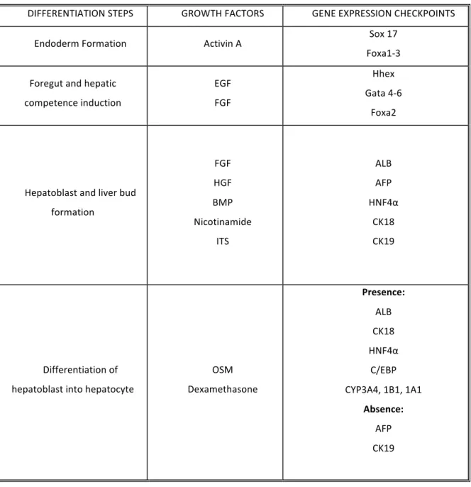
Table 1. Summary of in vitro differentiation protocols mimicking the in vivo liver development; gene expression checkpoints after each step of differentiation.
DIFFERENTIATION STEPS GROWTH FACTORS GENE EXPRESSION CHECKPOINTS
Endoderm Formation Activin A Sox 17
Foxa1-‐3
Foregut and hepatic competence induction EGF FGF Hhex Gata 4-‐6 Foxa2
Hepatoblast and liver bud formation FGF HGF BMP Nicotinamide ITS ALB AFP HNF4α CK18 CK19 Differentiation of hepatoblast into hepatocyte
OSM Dexamethasone Presence: ALB CK18 HNF4α C/EBP CYP3A4, 1B1, 1A1 Absence: AFP CK19
Embryonic stem cells (ESCs)
Embryonic stem cells are derived from the inner cell mass of blastocysts and meet all the criteria for stem cells.
In the literature, protocols of differentiation of ESC into HLC are decribed. In summary, they consist in the differentiation into endoderm with Activin A and Wnt [47]. Then,
hepatoblast formation is induced by FGF and BMP, and maturation into hepatocytes is normally induced by HGF and OSM [16-‐19] (Table 1). Duan et al. (2007) proved the possibility of achieving differentiated ESC expressing liver-‐specific genes and exhibiting liver specific functions [19]. Moreover, Brólen et al. (2010) obtained HLC expressing thw hepatic markers: CYP3A4, CYP1A2, CYP2C9, α-‐1-‐antitrypsin, AFP, HNF4α, CK18, ALB and exhibiting elevated urea secretion and glycogen accumulation. Finally differentiated cells had the ability to metabolize phenacetin, midazolam and diclophenac via the phase I cytochrome P450 enzymes, CYP1A, CYP3A and CYP2C, respectively [48]. However, increased tumorogenicity of these cells in in vivo models raise some problems in terms of safety [4, 49].
Adult stem cells (ASCs)
Adult stem cells are multipotent stem cells present in many adult tissues, generating much less controversy among the scientific community. The liver contains a population of adult stem cells called oval cells. Liver progenitor cells are useful in the study of liver development and liver diseases, however, the stem cell isolation is difficult and provides low numbers of cells [4]. For oval cells to differentiate into HLCs, total confluence and addition of HGF and EGF or fibroblast growth factor-‐4 (FGF-‐4) is required, however a mature phenotype has not yet been achieved [50].
Induced Pluripotent Stem Cells (iPSCs)
As it was proven for the first time by Takahashi et al. (2006,2007), it is possible to obtain pluripotent stem cells from human and mouse reprogrammed somatic cells [51-‐52]. This is achieved through the intregation of the pluripotency genes OCT3/4, SOX2, KLF4 and c-‐ MYC in the cell genome. Induced pluripotent stem cells (iPSCs) share many characteristics with ESCs and their use is ethically acceptable [51]. Differentiation procedures of iPSCs into HLCs are similar to protocols described to differentiate ESCs [23-‐24, 53]. Sullivan et al. showed the differentiation of iPSCs into cells exhibiting
hepatic markers ALB, AFP, HNF4α, CYP71A, and with the capacity of secreting the plasma, and with CYP1A2 and CYP3A4 activity [23].
However, there are some problems concerning iPSCs, such as the variability caused by differences in the iPSCs reprograming that can be the origin of the heterogeneity of the HLCs obtained [1].
Fetal stem cells
Fetal stem cells are a more primitive population of stem cells that can be isolated from fetal tissues such as fetal blood as well as from umbilical cord blood and matrix. The use of fetal stem cells have advantages over ESCs and ASCs since their use is more ethically acceptable, have an enhanced multipotency and are less immunogenic than ASCs [54]. Mesenchymal stem cells (MSCs) are multipotent stem cells that can be isolated from fetal tissues such as umbilical cord blood and Wharton’s jelly1. Many adult tissues also possess populations of MSC: adipose tissue, liver, muscle, bone marrow and dental pulp [55].
MSCs from different sources have been studied and each type varies in their proliferative and multilineage potential. However, the lack of a specific cell surface marker leads to difficulties in identifying this cell population.
Therefore, the Mesenchymal and Tissue Stem Cell Committee of the International Society for Cell Therapy (ISCT) proposes a set of standards to define human MSCs [56]:
1) MSC must be plastic-‐adherent when maintained in standard culture conditions using tissue culture flasks.
2) More than 95% of the MSC population must express CD105, CD73, CD90 and lack the expression of hematopoietic antigens CD45, CD34, CD14 or CD11b, CD79a or CD19 and HLA-‐DR molecules, not expressed on MSC unless stimulated (e.g. by IFN-‐γ).
3) Under specific stimuli, cells must be able to differentiate into osteoblasts, adipocytes, and chondroblasts in vitro.
These criteria apply only to human MSCs [56].
MSCs, due to their immunosuppressive and anti-‐inflammatory characteristics, were considered a suitable model to cellular biology studies, tissue engineering and therapeutic application. Therefore, in the past few years their use has grown as well as the search of new sources, which can provide a higher cell number and less epigenetic damaged cells [55].
The differentiation process of MSCs is slightly different than ESCs or iPSC because MSCs are derived from mesoderm. However the reason these cells can be directed to hepatic fate is due to the expression of hhex gene in their genome. Thus, the endoderm induction step is not present in the majority of protocols described [20, 57-‐58].
UCX® cells are Wharton’s Jelly derived human umbilical cord matrix Mesenchymal stem cells (ucmMSC). These cells meet the criteria for multipotent stem cells listed above [59]. UCX® isolation procedure allows for high numbers of cells, with less epigenetic damage [26-‐27]. Comparing with other adult sources of MSC, ucmMSCs have greater number of passages to senescence and shorter doubling times, which reflect the relatively primitive nature of these MSCs. [60].
UCX® have the ability to suppress T-‐cell proliferation in a more significant way than MSCs from other sources such as bmMSCs, and also have the capacity of convert naive CD4+ CD25+ T-‐cells into became regulatory by expressing the FOXP3 transcription factor, causing an immunosuppressive effect. Moreover, UCX® have anti-‐inflamatory effects in arthritis models in vivo [59].
Hence, UCX® are a great candidate to use as an alternative source in hepatic differentiation procedures to obtain models for in vitro toxicology. In spite of the advantages of ucmMSCs, to our knowledge, only one work, Campard et al. (2008), analysed the potential of these cells in the differentiation into HLC. The protocol described here was based in 3 steps that mimic the in vivo liver embryogenesis (see Annex 1). With this protocol, a population of differentiated cells with hepatocyte-‐like characteristics was obtained. Differentiated ucmMSC expressed hepatic markers: albumin, Glucose-‐6-‐phosphate, tryptophan 2,3-‐dioxygenase, α-‐antitrypsin, tyrosine