EVALUATION OF THE PHYSIOLOGICAL AND MOLECULAR
IMPACT OF IRON DEFICIENCY IN TWO LEGUME SPECIES
Inês Valadares Serrão Branco Mendes
EVALUATION OF THE PHYSIOLOGICAL AND MOLECULAR IMPACT
OF IRON DEFICIENCY IN TWO LEGUME SPECIES
AVALIAÇÃO DO IMPACTO FISIOLÓGICO E MOLECULAR DA
DEFICIÊNCIA DE FERRO EM DUAS ESPÉCIES DE LEGUMINOSAS
Thesis presented to Escola Superior de Biotecnologia of the Universidade Católica
Portuguesa to fulfill the requirements of Master of Science degree in Food Engineering
Inês Valadares Serrão Branco Mendes
Place: Centro de Biotecnologia e Química Fina, Escola Superior de Biotecnologia
Supervision: Marta Wilton de Vasconcelos, PhD Co-supervision: Carla Sancho dos Santos, MSc
Resumo
As leguminosas apresentam um papel socio-económico importante e fazem parte da nutrição humana e animal. As suas sementes comestíveis são uma fonte de micronutrientes, com alto conteúdo proteico.
Apesar do ferro (Fe) ser o quarto elemento mais abundante na superfície terrestre, a sua baixa solubilidade torna-o pouco biodisponível para as plantas, baixando o conteúdo de Fe nas suas sementes. Em países pobres, onde as sementes são componentes predominantes das dietas, o acesso a fontes de nutrientes variadas e fortificadas é limitado, aumentando a prevalência de doenças associadas à subnutrição, tal como a Anemia por Deficiência de Ferro. Várias estratégias têm sido utilizadas para atenuar esta doença, sendo uma delas a biofortificação. Contudo, para aumentar o conteúdo nutricional das sementes é necessário perceber os mecanismos relacionados com as vias dos minerais até às mesmas. Assim, genes relacionados com a absorção (FRO2, IRT1), transporte (NRAMP3, VIT1, YSL1) e armazenamento (Ferritin) do Fe, bem como novos genes candidatos (GCN2 and WEE1), foram estudados, em duas espécies de leguminosas utilizadas na nutrição humana e animal: Glycine max e Medicago sp. Este trabalho foca também os mecanismos fisiológicos comuns relativos à deficiência de Fe destas duas espécies, crescidas na presença e na ausência deste nutriente. Neste trabalho, ambas as espécies revelaram-se suscetíveis à clorose por deficiência de ferro (IDC), desenvolvendo vários sintomas característicos, tais como redução do crescimento, amarelecimento das folhas e desenvolvimento de raízes secundárias. Os genes FRO2 e IRT1 apresentaram níveis de expressão similares, o que sugere que o IRT1 é co-regulado pelo FRO2. Quanto ao transporte,
NRAMP3 e VIT1 apresentaram padrões de expressão opostos, sendo estes mais elevados nas
folhas. YSL1 e Ferritin apresentaram maior expressão na suficiência de Fe, em ambos os tecidos. Os resultados obtidos para o GCN2 indicam que este gene participa no metabolismo do Fe, visto que na falta de Fe é sobre-expresso, principalmente nas raízes. Quanto ao WEE1, os resultados sugerem que este gene é sub-expresso em ausência de Fe onde o crescimento é reduzido. Quando sumetidas a condições de stress (ausência de Fe), ambas as espécies apresentaram um impacto nutricional semelhante, com diminuição do conteúdo nutricional, e com maior concentração de zinco na parte aéria e de Fe nas raízes.
Este trabalho contribui para uma melhor compreensão, tanto dos mecanismos de resposta à deficiência de Fe, como de novos genes relacionados com o metabolismo do Fe, crucial não só para combater o IDC e consequente impacto económico, mas também para o aumento do conteúdo nutricional de leguminosas importantes na nutrição humana e animal.
Abstract
Legume grains have an important socio-economical role, being highly utilized in human and animal nutrition. They are mainly cultivated for their edible seeds, sources of micronutrients, and their high protein content.
Although iron (Fe) is the fourth most abundant element in the earth’s crust, its limited solubility makes it poorly bioavailable for plants, lowering the Fe content of their seeds. In poorer countries, where seeds are the main components of their diets, the access to diverse food sources and fortified food sources is often limited, increasing the prevalence of several diseases associated with undernourishment, namely Iron Deficiency Anemia (IDA). Various strategies are being used to withstand this disease and one of them is biofortification. However, to increase the seeds mineral content, one must understand the mechanisms underlying the minerals pathway to the plants edible parts. Thus, Fe related genes involved in nutrient uptake (FRO2,
IRT1), transport (NRAMP3, VIT1, YSL1), and storage (Ferritin), as well as novel candidate
genes (GCN2 and WEE1), were studied, in two significant crops utilized in human and animal nutrition: Glycine max and Medicago sp. This work also focused on the common physiological mechanisms underlying the response to Fe deficiency, of these two species, grown under Fe-deficient and Fe-sufficient conditions. In the current work, both species were very susceptible to Iron Deficiency Chlorosis (IDC), developing several characteristic symptoms, such as yield reduction, yellowing of leaves and development of secondary roots. Fe acquisition genes, FRO2 and IRT1, presented similar expression patterns, suggesting that IRT1 is co-regulated with
FRO2. In what concerns Fe transport, NRAMP3 and VIT1, showed opposite functions and
appeared to have a stronger expression in the leaves. YSL1 and Ferritin presented higher expression levels under Fe-sufficient conditions in shoots and roots. The results obtained for
GCN2 strongly indicate that this gene plays a role in Fe metabolism, since in the lack of Fe it
is over-expressed, mainly in roots. In what concerns to WEE1, the results show that this gene seems to have a more important role in Fe-sufficient roots, being under-expressed in stress situations, where growth is diminished. When submitted to stress conditions, both species presented a similar nutritional impact, with a decreased nutrient content, where Zn predominates in the aerial part, whereas Fe predominates in the roots.
This work contributes to an enhanced understanding, on Fe deficiency response mechanisms, as well as new Fe-metabolism related genes, crucial not only to combat IDC crop devastation and consequent economic damage, but also to increase Fe content in the edible parts of legume plants in order to improve human nutrition and health.
Acknowledgements
Foremost, I would like to express my sincere gratitude to my supervisor, Dra. Marta Vasconcelos, for giving me the opportunity to work in her research group and for sharing with me her vast wisdom and knowledge, broadening my horizons.
Special thanks to Carla, who is an enormous example for me, and have been always supportive no matter what, with all her patience and knowledge, helping me when I most needed.
To all PlanTech group, thank you for helping me throughout this stage. You helped me willing to work every day and improve my work with all the brainstorming and knowledge that we’ve shared with each other.
To my parents, and my sister, for all your love and support through my academic life.
To my grandparents, who always made me believe that I can successfully achieve all my established goals with persistence, resilience and faith.
To all my special friends, for always remembering me to be patient, for never letting me lose confidence in myself and for being by my side, no matter what.
Contents
RESUMO ... III ABSTRACT ... V ACKNOWLEDGEMENTS ... VII CONTENTS ... IX LIST OF FIGURES ... XI LIST OF TABLES ... XIII1 INTRODUCTION ... 15
1.1 LEGUMES GRAINS: THE IMPORTANCE TO HUMAN AND ANIMAL NUTRITION ... 15
1.1.1 Soybean and alfalfa ... 16
1.2 IRON ... 17
1.2.1 Anaemia: a major disorder around the world ... 17
1.2.2 Biofortification: how legume plants can improve our health ... 18
1.2.3 From root to seed: the mechanisms are still unclear ... 19
2 OBJECTIVES ... 25
3 MATERIALS AND METHODS ... 27
3.1 PLANT GROWTH CONDITIONS ... 27
3.2 TOTAL CHLOROPHYLL EXTRACTION AND QUANTIFICATION ... 27
3.3 MINERAL ANALYSIS ... 28
3.4 RNA EXTRACTION ... 28
3.5 GENE EXPRESSION ANALYSIS ... 29
3.6 STATISTICAL ANALYSIS ... 30
4 RESULTS AND DISCUSSION ... 33
4.1 IDC SYMPTOMS: VISIBLE RESPONSE MECHANISMS ... 33
4.2 NUTRITIONAL IMPACT OF LOW FE AVAILABILITY ... 36
4.3 GENE EXPRESSION ANALYSIS ... 38
4.3.1 Fe acquisition: FRO2 and IRT1 ... 38
4.3.2 Fe transport: NRAMP3, VIT1 and YSL1 ... 40
4.3.3 Fe storage: Ferritin ... 43
4.3.4 Novel genes: GCN2 and WEE1 ... 44
5 CONCLUSION ... 47
6 FUTURE WORK ... 49
List of Figures
Figure 3.1 - Germination conditions of M. truncatula (A) and G. max (B) seeds. ... 27
Figure 3.2 - Schematic representation of the methodology applied in this work. ... 31
Figure 4.1 - Shoot and root length (A) and weight (B) of G. max and M. truncatula grown
hydroponically in Fe-sufficient (●) and Fe-deficient (●) conditions. Means +SE separation were performed independently for each structure, using 12 independent replications. Different letters represent significant differences between samples (p < 0.05). ... 34
Figure 4.2 – Number of secondary roots of G. max and M. truncatula grown hydroponically in
Fe-sufficient (●) and Fe-deficient (●) conditions. Data are means +SE of 12 independent replications. Different letters represent significant differences between samples (p < 0.05). . 34
Figure 4.3 - G. max plants grown hydroponically in Fe-deficient (left) and Fe-sufficient (right)
conditions. ... 35
Figure 4.4 - Chlorophyll concentration of G. max and M. truncatula grown hydroponically in
Fe-sufficient (●) and Fe-deficient (●) conditions. Data are means +SE of 12 independent replications. Different letters represent significant differences between samples (p < 0.05). . 36
Figure 4.5 – Iron (A) and Zinc (B) content of G. max and M. truncatula grown hydroponically
in Fe-sufficient (●) and Fe-deficient (●) conditions. Data are means +SE of 3 independent replications. Different letters represent significant differences between samples (p < 0.05). . 37
Figure 4.6- Root Fe uptake related genes FRO2 (A) and IRT1 (B) fold of expression, in roots
and shoot tissues, of G. max and M. truncatula, grown in Fe-sufficient (●) and Fe-deficient (●) conditions. Roots in Fe-deficient conditions were used as comparison term and 18S was used as internal control. ... 39
Figure 4.7 – Fe transporter genes NRAMP3 (A), VIT1 (B) and YSL1 (C) fold of expression, in
roots and shoot tissues, of G. max and M. truncatula, grown in Fe-sufficient (●) and Fe-deficient (●) conditions. Roots in Fe-deficient conditions were used as comparison term and 18S was used as internal control. ... 41
Figure 4.8 – Fe storage related gene Ferritin fold of expression, in roots and shoot tissues, of G. max and M. truncatula, grown in Fe-sufficient (●) and Fe-deficient (●) conditions. Roots in
Fe-deficient conditions were used as comparison term and 18S was used as internal control. 43
Figure 4.9 – Novel candidate genes GCN2 (A), WEE1 (B) fold of expression, in roots and shoot
tissues, of G. max and M. truncatula, grown in Fe-sufficient (●) and Fe-deficient (●) conditions. Roots in Fe-deficient conditions were used as comparison term and 18S was used as internal control ... 44
List of Tables
Table 3.1 – Forward and reverse primer sequences used in quantitative Real-Time PCR
1 Introduction
1.1 Legumes grains: the importance to human and animal nutrition
The Fabaceae, or legumes, broadly defined by their unusual flower structure, podded fruit and the ability to form nodules with rhizobia, economically represent the second most important family of crop plants after Poaceae (grass family). The unique capacity of legumes to obtain fixed nitrogen from the symbiotic nitrogen-fixing bacteria offers a growth advantage, as soil nitrogen often limits plant growth (Graham and Vance, 2003). Not only is the symbiosis beneficial to the legume, but also symbiotic nitrogen fixation introduces approximately 40 million tons of nitrogen into agricultural soils each year (Herridge et al., 2008).
The legumes are highly diverse and can be divided into three subfamilies: Mimosoideae,
Caesalpinioideae, and Papilionoideae (Doyle and Luckow, 2003). Among these, the Papilionoideae subfamily contains nearly all economically important crop legumes, including
soybean (Glycine max), peanut (Arachis hypogaea), mungbean (Vigna radiata), chickpea (Cicer arietinum), lentil (Lens culinaris), common bean (Phaseolus vulgaris), pea (Pisum
sativum), and alfalfa (Medicago sativa) (Zhu et al., 2005).
Legume grains have been consumed by humans since the earliest practice of agriculture and have been used both for their medicinal, cultural as well as nutritional properties, providing an important source of protein and oil, which can also be converted into biodiesel (Libault et
al. 2010). Legumes also have an important socio-economical role, being highly present in the
Mediterranean diet (Santos et al., 2013). They are cultivated for their edible seeds, sources of micronutrients, such as iron (Fe) and zinc (Zn), and their high protein content, being one of the most important foods, for humans and animals (Vasconcelos and Grusak, 2006). On a worldwide basis, legumes contribute with about one third of humankind’s protein intake, while also serving as an important source of forage for animals and of edible and industrial oils (Graham and Vance, 2003). There are some population groups where 100% of their diet is plant based, being in most cases not a matter of personal choice, but of economic constraints, due to the high cost of proteins of animal origin and their inaccessibility by the poorer segment of the population (Tharanathan and Mahadevamma, 2003).
In intensive animal and milk production, where grain crops are major feed sources, forage legumes are required to maintain animal health (Jezierny et al., 2010). This is why legume production is so important for helping to improve human and animal health and nutrition, attenuating the incidence of several diseases, especially the ones related to micronutrient deficiency.
1.1.1 Soybean and alfalfa
The Food and Agriculture Organization (FAO) statistics for 2012 show that about 253 million metric tonnes of soybean were produced across the world, ranking it one of the world’s top commodity production. Much of the world's protein and oil comes from soybean and, in fact, soybean contains more protein (40%) than any other ordinary food source, including meat, cheese and fish, and high oil content (20%) (Krishnan, 2005; Bolon et al., 2010). Soybean is considered an important food source worldwide, especially in the developing countries, for its nutritional quality, easy cultivation, low cost and high production, and is also an excellent raw material for the production of derived food (e.g. milk, sour cream) thanks to the versatility of application of its products in food and feed (Silva et al., 2006). The appropriate addition of soy products results in lower calorie food products, with reduced lipid and high protein content, proper nutritional needs of adults, besides preserving the physical and sensory characteristics of traditional product (Dhingra and Jood, 2001). This feature allows soybean to be an important alternative food for the population in general, mostly for people who cannot or do not want to consume animal products and specific substances (e.g. lactose intolerant and vegetarians), as well as malnourished populations.
Moreover, G. max is a good crop to study genetic and molecular mechanisms since, in 2010, its genome was sequenced, assembled and published (Schmutz et al., 2010). This discovery enabled the advance of studies of genetic and molecular analysis, contributing to a better understanding of nutritional problems related to this legume.
In ruminant nutrition, legume forages such as alfalfa have a higher feeding value than other typical grasses and cereals commonly used in animal feeds, because of their rapid particle degradation, faster rumen fermentation and reduced rumen retention time, besides having greater voluntary intake, resulting in increased dry matter intake, growth rate and milk yield (Dewhurst et al., 2009). Alfalfa is grown all over the world on an estimated 32 million hectares of land (Yuegao and Cash, 2009). Crude protein content in alfalfa ranges between 18-25 %, with an oil and mineral proportion content of 1.78% and 9.87%, respectively (Katić et al., 2009).
A closely related species to M. sativa is Medicago truncatula. Although M. truncatula is not as used in animal feeding as M. sativa, it has been chosen as a model species for genomic and molecular studies in view of its rapid life cycle, small diploid genome, self-fertilization, high seed production and high transformation efficiency, reduced size and easiness of growing (Trieu et al., 2000).
To be convenient as a model for legume genomics, it is essential that M. truncatula exhibits genome conservation with other crop legumes. Choi et al. (2004) made a detailed comparison between M. truncatula and M. sativa, and observed that marker relationships were uniformly syntonic. In addition, Bell et al. (2001) reported that genes from M. truncatula share very high sequence identity to their counterparts from alfalfa, so it serves as an excellent, genetically tractable model for alfalfa, serving also as a model organism for soybean and other economically important legumes.
1.2 Iron
1.2.1 Anemia: a major disorder around the world
Although the effort on poverty reduction spearhead by the United Nations and numerous civil society organization is high, malnutrition, hunger and food insecurity are still affecting large portions of the global population, which consequently affects human and economic health and development (Charles, 2012). In most poor countries, the access to diverse food sources (quality and quantity) and fortified food sources, as well as the access to health services and interventions (e.g. supplementation) is often limited, increasing the prevalence of several diseases associated with undernourishment (e.g. anemia) (Balarajan et al., 2011).
The prevalence of anemia is estimated at 9% in countries with high development, whereas in countries with low development the prevalence is 43% (McLean et al., 2009), with Africa and Asia accounting for more than 85% of the absolute anemia burden in high-risk groups (Balarajan et al., 2011). Children and women of reproductive age are most at risk, with global anemia prevalence estimated in 2011 ot 43% in children younger than 5 years, 38% in pregnant women and 29% in non-pregnant women aged 15–49 years (Stevens et al., 2013).
Anemia is a condition characterized by reduction in hemoglobin concentration, diminished red blood cells, or packed-cell volume, leading to the subsequent diminishing in meeting the oxygen demands of tissues. Nutritional anemia results from insufficient bioavailability of hematopoietic nutrients (e.g. including folate, vitamin B12 and vitamin A) needed to meet the demands of hemoglobin and erythrocyte synthesis (Semba and Bloem, 2008). Anemia can also be caused by acute and chronic inflammation, parasitic infections, and inherited or acquired disorders that affect haemoglobin synthesis, red blood cell production or red blood cell survival. Iron deficiency is among the most common nutritional problems of human populations throughout the world, affecting more than two billion people (Bouis and Welch, 2010), which is caused by dietary iron deficiency, iron mal-absorption, and blood losses.
1.2.2 Biofortification: how legume plants can improve our health
Various strategies are being used to withstand Iron Deficiency Anemia (IDA) (Bouis and Welch, 2010). One of them is supplementation with Fe, which can be practiced at health centers through a liquid or injectable medicine. However, this strategy has some drawbacks such as the taste of the medicine, teeth staining and need for an outreach as well as a delivery mechanism (Frossard et al., 2000). Another approach to attenuate IDA in susceptible populations is fortification, usually done with flours produced from cereals, but tends to be expensive and is not an option for whole grains, like legumes (Blair et al., 2010). Although these interventions have been considerably successful in the past, they have not been able to erradicate the problem, mainly owing to the significant and expensive distribution logistics required which are insufficiently developed in poorer countries (Beyer, 2010). Hereupon, innovative, cost-effective, accessible and long-lasting treatment and prevention options are needed (Charles, 2012).
An advantageous alternative to the techniques referred above is to increase the concentration of Fe in diets through biofortification. Biofortification is characterized by the ‘fortification’ of agronomical important crop plant tissues by means of their own biochemical capacity (Beyer, 2010). In other words, biofortification is the process by which the nutritional quality of staple crops is enhanced, using the best traditional breeding practices and modern biotechnology methods (Nestel et al., 2006). This strategy implicitly targets poorer households, where staple foods predominate in their diets.
One of the major advantages of biofortified crops is that they capitalize on the regular daily intake of a consistent and large amount of food staples, by all family members (Nestel et
al., 2006). Another major advantage is that, after the one-time investment in order to develop
seeds that fortify themselves, recurrent costs are low, and germplasm can be shared internationally, making biofortification a cost-effective approach (Beyer, 2010). Besides that, once the biofortified crop system is placed and stabilized, it is highly sustainable, and nutritionally improved varieties can still be grown and consumed, even if government attention and international funding for micronutrient issues fade (Nestel et al., 2006). Probably the major advantage of biofortification, when compared to other approaches, is that it provides feasible means of reaching undernourished populations in relatively remote rural areas and to heath care centers where supplementation programs are made. In an environmentally beneficial way, biofortification may have important spinoff effects for increasing farm productivity in developing countries.
The seeds mineral content depends on the uptake from the rhyzosphere into the roots, translocation to the transpiring shoots trough xylem, transference into leaves or other tissues, and finally, translocation into the seeds trough phloem. An incomplete understanding of the pathways and the rate limiting steps involved in translocating minerals to the seeds represents a major challenge of biofortification (Waters and Sankaran, 2011). Thus, in order to develop a successful biofortification strategy, one must identify the key molecular players involved in nutrient uptake, transport and storage, preferably in crops heavily used in human and animal diets.
1.2.3 From root to seed: the mechanisms are still unclear
Although Fe is the fourth most abundant element in the earth’s crust, its limited solubility makes it poorly bioavailable for plants (Morrissey and Guerinot, 2009), which consequently results in seeds with insufficient Fe content to meet the body’s requirements. Fe availability is dictated by the soil redox potential and pH. In aerobic or alkaline soils, Fe is readily oxidized, and is predominately in the form of insoluble ferric oxides. At lower pH, the ferric Fe is free from the oxide, and becomes more available for uptake by roots (Morrissey and Guerinot, 2009). Because 30% of the world's cropland is too alkaline for optimal plant growth (Marschner, 1995) and some staple crops, like rice and soybean, are very susceptible to Fe deficiency (Takahashi et al., 2001), much research has focused on how plants cope with Fe limitation. Crops growing in alkaline soils fail to accumulate Fe in edible organs causing nutrient deficiency in humans (Gómez-Galera et al., 2010).
Plants require Fe for innumerous life-sustaining processes from respiration to photosynthesis, where it participates in electron transfer through reversible redox reactions, cycling between Fe2+ (ferrous) and Fe3+ (ferric) form. Fe homeostasis in plants is achieved through very dynamic processes requiring proteins and small organic molecules in order to take up the metal from the soil, to transport it throughout the plant, to compartmentalize it in the cells, and ultimately to buffer and to store it in case of excess (Briat et al., 2009).
Insufficient Fe uptake leads to Fe deficiency chlorosis (IDC) symptoms, such as yellowing of the upper leaves, interveinal chlorosis and stunted growth, as well as reduction of crop yields (Prasad et al., 2003; Kim and Guerinot, 2007). Besides influencing chlorophyll synthesis, IDC also lowers the concentrations of Fe in the seeds and other harvested tissues (Grusak, 1999), affecting both farmer profit and the nutritional value of plant products (Vasconcelos and Grusak, 2013). In this context, the acquisition, usage and detoxification of
Fe pose a considerable challenge to cells and organisms, which have evolved sophisticated mechanisms to satisfy their metabolic needs and minimize the risk of toxicity (Domenico et al., 2008; Andrews, 2008; Hentze et al., 2010).
In order to uptake Fe from the soil, dicotyledonous plants, such as soybean and alfafa, utilize Strategy I, which is characterized by the requirement of reducing Fe3+ to Fe2+ before its uptake. The reduction is carried out by a plasma membrane-bound ferric reductase, the Ferric Reduction Oxidase (FRO). This reduction step appears to be the rate-limiting stage in Fe uptake in Arabidopsis thaliana (Connolly et al., 2003), a model species commonly used in plant biology. In fact, the transgenic overexpression of ferric chelate reductases in the roots of rice, tobacco and soybeans has been successful in increasing tolerance to Fe-limiting conditions (Vasconcelos et al., 2006; Ishimaru et al., 2007). The FRO family of metal reductases contains seven additional members in Arabidopsis spp. (Wu et al., 2005; Mukherjee et al., 2006) however FRO2 is thought to be the main Fe3+ chelate reductase in roots and over- expressed in the epidermal cells of Fe-deficient roots (Kim and Guerinot, 2007). Plants overexpressing
FRO2 are resistant to low Fe growth conditions (Connolly et al., 2003).
The expression of FRO genes in various locations suggests that different sets of FRO proteins are participating in Fe uptake in different plant tissues. For example, FRO2, FRO3 and
FRO5 are expressed in roots, and FRO3 is predominantly expressed in the vascular cylinder of
roots, suggesting a role in Fe re-absorption from the root apoplast (Kim and Guerinot 2007); and FRO6, FRO7 and FRO8 are described as shoot-specific (Kim et al., 2006).
After its uptake, Fe2+ is transported into the plant to leaves and seeds by specific membrane transporters (Grotz and Guerinot, 2006), such as the Iron-Regulated Transporter 1 (IRT1), a member of the Iron-Regulated Transporter-Like Protein (ZIP) gene family (Waters et
al., 2002; Walker and Connolly, 2008; Ivanov et al., 2011). IRT1 is expressed in the epidermal
cells of roots and is localized in the plasma membrane (Kim and Guerinot, 2007). Both FRO2 and IRT1 genes are up-regulated under Fe deficiency and are activated by specific bHLH transcription factors, which are also up-regulated by Fe deficiency (García et al., 2012).
Once Fe enters the root symplast, it must be bound by chelating compounds, such as nicotianamine (NA) and citrate. Fe-chelator complexes then move through intercellular connections into the stele along the diffusion gradient. A broad spectrum of transporters has been characterized in model plants including the Natural Resistance Associated Macrophage (NRAMP) family protein, the Vacuolar Iron Transporter (VIT), and the Yellow Stripe 1-Like (YSL). Plant NRAMP proteins are generally involved in Fe import into the cytoplasm (Brear
2010; Lanquar et al.,2010; Sasaki et al., 2012), cobalt (Cailliatte et al., 2010), cadmium (Sasaki
et al., 2012), and aluminum (Xia et al., 2010), having a broad specificity. AtNRAMP3 and AtNRAMP4 are H+ metal symporters responsible for Fe andMn mobilization from the vacuole (Lanquar et al., 2010). VIT1 is involved in the uptake of Fe2+ into the vacuole for storage (Brear
et al., 2013). YSL transporters, on the other hand, are likely involved in the transport of Fe2+ -NA complexes and the genes they encode are expressed in various tissues, suggesting roles in Fe uptake at diverse locations (Kim et al., 2006).
Fe must also be transported through the phloem bounded to chelators, because the transpiration flow in the xylem vessels is inefficient in developing organs such as the apex, seeds and root tips (Kim and Guerinot, 2007). Fe remobilization from older leaves to younger
leaves also takes place via phloem transport (Kim et al., 2006).In 1993, Stephan and Scholz suggested that Fe traveled in the phloem as a Fe-NA complex, however, later on, this idea became controversial (Schmidke et al. 1999), but NA is still though to play a role in Fe loading and, probably, unloading of the phloem.
Free Fe is toxic since it facilitates the generation of highly reactive oxygen species (ROS).ROS can damage cellular constituents and, therefore, Fe homeostasis needs to be strictly controlled to avoid Fe deficiency and toxicity (Liao et al., 2012). Therefore, storage proteins play an important role in Fe homeostasis, since they assure that ferric Fe is in a way that is still bio-available in case of cellular needs but yet nonreactive with oxygen (Briat et al., 2010). Ferritins are a broad superfamily of ubiquitous Fe storage and detoxification proteins, found in the entire living kingdom, except in yeast (Andrews et al., 2003; Briat et al., 2006; Arosio et
al., 2008). Ferritin naturally occurs in plant tissues such as cotyledons, root and shoot apices,
cells of vessels, reproductive cells and senescing cells, and are abundant when plants are grown under high Fe conditions (Briat et al., 2010). Besides their role in Fe storage, ferritins are also involved in protection against oxidative stress through their potential detoxification properties of excess iron, dioxygen and, to some extent, hydrogen peroxide (Zhao et al., 2002; Theil et
al., 2006; Arosio et al., 2008).
The pathways and mechanisms that regulate the expression of these Fe-related genes are still unclear. Several published reports (Waters et al., 2007; Bacaicoa et al., 2011; Lingam
et al., 2011; Meiser et al., 2011; Ramírez et al. 2011; Romera et al., 2011; Wu et al. 2012)
suggest that some hormones, such as auxin, ethylene and nitric oxide (NO), can up-regulate the expression of these genes, in plants grown in Fe-deficient conditions. However, in plants grown with high levels of Fe, these hormones showed almost no effect in the expression of Fe-related genes, suggesting that their up-regulation does not exclusively depend on activator hormones,
but also on Fe availability (Lucena et al. 2006; Graziano and Lamattina, 2007; Chen et al., 2010; García et al. 2011).
Another question that is still unsolved, is how Fe reacts to repress gene expression and which pool of Fe (e.g. from root, phloem, intracellular, apoplastic, etc.) is monitored by the plant to mediate this control. Maas et al. (1998) proposed phloem Fe as an inhibitor of Fe deficiency responses in Strategy I plants, and they found that the application of Fe-EDTA to leaves decreased some of the Fe deficiency responses, such as proton extrusion and ferric reductase activity. Based on these results they proposed that leaves could modulate Fe deficiency responses through phloem Fe, leading to suppression of gene expression under Fe-sufficient conditions (Maas et al., 1998). Beyond this, there are studies also suggesting that application of Fe, chelated to other compounds (e.g. citrate), to leaves of Fe-deficient plants suppressed root Fe deficiency responses (Venkatraju and Marschner, 1981; Romera et al., 1992; Enomoto et al., 2007).
Even though much has been learned about the physiology of Fe uptake in model species, which are relevant to human and animal nutrition, there is no clear understanding of the physiology of tolerance to Fe deficiency in crop species such as soybean and alfalfa, and this has hampered breeding programs (Vasconcelos and Grusak, 2013). Since increasing the Fe uptake in the roots can augment Fe concentrations in the leaves, it is possible that some of this additional Fe may be remobilized to the grains, which would help in biofortification efforts that aim at enhancing seed Fe levels (Santos et al., 2013). However, the increased Fe translocation from shoots to seeds still remains one of the major bottlenecks in most biofortification programs (White and Broadley, 2005), and the answer to this may be in the identification of new candidate genes.
1.2.3.1 GCN2 and WEE1: novel candidate genes
To clearly understand the mechanisms of Fe uptake and translocation to the edible parts of the plant, the search for new candidate genes and new molecules that could be involved in the process should be intensive. The quick understanding of these mechanisms is essential to build a successful biofortification program, in order to combat malnutrition around the world.
It is known that the phosphorylation of the α subunit of the eukaryotic translation initiation factor 2 (eIF2α) provides a key mechanism for down-regulating protein synthesis in response to nutrient starvation or stresses (Wek et al., 2006; Hinnebusch, 2005). In mammals, GCN2 is the primary eIF2α-kinase in response to nutrient limitation (Zhang et al., 2002).
Studies in mouse (Dang Do et al., 2009) showed that, in stress situations, the expression of proteins encoded by mRNAs related to eIF2α phosphorylation, namely GCN2, is increased, consequently enhancing Fe uptake.
A GCN2-like ortholog (AtGCN2) has been identified in A. thaliana (Zhang et al., 2003) and was shown to be activated by amino acid deprivation conditions (Zhang et al., 2008), as well other stress stimuli, such as purine deprivation, UV light, cold shock and wounding (Lageix et al., 2008). The activation of GCN2 protein following wounding or exposure to methyl jasmonate,1-Aminocyclopropane-1-carboxylic acid (ACC) and salicylic acid, further suggests that this enzyme could play a role in plant defense responses to insect herbivores and may represent a key but yet uncharacterized player linking biotic and abiotic stresses (Lageix
et al., 2008). Lageix et al. (2008) confirmed that AtGCN2 is essential for plant growth in stress
situations, by introducing an intact copy of AtGCN2 in the gcn2 mutant line and observing that plants grow identically to wild-type seedlings during amino acid deprivation. To this date, there are no published studies on the role of GCN2 on Fe uptake, which makes the study of this gene an important innovation in Fe nutrition in plants.
Ataxia Telangiectasia Mutated (ATM) and RAD3-Related (ATR) proteins are the main regulators of the DNA damage pathway and perform functions in plants very similar to those of their orthologs in mammals (Garcia et al., 2003; Culligan et al., 2004). These proteins sense DNA damage and induce the coordinated expression of DNA repair and cell cycle–arresting genes. It has been known that WEE1 kinase is a key protein for cell cycle arrest in plants at times of DNA damage and interferes directly with cell cycle progression phosphorylating Cyclin-dependent kinases (CDKs) and preventing the cell from entering mitosis (Jones R. et
al., 2013). Plants lacking a functional WEE1 do not accumulate phosphorylated CDKs, thereby
entering mitosis with incompletely replicated or damaged DNA, with growth arrest as a consequence (De Schutter et al., 2007; Jones R. et al., 2013). Recent works (Santos et al., 2012) suggest that WEE1 may have a role in Fe homeostasis, in M. truncatula plants, as this gene was found to be up-regulated in Fe deficient plants, using a transcriptomic approach. However, confirmation of this observation is lacking, which makes it relevant to study this gene, in order to understand how its expression if affected by Fe deficiency and which mechanisms is WEE1 associated with.
2 Objectives
This project aims at identifying genes that are important in the uptake of Fe in two significant crops utilized in human and animal nutrition, G. max and M. truncatula, as well as evaluating the impact of low Fe availability on mineral accumulators in the plant food. It also aims at unravelling the common mechanisms underlying the response to Fe deficiency at a physiological and molecular level, of these two species, grown under Fe-deficient (0 M Fe3+ -EDDHA [ethylenediamine-N,N’bis(o-hydroxyphenyl)acetic acid]) and Fe-sufficient (20 M Fe3+-EDDHA) hydroponic conditions. To this end,
The specific goals of this work are:
- To analyze the common morphological responses of the two species in study, such as plant weight, root length and secondary structure development;
- To understand how Fe deficiency affects photosynthetic pigment accumulators. - To quantify and analyze, at a molecular level, selected genes related to Fe metabolism, in order to understand which genes are over expressed or repressed by the plant in Fe stress conditions.
- To evaluate the impact of Fe deficiency on Fe and Zn accumulation, in plants submitted to both treatments, in order to understand how Fe deficiency affects nutrient content. - To identify novel genes that may be involved in Fe uptake or translocation and that may be candidates for improved biofortification strategies.
3 Materials and Methods
3.1 Plant growth conditionsPlants of Medicago truncatula cultivar Luzerna revilheira and Glycine max cultivar Williams 82, were grown in an Aralab Fitoclima 10000EHF with 16h day / 8h night photoperiod. The temperature was kept at 20ºC during the light period, with 350 mol s-1 m-2 of photon flux density, and at 18ºC during the dark period, with 75% of relative humidity.
Scarified seeds of M. truncatula were germinated in 1.2% agar (Fig. 3.1-A) and of
G.max were rolled in filter paper and placed vertically in a solution of 250 mM CaCl2 (Fig.3.1-B), at 25ºC, in the dark.
Germinated seeds of M. truncatula and G. max with 5 cm and 15 cm length, respectively, were then transferred to hydroponic solutions with different Fe treatments: Fe-sufficiency (20 M Fe3+-EDDHA) (+Fe) and Fe deficiency (0 M Fe3+-EDDHA) (-Fe). The standard solution for hydroponic growth of M. truncatula contained: 3 mM KNO3; 1 mM Ca(NO3)2; 0.5 mM MgSO4.7H2O; 0.5 mM NH4H2PO4; 0.75 mM K2SO4; 25 M CaCl2; 25 M H3BO3; 2 M MnSO4; 2 M ZnSO4.H2O; 0.5 M CuSO4.H2O; 0.5 M MoO3; 0.5 M NiSO4. The conditions used for G. max included: 1.2 mM KNO3; 0.8 mM Ca(NO3)2; 0.3 mM MgSO4.7H2O; 0.2 mM NH4H2PO4; 25 M CaCl2; 25 M H3BO3; 0.5 M MnSO4; 2 M ZnSO4.H2O; 0.5 M CuSO4.H2O; 0.5 M MoO3; 0.1 M NiSO4. Both hydroponic solutions were buffered by the addition of 1mM MES, pH 5.5.
After 14 days (14d) of hydroponic growth, the length and weight of shoots and roots was measured and the number of secondary roots was counted.
3.2 Total chlorophyll extraction and quantification
The total chlorophyll extraction and quantification was performed according to an optimized method from Abadía et al. (1984).
A
B
In short, 12.5 ml of 4 g/L solution of CaCO3 in methanol were added to 0.5 g of fresh leaves. After an incubation period of 48 hours in the dark, at room temperature, the material was white due to the loss of the pigments to the methanolic solution. One ml of the supernatant was diluted in 25 ml of deionized water and absorbance measures were taken at 663 nm and 645 nm. Spectrophotometric chlorophyll quantification of total chlorophylls was calculated following the equation 3.2.1.
Total Chlorophylls = (8.02 · A663 + 20.21 · A645)
0.0125 x Dilution Factor
Fresh weight (g) (3.2.1)
3.3 Mineral Analysis
Three replicates of 200 mg of dried shoot and root of G. max and M. truncatula were mixed with five milliliters of 65% HNO3 in a Teflon reaction vessel and heated in a SpeedwaveTM MWS-3+ (Berghof, Germany) microwave system. Digestion procedure was conducted in five steps: 130°C for five minutes; 160°C for 10 min; 170°C for 10 min; 100°C for two minutes and 100°C for two minutes. Leaf tomato standard (Sigma) was digested to ensure digestion efficiency. The resulting clear solutions of the digestion procedure were then brought to 20 mL with ultrapure water for further analysis.
The determination of mineral concentration was performed using the Inductively Coupled Plasma – Optical Emission (ICP-OE) spectrometer Optima 7000 DV (PerkinElmer, Massachusetss, USA) with radial configuration.
3.4 RNA extraction
To extract RNA from leaves and roots, the Qiagen RNeasy Plant Mini Kit (USA, #74904) was used. Briefly, roots and leaves were grounded with liquid nitrogen, until a fine powder was obtained. To 75 mg of leaf powder and 100 mg of root powder, RLC buffer and RLT buffer were added, respectively, with 1% of β-mercaptoethanol. Roots and leaves were vortexed at 56ºC for 3 minutes and at room temperature (RT) for 6 minutes, respectively. The lysates were transferred to a QIAshredder spin column sitting in a 2 ml collection tube. Roots and shoots were centrifuged at maximum speed for 5 and 10 minutes, respectively. After the flow through was transferred to another 1.5 ml tube, 225 µl and 375 µl of 100% ethanol was added to roots and leaves respectively. Samples were transferred to an RNeasy mini spin
column sitting in a 2 ml collection tube, and they were centrifuged for 15 seconds at 10000 rpm. The column was then washed twice with 500 µl of RW1 buffer, centrifuged for 15 seconds, and then washed again with 500 µl of RPE buffer and centrifuged for another 15 seconds. RNeasy column was finally transferred to a 1.5 ml tube and RNA was eluted with 30-50 µl of sterilized water.
Possible DNA contamination was cleaned with Turbo DNA-free kit (Ambion, Austin, TX, USA), accordingly to manufacturer’s instructions. RNA quality and quantity were checked with UV-spectophotometry, using a nanophotometer (Implen, Isaza, Portugal).
3.5 Gene expression analysis
About two µg of total RNA were used to synthesize cDNA, using the First Strand cDNA Synthesis Kit (Fermentas), following manufacturer’s instructions. In short, one µl of Random Hexamer primer and eight µl of nuclease-free water were added to two µg of RNA. To this mixture it was added four µl of 5X Reaction Buffer, one µl of RiboLock RNase Inhibitor (20 u/µl), two µl of dNTP Mix (10 mM) and two µl of M-MuLV Reverse Transcriptase (20 u/µl), making a total volume of 20 µl. The mixture was finally incubated at 37ºC for 60 min, and then heated at 70ºC for 5 min.
Candidate genes were selected according to their established (FRO2, IRT1, NRAMP3,
VIT1, YSL1, Ferritin) or possible (WEE1 and GCN2) role on Fe metabolism. Primers targeting
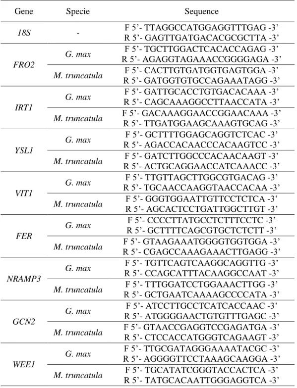
Fe stress metabolism were designed using Primer3 (Frodo.wi.mit.edu), specifying an expected PCR product of 100-200 bp and primer annealing temperatures between 56 ºC and 58 ºC. The sequences are presented in table 3.1. Quantitative Real-Time PCR (qPCR) reactions were performed on a Chromo4 thermocycler (Bio-Rad, CA, USA). Amplifications were carried out using 1.25 μM of the specific primers and mixed to 12.5 μL of 2xPCR iQ SYBR Green Supermix (Bio-Rad) and 100 ng of cDNA in a final volume of 25 μl. Three technical replicates were performed for each gene tested in qPCR reactions, as well as for controls. The amplification of all genes was performed accordingly to Han et al. (2013). Briefly, samples were heated at at 95 ºC for 10 min, followed by 40 cycles of 95 ºC for 15 s, 58 ºC for 15s and 68 ºC for 15s. Melt curves profiles were analysed for each gene tested. The comparative CT method (ΔΔCT) (Livak, & Schmittgen, 2001) was utilized for the relative quantification of gene expression value of Fe stress related genes using the 18S rRNA gene as the housekeeping gene (Opticon Monitor 3 Software, Bio-Rad).
Table 3.1 – Forward and reverse primer sequences used in quantitative Real-Time PCR reactions.
3.6 Statistical Analysis
The obtained data concerning shoot and root length and weight, number of secondary roots, chlorophyll concentration, and Fe and Zn content, were analyzed using GraphPad InStat for Windows (Version 3.05, 16 bit, GraphPad Sotware, Inc.). Treatment differences were tested by t-test (p < 0.05).
Gene Specie Sequence
18S - F 5’- TTAGGCCATGGAGGTTTGAG -3’ R 5’- GAGTTGATGACACGCGCTTA -3’ FRO2 G. max F 5’- TGCTTGGACTCACACCAGAG -3’ R 5’- AGAGGTAGAAACCGGGGAGA -3’ M. truncatula F 5’- CACTTGTGATGGTGAGTGGA -3’ R 5’- GATGGTGTGCCAGAAATAGG -3’ IRT1 G. max F 5’- GATTGCACCTGTGACACAAA -3’ R 5’- CAGCAAAGGCCTTAACCATA -3’ M. truncatula F 5’- GACAAAGGAACCGGAACAAA -3’ R 5’- TTGATGGAAGCAAAGTGCAG -3’ YSL1 G. max F 5’- GCTTTTGGAGCAGGTCTCAC -3’ R 5’- AGACCACAACCCACAAGTCC -3’
M. truncatula R 5’- ACTGCAGGAACCATCAAACC -3’ F 5’- GATCTTGGCCCACAACAAGT -3’
VIT1
G. max R 5’- TGCAACCAAGGTAACCACAA -3’ F 5’- TTGTTAGCTTGGCGTGACAG -3’ M. truncatula F 5’- GGGTGGAATTGTTCCTCTCA -3’
R 5’- AGCACTCCTGATTGGCTTGT -3’
FER
G. max F 5’- CCCCTTATGCCTCTTTCCTC -3’
R 5’- GCTTTTCAGCGTGCTCTCTT -3’
M. truncatula R 5’- CGAGCCAAAGAAACTTGAGG -3’ F 5’- GTAAGAAATGGGGTGGTGGA -3’
NRAMP3 G. max F 5’- TGTTCAGTCAAGGCAGGTTG -3’ R 5’- CCAGCATTTACAAGGCCAAT -3’ M. truncatula F 5’- TTTGGATCCTGGAAACTTGG -3’ R 5’- GCTGAATCAAAAGCCCCATA -3’ GCN2
G. max R 5’- ATGGGGAACTGTGTTTGAGC -3’ F 5’- ATCCTTGCCTCATCACCAAC -3’ M. truncatula F 5’- GTAACCGAGGTCCGAGATGA -3’ R 5’- CTCCACCATGGGTCAGAAGT -3’
WEE1
G. max F 5’- TTGCGATAGGGAAAATACGC -3’
R 5’- AGGGGTTCCTAAAGCAAGGA -3’
M. truncatula F 5’- TGCATATCGGGTACCACTCA -3’
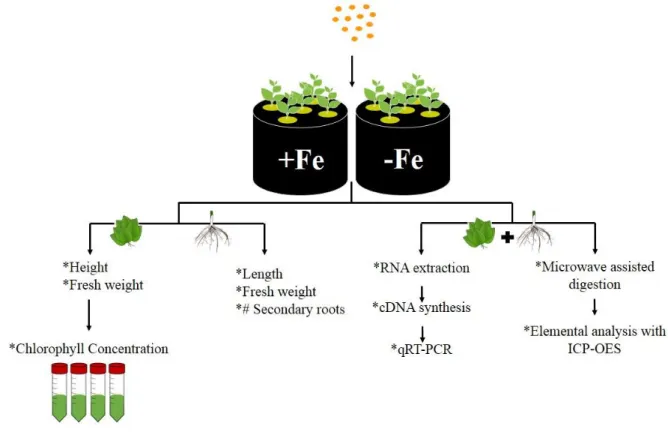
Figure 3.2 summarizes all the methodologies utilized in this work to evaluate the mechanisms underlying the response to Fe deficiency at a molecular and physiological level, of G. max and M. truncatula plants grown in Fe-sufficiency and Fe-deficiency.
4 Results and Discussion
From all the strategies being used to withstand Iron Deficiency Anemia, biofortification seems to be the one with greater advantages and it may have important spinoff effects for increasing farm productivity in developing countries. Thus, Fe is not synthesized in the plant, it must be absorbed from the rhyzosphere. The amount of Fe that a plant food can accumulate in its edible tissues will be directly dependent on the uptake of Fe from de soil. It is crucial to analyze the morphological mechanisms developed by the plant in response to iron limitations in the soil, in order to understand how the plant is nutritionally affected by Fe deficiency. The growth, the development of secondary structures and the chlorophyll levels of soybean and M.
truncatula plants grown in Fe-sufficiency and Fe deficiency was evaluated.
The seed mineral content depends on the uptake of minerals into the roots, translocation to shoots, and efficient transference to the seeds. Increasing nutrient concentration in the seeds, via genetic manipulation or conventional breeding, also requires a better understanding of these pathways, especially the rate limiting steps involved in loading minerals to the seeds. Therefore, in order to develop a successful biofortification strategy, one must identify the key molecular players involved in nutrient uptake (e.g. FRO2 and IRT1), transport (e.g. NRAMP3, VIT1 and
YSL1) and storage (e.g. Ferritin), as well as novel candidate genes, that could have important
roles in Fe metabolism (e.g. GCN2 and WEE1).
4.1 IDC symptoms: visible response mechanisms
When Fe is scarce, plants develop several morphological and biochemical changes that lead to an increase in their Fe uptake capacity. One of the main consequences of IDC is the yield reduction that leads to plants with diminished length and growth. In order to study how Fe affects plant development, these parameters were measured, in both G. max and M.
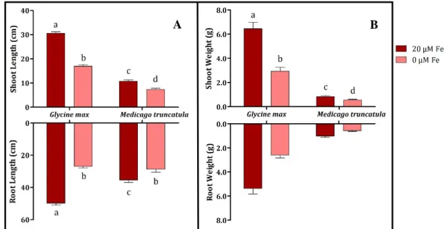
Figure 4.1 - Shoot and root length (A) and weight (B) of G. max and M. truncatula grown hydroponically in Fe-sufficient (●) and Fe-deficient (●) conditions. Means +SE separation were performed independently for each structure, using 12 independent replications. Different letters represent significant differences between samples (p < 0.05).
In general, both species behaved similarly when faced with the absence of Fe. Both plants developed visible IDC symptoms, resulting in a drastic yield reduction. Root and shoot length, decreased 45.6% and 44.5%, respectively, for G. max, and 18.8% and 31.8% for M.
truncatula (Figure 4.1-A). In what concerns root and shoot weight, G. max had 51.7% and
54.6% lower fresh weight in roots and shoots, respectively, under Fe deficiency, which was more pronounced that M. truncatula that had a reduction of 44.7% and 31.7% (Figure 4.1-B).
Another important characteristic associated with the absence of Fe is the development of secondary structures. Thus, secondary roots were counted and compared between the two treatments in both species (Figure 4.2).
A B
Figure 4.2 – Number of secondary roots of G. max and M. truncatula grown hydroponically in Fe-sufficient (●) and Fe-deficient (●) conditions. Data are means +SE of 12 independent replications. Different letters represent significant differences between samples (p < 0.05).
As previously seen by others (Schmidt, 1999; Andaluz et al., 2009), plants submitted to Fe deficiency showed swelling of root tips and increased the number of secondary structures, namely an average of more 40% for G. max and 30% more for M. truncatula. The increased number of secondary structures helps the plant in augmenting the absorbable area for Fe uptake, and the scavenging of Fe in the rhyzosphere (Schmidt, 1999). Since the surface of root hairs can represent up to 70% of the total root surface area (López-Bucio et al., 2003), the relevance of root hairs in nutrient uptake is crucial.
At shoot level, when plants were faced with Fe-deficiency, they developed visible symptoms of chlorosis - e.g. yellowing of leaves - whereas plants grown in Fe-sufficient conditions remained green throughout the experiment, as it can be seen in figure 4.3.
Figure 4.3 - G. max plants grown hydroponically in Fe-deficient (left) and Fe-sufficient (right) conditions.
The yield reduction observed, as previously reported by others (Jolley et al., 1996), is according with the expected, since Fe is essential for several mechanisms that regulate plants growth. The absent of Fe inhibits chloroplast biogenesis and chlorophyll biosynthesis, leading to the development of chlorosis, especially in younger leaves (Henriques et al., 2002).
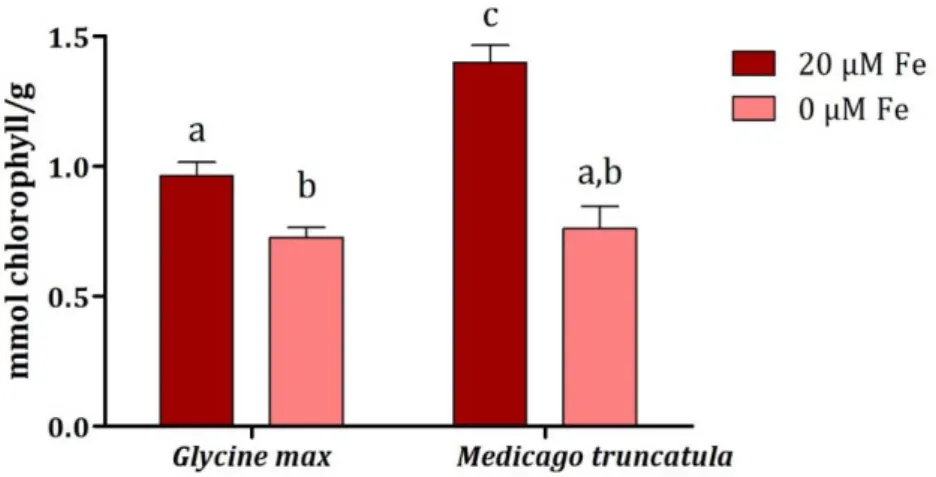
To quantify the visible symptoms observed (as in Figure 4.3) chlorophyll levels of both species were measured (Figure 4.4).
Figure 4.4 - Chlorophyll concentration of G. max and M. truncatula grown hydroponically in Fe-sufficient (●) and Fe-deficient (●) conditions. Data are means +SE of 12 independent replications.Different letters represent significant differences between samples (p < 0.05).
When challenged with the lack of Fe, both plants presented lower chlorophyll levels, namely 24.6% less in G. max and 45.6% less in M. truncatula (Figure 4.4), corroborating the visible symptoms observed in figure 4.1.3. Fe starved plants may be expected to be more prone to oxidative damage since Fe is a constituent of enzymes associated with the cellular antioxidant system, e.g. ascorbate peroxidase, catalase, peroxidase, and Fe-superoxide dismutase (Kumar
et al., 2010). When in lack of this nutrient, these proteins would not function properly, leading
to the accumulation of ROS, to oxidative stress, and to lower chlorophyll levels, as observed in figure 4.4.
As leaves are affected by root Fe concentration, it is possible that the seeds have a similar behavior. However, root Fe uptake capacity is linked with the solubilization of Fe in the rhyzosphere by the plant’s root Fe reductase activity, which is necessary to convert the less soluble Fe3+ to the more soluble Fe2+. Blair et al. (2010) have concluded that, at least in common bean, high seed Fe accumulation is generally correlated with higher Fe reductase activity. This might suggest a relation between root uptake and seed loading of Fe in common bean. Therefore, the supplementation of Fe at root level may help biofortification programs, since some of that additional Fe could also be remobilized to the grains.
4.2 Nutritional impact of low Fe availability
In order to understand how Fe deficiency affects the nutritional composition of Fe and Zn in soybean and M. truncatula, Fe and Zn were quantified by ICP-OES, after the 14d trial (Figure 4.5).
Figure 4.5 – Iron (A) and Zinc (B) content of G. max and M. truncatula grown hydroponically in Fe-sufficient (●) and Fe-deficient (●) conditions. Data are means +SE of 3 independent replications. Different letters represent significant differences between samples (p < 0.05).
Under Fe deficiency, both G. max and M. truncatula, showed lower Fe content, in particular at the shoot level. The implications of this observation are that when grown under Fe deficiency, crop plants will be nutritionally poorer, especially when considering the aerial parts (which include leaf, stem and ultimately, seeds). As Zn shares common transporters to the ones utilized for Fr uptake, and since Zn is also an essential nutrient for human and animal health, Zn content was also analyzed in both plant species. As expected, Zn content was negatively impacted in plants grown under Fe deficiency (Figure 4.5-B). This shows that the impact of Fe deficiency on the plants nutritional status will go beyond the levels of Fe in the edible plant parts to other transportable cations such as Zn and possibly Calcium (Ca), Magnesium (Mg) and Manganese (Mn). This has already been observed in previous studies conducted in rice (Sperotto et al., 2012) and soybean (Vasconcelos et al., 2010).
When a great amount of Fe is available around the root surface, plants tend to uptake this important nutrient and it will be storaged when is no longer needed. In optimal growth conditions, since G. max is much more susceptible to IDC than M. truncatula, the Fe was mobilized to the storage spots in the aerial part of the plant, thus presenting higher Fe content in the leaves (Figure 4.5-A). Regarding the root levels of Fe and Zn, it can be seen that M.
truncatula presented higher Fe content in sufficient conditions, as expected. However, when Fe
is absent, the uptake of Zn also decreases, suggesting that Fe may act as a co-factor in Zn uptake at root level. On the other hand, G. max presented similar values of Zn in the roots in both
conditions. When the plant is faced with lack of Fe, it mobilizes the internal Fe to the roots (Figure 4.5-A), as an internal signal to induce Fe uptake, which leads to the development of IDC symptoms in the leaves, as described before. Faced with that, plants uptake Zn and mobilize it to the shoots, possibly to counterwork IDC, which can be observed in the obtained results (Fig.4.5-B), where G. max presents higher Zn content than Fe in shoots under Fe deficiency.
4.3 Gene Expression Analysis
Since the crops do not all respond to Fe stress equally, it is essential to understand the mechanisms underlying the translocation of Fe from roots and leaves to seeds, in order to regulate Fe accumulation in seeds and design an efficient biofortification program. To this end, it is essential to look at the molecular regulators of these processes, and to identify the rate limiting steps, at a molecular level. Thus, the expression of several selected genes related to Fe metabolism was evaluated.
4.3.1 Fe acquisition: FRO2 and IRT1
The root Fe uptake related genes FRO2 and IRT1 are extremely important since they participate in one of the most critical steps concerning plant defense mechanisms to Fe deficiency, and control the efficiency of Fe uptake. The present work suggests that G. max and
Figure 4.6- Root Fe uptake related genes FRO2 (A) and IRT1 (B) fold of expression, in roots and shoot tissues, of G. max and M. truncatula, grown in Fe-sufficient (●) and Fe-deficient (●) conditions. Roots in Fe deficient conditions were used as comparison term and 18S was used as internal control.
FRO2 is responsible for reducing Fe3+ to Fe2+, being mainly expressed by the roots, the organ responsible for mineral uptake. Studies show that roots are responsible for Fe deficiency sensing (Vigani et al., 2013). Thus, when M. truncatula was submitted to stress conditions, the expression of FRO2 in roots increased, as expected (Figure 4.6-A). Once the plant is faced with this stress, it needs to increase the rate of nutrient uptake, to compensate the lack of Fe and, for that, root membrane reductases must increase their activity and their uptake rate. When Fe was present in sufficient amounts, M. truncatula had almost no FRO2 expression: since from the beginning of the trial, plants have been in optimal conditions, maybe they captured sufficient Fe to meet their daily requirements, inhibiting the FRO2 gene and decreasing Fe uptake in order to avoid Fe toxicity.
On the other hand, G. max presented an opposite expression pattern, showing higher expression in roots, especially in Fe-sufficiency, decreasing 2.98 fold when submitted to the stress condition (Figure 4.6-A). Blair et al. (2010), observed that in some of the genotypes studied, Fe reductase activity, which is directly linked to FRO2 expression, in Fe deficiency conditions, showed lower values than plants grown in Fe-sufficiency. This suggests that the expression may be reduced due to a need for Fe in the FRO2 enzyme itself. As previously seen
in figure 4.5, G. max roots, presented higher Fe content in Fe-deficient conditions, which would enhance the reductase system, that seem to need some Fe to be activated. Fe reductase is known to be the rate-limiting enzyme for Fe uptake since Fe transporters, such as IRT1, do not reach saturation at normally achieved concentrations of Fe2+ (Grusak et al., 1990). If there is no Fe being reduced, IRT1 should consequently present lower activity in G. max Fe-deficient roots, which is clearly observed in figure 4.6-B.
In addition to transporting Fe2+, IRT1 non-selectively mediates the uptake of several divalent transition metal cations into the root symplasm, thus acting as a major pathway for the influx of Zn2+ (Vert, G. 2002). In general, the levels of expression of IRT1 in both species and in both tissues are very similar to those obtained for FRO2, suggesting that IRT1 is co-regulated with this gene, as previously seen by others for A. thaliana (Kim and Guerinot, 2007). If FRO2 gene is more activated, it will reduce more Fe, and consequently IRT1 will transport more Fe into root tissues, showing a higher level of expression. If, on the other hand, Fe is not being reduced by FRO2, IRT1 has no need to be activated, showing low expression levels.
Studies on Fe homeostasis (Vert, G. 2002) showed evidence for long-distance systemic signalling of metal status in different crop species. Experiments with Arabidopsis, in which half the root system of was placed in Fe-deficient hydroponic medium and the other half was placed in Fe-replete medium, showed that IRT1 and FRO2 transcript levels are regulated by both local Fe levels in the apoplast and by a shoot-derived systemic Fe deficiency signal (Vert, G. 2002).
4.3.2 Fe transport: NRAMP3, VIT1 and YSL1
After Fe is transported into the roots by IRT1, the transport of this nutrient across the plant is another crucial step that needs to be well known to efficiently develop a biofortification strategy. Fe transporter families, such as VIT, NRAMP and YSL, are extremely important in Fe metabolism, as they assure that Fe is efficiently delivered to shoots, other plant edible parts and storage organs.
Figure 4.7 – Fe transporter genes NRAMP3 (A), VIT1 (B) and YSL1 (C) fold of expression, in roots and shoot tissues, of G. max and M. truncatula, grown in Fe-sufficient (●) and Fe-deficient (●) conditions. Roots in Fe deficient conditions were used as comparison term and 18S was used as internal control.
NRAMP3 and VIT1 have contrasting functions: while the first is responsible for the
remobilization from the vacuole (Lanquar et al., 2005), the second is responsible for the Fe loading in the vacuole (Kim et al., 2006).
In figure 4.7 it can be seen that these genes are mainly expressed in shoots, whereas in roots they present lower expression. In shoots, an increase of NRAMP3 expression of 2.08 and 1.17 fold was observed for G. max and M. truncatula, respectively, when submitted to Fe deficiency (Figure 4.7-A). Studies in A. thaliana demonstrate that NRAMP3 is an H+ metal symporter responsible for Fe and Mn remobilization from the vacuole, a crucial step during early seedling development (Lanquar et al., 2010). Thus, the results obtained here are coherent because, when under Fe deficiency, plants need more remobilization of Fe to respond to their needs, which means that NRAMP3 will be more expressed. In roots, the expression levels were
lower and similar in both conditions, for both species (Figure 4.7-A), again supporting the hypothesis that this gene’s role is predominant in the leaves, the principal storage organ of Fe.
Knowing the role of VIT1, the expression of this gene is expected to be higher in Fe-sufficient leaves, where there is a need for Fe storage, which was clearly observed in both species (Figure 4.7-B). In shoots, the expression levels were 1.78 and 1.56 fold higher in Fe sufficient leaves, for G. max and M. truncatula, respectively (Figure 4.7-B). These results are in accordance with previous reports, because when Fe is in sufficient conditions, plants activate
VIT1 to increase Fe2+ uptake into the vacuole for storage purposes (Brear et al., 2013), in order to minimize waste, and reduce the risk of oxidative damage. In roots, M. truncatula had similar expression for both conditions, whereas in G. max the expression increased 3.57 fold in Fe deficiency (Figure 4.7-B).
Studies in A. thaliana (Kim and Guerinot, 2007) demonstrate that AtNRAMP3 and AtVIT1 mutants present arrested seedling growth on plants grown on Fe-limiting soil. Moreover, Zhang et al. (2012) reported that the disruption of the rice VIT orthologues (OsVIT1 and OsVIT2) increased Fe and Zn accumulation in rice seeds and decreased Fe and Zn in the leaves. Taken together, these results suggest that vacuoles are an important site of Fe storage and Fe remobilization during germination, and without leaf accumulation, there is enhanced Fe and Zn translocation to the seed, providing one successful strategy to biofortify Fe and Zn.
Besides NRAMP3 and VIT1, YSL1 has an important role in Fe metabolism, being involved in the transport of the Fe2+-NA complexes (Kim et al., 2006). This complex, as mentioned in the introduction section is hypothesized as the main transportable Fe form in the phloem.
Hence, it was expected that the expression levels of YSL1 would be higher in Fe-sufficient conditions, because when there is more amount of Fe, more transporters are needed to satisfy the plant’s demand. Both species presented the expected results in both studied tissues (Figure 4.7-C), since both presented higher YSL1 relative expression values under Fe-sufficient conditions. Similar results in both tissues suggest roles in Fe translocation at diverse plant organs (Kim et al., 2006). Jean et al. (2005) showed that by knocking out AtYSL1, the levels of NA and Fe in leaves and seeds decreased, as well as the germination rate, even when plants were grown in the presence of an excess of Fe, suggesting that Fe and NA levels in seeds rely in part on YSL1 function. Thus, as well as NRAMP3 and VIT1, YSL1 transporter may also play a crucial role in the control of the amount of Fe translocated to the seeds. Overexpressing this
gene could lead to an increased translocation of Fe to the edible plant parts, resulting in seeds with higher Fe concentration.
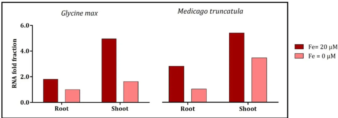
4.3.3 Fe storage: Ferritin
Storage proteins like Ferritin play an important role in Fe homeostasis, assuring that Fe in excess is in a bio-available way in case of cellular needs but yet nonreactive with oxygen (Briat et al., 2010).
Figure 4.8 – Fe storage related gene Ferritin fold of expression, in roots and shoot tissues, of G. max and M. truncatula, grown in Fe-sufficient (●) and Fe-deficient (●) conditions. Roots in Fe deficient conditions were used as comparison term and 18S was used as internal control.
Both G. max and M. truncatula down-regulated the Ferritin gene under stress conditions. In shoots, when submitted to Fe deficiency, the expression decreased 3.06 and 1.56 fold, for G. max and M. truncatula, respectively (Figure 4.8). At root level, the expression decreased 1.81 and 2.73 fold, for G. max and M. truncatula, respectively (Figure 4.8). These results are in accordance with the expected since Ferritin plays an important role in Fe storage, so when there is sufficient amount of Fe within the plant, it needs to be stored, and Ferritin expression increases. Thus, this protein manages the insolubility and potential toxicity of Fe in the presence of oxygen, being involved in oxidative protection by sequestering free Fe (Lobreaux, 1995). By manipulating the expression of the vacuolar Fe transport systems AtVIT1,
AtNRAMP3, and AtNRAMP4, Ravet et al. (2009) have shown that the control of Fe fluxes
between vacuoles and plastids is an important factor of Fe homeostasis in seeds.
One of the strategies studied for Fe biofortification has been the overexpression of the
Ferritin gene (Vasconcelos et al., 2003). However, it has been shown that, independently of Fe
content increase in shoots, the content in the grain does not increase proportionally (Drakakaki
2012). Besides, Ravet et al (2009) have shown that when Fe is loaded into seeds, at its maximum rate, Ferritin gene expression is almost lacking. Therefore, when designing new biofortification approaches, it is crucial to study all stages of development and the genes involved in each phase. Down-regulating Ferritin gene expression in leaves could allow more Fe storage in the seeds. However, one must be careful with the possible negative consequences in terms of oxidative stress.
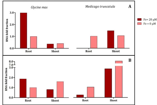
4.3.4 Novel genes: GCN2 and WEE1
Even though several gene families are known to be involved in the Fe uptake, transport and storage, there are still many undiscovered genes that may have important roles in these processes. Therefore, to efficiently construct a biofortification strategy it is worthwhile to find candidate genes that could have an important role in Fe metabolism. To this end, two novel genes were studied in the current work: GCN2 and WEE1. To ascertain their putative role in mineral nutrition, the expression of these selected genes was evaluated (Figure 4.9).
Figure 4.9 – Novel candidate genes GCN2 (A), WEE1 (B) fold of expression, in roots and shoot tissues, of G. max and M. truncatula, grown in Fe-sufficient (●) and Fe-deficient (●) conditions. Roots in Fe deficient conditions were used as comparison term and 18S was used as internal control.