Determination of
macrophage
activation profile
and T cell
populations in the
context of
congenital
toxoplasmosis
Maria Miguel Ferreira Pinto Menezes de
Castro
Mestrado em Bioquímica
Departamento de Química e Bioquímica 2016
Orientador
Professora Doutora Margarida Borges, Professora auxiliar da
Faculdade de Farmácia da Universidade do Porto
Agradecimentos
Em primeiro quero agradecer à minha orientadora Professora Doutora Margarida Borges, por me ter proporcionado a oportunidade de integrar o seu grupo de investigação, pela orientação, apoio prestado, pelo conhecimento que me transmitiu, a disponibilidade e o tempo dedicado. Gostaria também de agradecer à Professora Dra. Natércia Teixeira pela oportunidade que me deu em trabalhar no seu grupo de investigação no laboratório de Bioquímica da Faculdade de Farmácia da Universidade do Porto. Agradeço à Professora Doutora Maria de São José Nascimento por ter disponibilizado o seu laboratório de cultura celular para a realização das culturas in
vitro do parasita utilizado neste estudo. Agradeço também ao Biotério do ICBAS e a
toda a equipa associada, à Professora Doutora Margarida Araújo, à Bárbara, à Salomé e à Cátia na obtenção dos animais gestantes, inoculação e colheita de órgãos, fundamental para a realização deste trabalho.
Quero agradecer à Doutora Tânia Silva por tudo o que me ensinou sobre culturas das HFF e do parasita, por toda a sua disponibilidade e apoio prestado. Agradeço à Mestre Carina pela amizade, pelo tempo que disponibilizou para me ensinar diversas técnicas e pelas horas extras que passamos no laboratório (até quando Portugal joga). Agradeço à Sónia e à Joaninha, pela amizade, por toda ajuda e disponibilidade sempre presente (colar etiquetas era sempre mais rápido com a vossa ajuda!).
Aos meus colegas e amigos que diariamente partilharam o laboratório comigo Renata, Fernanda, Bárbara, Tiago e João, por toda a amizade ao longo deste ano, pelo apoio, por terem estado sempre presentes, por todos os almoços partilhados e todas as brincadeiras que fizeram com que os dias no laboratório passassem a correr. Gostava de agradecer à Doutora Cristina, à Doutora Marta, ao Doutor Bruno, à Doutora Sandra e à Doutora Susana por todo apoio, pelo bom ambiente que está sempre presente no laboratório, pelas palavras amigas e pela disponibilidade sempre prestada.
Agradeço a todos os professores do departamento de Bioquímica FFUP por toda a disponibilidade e pelo bom ambiente. Agradeço à Dona Casemira e à Ana Paula, “pilares” fundamentais no laboratório de Bioquímica, e também à Cristina do laboratório de Microbiologia por todo o apoio e disponibilidade.
Quero agradecer às minhas meninas do 404 da RUCA I por toda a paciência e por todas as horas que me ouviram a falar da tese, no fundo por toda a amizade ao longo destes dois anos. Agradeço à Inês pela amizade, pelo apoio e pelo que partilhamos nestes dois anos, incluindo as dificuldades e os bons momentos das nossas teses. Gostava também de agradecer aos meus “meninos e meninas” do mestrado com quem neste ano partilhei muitos jantares, cafés e almoços na faculdade.
Não poderia deixar de agradecer às minhas duas grandes amigas, Ana Isabel e Daniela por estes 6 anos de amizade, pois apesar dos Km que nos separaram nestes últimos dois anos, estão sempre por perto para podermos partilhar juntas os bons e os maus momentos. Aos meus amigos Lurdes, João, Diogo, Francisco, Joana, Ana e a todos os outros amigos candidatos, pela força, apoio e oração neste ano. Aos meus amigos de Aldoar pela força, apoio e compreensão nesta última fase. Aos amigos do CREU-IL, especialmente, à Ana pela amizade, pelos lanches e pela força que sempre me transmitiu.
Por último, agradeço aos meus pais, em especial à minha mãe, por todo o apoio, compreensão, paciência, pelo esforço que fizeram ao longo dos últimos 6 anos para eu conseguir continuar a estudar, por me deixarem ser quem sou, por estarem sempre presentes para me ouvir e pelos bons conselhos. Obrigada por tudo!
Resumo
O Toxoplasma gondii é um parasita humano bem-sucedido que infecta milhões de pessoas em diversos países. Em indivíduos imunodeprimidos, o parasita causa uma doença severa com diversas manifestações clinicas. No entanto, os indivíduos imunocompetentes são assintomáticos, podendo este parasita persistir no hospedeiro por um longo período de tempo. Nas mulheres grávidas, uma primo-infeção com T.
gondii pode levar a severas manifestações clinicas, como por exemplo, ao aborto ou a
malformações do feto, como coriorretinite, hidrocefalia e encefalites. Assim, é importante entender os mecanismos e as células imunológicas envolvidas na resposta à infeção com este parasita tanto a nível sistémico como a nível da interface feto-materna. Neste trabalho, tivemos como principais objetivos: determinar o efeito da infeção nos parâmetros básicos da função reprodutiva durante a gravidez; quantificação do T. gondii em vários tecidos e células; e a análise de neutrófilos, macrófagos e populações de células T nas células esplénicas e do exsudado peritoneal. Para a infeção, usou-se uma estirpe do tipo II de T. gondii e murganhos Balb/c gestantes e não gestantes. Análises macroscópicas foram realizadas para avaliar os locais de implantação. Para a quantificação do parasita foi realizada a técnica de Real-Time PCR quantitativo. As populações de neutrófilos, macrófagos e células T foram avaliadas por citometria de fluxo. Os resultados obtidos neste trabalho demonstraram, que após 5 dias de infeção com T. gondii Me49 não ocorreu a disseminação do parasita desde do local da infeção (cavidade peritoneal) até ao pulmão, coração e rim. Foi no entanto, possível confirmar a disseminação sistémica do parasita após 7 dias de infeção. Neste trabalho foi possível efetuar a deteção e quantificação de células do exsudado peritoneal - T. gondii+ pela técnica de citometria de fluxo para 5 e 7 dias após infeção. A análise da expressão de moléculas MHCII por citometria de fluxo, indicou um aumento significativo da expressão de MHCII nos small
peritoneal macrophages comparativamente aos large peritoneal macrophages, nos
murganhos não gestantes ou gestantes, não infetados. Foi demonstrado que após 7 dias de infeção a população dos large peritoneal macrophages é inexistente em murganhos não gestantes ou gestantes. Ao nível do baço, foi possível verificar uma diminuição significativa da percentagem da população CD11b+F4/80+MHCII+ e uma
diminuição na expressão de MHCII nestas células, no dia 12 de gravidez durante a infeção com T. gondii. Estes resultados sugerem que T. gondii poderá influenciar vias dependentes da gravidez, interferindo com a capacidade da apresentação antigénica.
Palavras-chave
Toxoplasmose congénita, Toxoplasma gondii, Macrófago, Neutrófilo, Célula T, MHCII, Arginase-1, NOS2, Balb/c, Gravidez
Abstract
Toxoplasma gondii is a successful human parasite, which infects millions of people in
several countries in the world. In immunosuppressed individuals, the parasite causes a severe disease with many clinical manifestations. However, immunocompetent individuals are asymptomatic, but this parasite persists for long periods of time. In pregnant women, T. gondii primo-infection can lead to a variety of severe clinical manifestations, like abortion or fetal malformations, as chorioretinitis, hydrocephalus and encephalitis. Therefore, is important to understand the immunological mechanisms and cell players involved in T. gondii immune response at the systemic level and maternal-fetal interface. In this work, the principal aims were: to determine the effect of infection in basic parameters of reproductive function in pregnancy; T. gondii quantification in tissues and cells; and neutrophil, macrophage and T cell populations analysis in spleen and peritoneal exudate cells. Infection using a type II strain of
T.gondii of pregnant and non-pregnant Balb/c mice, allowed the follow-up of
pregnancy. Macroscopic analyses were performed to evaluate implantation sites. The quantification of the parasite was performed by quantitative Real-Time PCR. Neutrophil, macrophage and T cell populations were evaluated by flow cytometry analysis. The results obtained, indicated that 5 days of infection were not sufficient for the parasite dissemination from the local of infection (intraperitoneal cavity) to the lung, heart and kidney. However, it was possible to confirm systemic parasite dissemination after 7 days of infection. The data obtained indicated that flow cytometry technique is sensible to detect peritoneal exudate cells-T. gondii+ at 5 and 7 days post-infection. MHCII expression was significantly increased in small peritoneal macrophages compared to large peritoneal macrophages from uninfected non-pregnant or pregnant mice. It was demonstrated that large peritoneal macrophages are inexistent after 7 days of T. gondii infection in non-pregnant or pregnant mice. At spleen level, it was found a significant decrease in the percentage of CD11b+F4/80+MHCII+ and a MHCII+
expression down-regulation in CD11b+F4/80+, at day 12 of pregnancy during T.gondii
infection. These results, suggest that T. gondii may be influencing pregnancy-dependent pathways, interfering with the capacity of antigen presentation.
Keywords
Congenital toxoplasmosis, Toxoplasma gondii, Macrophage, Neutrophil, T cell, MHCII, Arginase-1, NOS2, Balb/c, Pregnancy
Table of Contents
Agradecimentos ... I Resumo ... III Abstract ... V List of figures ... IX List of tables ... XI List of Abbreviations ... XIIIntroduction ... 1
1. Toxoplasma gondii ... 1
2. Parasite morphology and Life-cycle ... 2
3. Transmission ... 4
3.1. Tissue cyst transmission ... 4
3.2. Congenital transmission ... 5
3.3. Oocysts transmission ... 5
3.4. Organ transplantation and laboratory infection ... 5
4. Animal hosts ... 6 5. Humans infection ... 7 5.1. Congenital toxoplasmosis ... 7 6. Epidemiology... 8 7. Diagnosis ... 10 8. Treatment ... 11
9. Prevention and control ... 12
10. Maternal-fetal interface (FMI) and immune cell populations ... 13
10.1. NK and Decidual NK cells ... 15
10.2. T cells ... 15
10.3. Decidual Macrophages ... 16
10.4. Dendritic cells ... 19
11. The immunobiology response to T. gondii ... 19
12. Animal study model ... 21
Aims ... 23
Materials and Methods ... 25
1. Parasite ... 25
2. Human Foreskin Fibroblast cells (HFF) in vitro infection ... 25
4. Collection of biological samples ... 26
5. Quantitative real-time PCR (q-PCR) ... 27
6. Flow cytometry analysis ... 27
7. Statistical analyses ... 29
Results ... 31
1. Maintenance of parasite culture ... 31
2. In vivo infection and pregnant Balb/c ... 32
3. Implantation units ... 34
4. Pregnancy induces splenomegaly independently of T. gondii infection... 34
5. Parasite load quantification ... 36
6. T cell populations ... 40
7. Macrophage and neutrophil populations ... 45
8. Arg-1 and NOS2 expression macrophages and neutrophils ... 47
9. MHC II expression in macrophages ... 49
Discussion ... 54
Conclusion ... 58
List of figures
Fig.1 -Life cycle stages of T. gondii……….. 2
Fig.2 - The different infectious stages of T.gondii life cycle ……… 3
Fig. 3- The life cycle of T. gondii……….. 4
Fig. 4- Different pathways of T. gondii infection and transmission………... 6
Fig. 5- Global status of T. gondii seroprevalence ……….. 9
Fig.6- Schematic representation of the maternal-fetal interface (FMI)……… 14
Fig.7 – Different types of macrophages polarization profile and the different subclasses of M2 profile………. 18
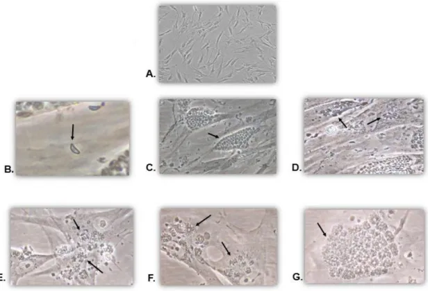
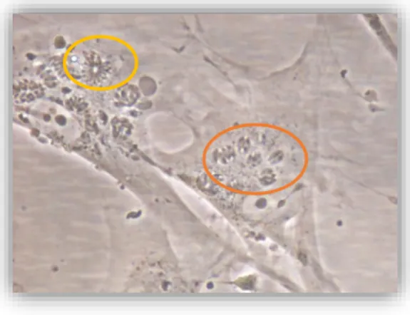
Fig. 8- In vitro culture of T. gondii-infected HFF cells……… 31
Fig. 9 - T. gondii- infected HFF cells……… 32
Fig. 10- Representative histogram overlay of PEC TOXO YFP+……… 33
Fig. 11- Macroscopic evaluation of Implantation units………. 34
Fig. 12- Pregnancy and T. gondii infection influence on spleen size and spleen cell numbers……….. 35
Fig. 13 –Quantification of PEC and spleen cells TOXO-YFP+ by flow cytometry……….. 40
Fig. 14 – Representative flow cytometry gating strategy used to define CD4+ and CD8+ T cells in PEC [A] and in spleen cells [B]………. 41
Fig. 15- Evaluation of T cell populations in PEC………. 42
Fig. 16 – Evaluation of T cell populations in spleen cells……….. 44
Fig. 17 –Representative flow cytometry gating strategy used to define granulocytes (CD11b+), neutrophils (CD11b+Ly6G+) and macrophages (CD11b+ Ly6G-F4/80+) in PEC [A] and spleen cells [B]……….. 46
Fig. 18– Evaluation of granulocytes (CD11b+), neutrophils (CD11b+Ly-6G+) and macrophages (CD11b+Ly-6G- F4/80+) in PEC [A] spleen cells percentage [B] and spleen cells number [C]……… 47
Fig. 19 – Representative flow cytometry gating strategy used to define spleen M1 (CD11b+F4/80+NOS2+) and M2 (CD11b+F4/80+Arg-1+) macrophages [A] and spleen neutrophils expressing NOS2 (CD11b+Ly6G+NOS2+ ) and Arg-1 (CD11b+Ly6G+Arg-1+) [B]……… 48
Fig. 20 – Evaluation of macrophages [A,B] and neutrophils expressing Arg-1 and NOS2……….. 49
Fig.21- Representative flow cytometry gating strategy used to define CD11bhighF4/80highMHCII+ (LPM) and CD11bintF4/80intMHCII+ (SPM) in PEC [A] Data
from E0-Ninf animal. [B] Data from a E14-Inf animal……… 50
Fig.22- Evaluation of MHCII in LPM and SPM……… 51 Fig.23- Representative flow cytometry gating strategy used to define MHCII
CD11b+F4/80+ spleen cells………. 52
List of tables
Table 1 – Risk of T. gondii congenital infection in different gestational age and
development of clinical signs in the infected offspring……….. 8
Table 2 – Toxoplasmosis manifestations and their treatment indications……….. 12
Table 3 –Strain mice and susceptibility of T. gondii acute infection……… 22
Table 4 – Number of pregnant Balb/c mice considered in this study………. 33
Table 5- Number of non-pregnant Balb/c mice considered in this study………... 33
Table 6- Parasite load quantification in E12-Inf and E0-5 days Inf mice……… 37
Table 7 - Parasite load quantification in E14-Inf and E0-7 days Inf mice………... 37
Table 8 - Parasite load quantification at FP interface and embryo from E12-Inf mice…. 38 Table 9- Parasite load quantification at FP interface and embryo from E14-Inf mice….. 39
List of Abbreviations
Abs – Antibodies
AIDS – Acquired immune deficiency syndrome Arg-1 – Arginase- 1
Balb/c - Balb\cByJ CTLs- Cytotoxic T cells DCs - Dendritic cells
dDCs - Decidual dendritic cells
DMEM - Dulbecco’s modified eagle medium DNA - Deoxyribonucleic acid
dNK - Decidual Natural Killer E- Day of gestation
ELISA- Enzyme-linked immunosorbent assay EVT – Extravillous trophoblast cells
FBS - Fetal bovine serum FMI- Maternal-fetal interface FP- Fetoplacental units
HFF- Human foreskin fibroblasts Ig - Immunoglobulin
IL - Interleukine
IFN-ϒ – Interferon gamma Inf – Infected
ip –Intraperitoneal
LAMP- Loop-mediated isothermal amplification LPM - Large peritoneal macrophages
MFI – Mean fluorescence intensity MHC- Major Histocompatibility complex MIP-Macrophage inflammatory protein NK – Natural Killer
dNK- Decidual natural killer cells NO – Nitric oxide
NOS2 or iNOS – Inducible nitric oxide synthase PBS – Phosphate-buffered saline
PCR - Polymerase chain reaction PEC - Peritoneal exudate cells
q-PCR - Quantitative real-time polymerase chain reaction ROI - Reactive oxygen intermediates
SAG-1 – surface-antigen 1
SPM - Small peritoneal macrophages
T. gondii – Toxoplasma gondii
Th - T helper cells TLR - Toll-like receptor TNF- Tumor necrosis factor Treg – Regulatory T cells
TGF- β - Transforming growth factor beta TOXO – Toxoplasma gondii
Introduction
1. Toxoplasma gondii
T.gondii is an important group of obligate intracellular protozoan of the Phylum Apicomplexa, subclass coccidian, order Eimeriorina and family toxoplasmatidae (1,2).
The cat constitutes the definitive host and other warm-blooded animals, such as humans, are the intermediate hosts (3). The first report of T.gondii occurred in 1908 by Charles Nicolle and Manceuax using tissues of hamster-like rodent, Ctenodactylus
gundi, which was used in leishmaniasis research in the North Africa. Initially, they
believed that the parasite found was Leishmania. However, they realized that it was a new organism and named it as T. gondii (4). This name derived from its crescent-shaped morphology of tachyzoite the Toxo means “arc form” in Greek and gondii derived from the first found host (5). At the same time another scientist, Alfonso Splendore, discovered a similar parasite in rabbits in Brazil (6).
In the next years, T. gondii was found in several other hosts, mainly in avian species (7). However, only in 1937 Sabin and Olitsky isolated, for the first time, viable
T. gondii from the animal host and in 1941, they proved that human T. gondii was
identical to the parasite present in other hosts (8). Later in 1940’s antibodies were detected and found to kill extracellular but not intracellular tachyzoites (8,9). In 1970 the life cycle of this parasite was discovered, allowing a better understanding of
T.gondii transmission (10). In the course of 1980’s and 1990’s various methods, were
developed to recognize genetic differences between T. gondii isolates from animals and humans (11–14). Many advances have been achieved due to T. gondii genes mapping helping in the search of better antigens for the diagnosis, prevention and the knowledge of disease mechanisms (15).
T.gondii infects millions of people in the world and is the most successful human
parasite (16). This parasite can cause severe clinical manifestations specifically in two distinct cases: immunodeficient individuals and primo-infected pregnant women. The primo-infection in pregnant women can lead to a congenital toxoplasmosis, with diverse clinical manifestations such as hydrocephalus, macrocephaly, encephalitis, seizures, calcifications, chorioretinitis, blindness in the fetus and may also result in intrauterine death and spontaneous abortion (17).
2. Parasite morphology and Life-cycle
T. gondii present three infectious stages, such as sporozoites which disseminate in
the environment within oocysts, tachyzoites which facilitate expansion during acute infection and bradyzoites which maintain the chronic infection being all the three stages haploid forms (Fig.1 ; 17,18).
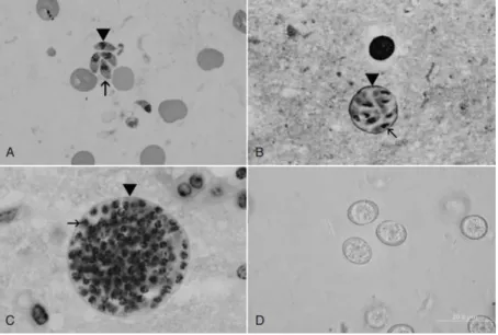
Fig. 1- Life cycle stages of T. gondii. A- Tachyzoites; B-Small tissue cysts; C- Tissue cysts; D- Unsporulated oocysts (2).
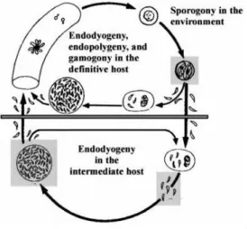
The life cycle of this parasite has both asexual (schizogony) and sexual (gametogony) reproduction, which occur in the small intestine of the definitive host (Fig.2; 19). Tachyzoites and bradyzoites divide asexually, while sporozoites are the parasite forms obtained after sexual reproduction (21).
The sexual phase or gametogony occurs throughout the small intestine and in the ileum. The microgamete (the male gamete) is biflagellate and fertilizes the macrogamete (the female gamete) within the enterocyte. The fertilization initiates oocysts wall formation, and the product of this sexual reproduction is the zygote. The oocysts are released into the intestinal lumen by rupture of the epithelial cells and thereafter excreted in cat feces (18). The oocysts release, from the epithelial cells, takes 3 to 10 days after ingesting of bradyzoites, >18 days after ingesting of sporulated oocysts and >13 days after ingesting of tachyzoites (22). Sporogony occurs outside of the host within 1-5 days after excretion, depending on atmospheric conditions, such as aeration, humidity, and temperature. Each oocyst contains two sporocysts, which contain four sporozoites (18).
Fig. 2- The different infectious stages of T.gondii life cycle, i.e. tachyzoites, bradyzoites contained in tissues cysts and sporozoites contained in sporulated oocysts (23).
The asexual phase can occur after ingesting the tissue cysts or infectious oocysts (23). After this ingestion by an intermediate host (such as humans), the sporozoites are released from the oocysts, penetrate the intestine, invade a range of different cell types and multiply asexually, as tachyzoites (24). This parasite form is crescent or oval and is present during acute infection and multiply rapidly in a wide variety of nucleated cell hosts, within the parasitophorous vacuole (21,25). The parasite invasion is due to an actin-based motility, being the formation of this vacuole derived from the host cell plasma membrane and secretion of parasite proteins (26). The tachyzoites divide every 6-9h by a process denominated endodyogeny, in which daughter cells form multiply within the mother cell (27). After replication, the host cells are disrupted, and the tachyzoites disseminated through the bloodstream, infecting many tissues, including the CNS, eye, skeletal, and heart muscles, placenta leading that way to diverse clinical manifestations (25). Finally in response to environmental conditions, the tachyzoites converts into bradyzoites forming the tissue cysts, present in different organs and tissues (Fig. 3; 27). Bradyzoites replicated slowly by endodyogeny, they are morphologically identical to tachyzoites and are considered as the latent form able to remain in the tissues for the entire host life (18,21,25).
Depending on several environment factors, the tissue cysts can rupture and bradyzoites forms can convert to tachyzoites resulting in the reactivation of infection. This reactivation may result in various clinical manifestations, as toxoplasmic encephalitis, an opportunistic disease which can occur in immunodeficient patients (18).
Fig. 3- The life cycle of T. gondii (2).
3. Transmission
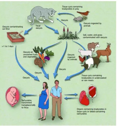
The transmission of Toxoplasma gondii may occur in three different ways: Tissue cyst transmission, congenital transmission, and oocyst transmission. However, it can also occur by organ transplantation or laboratory infection (Fig. 4). In the next subtitles it each of the routes of transmission will be exposed.
3.1. Tissue cyst transmission
In 1954 Weinman and Chandler suggested that transmission could occur through the ingestion of undercooked meat. Later on, in 1960, Jacobs et al. provided evidence to support this idea, demonstrating that T. gondii was resistant to proteolytic enzymes due to T. gondii forms derived from cysts (29). Nowadays, we know that the primo-infection of cats can occur by eating infected prey, such as rodents, birds, initiating the sexual reproduction and production of oocysts in small intestine (30). Although, intermediate hosts can also acquire toxoplasmosis via ingestion of tissue cysts (20)
Intermediate hosts, such as pigs, sheep, and goats, can present tissue cysts in muscle and brain. Therefore, Human can acquire the infection with T. gondii through the ingestion or handling of undercooked or raw meat from infected animals (25). After ingestion, the cyst wall can usually suffer a rupture by proteolytic enzymes in the stomach, occurring the release of bradyzoites, which are resistant to proteolytic digestion, and therefore they survive and initiate the infection in the small intestine (20).
3.2. Congenital transmission
In 1939, three pathologists, Wolf, Cowen and Paige, identified the first case of Human congenital toxoplasmosis. They have then reported that the T.gondii infection was associated with encephalomyelitis, hydrocephalus, chorioretinitis and encephalitis in the developing fetus (31). Later on, these clinical manifestations were found in other animals species, such as sheep, goats, and rodents (32).
3.3. Oocysts transmission
For many years, the infection with T. gondii has been closely connected to carnivorism or congenital transmission. However, doesn’t explaining the widespread infection in vegetarians and herbivores. Therefore, in 1965, Hutchison discovered, the oocysts, the parasite form present in the cat feces (33). This new form was able to transmit the infection to mice and other intermediate hosts, being stable and able to survive for up to 12-18 months in the environment, depending on the climatic conditions (34).
The cat is able to excrete up 10 million of oocysts per day, which sporulates in 1 to 5 days, becoming infective (19). The oocyst form can contaminate soil, water, and food, like vegetables and fruits. Therefore, transmission can occur by the ingestion of contamined water, vegetable and fruits (35).
3.4. Organ transplantation and laboratory infection
Another route of transmission, which is less frequent is by the organ transplantation. Heart, heart-lung, kidney, liver, or liver-pancreas transplantation, can lead to the transmission of T. gondii by a seropositive donor to a seronegative receptor (25). In the laboratory, the technician can be infected by contacting with contaminated needles, glassware or infected tissues or blood derived products (25).
Fig. 4- Different pathways of T. gondii infection and transmission (36).
4. Animal hosts
T. gondii can cause infection, a severe disease and even mortality in diverse
species of domestic and wild animals (37). In livestock animals, such as pigs, sheep and goats were, frequently, observed tissue cysts of T.gondii (23). In sheep and goats, the parasite may cause embryonic death and resorption, fetal death, abortion, stillbirth and neonatal death (38). However, in the case of cattle and horses, both presented resistance to the parasite (39). T. gondii infection is also very frequently in sea mammals, such as seals, dolphins, sea lions and others (37). Clinical toxoplasmosis has also been reported in several animals: carnivores (chinchillas, mink, ferrets, foxes, raccoons, skunk and black bears), wild pigs, wild felids, small mammals (rats, mice, rabbits, guinea pigs, squirrels), monkeys, marsupials and birds (38).
5. Humans infection
In immunocompetent individuals the primo-infection with T.gondii is asymptomatic, but about 10 % of infected individuals became symptomatic, with the development of lymphadenopathy (40), accompanied by fever, malaise, night sweats, myalgias, sore throat, maculopapular rash, abdominal pain, hepatosplenomegaly and small numbers of atypical lymphocytes (41).
In immunosuppressed patients, the clinical manifestation is more severe with splenomegaly, polymyositis, dermatomyositis, chorioretinitis, myocarditis, pneumonitis, hepatitis, encephalitis and multi-system organ failure (42). Ocular toxoplasmosis is the most common cause of chorioretinitis (43) and can be acquired congenitally, but it can also be developed at the post-natal period or can be a result of infection reactivation in immune-compromised and/or pregnant individuals (44). The toxoplasmic chorioretinitis cause decreased vision, blindness or glaucoma. (43). In the case of acute chorioretinitis symptoms like blurred vision, scotoma, pain, photophobia, epiphora or loss of central vision may occur (41). Encephalitis is the most frequent manifestation of
T.gondii disease characterized by a unifocal or multifocal lesion at the central nervous
system in immunosuppressed individuals (45,46). The symptomatology consists of headache, fever, psychomotor or behavioral changes, confusion, lethargy, hemiparesis, seizures, ataxia and cranial nerve palsies (46). AIDS positive individuals represent the majority of immunosuppressed patients exhibiting this clinical form (47).
5.1. Congenital toxoplasmosis
Congenital toxoplasmosis can occur when a woman acquires T.gondii infection for the first time during pregnancy. The infection occurs through the transplacental passage of the tachyzoites stage, thereby infecting the fetus (vertical transmission) (48). In the case of immunocompetent women that had been infected previously to their pregnancy, they are rarely reinfected (48). Although, it is described that reactivation of T. gondii latent infection occurs in immunocompromised women during pregnancy (49).
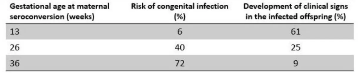
The severity and incidence of congenital infection depends the amount of weeks of pregnancy in which the woman acquires the infection. In the first weeks the risk of transmission is low (5-25%) but the offspring presents severe clinical signs, which can result in spontaneous abortion, hydrocephaly, and mental retardation. Otherwise, in later weeks of pregnancy the risk of transmission increase (to 90%), but with less
clinical signs, such as chorioretinitis and potential blindness (Table 1; 19,49). Although some other clinical manifestations can occur, including miscarriage, fetal developmental retardation, encephalitis, microcephaly, intracranial calcifications, strabismus and epilepsy (51).
The global maternal-fetal transmission rate in immunocompetent women who acquires primary T. gondii infection in pregnancy is approximately 29% (52) and in Europe, congenital toxoplasmosis affects normally 1 to 10 in 10 000 newborn babies (53). These numbers indicate that T. gondii infection is a worldwide problem and more research work is needed to understand parasite transmission and how the mother immune response can be determinant in controlling congenital toxoplasmosis.
Table 1 – Risk of T. gondii congenital infection in different gestational age and development of clinical signs in the infected offspring. Adapted from (49).
6. Epidemiology
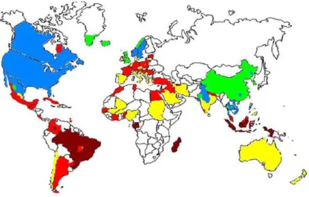
T. gondii infects approximately 25 to 30% of the human population in different
areas of the world (Figure 6; 24). The seroprevalence depends on various factors, such as the environmental temperature. So in temperate countries, the range varies from 10 to 50% and in tropics countries the range reaches over 80% (54). The lower seroprevalence ranges 10 to 30% have been reported in South East Asia, in Sahelian countries of Africa, as well as in cold climate areas such as Northern Europe and North America. However, moderate seroprevalences between 30 to 50% have been observed in countries of Southern and Central Europe, but high incidences have been observed in Latin America and tropical African countries (55).
In the United States, a study of National Health and Examination Nutrition Survey (NHANES) demonstrate that T. gondii incidence decreased in U.S.-born persons aged between 12 to 49 years, from 14.1 % between 1988 to 1994 and to 9% in 1999 to 2004 (56). Additionally, recent studies have shown that prior infection to pregnancy is
common, however when congenital toxoplasmosis occurs as prior infection is relatively uncommon and estimated from 400 to 4000 cases per year of this disease (57).
In Europe, it is observed a declining in the T. gondii seroprevalence in pregnant women. In France, the seroprevalence in pregnant women were about 80% in the early 1960’s, around 66% in the 1980’s, 54% in 1995 and 44% in 2003. At the same time, the average age rate of pregnant women enhanced (58). In the Netherlands, the seroprevalence declined from 35,2% in 1995-1996 to 18,5% in 2006- 2007 in women of reproductive age (59).
Fig. 5- Global status of T. gondii seroprevalence .Dark red corresponding to prevalence about 60%, light red to 40-60%, yellow 20-40%, blue 10-20 % and green < 10% of prevalence. The white zones equal absence data (60).
Toxoplasmosis can also occurs among worldwide laboratory workers. A study based on a total of 47 laboratory-acquired cases have reported 81% of them as symptomatic cases (61). Many factors can affect seroprevalence in humans in different countries as climatic factors, influencing the survival of oocysts in the environment. Several anthropogenic factors including economic, social or cultural habits, dietary habits (hand washing, method of cooking meat, vegetable cleaning and kinds of meat or vegetables consumed, etc.), quality of water and sanitation coverage (55). However, in the case of infection in Europe, for instance, the high prevalence was justified by the choice of people for eating raw or undercooked meat. Nevertheless, high prevalence in Central America and other developing countries has been attributed to the low socio-economics status and frequency of stray cats in a climate favouring survival of oocysts (62).
Another reason for the difference in prevalence in the several countries is the existence of three different genotypes, designated type I, II and III. Type I strain is
related to high-level virulence in mice and humans, and has been reported in patients with ocular toxoplasmosis, congenital disease, and AIDS patients. Type II is considered nonvirulent for mice but most commonly associated with human’s infections in Europe and North America reported in patients with congenital diseases and AIDS patients. Type III is nonvirulent for mice, but it is the most common strain in animals and detected in lower degree in humans (63).
In conclusion, the parasite infects billions of people worldwide and the disease constitutes a public health problem. Therefore, it is necessary more and regular diagnosis in susceptible patients (pregnant and immunosuppressed people) and the search of more efficient treatment without secondary effects.
7. Diagnosis
The diagnosis of T.gondii can accomplish the three main assays:
Serological assays: is the primary method of diagnosis consisting on the detection of specific antibodies to T. gondii. For the IgG antibodies detection the test used is the Dye test (DT), the enzyme-linked immunosorbent assay (ELISA), the Modified agglutination test (MAT), the indirect fluorescent antibody test (IFAT). The test used to measure the IgM antibody are double-sandwich or capture IgM-ELISA kits, the IFAT and the IgM-ISAGA. This test is still used by most laboratories to determine if a patient has been infected recently or in the distant past. Thus, IgM antibodies can appear earlier in patients with primo-infection, but decline more rapidly than IgG antibodies do. IgA antibodies can be detected in sera of infected adults or congenitally infected infants by the ELISA method. IgA antibodies can persist for many months or more than a year in the patient. Lastly, the IgE antibodies may be detectable by ELISA in congenitally infected infants, children with congenital toxoplasmic chorioretinitis and acutely infected adults. The period of IgE seropositivity is shorter compared to IgM or IgA antibodies being useful for identifying recently acquired infections (64). Although the diversity of serological tests, it's difficult to estimate when infection occurred in the case of maternal infection being dificult to manage the risk to the fetus properly. Recent assays have been proposed as the association of a sensitive test detecting T. gondii-specific IgM antibodies and allowing the measurement of the avidity of T.gondii IgG antibodies constituing a predictive assay to determine the time of infection. This assay has been denominated VIDAS avidity assay (65). Other recent methods, such as flow
cytometry analyses allow the detection of T. gondii antibodies, and proved to be quantitative and highly sensitive as compared with commonly used assays (66).
Molecular assays: The polymerase chain reaction (PCR) can detect infection by amplification of the T. gondii DNA in various body fluids (as amniotic fluids or cerebrospinal fluid) and tissue samples. This technique is useful in diagnosing congenital, cerebral, ocular and disseminated toxoplasmosis (67–69). Other recent and promising method is the loop-mediated isothermal amplification (LAMP). This approach is highly specific, sensitive and very useful in the diagnosis of parasite infections. Using a diverse type of samples like blood samples, water and urine (70,71). The LAMP assay may represent a useful and practical tool for the current diagnosis and therapeutic evaluation of human toxoplasmosis (71).
Image assay: it is frequently recommended ultrasound for women with suspected or confirmed acute infection acquired during or shortly before gestation. This imaging assay shows the presence of fetal abnormalities including splenomegaly, hydrocephalus, brain or hepatic calcification, and other lesions (49). To detect cerebral toxoplasmosis is actually used the magnetic resonance imaging (MRI), computed tomography (CT) or the positron emission tomography (PET) scans. These assays allow the monitorization of cerebral toxoplasmosis treatment (72,73).
8. Treatment
The current therapy of toxoplasmosis is based on drugs proved to be active against tachyzoites (the parasite form presents in the acute stage of toxoplasmosis), but not active to eliminate the tissue cysts stage (parasite form of chronic toxoplasmosis). Nowadays, the treatment recommended for acute toxoplasmosis is a combination of drugs: pyrimethamine and sulfadiazine plus folic acid (73; Table 2). The treatment of congenital toxoplasmosis may be applied at the prenatal period to prevent the risk of mother-to-child transmission and at the postnatal period. Thus, if the maternal infection is detected before 18 weeks of gestation, the treatment recommended is spiramycin, to prevent vertical transmission of the parasite. After 18 weeks of pregnancy, the treatment recommended is pyrimethamine, sulfadiazine plus folic acid, avoiding a reduction of platelet count (74; Table 2).
Table 2 – Toxoplasmosis manifestations and their treatment indications. Adapted from (76).
Other studies using different drugs as sulphonamides, pyrimethamine, trimethoprim, and clindamycin, alone or in combination, as well as ponazuril proved to be active for domestic cat treatment (77).
Despite, the number of existent therapies, these are limited and not specific for toxoplasmosis. It is necessary the discovery of the new drugs able to eradicate the parasite form responsible for chronic toxoplasmosis and safer for the human organism. Recent studies have tested new drugs with a selective action on isoprenoid pathway, inhibitors of T.gondii enzymes as dihydrofolate reductase/thymidylate synthase and T.
gondii adenosine kinase and T. gondii histone deacetylase (74,78). These new drugs
may be a new promise and future treatment for T. gondii infection.
9. Prevention and control
Prevention of human toxoplasmosis can be done in two different ways:
Primary prevention: Seronegative women should be informed about how to prevent toxoplasmosis during pregnancy (79). The implementation of simple strategies in daily life to avoid infection such as: soap washing of kitchen surfaces, the utensils and the hands after having contacted with unwashed fruits or vegetables or/and raw meat; wearing gloves when gardening or changing cat litter; fruits and vegetables should be peeled or washed before eating (57). Meat should be cooked at a high temperature (67ºC) to kill the parasite (80). The parasite can also be killed by exposure to extreme cold temperature (-13ºC) (81).
Secondary prevention: The prenatal and postnatal screenings allow the detection and treatments of recent T. gondii infection in pregnant women
able to reduce congenital toxoplasmosis prevalence (82). In European countries such as France, Austria, Switzerland, Germany, Italy and Belgium, applied programs have also shown the decrease of the prevalence of this disease (82,83). Serological test is regularly realized during pregnancy to detect T. gondii infection (82).
In conclusion, the application of educational tools are necessary and extremely useful on the sensibilization to this disease, minimizing the risk of infection. Additionally, it is necessary the implementation of more effective screening programs in childbearing age or pregnant women.
10.
Maternal-fetal
interface
(FMI)
and
immune
cell
populations
The maternal immune system, during a healthy pregnancy, changes in order to tolerate semi-allogeneic fetus without compromising the ability to protect the mother and the fetus against infection.
Pregnancy implies an intimate association between the mother and developing fetus. In a typical situation, maternal tissues are not contacting directly with the developing fetus. This intimate connection is formed by the maternal-fetal interface (FMI), which included the maternal and fetal components, such as the decidua and the placenta, respectively (84).
The maternal tissue, the decidua, is formed by a process called decidualisation. This process is vital for human pregnancy and responsible for providing a maternal immune tolerance, protection of the fetus and the modulation of the placentation (85). Placenta is a multifaceted organ that plays an essential role in maintaining and protecting the developing fetus. Placenta allows the nutrients apport from the mother to the fetus, substance secretion from the fetus, constituting a barrier to the fetus against pathogens and the maternal immune system, being an active endocrine organ too. It also has the capacity of synthesizing and secreting growth factors, hormones, cytokine and other bioactive products (86).
In the placenta, there are specialized cells denominated trophoblasts, which play a major role in the implantation and development of the maternal-fetal interface (87). These fetal trophoblast cells are in close contact with maternal immune cells and the reason for this process is intimately associated with the Extravillous Trophoblast cells
(EVT), which invade the maternal uterine mucosa, namely the decidua during the pregnancy. Trophoblast invasion assures placenta anchoring within the uterine wall and the access of the fetus to the maternal vascular system ensuring the supply of oxygen and nutrients. In the first trimester of pregnancy the invasive capacity of EVT is stronger, but declines afterwards (88). Trophoblasts cells also play an essential role in materno-fetal infection and have importance concerning parasite transmission, because of their location between maternal and fetal blood circulation (89).
During normal pregnancy, the maternal immune system can tolerate the paternal antigens present in placental tissue without immune rejection (84). To ensure materno-fetal tolerance a specific immune cell population plays a crucial role denominated regulatory T cells (Treg) (90). However, this tolerance to fetal antigens can occur in the presence of a variety of other cell population.
The immune populations present at the maternal-fetal interface are of maternal origin. Decidua is an active immunological tissue constituted by a broad population of maternal immune cells dynamically modifying in composition from ovulation throughout the course of pregnancy (91). More specifically the major population of decidual leukocytes present in the first trimester of pregnancy are: NK cells (~70%), Macrophages (~20%), T cell (~10-20%) including the regulatory T cells (Treg) and a small population of dendritic cells (DCs) (Figure 7; 88).
Fig. 6 – Schematic representation of the maternal-fetal interface (FMI). MΦ: macrophages; NK: Natural killer cells; T: T cells; Treg: regulatory T cells; DC: dendritic cells; EVT: extravillous trophoblast cells (93).
10.1. NK and Decidual NK cells
NK cells play an important role in host defense, killing tumor and/or fetus virally infected cells (94) by secreting a variety of cytokines (95). In the T. gondii, context NK cell contributes to the initial host resistance parasite through the production of the effector cytokine IFN-ϒ (96). The development of NK cells occurs in the bone marrow, migrating after to the spleen, peripheral blood, and liver. Nevertheless, this cells can migrate into tissues in response to infectious and inflammatory stimulus (97).
NK cells constitute the dominant maternal immune cell population in the first trimester of decidua. After the first trimester, decidual NK cell (dNK) numbers decrease becoming nearly absent at the term of pregnancy (95,98). The origin of dNK is not established, but several explanations have been proposed, such as: the maturation of endometrial NK cells in response to pregnancy-associated factors; the differentiation from hematopoietic precursors present in the decidua in response to decidual stromal factors; and the recruitment of peripheral blood NK cells that differentiate locally into dNK cells (99). dNK cells are in close contact with invasive placental trophoblasts, allowing the frequent interaction with other receptors (95). Specifically, dNK produce chemokines that attracting invasive trophoblasts, while trophoblasts express the receptors for that dNK produce chemokines (94). Thus, these cells have two important functions: helping the migration of trophoblast and promoting blood flow at the maternal-fetal interface (98). dNK cells are responsible for the secretion of high levels of the angiogenic factors: endothelial growth factor (VEGF), angiopoietin-2 and placental growth factor (PIGF), as promoting angiogenesis the maternal-fetal interface and vasculogenesis on the fetal side (95,99). Additionally, dNK cells have the capacity to produce cytokines and growth factors such as tumor necrosis factor-α (TNF-α), interleukin (IL)-10 and (IL)-1 β, transforming growth factor (TGF-β1), leukemia inhibitory factor (LIF), interferon gamma (IFN-ϒ) and granulocyte-macrophage colony-stimulating factor (GM-CSF) (98).
10.2. T cells
T cells have an important role in immunostimulation and immunoregulation (90). A variety of subpopulations expressing the CD4 marker (CD4+ T cells) exist such as
helper T cells (Th), type 1 (Th1) and type 2 (Th2), and regulatory T cells. Other subpopulations express CD8 marker (CD8+ T cells) denominated by cytotoxic T cells
pathogens and have the capacity to produce IFN-ϒ. The presence and profile of these cells subpopulations in the maternal-fetal interface can increase decidual inflammation leading to the detriment of pregnancy success (102).
The primarily function of Th1 cells is the destruction of intracellular pathogens and virus-infected cells, and are responsible for the production IL-2 and IFN-ϒ (101). These cells can be considered as the major players protecting fetal survival but may also lead to pregnancy pathologies. Otherwise, Th2 cells have functions in the eradication of helminths and allergic reactions, producing IL-4, IL-13 and IL-5 (102). However, these cells during pregnancy have the capacity to overrules Th1 response, constituting a major protection to the fetus from maternal Th1 cell response (103). It has been observed that Th1 cells are associated with spontaneous abortions or pregnancy pathologies (104,105). It have also been reported the presence of Th2 cells in abortion cases (106,107). Thus Th1/Th2 paradigm explanation is insufficient and another explanation emerged to understand the rejection of fetus by maternal immune cells (90). Recently Th1/Th2/Th17 and regulatory T (Treg) cells paradigm has been proposed (108). The Th17 cells also express CD4 marker playing an important role in the acute inflammatory response being involved in host defence against extracellular pathogens, such as bacteria, fungi or viruses. They are responsible for the production of pro-inflammatory cytokine IL-17 (102). An increase of Th17 subpopulation at systemic level has been suggested to be harmfull for the pregnant maintenance (109).
Although, regulatory T (Treg) cells play an essential role in prevention of autoimmune disease, they are also able to modulate immune responses during infection and exhibit a role in maintaining immune homeostasis (110). Treg cells have an important role in pregnancy, leading to the maintenance of the fetal immune tolerance (111). A decline or dysfunctional capacity of Treg cells during pregnancy are closely associated with the occurrence of spontaneous abortion or other complications (112). In human pregnancy during the first trimester, the Treg cells numbers are increased in decidua peaking during the second trimester and lastly declining postpartum (113).
10.3. Decidual Macrophages
Decidual macrophages are the second most abundant leukocyte population in the decidua, approximately 20% of total leukocytes (92). However, unlike NK cells, the number of macrophages remains high throughout pregnancy (114).
Macrophages are involved in a diversity of important processes for a successful pregnancy such as placental cell invasion, tissue remodeling, immune-cell activities and angiogenesis (115). Decidual macrophages in association with dNK cells, contribute to the migration of trophoblast and help the remodeling process of spiral arteries (93). Otherwise, these cells have a relevant function in apoptotic cells and debris clearance originated during placental and vascular remodeling (116).
Although the diverse roles of these cells, they have an essential role at the maternal-fetal interface, in the defence against pathogens protecting the fetus from infection (117).
Macrophages Activation
Macrophages were regarded as phagocytic cells responsible for pathogens elimination and tissue homeostasis in a broad range of organisms. They have functions in both adaptive and innate immunity, being the major-antigen specific cells exhibiting different activation profiles (118).
Macrophages and dendritic cells (DCs) express a diversity of pattern-recognition receptors, such as Toll-like receptors (TLRs), allowing the pattern-recognition of pathogens (119). These receptors have an essential role in innate immunity and are responsible for the induction of pro-inflammatory cytokines production and upregulation of co-stimulatory molecules (120).
Activation states of macrophage can be divided in M1 (classically activation) or M2 (alternative activation). Classically activated macrophages can be induced by IFN-ϒ alone or in combination with lipopolysaccharide (LPS) and TNF-α. M1 macrophages have a microbicidal and tumoricidal capacity producing high levels of pro-inflammatory cytokines and mediators (121). Alternatively activated macrophages can be induced by IL-4, IL-13, IL-10 and TGF-β (122), having an important role in parasite clearance, tissue remodeling, inflammatory dampening, angiogenesis, allergy response, tumor promotion and immunoregulation (123). M2 macrophages can be divided into three subclasses: M2a induced by IL-4 and IL-13, M2b induced by exposed to TLR- receptors agonists or immune complexes and M2c induced by IL-10 and glucocorticoids (124). A study has proposed a fourth type of M2 macrophages profile, the M2d, which has similar features to tumor-associated macrophages (TAMs) and is characterized by an IL-10high IL-12low profile (Fig.7; 127).
Fig.7 – Different types of macrophages polarization profile and the different subclasses of M2 profile. Adapted from (126)
M1 and M2 macrophages are in accordance with Th1/Th2 paradigm, because the Th1 cells can produce IFN-ϒ, which causes M1 activation. Otherwise, Th2 cells can produce IL-4 and IL-10 associated with M2 activation (127). However, it is notable that M1/M2 macrophages occur in all animals, whether they have T cells or not (128). More specifically in case of the presence of pathogens or altered self-antigens, M1/M2 macrophages response has an important role in induced Th1/Th2 responses (127).
It is known that macrophages have a flexibility in their programming, which means that, they have a capacity of switching from activation state to another in response to a variety of signals and factors depending on the micro-environmental conditions (129,130). Besides, recent in vivo studies have related that macrophages can also express, simultaneously, cell M1 and M2 markers in the context of T. gondii infection. (131). In accordance, M1/M2 macrophages can be associated with two opposing pathways for arginine metabolism. Arginine can be metabolized via inducible nitric oxide synthase (NOS2) to NO and citrulline, which is associated with M1 profile. Otherwise, arginine can also be metabolized via arginase 1 (Arg-1) to ornithine and urea, which is associated with M2 profile (132).
Actually, it is known that pregnancy is an active, functional and highly controlled immunologic process (133). A successful implantation is characterized by a transient inflammatory response initiated by cytokines and prostaglandins. In the peri-implantation period, decidual macrophages present mostly a M1 profile. However,
when trophoblast establishes a link to the endometrium and invades the uterine stromal, decidual macrophages initiate a transition to mixed M1/M2 profile (134). This mixed M1/M2 profile is maintained through the first trimester and in an initial phase of the second trimester (133). After placental development, the activation profile of decidual macrophages changes toward to M2 profile avoiding rejection of the fetus and allowing the fetal growth until parturition (115).
A recent in vivo study in rats demonstrated that T. gondii acute infection was associated with M1 profile, interfering with physiological balance of pregnancy and possibly leading to maternal pregnant failure, such as abortion (135).
10.4. Dendritic cells
Dendritic cells (DCs) are a type of antigen presenting cells (APC) with the capacity to activate maternal T cells with fetal/placental specificity. These cells have a capacity of recognizing pathogens, through the pattern-recognition receptors, as Toll-like receptors (136). dDCs are present in the first trimester of pregnancy, specifically of the decidua, (92) declining after implantation and remaining a few cell numbers near the developing placenta. The reduction of decidual DCs is critical for fetomaternal tolerance, being also responsible, in specifically conditions for inducing a T cell response to placental antigens and consequently leading to the failure of pregnancy (136). One possible explanation is the expression of indoleamine 2,3 dioxygenase (IDO) by DCs, at the maternal-fetal interface suppressing T-cell responses (137,138). Studies using decidua from early human pregnancy, have shown an increase of IL-12 production by myeloid dDCs, inducing a Th2 response and, consequently, leading to pregnancy maintenance (98).
11.
The immunobiology response to T. gondii
T. gondii is frequently acquired by oral ingestion and actively crosses the intestinal
epithelium, disseminating to other organs, tissues, and cells (139). The intestinal epithelium is the first line of defense against T.gondii (24). The infected cells have the capacity to secrete cytotoxic molecules which attract diverse immune cells, such as DCs, macrophages, neutrophils and T cells (24,140).
Neutrophils are important phagocytic cells that in presence of pathogens are rapidly recruited from bloodstream to the site of infection. These cells kill pathogens by releasing an anti-microbial compounds from their granules and production of ROI and NO (141). Furthermore, activated neutrophils can also produce a diversity of
chemokines, such as IL-8, macrophage inflammatory protein-1α (MIP-1α) and MIP-1β. The secretion of these MIP chemokines attract T cells, macrophages, monocytes and DCs (141). During the T. gondii infection early neutrophil induction is dependent on IL-17 signaling, which permits to eliminate a large number of parasites in the initial stage (142).
The maturation and activation of DCs in response to infection have a fundamental role in the initiation of innate immunity and consequently in adaptive immunity development. This maturation is characterized by an up-regulation of the expression of MHC molecules and diverse co-stimulatory molecules increasing the proliferation T cells (143). In response to microbial stimuli, DCs are also able to carry microbial antigens from the local of infection to the spleen accumulating in the T cells areas. Additionally, DCs are responsible for antigen-presenting activity leading to the polarisation of Th responses toward Th1 by the production of IL-12 (144,145). Indeed, DCs are the main source of IL-12 in response to T. gondii infection (146).
Macrophages, such as DCs, provide a first line of defense against initial dissemination and/or growth of infectious parasite (147). In the presence of T. gondii macrophages, DCs and neutrophils are important source of IL-12 in the initial stage (148). IL-12 is essential in the stimulation of NK cells and T cells to produce IFN-ϒ, as well as, promoting the proliferation of CD4+ and CD8+ T cells (144).
NK cells are the major source of IFN- ϒ and are able to recognize and kill infected cells (149). They can migrate to lymphoid tissue at the site of infection, where they release IFN- ϒ inducing classically activate macrophages with enhanced MHC class II expression (97). The classical activation is essential to the control of intracellular T.
gondii infection, leading to the production of inflammatory cytokines, such as TNF, and
NO.
There are two essentials signals for classical activation of macrophages. In the first place IFN-ϒ is release by NK or/and T cells and prevented the macrophages. Then, in response to the activation of pattern recognition receptors (PRRs) by pathogens-associated molecules patterns (PAMPs), the endogenous TNF is produced by macrophages and completes the activation (24). After macrophages activation, they can migrate to the local of infections, phagocytose and destroy pathogens by the release of ROI, NO and lysosomal enzymes, as well as by inducing nutrient deprivation mechanisms (147,150). However, the overproduction of inflammatory cytokines and NO can promote severe pathology, thus the activity of these cells need be tightly regulated. So, signals provided by macrophages or surrounding cells, may
down-regulated macrophages activation resulting in the production of anti-inflammatory cytokines, including IL-10 and TGF- β (150).
CD4+ and CD8+ cells are important in the host survive during a chronic infections.
CD8+ cells have a main role as effector lymphocytes against the parasite (151). T.
gondii induces CTLs that have an ability to lyse infected cells (152). Otherwise, CD4+
are essential to regulate immune response to T. gondii and have the capacity to release several cytokines (153).
12.
Animal study model
Murine infection with T. gondii has demonstrated to be one of the most powerful in
vivo models for the study of fundamental aspects of immune mechanisms controlling
intracellular parasite infections (154). Although, important parameters for the development of infection must be considered as, the strain of parasite, the mouse strain and the infection route (Table 3).
Mice and rats models are frequently used in the study of fetal-placental development. Obviously, there are several differences between rodents models and humans: the decidualization process in the case of humans can happen spontaneously during the late secretory phase of menstrual cycle, but in rat models this process occurs only in response to implantation or an artificial stimulus (155); the time of gestation in rodents models is approximately between 21-22 days and in humans are 9 months (92,155); and another critical difference is that placental trophoblast does not invade deeply in decidual arterioles in the murine model (92).
However, the murine and human pregnancy are similar in some events, such as placentation and accumulation of dNK cells (92). Although, NK cells are abundant in human and rat decidua, in the case of macrophages, they are more abundant in human decidua than in murine decidua (92,156).
Balb/c mice as human don’t have the capacity to transmit T. gondii to the offspring during the chronic phase of infection. Vertical transmission occurs when exposition to the parasite occurs in specific conditions during pregnancy (157). Thus, this strain is commonly used in the study of vertical transmission of T. gondii. Normally, the T. gondii infection occurs in the first period of pregnancy (until 7 days of pregnancy) considering the first day of pregnancy as the day on which the presence of vaginal plug occur (155,157). Like in humans, the infection in the early stage can result in embryo loss
due to abortion or resorption, while infection in the second period (7- 14 days) of pregnancy can lead to congenital toxoplasmosis (157).
As the pregnancy period time is very short (only 21 days), infection in later period (14-21 days) typically, are not ethical approved by the competent European Entities (DGV;160).
Aims
During the last years, several scientific advances, have been done to understand the immune response to T. gondii. However, there are fewer studies describing how maternal immune response during pregnancy may have a role in the pathology associated with congenital toxoplasmosis. A diversity of immune cell populations are described, as having an important role at the maternal-fetal interface. It is also known that under environmental conditions modification, like with infection, the activation profile of some of these cell populations, as macrophages and associated T cell responses might be implicated in pathological consequences for the fetus.
In this study, it was used Balb/c mice, a murine strain commonly used as a animal model of vertical transmission of T. gondii, allowing the follow-up of pregnancy. We aimed to understand the diversity of cell population present during T. gondii infection and pregnancy.
Specifically, we proposed to determine:
- the effect of infection on basic parameters of reproductive function in pregnancy, particularly in the implantation sites number.
- T.gondii quantification in liver, kidney, heart, lung, and spleen, decidua/placenta
(E12), decidua (E14) and placenta (E14), embryo (E12, E14), PEC and spleen cells.
- Ex-vivo analysis of neutrophils, macrophages and lymphocytes at PEC and
Materials and Methods
1. Parasite
The T. gondii strain used in this study was the Yellow Fluorescence protein (YFP), expressing type II strain, corresponding to the ME49 clone, kindly provided from Doctor Marcus Meissner laboratory (Parasitology department of the Faculty of Medicine, Heidelberg University, Germany).
2. Human Foreskin Fibroblast cells (HFF) in vitro infection
Viable tachyzoites were obtained by in vitro infection of HFF cells provided from American Type Culture Collection (ATCC). Cells culture were grown in Dulbecco’s Modified Eagle Medium (DMEM; Life Technologies, Paisley, UK) supplemented with L-glutamine (2mM), fetal bovine serum (10%), 100 U/ml penicillin and 100 µg/ml streptomycin. Cell culture were maintained in 75 cm2 and 25 cm2 flasks at 37 ◦C, in
humidified atmosphere of 5% CO2. Once confluent, HFF were either kept for
maintaining T. gondii or split for maintenance of the HFF stocks.
For the maintenance of in vitro parasite cultures, the inoculum concentration obtained, was estimated depending on the microscopically observation of the density of intracellular tachyzoites, the size of the cell culture flask used, and on the parasite numbers needed for the maintenance of the parasite and/or in vivo infection. Specifically, when the majority of fibroblasts exhibited multiple intracellular tachyzoites, namely in rosettes, fibroblast cells were detached using a cell scraper and disrupted using a 25G needle and syringe. Cell suspension was filtered using a 0.45 µm filter and centrifuged at 500g for 6 minutes at room temperature, in order to eliminate HFF cell debris and resuspended in 1 ml of saline solution (0.9% NaCl). Tachyzoites were counted using a Neubauer chamber. When using a confluent 75 cm2 flask of HFF,
infection was done using 5x10⁶ to 6x10⁶ viable tachyzoites for an incubation period of
48h to 72h. When using a confluent 25cm2 flask of HFF, infection was done using
2x10⁵ to 5x10⁵ viable tachyzoites for an incubation period of 48h to 72h. Parasites were


![Fig. 10- Representative histogram overlay of PEC TOXO YFP + [A] E0 7 days Inf (blue), E0-5 days Inf (red) and E0-Ninf (orange)](https://thumb-eu.123doks.com/thumbv2/123dok_br/15702138.1067400/49.892.155.726.281.500/fig-representative-histogram-overlay-pec-toxo-ninf-orange.webp)
