Vaccine 25 (2007) 7674–7686
Immunogenicity of a killed
Leishmania
vaccine
with saponin adjuvant in dogs
Rodolfo Cordeiro Giunchetti
a,b, Rodrigo Corrˆea-Oliveira
b, Olindo Assis Martins-Filho
c,
Andr´ea Teixeira-Carvalho
a,c, Bruno Mendes Roatt
a, Rodrigo Dian de Oliveira Aguiar-Soares
a,
Juliana Vitoriano de Souza
a, N´adia das Dores Moreira
a, Luiz Cosme Cotta Malaquias
d,
Luciana Lisboa Mota e Castro
b, Marta de Lana
e,f, Alexandre Barbosa Reis
a,b,f,∗aLaborat´orio de Imunopatologia, N´ucleo de Pesquisas em Ciˆencias Biol´ogicas/NUPEB, Instituto de Ciˆencias Exatas e Biol´ogicas,
Universidade Federal de Ouro Preto, Ouro Preto, Minas Gerais, Brazil
bLaborat´orio de Imunologia Celular e Molecular, Instituto de Pesquisas Ren´e Rachou, Funda¸c˜ao Oswaldo Cruz, Belo Horizonte, Minas Gerais, Brazil cLaborat´orio de Doen¸ca de Chagas, Instituto de Pesquisas Ren´e Rachou, Funda¸c˜ao Oswaldo Cruz, Belo Horizonte, Minas Gerais, Brazil
dN´ucleo se Pesquisa em Imunologia, Universidade Vale do Rio Doce, Governador Valadares, Minas Gerais, Brazil eLaborat´orio de Doen¸ca de Chagas, N´ucleo de Pesquisas em Ciˆencias Biol´ogicas/NUPEB, Instituto de Ciˆencias Exatas e Biol´ogicas,
Universidade Federal de Ouro Preto, Ouro Preto, Minas Gerais, Brazil
fDepartamento de An´alises Cl´ınicas, Escola de Farm´acia, Universidade Federal de Ouro Preto, Ouro Preto, Minas Gerais, Brazil
Received 7 July 2007; received in revised form 2 August 2007; accepted 7 August 2007 Available online 24 August 2007
Abstract
Cellular and humoral immune responses of dogs to a candidate vaccine, composed ofLeishmania braziliensispromastigote protein plus saponin as adjuvant, have been investigated as a pre-requisite to understanding the mechanisms of immunogenicity against canine visceral leishmaniasis (CVL). The candidate vaccine elicited strong antigenicity related to the increases of anti-LeishmaniaIgG isotypes, together with higher levels of lymphocytes, particularly of circulating CD8+T-lymphocytes andLeishmania chagasiantigen-specific CD8+T-lymphocytes. As indicated by the intense cell proliferation and increased nitric oxide production duringin vitrostimulation byL. chagasisoluble antigens, the candidate vaccine elicited an immune activation status potentially compatible with effective control of the etiological agent of CVL. © 2007 Elsevier Ltd. All rights reserved.
Keywords: Canine visceral leishmaniasis; Saponin adjuvant; Cell and humoral immune response
1. Introduction
Canine visceral leishmaniasis (CVL) is caused by Leish-mania (L.) chagasi (syn. Leishmania (L.) infantum) and represents a major veterinary and public health problem in various regions of the New World and in countries around the Mediterranean basin[1]. In the endemic areas of Brazil, the
∗Corresponding author at: Laborat´orio de Imunopatologia, N´ucleo de
Pesquisas em Ciˆencias Biol´ogicas, Universidade Federal de Ouro Preto, Campus Universit´ario, Morro do Cruzeiro, 35.400-000, Ouro Preto, Minas Gerais, Brazil. Tel.: +55 31 3559 1694; fax: +55 31 3559 1680.
E-mail addresses:giunchetti@nupeb.ufop.br(R.C. Giunchetti), alexreis@nupeb.ufop.br(A.B. Reis).
prevalence of CVL reportedly ranges from 5 to 35%[2]. The dog is the most important domestic reservoir of the etiological agent of the human visceral leishmaniasis, and for this reason the current strategy for managing the disease in man includes the detection and elimination of seropositive dogs alongside vector control and therapy for individual cases[3]. Thus, in the past 5 years the Brazilian Ministry of Health has directed the screening of around two million dogs and the elimination of more than 160,000 seropositive animals, but the incidence of human VL has not been reduced to an acceptable level[4]. These approaches have not been entirely effective, however, partly because of the resistance by dog owners to acquiesce in the euthanasia of their infected pets[5,6].
In this context, a vaccine against VL would be an impor-tant tool in the control of CVL, and would also dramatically decrease the infection pressure of L. chagasi for humans [7]. Although an effective vaccine against human VL is not yet available, much effort has been expended in this area in recent years and several candidate vaccine antigens have been studied extensively in mice [8–14]. However, results obtained with a vaccine against VL that has been designed and tested using a mouse model cannot necessar-ily be extrapolated to other species [15]. This situation is well-illustrated by reference to the vaccine developed by Dunan et al.[16], which was effective in murine models but offered no protection against CVL [17]. Ideally, a vaccine designed to protect dogs should be developed using a canine model.
A recent strategy for the development of a vaccine against leishmaniasis has been based on the use of purified frac-tions from parasite extracts (FLM antigen) or from parasite cultures (excreted/secreted antigens), and some encouraging results have been reported[18–21]. However, vaccines pre-pared from whole parasites antigenic extracts still remain a reliable perspective considering their broad spectrum of antigenicity, cost and safety, and a number of such vac-cines have been tested [22,23]. In phase I and II clinical trials, Mayrink et al.[24]demonstrated enhanced lympho-cyte proliferation and significant protection against infection byLeishmaniain Brazilian dogs that had received merthio-lated, ultrasound-disrupted promastigotes ofL. braziliensis
together with Bacillus Calmete-Gu´erin (BCG). Strong cel-lular proliferation to soluble Leishmania antigens (SLA) has also been reported in dogs vaccinated with autoclaved
L. major promastigotes (ALM) plus BCG [25]. Addition-ally, a vaccine composed of L. infantum promastigotes that had been freeze/thawed and then emulsified with Fre-und’s complete adjuvant, induced high parasiticidal activity and increased the formation of nitric oxide (NO) in the macrophages of treated dogs[26]. Moreover, a single dose of a vaccine composed of aluminium hydroxide (alum)-precipitatedL. major(alum-ALM) plus BCG has been shown to be safe and to decrease the incidence of CVL from 12 to 3.7%, which is equivalent to a 69.3% efficacy rate [27].
Considering the promising results obtained using Leish-mania crude antigen vaccines [26], together with the somewhat simpler facilities required in their manufacture and the lower production costs involved, a killed Leishma-nia crude antigen vaccine could be useful in the control of CVL in endemic areas of developing countries. How-ever, in most of the studies published, the detailed immune status of the dogs following vaccination was not eval-uated, probably owing to the lack of specific reagents and standardised methods by which to investigate canine cell biology. In the present paper, we present a detailed analysis of the immunogenicity/antigenicity of a CVL vac-cine composed ofL. braziliensis antigens plus saponin as adjuvant.
2. Material and methods
Details of the study were presented to and approved by the Ethical Committee for the Use of Experimental Animals of the Universidade Federal de Minas Gerais, Belo Horizonte-MG, Brazil.
2.1. Design of vaccine
Promastigotes ofL. braziliensis(MHOM/BR/75/M2903) were maintained in in vitro culture in NNN/LIT media as described previously [24]. Parasites were harvested by centrifugation (2000×g, 20 min, 4◦C) from 10-day-old
cul-tures, washed three times in saline buffer, fully disrupted by ultrasound treatment (40 W, 1 min, 0◦C), separated into
aliquots and stored at−80◦C until required for use. Protein
concentration was determined according to the method of Lowry et al.[28]. The vaccine described is registered at the Instituto Nacional da Propriedade Industrial (Brazil) under patent number PI 0601225-6 (17 February 2006).
2.2. Study animals and vaccination
Twenty-five male and female mongrel dogs that had been born and reared in the kennels of the Instituto de Ciˆencias Exatas e Biol´ogicas, Universidade Federal de Ouro Preto, Ouro Preto, Minas Gerais, Brazil, were treated at 7 months with an anthelmintic and vaccinated against rabies (Tecpar, Curitiba-PR, Brazil), canine distemper, type 2 aden-ovirus, coronavirus, parainfluenza, parvovirus and leptospira (Vanguard®HTLP 5/CV-L; Pfizer Animal Health, New York, NY, USA). The absence of specific anti-Leishmania antibod-ies was confirmed by indirect fluorescence immunoassay.
Experimental dogs were treated within four experimen-tal groups as follows: (i) control group C (n= 10) received 1 ml of sterile 0.9% saline; (ii) LB group (n= 5) received 600g ofL. braziliensispromastigote protein in 1 ml sterile 0.9% saline; (iii) Sap group (n= 5) received 1 mg of saponin (Sigma Chemical Co., St. Louis, MO, USA) in 1 ml sterile 0.9% saline; and (iv) LBSap group (n= 5) received 600g of
L. braziliensispromastigote protein and 1 mg of saponin in 1 ml sterile 0.9% saline. In each case animals received three subcutaneous injections in the right flank at intervals of 4 weeks.
2.3. Local and/or general reactions upon immunisation
2.4. Blood sample collection
Peripheral blood (5 ml) was collected from the jugular vein of each dog and transferred to tubes containing suffi-cient EDTA to produce a final concentration of 1 mg/ml. The absolute count of lymphocytes in each sample was obtained using a Coulter (Miami, FL, USA) model MD18 instrument. Blood samples were stored at room temperature for up to 12 h prior to processing.
2.5. Humoral immune response
Immunogenicity was evaluated by the determination of antibodies against a soluble lysate of L. chagasi antigen (MHOM/BR/1972/BH46) (SLcA) according to the con-ventional enzyme-linked immunosorbent assay (ELISA) described by Reis et al.[29,30]. SLcA was coated onto 96-well microplates (MaxiSorpTM, Nalge Nunc Intl., Rochester, NY, USA) at a concentration of 10g/well, the serum sam-ples were added at 1:80 dilution, the wells were washed and peroxidase-conjugated goat anti-dog IgG1 or sheep anti-dog IgG and IgG2 (Bethyl Laboratories Inc., Montgomery, TX, USA) added at dilutions of 1:1000, 1:8000 and 1:16,000, respectively. The wells were then washed, substrate and chro-mogen (o-phenylenediamine; Sigma–Aldrich Co., St. Louis, MO, USA) added, and the absorbance read at 492 nm on a Multiskan®MCC 340 (Labsystems, Helsinki, Finland)
auto-matic microplate reader.
2.6. Immunophenotyping
Unlabelled canine monoclonal antibodies (mAbs) anti-CD5 (rat-IgG2a: clone YKIX322.3), anti-CD4 (rat-IgG2a: clone YKIX302.9), anti-CD8 (rat-IgG1: clone YCATE55.9) were used in an indirect immunofluorescence procedure in which pooled normal rat serum (diluted 1:6000) was employed as isotypic control and fluorescein isothiocyanate (FITC)-labelled IgG sheep anti-rat polyclonal antibody was used as the secondary antibody. Non-specific binding of the second-step reagent was blocked with pooled normal sheep serum in phosphate-buffered saline (PBS) containing 10% of foetal bovine serum (Gibco, Grand Island, NY, USA).
FITC-labelled mouse anti-human-CD21 (mouse-IgG1: clone IOBla), phycoerythrin (PE)-Cy5-conjugated mouse anti-human-CD14 (mouse-IgG2a: clone T ¨UK4), and RPE-conjugated mouse anti-mouse MHC-I (mouse-IgG2b: clone 2G5) mAbs were used in a direct immunofluorescence pro-cedure. In an attempt to identify optimal dilutions for each assay, mAbs were previously titred in a solution of PBS con-taining 1% bovine serum albumin and 0.1% sodium azide. Unlabelled mAbs, anti-CD14 and anti-MHC-I mAbs were purchased from Serotec (Oxford, UK) and anti-CD21 was from Immunotech Co. (Marselle, France).
Microplate assays for immunophenotyping canine whole blood leukocytes (WBL) in both fresh blood samples and peripheral blood mononuclear cells (PBMC) obtained after
in vitrostimulation were carried out as described by Reis et al.[31].
2.7. Flow cytometry
Flow-cytometric measurements were performed on a FACScan instrument (Becton Dickinson, Moutain View, CA, USA) interfaced to an Apple G3 workstation. Cell-Quest soft-ware (Becton Dickinson) was used in both data acquisition and analysis. A total of 15,000 events were acquired for each preparation. Canine WBL were selected on the basis of their characteristic forward (FSC) and side (SSC) light-scatter dis-tributions. Following FSC and SSC gain adjustments, the lymphocytes were selected by gating on the FSC versus SSC graph. Fluorescence was evaluated from FITC and PE-Cy5 spectra on FL1 or FL3 single-histogram representations. Monocytes were analysed by fluorescence intensity detection on single histograms obtained directly from ungated leuko-cytes. For data analysis, a marker was set as an internal control for non-specific binding in order to encompass >98% of unla-belled cells: this marker was then used in all data analysis for a given animal. The results were expressed as the percent-age of positive cells within the selected gate for cell surface markers presenting bimodal distribution (CD5, CD4, CD8 and CD21). Semi-quantitative analyses were carried out for the cell surface marker (MHC-I) that exhibited a unimodal distribution in order to evaluate differential expression, and the results were expressed as mean fluorescence channel (MFC) on a log scale. Data were also expressed as absolute counts in order to allow the normalization of values obtained from groups whose overall leukocyte counts were different. The absolute counts for lymphocytes and monocytes were calculated as: (global leukocyte counts×percentage of lym-phocytes or monocytes in hematoscopy)/100. The absolute counts for lymphocyte subsets and monocytes were further calculated as: (absolute lymphocyte counts×percentage of fluorescent positive cells within lymphogate)/100 and (global leukocyte counts×percentage of fluorescent positive cells within ungated monocytes)/100.
2.8. In vitro assays
PBMC were isolated from 20 ml samples of heparinised blood that had been layered onto 10 ml of Ficoll–Hypaque density gradient (Histopaque®1.077; Sigma Chemical Co.) and centrifuged at 450×g for 40 min at room temper-ature. The separated PBMC were resuspended in Gibco RPMI 1640 medium, homogenised, washed twice with RPMI 1640, centrifuged at 450×g for 10 min at room tempera-ture, homogenised and finally resuspended in RPMI 1640 at 107cells/ml.
For in the in vitro assays, the cell culture medium comprised RPMI 1640 supplemented with streptomycin (100 mg/ml), penicillin (100 U/ml), l-glutamine (2 mM),
performed in 96-well flat-bottomed tissue culture plates (Corning, New York, NY, USA), each well containing 150l of supplemented RPMI medium. Aliquots (25l) of PBMC (2.5×10⁵cells/well) were added to triplicate wells together with 25l of vaccine soluble antigen (VSA; L. braziliensis, 25g/ml) or 25l of SLcA (25g/ml), obtained according to Reis et al.[29,30], for the antigenic stimulus assays. For the mitogenic stimulus assays, 25l aliquots of PBMC (2.5×10⁵ cells/well) were added to triplicate wells together with 25l of phytohaemagglutinin (PHA; 2.5g/ml; Sigma–Aldrich Chemie Gmbh, Taufkirchen, Ger-many). Incubations were carried out in a humidified 5% CO2
atmosphere at 37◦C for 3 days (mitogenic-stimulated
cul-tures) or 5 days (antigenic-stimulated culcul-tures). Six hours prior to the termination of the culture, 1Ci of3H-thymidine (Sigma Chemical Co.) was added to each well and the cells were subsequently harvested onto glass fibre filters. The incorporation of radioactivity was determined by liquid scintillation counting. Control assays were prepared exactly as above, employing 25l aliquots of PBMC (2.5×10⁵ cells/well) but with 25l of RPMI 1640 medium replac-ing the stimulant, and were incubated for the appropriate time. Proliferation responses were expressed in terms of mean counts per minute in triplicate wells, whilst the stimula-tion index was calculated as: (mean proliferastimula-tion response of cultures stimulated by VSA or SLcA/mean proliferation response of unstimulated cultures).
In order to investigate the immunophenotypic features, PBMC were cultured in 48-well flat-bottomed tissue culture plates (Costar, Cambridge, MA, USA), each well contain-ing 650l of supplemented RPMI medium. Aliquots (50l) of PBMC (5.0×10⁵ cells/well) were added to triplicate wells together with 100l of VSA (25g/ml) or 100l of SLcA (25g/ml). Control assays were prepared as above but employing 50l aliquots of PBMC (5.0×10⁵cells/well) and 100l of RPMI 1640 medium replacing the stimulant. Incu-bation was carried out in a humidified 5% CO2atmosphere
at 37◦C for 5 days, after which the PBMC were removed for immunophenotyping and the supernatants were collected for further assay as described below.
2.9. NO levels
As an indirect measurement of NO productionin vitro, nitrite levels were determined in the supernatants of PBMC cultures using the Griess reaction[32,33]. Briefly, a 100l aliquot of Griess reagent (0.1% naphthylethylenediamine dihydrochloride, 1% sulphanylamide and 5% phosphoric acid; Sigma Chemical Co.) was added to the culture super-natant and, following 10 min incubation in the dark at room temperature, the absorbance was measured at 540 nm in an automatic microplate reader. Each sample was assayed in duplicate and the concentration of nitrite was determined by interpolation from a standard curve constructed using sodium nitrite solutions of known concentration in the range 0–100mol/l. Data were expressed as means of the NO
pro-duction index (antigen-stimulated culture/control culture) in order to compare values in culture supernatants prepared at T0 (immediately prior to the application of the first dose of vaccine) and T3 (15 days after the application of the third dose of vaccine).
2.10. Statistical analyses
Statistical analyses were performed using Prism 4.0 soft-ware package (Prism Softsoft-ware, Irvine, CA, USA). Normality of the data was demonstrated using a Kolmogorov–Smirnoff test. One-way analysis of variance (ANOVA) and Tukey post-tests were used for determining the differences between groups in terms of humoral immune responses and immunophenotypic profiles. Student’s t-tests were used to evaluate differences in mean values determined inin vitro
assays of stimulated cultures and control cultures prepared at T0 and T3. Pearson’s rank correlation was employed to inves-tigate associations between phenotypic features in circulating leukocytes or between phenotypic features and cell prolifer-ation. In all cases, differences were considered significant whenPvalues were <0.05.
3. Results
3.1. Local induration in dogs that had received saponin as adjuvant represented the major adverse reaction observed
Vaccination was not associated with hyperthermia, pain, fever, lymphadenopathy or any other general adverse reac-tions. Moreover, no local adverse reactions were observed in vaccinated dogs, with the exception of mild local induration reactions in some dogs vaccinated with preparations contain-ing saponins (Table 1). Such nodules were most commonly observed after the second injection of vaccine, but did not result in the formation of ulcerated lesions.
Table 1
Local alterations in the inoculum region measured 72 h after saponin orL. braziliensisvaccine + saponin inoculation
Group Animal code Nodule size (cm)
T1a T2b T3c
Sap (n= 5)
C03 – – –
C05 2.5×1.5 4.0×4.5 –
C14 – – –
C20 – 2.0×3.0 2.0×2.0
C27 – – –
LBSap (n= 5)
C04 – 2.0×2.0 –
C30 – – –
C36 – – –
C12 – – 5.0×4.0
C19 – – –
3.2. LBSap elicited an intense immunogenic reaction that was characterised by elevated levels of IgG1 and IgG2 antibodies to Leishmania, the association of which signified a mixed Th1/Th2 immune response
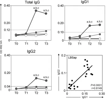
Significant (P< 0.05) increases in the serum levels of
anti-Leishmaniatotal IgG at T2 (15 days after the application of the second dose of vaccine) and T3 were observed in dogs of the LBSap group compared with those of the Sap, LB and control (C) groups (Fig. 1, upper left panel). Further analysis demonstrated that the levels of both anti-LeishmaniaIgG1 (Fig. 1, upper right panel) and IgG2 (Fig. 1, lower left panel) were enhanced (P< 0.05) in the LBSap group at both T2 and T3 compared with the other groups. Moreover, there was a positive association between IgG1 and IgG2 levels (Fig. 1, lower right panel).
3.3. LBSap elicited an increase in the numbers of circulating CD5+T-lymphocytes, mainly as CD4+and CD8+T-cell subsets, and CD21+B-lymphocytes
A preliminary comparative analysis of the cell profiles of the treatment groups showed that at T1 (15 days after the application of the first dose of vaccine) there was a
sig-Fig. 1. Comparative immunogenicity in the different treatment groups: C (control;); Sap (saponin;); LB (killedL. braziliensisvaccine;); LBSap (killedL. braziliensisvaccine plus saponin;䊉). Upper left and right panels and lower left panel show anti-L. chagasitotal IgG, anti-L. chagasiIgG1, and anti-L. chagasiIgG2, respectively: thex-axis displays the times at which the assays were conducted (T0: prior to the first dose; T1: 15 days after the first dose; T2: 15 days after the second dose; and T3: 15 days after the third dose) and they-axis represents the mean ELISA absorbance values determined at 492 nm in serum samples diluted 1:80. Significant differences (P< 0.05) between the LBSap group and the control C, Sap, and LB groups are indicated, respectively, by the letters a, b, and c. The lower right panel shows the correlation between IgG1 and IgG2 in the LBSap group and the Pearson’s correlation indexes (r) atP< 0.05 are shown in figure.
nificant (P< 0.05) reduction in the numbers of circulating lymphocytes in the Sap and LB groups compared with the control group C (Fig. 2, upper panel). Detailed investiga-tion of the immunophenotypic features of the lymphocytes (Fig. 2, middle panels) revealed that at T1 there was an increase (P< 0.05) in the number of circulating CD5+ T-lymphocytes in the LBSap group compared with the Sap group, and significantly (P< 0.05) higher counts of both CD4+ and CD8+ T-cell subsets and CD21+ B-cells in the LBSap group compared with the Sap and control C groups. Furthermore, from the absolute counts obtained at T1, T2 and T3, it was possible to demonstrate positive correlations within the LBSap group with respect to CD5+T-cells versus
CD21+ B-cells (P= 0.0024; r= 0.6395), and CD8+ versus CD4+ T-cells (P< 0.0001; r= 0.8533) (Fig. 2, middle pan-els), and with respect to CD4+ (P= 0.0002;r= 0.7422) and CD8+ (P< 0.0001;r= 0.8420) versus CD5+T-cells (Fig. 2, lower panel).
3.4. In vitro cell proliferation in the presence of antigenic stimuli was increased intensely following vaccination with LBSap but reduced significantly after treatment with LB
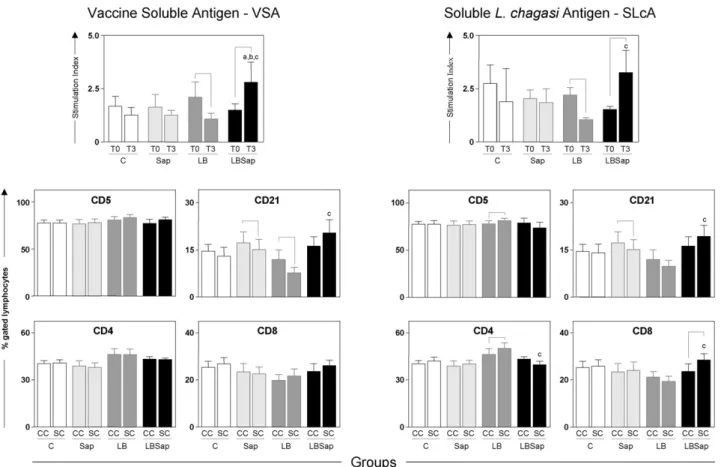
In vitrocell reactivities were determined in the presence of the VSA in order to evaluate memory lymphoprolifera-tive immune response and in the presence of SLcA in order to investigate possible lymphoproliferative homology with the complete antigenic repertoire of the etiological agent of VL (Fig. 3, upper panel). The lymphoproliferative activity at T3 compared with that at T0 in the LB group was reduced (P< 0.05) in the presence of either stimulus. In contrast, the LBSap group displayed significant (P< 0.05) increases in stimulation index at T3 compared with T0 in the pres-ence of both stimuli. Additionally, at T3 the values recorded in the presence of VSA was significantly (P< 0.05) higher than those observed in the LB, Sap and control C groups. Analysis of lymphoproliferative immune response and in the presence of SLcA also demonstrated at T3 a significant increase (P< 0.05) in comparison to that observed in the LB group.
The non-specific lymphoproliferative response was eval-uated as stimulation index (SI) of PHA mitogen-induced cell proliferation. Our data confirmed the high degree of cell via-bility as demonstrated by outstanding SI values observed in the mitogenic-stimulated cultures that ranged from mean values of 8 to 15 (data not shown).
3.5. Vaccination with LBSap increased the frequencies of CD21+B-lymphocytes and CD8+T-lymphocytes and decreased the levels of CD4+T-cells in
antigen-stimulated in vitro cell proliferation cultures
Fig. 3. Cell proliferation response of peripheral blood mononuclear cells after stimulation with vaccine soluble antigen (VSA) (upper left panel) and soluble L. chagasiantigen (SLcA) (upper right panel). The middle and lower panels show the immunophenotypic profile ofin vitroperipheral blood mononuclear cells following stimulation with vaccine soluble antigen (VSA) (left panels) and solubleL. chagasiantigen stimulation (right panel) determined at T3 for treatment groups: C (control;); Sap (saponin; ); LB (killedL. braziliensisvaccine; ); LBSap (killedL. braziliensisvaccine plus saponin;). The results are expressed as the mean frequencies of CD5+, CD21+, CD4+and CD8+cells in the non-stimulated cultures (CC; controls) and in the stimulated cultures (SC). Significant differences (P< 0.05) between values measured at T0 (before the first dose) and T3 (15 days after the third dose) are indicated by connecting lines, and between the LBSap and the control C, Sap, and LB groups at T3 are represented by the letters a, b, and c, respectively.
nificantly (P< 0.05) reduced frequencies of CD21+ B-cells were observed in the Sap and LB groups compared with those in the non-stimulated control cultures. Moreover, fol-lowing stimulation with VSA, the LBSap group exhibited a higher (P< 0.05) frequency of CD21+ B-lymphocytes com-pared with the LB group. Similar results were obtained when
in vitro cultures were stimulated with SLcA (Fig. 3, right middle and lower panels), indicating that the frequency of CD21+ B-cells was decreased (P< 0.05) in the Sap group compared with the control cultures, but increased (P< 0.05) in the LBSap group compared with the LB group. Moreover, although no differences had been observed in the frequen-cies of T-cells in assays involving VSA stimulation, when cultures were stimulated with SLcA, the LB group exhibited a higher frequency of CD5+T-cells compared with the con-trol C group, and this was mainly due to increased (P< 0.05) levels of CD4+T-cells. Additionally, compared with the LB group, the LBSap group presented lower (P< 0.05) levels of CD4+T-cells and an increased (P< 0.05) frequency of CD8+ T-cells.
3.6. LBSap vaccination gave rise to an increase in the levels of circulating CD14+monocytes and an associated up-regulation of MHC-I expression by lymphocytes
3.7. The correlations observed between cell
proliferation in antigen-stimulated in vitro cultures and the levels of circulating CD14+monocytes and CD21+ B-lymphocytes indicate the possibility of distinct APC profiles during LBSap vaccination
In order to identify which APC imparts the major contribu-tion during specificin vitroantigenic stimulation, correlation analyses were performed using data derived from the LBSap group (Fig. 4, middle panel). The results demonstrate a pos-itive association between CD14+monocytes andin vitrocell proliferation in cultures stimulated with VSA (P= 0.0306;
r= 0.7541), and a negative correlation in cultures stim-ulated with SLcA (P= 0.0229; r=−0.8924). In contrast, CD21+ B-cell counts exhibited a negative association with cell proliferation in VSA-stimulated cultures (P< 0.0001;
r=−0.8764), and a positive association in cultures stimulated with SLcA (P= 0.0460;r= 0.7630). These data provide evi-dence for a distinct APC according to antigen stimulus, and suggest that CD14+monocytes might act as APC in vaccine antigenic stimulation whilst CD21+ B-cell could fulfil this role in SLcA stimulation.
3.8. LBSap enhanced NO production in SLcA-stimulated in vitro cell proliferation cultures
Since the production of NO is considered to be a key ele-ment in killing mechanisms that mediate the elimination of intracellular pathogens, the levels of anti-microbial oxidant produced by in vitro antigenic-stimulated PBMC derived from dogs vaccinated with LBSap were determined. Inter-estingly, higher (P< 0.05) levels of the reactive NO radical (nitrite) were recorded in the supernatant of SLcA-stimulated cultures at T3 compared with T0 (Fig. 4, lower panel), sug-gesting an outcome related to the Th1 immune response.
3.9. Positive correlations were observed between cell proliferation in antigen-stimulated in vitro PBMC cultures and the levels of CD4+and CD8+ T-lymphocytes only in the LBSap group
In an attempt to determine whether the immunophenotypic features ofin vitroPBMC cultures subjected to antigen stimu-lation were associated with a specific cell profile, we analysed the levels of association between cell type and proliferation within all four groups (Fig. 5). Data analysis demonstrated positive associations only in the LBSap group, and specifi-cally between CD4+ and CD8+ T-cells and cell reactivity in both VSA- and SLcA-stimulated cultures.
4. Discussion
Canine visceral leishmaniasis, which resembles human visceral leishmaniasis with respect to many of its symptoms, is a severe chronic disease that is often fatal [29,34]. The
natural history of CVL has been well described, particu-larly in regard to the parasite load in different tissues and the immunopathological changes according to progression of clinical forms[29,30,35–37]. These data provide support for the improvement of the tools employed in the evaluation of both chemotherapies and vaccines that have been devel-oped for CVL. Unfortunately, different treatment strategies have failed to achieve a consistent parasitological cure for CVL owing to the presence of latently infected cells[38,39]. In this context, a dog vaccine may be the most practical and effective method by which to reduce the incidence of human VL, and it might also permit a similar vaccine to be developed for humans[12,14,15,40].
All of these features point to immunoprophylaxy as a promising alternative for prevention of CVL. For this rea-son a considerable effort has been dedicated to studies on immune responses in CVL, and severalLeishmaniaantigens implicated in these responses have been reported[14,15,41]. There is a major consensus thatL. chagasi/infantumantigens display a potent immunosuppressive potential that would be deleterious for the immunoprotection against CVL. Several studies have reported the potential of L. chagasi antigens to trigger immunosuppression by blocking thein vitro lym-phoproliferative response to Leishmania antigens as well as the synthesis of pro-inflammatory cytokines by antigen-presenting cells [42,43]. The use of purified Leishmania donovaniandL. infantumantigens has been also proposed to overcome this immunosuppressive effect of L. chagasi
antigens[18–20]. Most studies, including those clinical tri-als with vaccine candidates to CVL immunoprofilaxis have been conduced using eitherL. amazonensis,L. braziliensis
orL. majorantigens. Previous studies from our group have demonstrated thatL. braziliensisantigen have a potent role in protectingL. chagasiinfection in dogs (unpublished data). Therefore, a critical question for screening and development of anti-leishmanial vaccines in CVL is to define Leishma-niaantigens and adjuvant systems that elicit a favorable and sustained cytokine environmentin vivo.
Considering the importance of immunoprofilaxis strate-gies for the control of leishmaniasis, and the lack of studies concerning the cellular and humoral events that occur dur-ing vaccination, we have attempted to evaluate the immune response of a promising new vaccine candidate against CVL composed ofL. braziliensisantigens plus saponin as adju-vant. The assessment of such information is an essential pre-requisite to the understanding of mechanisms related to immunogenicity elicited by candidate vaccines.
Fig. 5. Correlations between cell proliferation (counts per minute) and frequencies of CD5+, CD4+and CD8+T-cells followingin vitroperipheral blood mononuclear cell cultures derived from the LBSap group (upper panels), and C (control), Sap (saponin) and LB (killedL. braziliensisvaccine) groups (lower panel), stimulated by vaccine soluble antigen (VSA; upper panel) and solubleL. chagasiantigen (SLcA; middle panel) and determined at T0 (before the first dose) plus T3 (15 days after the third dose) and the Pearson’s correlation indexes (r) atP< 0.05 are shown in figure.
of saponin as vaccine adjuvant. Some side effects that have been reported following the use of saponin adjuvants include some non-specific immune reactions, loss of hair at the site of injection, anorexia, apathy, vomiting and diarrhoea[44–46]. However, as saponin induces the development of strong CD8+ T-lymphocytes cytotoxicity[47], its use as adjuvant has been included in several veterinary vaccines[44].
In the present study, the evaluation of immunogenicity of LB and LBSap revealed that animals treated with LB together with saponin adjuvant presented higher (P< 0.05) amounts of anti-Leishmaniatotal IgG that were associated
sug-gested that a mixed immune response is triggered by LBSap vaccination.
The intense humoral immune response demonstrated in the LBSap group was synchronous with increased (P< 0.05) counts of circulating CD21+ B-cells following the first immunisation, resulting in differentiation of plasmacytes and higher levels of immunoglobulin secretion. Additionally, the higher numbers (P< 0.05) of circulating CD5+T-cells in the LBSap group were positively correlated with CD21+B-cells suggesting a possible cooperation between T- and B-cells during the immunisation sequence.
Protection against infection byLeishmaniarelies on the cell-mediated immune response, which implies that a suc-cessful immunisation protocol should be able to activate cell-mediated immunity in the immunised animal[23,53,54]. In the present study, the increase in CD5+ T-cells in the LBSap group was associated with increased levels of CD4+ and CD8+ T-cells. Moreover, positive correlations between CD5+ T-cells and the CD4+ and CD8+ T-cell subset, and between CD4+ and CD8+ T-cells, further suggested signif-icant cooperation at the cellular level as has been observed during early clonal expansion and the generation of primary CD8+ cytotoxic effectors[55]. Additionally, the augmenta-tion of circulating CD8+ T-cells during on-going CVL has been previously described as the major phenotypic feature of the asymptomatic disease in dogs bearing low parasite loads [37]. Thus, our findings support the hypothesis that CD8+T-lymphocytes play a role in protective immunity
dur-ingLeishmaniainfection as has been suggested previously for CVL[37,56].
In order to evaluate the activation status of lymphocytes during the immunisation protocol, the expression of MHC-I was studied. The finding of a higher expression of MHC-I in lymphocytes in the LBSap group indicated an improvement in the activation status of lymphocytes during the immu-nisation protocol. It has been proposed that an increase in the expression of MHC-II may reflect an antigenic priming-related immunological event [37,57]. Consistent with this hypothesis, we have demonstrated that dogs with asymp-tomatic CVL displayed an enhanced activation status of circulating lymphocytes (as determined from MHC-II)[37], together with lower overall tissue parasitism[30,37]. How-ever, data from the present study highlight the importance of MHC-I expression as an activation marker in lymphocytes. In fact, the up-regulation of MHC-I and -II are both related to the presence of IFN-␥[58,59]. Consistent with this hypoth-esis, the evaluation of APC revealed increased numbers of circulating CD14+monocytes in the LBSap group that were associated with increased MHC-I expression in lymphocytes. On the basis of these results, it is possible to speculate that this association would represent the interactions between innate and adaptive immune responses, reflecting in improvement in activation status during LBSap immunisation.
Aiming to determine whether the candidate vaccine would activate PBMC underin vitroantigenic stimulation, we mea-sured the stimulation index at T0 and T1 in cells derived from
LBSap and LB dogs. Higher cell reactivities following stim-ulation by either VSA or SLcA were recorded for the LBSap group after the third immunisation, whereas the stimulation index in the LB group was lower at T3 compared to T0. Thus, our findings support the hypothesis that PBMC proliferation in the LBSap group is associated with the recognition ofL. chagasiantigen, and suggest that this vaccine may be of value against the etiological agent of VL.
Further analyses were carried out in order to investigate the profile of APC inin vitrocultures of PBMC from the LBSap group subjected to antigenic stimuli. Whilst cell prolifera-tion exhibited a positive associaprolifera-tion with circulating CD14+
monocytes in the presence of VSA, a negative correlation was observed upon SLcA stimulation. In contrast, a negative association between CD21+B-cells and cell proliferation was observed in the presence of VSA, and a positive association upon SLcA stimulation. These findings indicate a distinct profile of APC in the cultures and establish that CD14+ mono-cytes and CD21+ B-cells are the major APC, respectively, duringin vitrostimulation with VSA and SLcA.
Whenin vitrocultures of PBMC derived from the LBSap group were stimulated with VSA or SLcA, increased lympho-proliferation activity was accompanied by a higher frequency of CD21+B-cells demonstrating the potential immunoglob-ulin isotypes produced. In contrast, no differences were observed in the frequency of CD5+ T-cells following in vitrostimulation, whilst reductions in the frequency of CD4+
T-cells and increases in the level of CD8+ T-cells were
associated with SLcA stimulus. These results support the hypothesis that CD8+ T-cells play a protective role in the mechanism of control ofLeishmaniaparasitism. Additional correlation analysis revealed that both CD4+ and CD8+ T-cells exhibited positive associations with cell proliferationin vitrofollowing stimulation with VSA or SLcA. These data suggest the possibility of stimulating antigen-specific T-cell subsets following LBSap immunisation that could contribute to improvements in the cellular immune response duringL. chagasiinfection.
The results obtained from the analysis of NO levels (determined as nitrite) in culture supernatants confirmed the hypothesis that LBSap immunisation induces a potential resistance profile againstLeishmania infection despite the higher nitrite levels observed inin vitroT3 cells stimulated by SLcA.
in antigenic repertoire, and this should potentially activate a stronger cellular response, mainly by T-lymphocytes, com-pared with that of purified subunit preparations or DNA vaccines.
The major findings in the present study point to a strong antigenicity of the candidate vaccine related to increased immunoglobulin isotypes, together with higher levels of lym-phocytes, particularly in circulating CD8+ T-lymphocytes and SLcA-specific CD8+ T-lymphocytes. In addition, the intense cell proliferation and NO production duringin vitro
stimulation by SLcA shows that LBSap vaccine elicited a potential immune activation status potentially compatible with effective control of the etiological agent of CVL. Further investigations will focus on the efficacy of the LBSap vacci-nation in protection against experimental challenge withL. chagasi.
Acknowledgments
The authors wish to express their appreciation of the hard work carried out by the kennel staff of the Federal Univer-sity of Ouro Preto and for their special dedication during the execution of this project. The authors are also grateful for the use of the facilities at CEBIO, Universidade Federal de Minas Gerais and Rede Mineira de Bioterismo (FAPEMIG). This work was supported by Fundac¸˜ao de Amparo `a Pesquisa do Estado de Minas Gerais, Brazil (FAPEMIG grant: CBB CBB9202), PAPES IIIb (FIOCRUZ/RJ/2002) and CAPES. OAMF, ML and RCO are grateful to CNPq for fellowships.
References
[1] Desjeux P. Leishmaniasis: current situation and new perspectives. Comp Immunol Microbiol Infect Dis 2004;27:305–18.
[2] Evans TG, Vasconcelos IA, Lima JW, Teixeira JM, McAullife IT, Lopes UG, et al. Canine visceral leishmaniasis in northeast Brazil: assessment of serodiagnostic methods. Am J Trop Med Hyg 1990;42:118–23. [3] Tesh RB. Control of zoonotic visceral leishmaniasis: is it time to change
strategies? Am J Trop Med Hyg 1995;52:287–92.
[4] Braga MD, Coelho IC, Pompeu MM, Evans TG, MacAullife IT, Teix-eira MJ, et al. Controle do calazar canino: comparac¸˜ao dos resultados de um programa de eliminac¸˜ao r´apida de c˜aes sororreagentes por ensaio imuno-enzim´atico com outro de eliminac¸˜ao tardia de c˜aes soror-reagentes por teste de imunofluorescˆencia indireta de eluato de papel filtro. Rev Soc Bras Med Trop 1998;31:419–24.
[5] Franca-Silva JC, da Costa RT, Siqueira AM, hado-Coelho GL, da Costa CA, Mayrink W, et al. Epidemiology of canine visceral leishmaniosis in the endemic area of Montes Claros Municipality, Minas Gerais State, Brazil. Vet Parasitol 2003;111:161–73.
[6] Palatnik-de-Sousa CB, dos Santos WR, Franca-Silva JC, da Costa RT, Reis AB, Palatnik M, et al. Impact of canine control on the epidemiology of canine and human visceral leishmaniasis in Brazil. Am J Trop Med Hyg 2001;65:510–7.
[7] Hommel M, Jaffe CL, Travi B, Milon G. Experimental models for leishmaniasis and for testing anti-leishmanial vaccines. Ann Trop Med Parasitol 1995;89(1):55–73.
[8] Webb JR, Kaufmann D, Campos-Neto A, Reed SG. Molecular cloning of a novel protein antigen ofLeishmania majorthat elicits a potent
immune response in experimental murine leishmaniasis. J Immunol 1996;157:5034–41.
[9] Wilson ME, Young BM, Andersen KP, Weinstock JV, Metwali A, Ali KM, et al. A recombinantLeishmania chagasiantigen that stim-ulates cellular immune responses in infected mice. Infect Immun 1995;63:2062–9.
[10] Webb JR, Campos-Neto A, Ovendale PJ, Martin TI, Stromberg EJ, Badaro R, et al. Human and murine immune responses to a novel Leish-mania majorrecombinant protein encoded by members of a multicopy gene family. Infect Immun 1998;66:3279–89.
[11] Dole VS, Raj VS, Ghosh A, Madhubala R, Myler PJ, Stuart KD. Immu-nization with recombinant LD1 antigens protects against experimental leishmaniasis. Vaccine 2000;19:423–30.
[12] Mauel J. Vaccination againstLeishmaniainfections. Curr Drug Targets Immune Endocr Metabol Disord 2002;2:201–26.
[13] Campos-Neto A, Porrozzi R, Greeson K, Coler RN, Webb JR, Seiky Y, et al. Protection against cutaneous leishmaniasis induced by recombi-nant antigens in murine and non-human primate models of the human disease. Infect Immun 2001;69:4103–8.
[14] Brodskyn C, de Oliveira CI, Barral A, Barral-Netto M. Vaccines in leishmaniasis: advances in the last five years. Expert Rev Vaccines 2003;2:705–17.
[15] Gradoni L. An update on antileishmanial vaccine candidates and prospects for a canineLeishmaniavaccine. Vet Parasitol 2001;100: 87–103.
[16] Dunan S, Frommel D, Monjour L, Ogunkolade BW, Cruz A, Quilici M. Vaccination trial against canine visceral leishmaniasis. Phocean Veterinary Study Group on Visceral Leishmaniasis. Parasite Immunol 1989;11:397–402.
[17] Monjour L, Vouldoukis I, Ogunkolade BW, Hetzel C, Ichen M, From-mel D. Vaccination and treatment trials against murine leishmaniasis with semi-purifiedLeishmaniaantigens. Trans R Soc Trop Med Hyg 1988;82:412–5.
[18] da Silva V, Borja-Cabrera GP, Correia Pontes NN, de Souza EP, Luz KG, Palatnik M, et al. A phase III trial of efficacy of the FML-vaccine against canine kala-azar in an endemic area of Brazil (Sao Goncalo do Amaranto, RN). Vaccine 2000;19:1082–92.
[19] Borja-Cabrera GP, Correia Pontes NN, da Silva VO, Paraguai de Souza E, Santos WR, Gomes EM, et al. Long lasting protection against canine kala-azar using the FML-QuilA saponin vaccine in an endemic area of Brazil (Sao Goncalo do Amarante RN). Vaccine 2002;20:3277–84. [20] Lemesre JL, Holzmuller P, Cavaleyra M, Goncalves RB, Hottin G,
Papierok G. Protection against experimental visceral leishmaniasis infection in dogs immunized with purified excreted secreted antigens ofLeishmania infantumpromastigotes. Vaccine 2005;23:2825–40. [21] Lemesre JL, Holzmuller P, Goncalves RB, Bourdoiseau G, Hugnet
C, Cavaleyra M, et al. Long-lasting protection against canine vis-ceral leishmaniasis using the LiESAp-MDP vaccine in endemic areas of France: double-blind randomised efficacy field trial. Vaccine 2007;25:4223–34.
[22] Jeronimo SM, Higgs E, Vedvick T, Mann BJ, Jernigan J, Petri WA, et al. Identification ofLeishmania chagasiantigens recognized by human lymphocytes. J Infect Dis 1995;172:1055–60.
[23] Ravindran R, Ali N. Progress in vaccine research and possible effector mechanisms in visceral leishmaniasis. Curr Mol Med 2004;4:697–709. [24] Mayrink W, Genaro O, Silva JC, da Costa RT, Tafuri WL, Toledo VP, et al. Phase I and II open clinical trials of a vaccine againstLeishmania chagasiinfections in dogs. Mem Inst Oswaldo Cruz 1996;91:695–7. [25] Lasri S, Sahibi H, Sadak A, Jaffe CL, Rhalem A. Immune responses in
dogs vaccinated with autoclavedLeishmania majorpromastigotes. Vet Res 1999;30:441–9.
[26] Panaro MA, Acquafredda A, Lisi S, Lofrumento DD, Mitolo V, Sisto M, et al. Nitric oxide production by macrophages of dogs vaccinated with killedLeishmania infantumpromastigotes. Comp Immunol Microbiol Infect Dis 2001;24:187–95.
autoclavedLeishmania majorvaccine mixed with BCG against canine visceral leishmaniasis in Meshkin-Shahr district, I.R. Iran. Vaccine 2004;22:4097–100.
[28] Lowry OH, Rosebrough NJ, Farr AL, Randall RJ. Protein measurement with the Folin phenol reagent. J Biol Chem 1951;193:265–75. [29] Reis AB, Martins-Filho OA, Teixeira-Carvalho A, Carvalho MG,
Mayrink W, Franca-Silva JC, et al. Parasite density and impaired biochemical/hematological status are associated with severe clinical aspects of canine visceral leishmaniasis. Res Vet Sci 2006;81:68–75. [30] Reis AB, Teixeira-Carvalho A, Vale AM, Marques MJ, Giunchetti
RC, Mayrink W, et al. Isotype patterns of immunoglobulins: hall-marks for clinical status and tissue parasite density in Brazilian dogs naturally infected byLeishmania(Leishmania)chagasi. Vet Immunol Immunopathol 2006;112:102–16.
[31] Reis AB, Carneiro CM, Carvalho MG, Teixeira-Carvalho A, Giunchetti RC, Mayrink W, et al. Establishment of a microplate assay for flow cytometric assessment and it is use for the evaluation of age-related phenotypic changes in canine whole blood leukocytes. Vet Immunol Immunopathol 2005;103:173–85.
[32] Green LC, Wagner DA, Glogowski J, Skipper PL, Wishnok JS, Tan-nenbaum SR. Analysis of nitrate, nitrite, and [15N]-nitrate in biological fluids. Anal Biochem 1982;126:131–8.
[33] Gutman SI, Hollywood CA. Simple, rapid method for determining nitrates and nitrites in biological fluids. Clin Chem 1992;38:2152. [34] Keenan CM, Hendricks LD, Lightner L, Webster HK, Johnson AJ.
Visceral leishmaniasis in the German shepherd dog. I. Infection, clinical disease, and clinical pathology. Vet Pathol 1984;21:74–9.
[35] Giunchetti RC, Mayrink W, Genaro O, Carneiro CM, Correa-Oliveira R, Martins-Filho OA, et al. Relationship between canine visceral leish-maniosis and theLeishmania(Leishmania)chagasiburden in dermal inflammatory foci. J Comp Pathol 2006;135:100–7.
[36] Lage RS, Oliveira GC, Busek SU, Guerra LL, Giunchetti RC, Correa-Oliveira R, et al. Analysis of the cytokine profile in spleen cells from dogs naturally infected byLeishmania chagasi. Vet Immunol Immunopathol 2007;115:135–45.
[37] Reis AB, Teixeira-Carvalho A, Giunchetti RC, Guerra LL, Carvalho MG, Mayrink W, et al. Phenotypic features of circulating leucocytes as immunological markers for clinical status and bone marrow parasite density in dogs naturally infected byLeishmania chagasi. Clin Exp Immunol 2006;146:303–11.
[38] Baneth G, Shaw SE. Chemotherapy of canine leishmaniasis. Vet Para-sitol 2002;106:315–24.
[39] Noli C, Auxilia ST. Treatment of canine Old World visceral leishma-niasis: a systematic review. Vet Dermatol 2005;16:213–32.
[40] Abranches P, Santos-Gomes G, Rachamim N, Campino L, Schnur LF, Jaffe CL. An experimental model for canine visceral leishmaniasis. Parasite Immunol 1991;13:537–50.
[41] Barbieri CL. Immunology of canine leishmaniasis. Parasite Immunol 2006;28:329–37.
[42] Carvalho EM, Badaro R, Reed SG, Jones TC, Johnson Jr WD. Absence of gamma interferon and interleukin 2 production during active visceral leishmaniasis. J Clin Invest 1985;76:2066–9.
[43] Peruhype-Magalhaes V, Martins-Filho OA, Prata A, Silva Lde A, Rabello A, Teixeira-Carvalho A, et al. Mixed inflammatory/regulatory cytokine profile marked by simultaneous raise of interferon-gamma and interleukin-10 and low frequency of tumour necrosis factor-alpha(+)
monocytes are hallmarks of active human visceral Leishmaniasis due toLeishmania chagasiinfection. Clin Exp Immunol 2006;146:124–32. [44] Rajput ZI, Hu SH, Xiao CW, Arijo AG. Adjuvant effects of saponins on animal immune responses. J Zhejiang Univ Sci B 2007;8:153–61. [45] Santos WR, de Lima V, de Souza EP, Bernardo RR, Palatnik M,
Palatnik de Sousa CB. Saponins, IL12 and BCG adjuvant in the FML-vaccine formulation against murine visceral leishmaniasis. Vaccine 2002;21:30–43.
[46] Parra LE, Borja-Cabrera GP, Santos FN, Souza LO, Palatnik-de-Sousa CB, Menz I. Safety trial using the Leishmune vaccine against canine visceral leishmaniasis in Brazil. Vaccine 2007;25:2180–6.
[47] Kensil CR. Saponins as vaccine adjuvants. Crit Rev Ther Drug Carrier Syst 1996;13:1–55.
[48] Fujiwara RT, Vale AM, Franca da Silva JC, da Costa RT, Quetz JS, Martins-Filho OA, et al. Immunogenicity in dogs of three recombinant antigens (TSA, LeIF and LmSTI1) potential vaccine candidates for canine visceral leishmaniasis. Vet Res 2005;36:827–38.
[49] Deplazes P, Smith NC, Arnold P, Lutz H, Eckert J. Specific IgG1 and IgG2 antibody responses of dogs toLeishmania infantumand other parasites. Parasite Immunol 1995;17:451–8.
[50] Bourdoiseau G, Bonnefont C, Hoareau E, Boehringer C, Stolle T, Cha-banne L. Specific IgG1 and IgG2 antibody and lymphocyte subset levels in naturallyLeishmania infantum-infected treated and untreated dogs. Vet Immunol Immunopathol 1997;59:21–30.
[51] Courtenay O, Quinnell RJ, Garcez LM, Shaw JJ, Dye C. Infectiousness in a cohort of Brazilian dogs: why culling fails to control visceral leish-maniasis in areas of high transmission. J Infect Dis 2002;186:1314–20. [52] Day MJ. Immunoglobulin G subclass distribution in canine leishman-iosis: a review and analysis of pitfalls in interpretation. Vet Parasitol 2007;147:2–8.
[53] Muller I, Pedrazzini T, Kropf P, Louis J, Milon G. Establishment of resistance toLeishmania majorinfection in susceptible BALB/c mice requires parasite-specific CD8+ T cells. Int Immunol 1991;3:587–97. [54] Melby PC, Yang YZ, Cheng J, Zhao W. Regional differences in the
cel-lular immune response to experimental cutaneous or visceral infection withLeishmania donovani. Infect Immun 1998;66:18–27.
[55] Castellino F, Germain RN. Cooperation between CD4+ and CD8+ T cells: when, where, and how. Annu Rev Immunol 2006;24:519–40. [56] Pinelli E. Cytokines in canine visceral leishmaniasis. In: Schijns VECJ,
Horzinek MC, editors. Cytokines in veterinary medicine. Utrecht: Utrecht University Press; 1997. p. 217–47.
[57] Cobbold S, Metcalfe S. Monoclonal antibodies that define canine homologues of human CD antigens: summary of the First Interna-tional Canine Leukocyte Antigen Workshop (CLAW). Tissue Antigens 1994;43:137–54.
[58] Whitley EM, Bird AC, Zucker KE, Wolfe LG. Modulation by canine interferon-gamma of major histocompatibility complex and tumor-associated antigen expression in canine mammary tumor and melanoma cell lines. Anticancer Res 1995;15:923–9.
[59] Radosevich TJ, Seregina T, Link CJ. Effective suppression of class I major histocompatibility complex expression by the US11 or ICP47 genes can be limited by cell type or interferon-gamma exposure. Hum Gene Ther 2003;14:1765–75.