MO
H
Degre
UN
FA
OLECULA
ERPESV
M
ee of Doc
Special
NIVERSI
ACULDA
AR PAT
VIRUS-Marta Be
ctor of Ph
ization in
IDADE D
ADE DE M
THOGEN
-4 LATE
ebiano A
hilosophy
n Biopatho
2009
DE LISBO
MEDICIN
NESIS O
ENT INF
Alenque
in Biome
ological S
OA
NA
OF MUR
FECTIO
r
edical Sci
Sciences
RID
ON
ences
As opiniões expressas nesta publicação são
da exclusiva responsabilidade do seu autor.
UNIVERSIDADE DE LISBOA
FACULDADE DE MEDICINA
M
OLECULAR
P
ATHOGENESIS OF
M
URID
H
ERPESVIRUS
-4
L
ATENT
I
NFECTION
by
Marta Bebiano Alenquer
(Recipient of a scholarship – SFRH/BD/17260/2004 from Fundação para a Ciência e Tecnologia)
Thesis submitted for the degree of
Doctor of Philosophy in Biomedical Sciences
(Specialization in Biopathological Sciences)
Supervised by Professor Doutor Pedro Simas,
Faculdade de Medicina da Universidade de Lisboa
A impressão desta dissertação foi aprovada pela Comissão
Coordenadora do Conselho Científico da Faculdade de Medicina de
Lisboa em reunião de 2 de Junho de 2009.
P
REFACEThe current thesis presents data obtained during my PhD research project, developed at Instituto de Medicina Molecular in the period of October 2005 to September 2008, under the supervision of Professor Doutor Pedro Simas (Faculdade de Medicina, Universidade de Lisboa). Part of this project was developed in collaboration with Doctor Philip Stevenson (Division of Virology, University of Cambridge), who received me in his laboratory from February to July 2008 and where the results presented in figures 5.1 to 5.4 were generated.
This thesis is organized in 7 chapters, which are preceded by a summary written in Portuguese and an abstract. Before the description of the results, an introductory review of the subject is provided in chapter 1 and the aims of the work are also detailed at the end of chapter 1. In chapters 2, 3, 4 and 5 the original data obtained during this research project is presented and discussed. Final considerations, which integrate the results presented in previous chapters as well as future directions, are presented in chapter 6. Finally, chapter 7 concerns the description of the methodologies and materials employed to carry out the presented work. The publications that resulted from the research carried out throughout the duration of this project are included in appendix 1 to 4.
The data presented in this dissertation was purely the result of work carried out by me and it is clearly acknowledged in the text whenever data or reagents produced by others were utilized. This work has not been submitted for any degree at this or any university.
P
UBLICATIONSAppendix 1
Pires de Miranda, M.*, Alenquer, M.*, Marques, S., Rodrigues, L., Lopes, F., Bustelo, X. R. & Simas, J. P. (2008). The Gammaherpesvirus m2 protein manipulates the Fyn/Vav pathway through a multidocking mechanism of assembly. PLoS ONE 3, e1654. *shared first authorship.
Construction of recombinant viruses and all animal experiments presented in this publication (figures 5 and 6 and table 1) were performed by me.
Appendix 2
Marques, S., Alenquer, M., Stevenson, P. G. & Simas, J. P. (2008). A single CD8+ T cell epitope sets the long-term latent load of a murid herpesvirus. PLoS Pathog 4, e1000177.
Data presented in this publication were obtained in collaboration. I provided technical assistance in the in situ hybridization experiments and helped in the analysis and discussion of the obtained results.
Appendix 3
Milho, R., Smith, C. M., Marques, S., Alenquer, M., May, J. S., Gillet, L., Gaspar, M., Efstathiou, S., Simas, J. P. & Stevenson, P. G. (2009). In vivo imaging of murid herpesvirus-4 infection. J Gen Virol 90, 21-32.
Data presented in this publication were obtained in collaboration. The set up of the luciferase-base imaging technique in the laboratory was done by me. Data on figures 2B, 3 and 5 were obtained by the first author with my collaboration/guidance.
Appendix 4
Gillet, L., Alenquer, M., Glauser, D. L., Colaco, S., May, J. S. & Stevenson, P. G. (2009). Glycoprotein L sets the neutralization profile of murid herpesvirus 4. J Gen Virol 90, 1202-14.
Data presented in this publication were obtained in collaboration. Data referring to SC-9E8 antibody on figure 6A were obtained by me.
A
CKNOWLEDGEMENTSEm primeiro lugar, quero agradecer ao meu supervisor Pedro Simas por me ter dado a oportunidade de realizar o doutoramento no seu laboratório. A sua orientação, conselhos, apoio e motivação ao longo destes quatro anos foram fundamentais.
Um agradecimento especial a todos os membros da unidade de Patogénese Viral pela sua amizade, ajuda e constante apoio. Em particular, agradeço à Sofia a sua inestimável ajuda, sugestões e inúmeras discussões científicas. Obrigada à Lénia, à Marta e à Filipa pela contribuição nos estudos in vitro de M2 que foram o ponto de partida para os meus estudos in vivo. Mais recentemente, o apoio e amizade da Teresa foram insubstituíveis.
Gostaria também de agradecer ao Instituto de Medicina Molecular por ter proporcionado todas as condições necessárias à realização do meu projecto de doutoramento. Este agradecimento é extensível aos investigadores e funcionários do instituto que contribuíram para este trabalho. Em particular, agradeço aos membros da unidade de Malária, com os quais partilhamos o laboratório, pelo excelente ambiente de trabalho, e à Ana Caetano pela ajuda nas experiências de citometria de fluxo
Some of the work presented in this thesis was carried out in the laboratory of Dr Philip Stevenson, to whom I am indebted for all his support and invaluable contribution into my research project during my visit to his laboratory at the University of Cambridge. To all virology members, in particular Stevenson’s group members and Helen, thank you for all the help and for making me feel at home in Cambridge.
Por fim, muito obrigada à minha família e aos meus amigos por me aturarem e apoiarem... eu sei que não tem sido fácil!
R
ESUMOA principal característica biológica dos herpesvírus é a sua capacidade de persistirem durante toda a vida do hospedeiro através do estabelecimento de infecções latentes. Durante a latência, os herpesvírus permanecem no núcleo da célula infectada sob a forma de um episoma, não havendo produção de viriões infecciosos. A expressão génica está limitada aos genes importantes para a manutenção do genoma viral e sobrevivência da célula hospedeira, permitindo ao vírus não ser reconhecido pelo sistema imunitário. No caso dos gama-herpesvírus, a fase de latência é estabelecida maioritariamente em células B e está associada ao desenvolvimento de doenças linfoproliferativas, fazendo com que o controlo destes vírus seja um objectivo clínico prioritário. Para persistirem nos hospedeiros, os gama-herpesvírus exploram o ciclo de vida normal das células B, induzindo a proliferação das células infectadas em centros germinativos e a sua posterior diferenciação em células B de memória de longa duração. Estes processos são controlados por proteínas virais que subvertem vias de sinalização da célula B ao regularem ou substituírem determinadas proteínas sinalizadoras. Assim, o estudo das proteínas modeladoras virais é de extrema importância, uma vez que estas têm um papel fundamental na patogénese dos gama-herpesvírus e estão potencialmente relacionadas com desenvolvimento de doenças linfoproliferativas. Os dois gama-herpesvírus humanos conhecidos até à data – vírus Epstein-Barr (EBV) e o vírus associado ao sarcoma de Kaposi (KSHV) – codificam proteínas membranares expressas durante a fase de latência que actuam como receptores constitutivamente activos, interferindo assim em vários aspectos da sinalização da célula B. Embora muitos estudos tenham abordado a função bioquímica destas proteínas, a estrita especificidade de hospedeiro dos gama-herpesvírus humanos tem limitado o estudo da função destas proteínas in
vivo. Assim, um dos maiores desafios no estudo dos gama-herpesvírus consiste em
relacionar a função bioquímica dos genes com o seu papel na infecção natural do hospedeiro.
Neste estudo utilizou-se a infecção de ratinhos de laboratório com o herpesvírus de murganho 4 (MuHV-4) como modelo experimental determinar, in vivo, a importância que a modelação da função das células B tem para o estabelecimento e manutenção de latência no hospedeiro. O MuHV-4 é um parasita natural de roedores selvagens, geneticamente relacionado com os agentes patogénicos EBV e KSHV e, à semelhança
destes, infecta latentemente células B, induzindo a sua proliferação em centros germinativos. Este modelo animal tem, portanto, um grande potencial para a compreensão de factores virais e do hospedeiro relevantes para a patogénese de gama-herpesvírus. Estudos bioquímicos realizados no nosso grupo demonstraram que a proteína M2 de MuHV-4 se encontra envolvida na modelação da função das células B. No entanto, M2, contrariamente às proteínas modelação de EBV e KSHV, não se comporta como um receptor celular constitutivamente activo, mas como uma proteína adaptadora. As funções adaptadoras de M2 promovem a formação de complexos celulares multi-proteicos que favorecem a aproximação entre enzimas e substratos, modelando deste modo vias de sinalização da célula B. Uma das vias de sinalização activadas por M2 é a via das proteínas Vav1/Rac1. M2, ao facilitar o encontro entre Vav1 e a cinase de tirosinas Fyn, induz a fosforilação de Vav1 por Fyn e, consequentemente, a activação da actividade catalítica de Vav1. Estudos recentes demonstraram que M2 está também envolvida na interacção e modulação da actividade de outras proteínas sinalizadoras, nomeadamente PI3K e PLCγ2.
O presente estudo teve como principal objectivo relacionar a função molecular de M2, enquanto modeladora de vias de sinalização de células B, com o seu papel na infecção
in vivo do hospedeiro. Para tal, construíram-se vírus recombinantes nos quais domínios
de M2 importantes para a sua função molecular foram inactivados através de mutações pontuais. Inicialmente, construíram-se vírus recombinantes que contêm mutações na região rica em prolinas (M2P2) ou em dois resíduos de tirosina susceptíveis de fosforilação por Fyn (M2Y). Estes dois domínios foram identificados em estudos bioquímicos realizados pelo nosso grupo como sendo essenciais para a ligação e activação de Vav1. Para avaliar a importância biológica destes domínios procedeu-se à infecção intranasal de ratinhos com os vírus recombinantes e analisou-se o comportamento dos vírus durante a fase de infecção latente, por comparação com a infecção de ratinhos com o vírus selvagem (WT) ou o vírus M2FS, que não expressa a proteína M2. Os vírus foram caracterizados durante o estabelecimento e manutenção da fase de latência através de três ensaios independentes mas complementares. Através de ensaios de reactivação ex vivo avaliou-se a infecção latente em esplenócitos totais. A frequência de infecção em células B do centro germinativo, que constituem o principal reservatório de latência do vírus, foi determinada após purificação desta população por citometria de fluxo, combinando ensaios de diluição limite com PCR em tempo real. A realização de experiências de hibridação in situ em secções do baço permitiu localizar
as células infectadas e monitorizar a sua cinética de proliferação no interior dos folículos linfóides. Os resultados obtidos pelos três ensaios foram concordantes entre si e revelaram que os domínios funcionais de M2 analisados são fundamentais, durante o estabelecimento de latência, para uma eficiente colonização dos folículos linfóides e estabelecimento de reacções do centro germinativo. Além disso, o fenótipo apresentado pelos vírus recombinantes com mutações pontuais em M2 durante o estabelecimento de latência é indistinguível do fenótipo obtido com o vírus que não expressa M2. Este facto vem reforçar a importância fisiológica, na infecção latente de MuHV-4 em células B, da modulação de vias de sinalização da célula B por M2.
A eliminação da expressão de M2 durante a fase de latência em ratinhos BALB/c origina dois fenótipos distintos: por um lado, durante o estabelecimento de latência, origina um atraso na colonização dos folículos e no início da proliferação das células infectadas nos centros germinativos; por outro lado, a longos tempos pós-infecção, observa-se um aumento exacerbado do número de células B do centro germinativo infectadas. Apesar da disrupção dos domínios funcionais de M2 reproduzir totalmente o comportamento do vírus que não expressa M2 durante o estabelecimento de latência, o fenótipo a longo prazo não é reproduzido. Assim, a longos tempos pós-infecção, os vírus recombinantes com mutações nos domínios funcionais de M2 comportam-se exactamente como o vírus WT. Estudos recentes realizados no nosso laboratório revelaram que esta incapacidade do vírus que não expressa M2 em cessar a proliferação das células do centro germinativo, não se deve a uma função molecular de M2 mas sim a um efeito imunológico. A proteína M2 contém um epítopo que é activamente reconhecido por células T citotóxicas (CTLs) e contribui para o controlo da infecção latente de MuHV-4. Na ausência do epítopo de M2, as células T citotóxicas não reconhecem as células infectadas e não conseguem controlar a amplificação da latência em células B do centro germinativo.
Para verificar se todas as funções moleculares de M2 importantes para o estabelecimento de latência em células B tinham sido identificadas, construiu-se um vírus recombinante duplo, com mutações pontuais não só nos resíduos de tirosina de M2 mas também no seu epítopo. O fenótipo deste vírus foi analisado após infecção de ratinhos e comparado com o do vírus deficiente para a expressão de M2. O comportamento dos dois vírus foi exactamente o mesmo, tanto durante estabelecimento de latência como na infecção a longo prazo. Esta observação indica que os domínios de M2 importantes para o estabelecimento e manutenção da latência foram identificados:
através das tirosinas e da região rica em prolinas, M2 modula vias de sinalização da célula B de forma a promover uma eficiente colonização dos folículos linfóides; o epítopo reconhecido por CTLs torna M2 indirectamente responsável pelo nível de carga viral a longo prazo.
Estudos bioquímicos recentes realizados no nosso laboratório demonstraram que os dois resíduos de tirosina fosforilados de M2 (Y120 e Y129) têm especificidades diferentes, ligando-se a moléculas sinalizadoras distintas. Além disso, a tirosina 120 parece ter um papel predominante, sendo essencial na ligação a Vav1, Fyn, PLCγ2 e PI3K. Para determinar a importância relativa das duas tirosinas durante a infecção in vivo de ratinhos, construíram-se novos vírus recombinantes com cada uma das tirosinas individualmente mutadas. Os resultados obtidos demonstraram que embora in vitro a tirosina 120 pareça ser mais importante na interacção com moléculas sinalizadoras, in
vivo ambas as tirosinas são necessárias para o normal estabelecimento de latência. Este
resultado sugere que a tirosina 129 interage com outras moléculas importantes para a modelação da função da célula B.
Embora o alvo preferencial para o estabelecimento de latência de MuHV-4 sejam as células B, durante o estabelecimento da fase latente este vírus é também capaz de infectar células dendríticas e macrófagos. Assim, no presente trabalho pretendeu-se ainda avaliar a importância da função de M2 na infecção de células dendríticas e macrófagos. Para tal, infectaram-se culturas de células dendríticas ou de macrófagos com o vírus deficiente para expressão de M2 ou com o vírus WT e avaliou-se o estabelecimento de infecções líticas e latentes por imunofluorescência. Além disso, infectaram-se ratinhos com os mesmos vírus e analisou-se a infecção de células dendríticas e macrófagos dos nódulos linfáticos mediastinais pouco tempo após a infecção. Em ambos os casos não foram detectadas diferenças na infecção entre o vírus WT e o mutante deficiente para a expressão de M2, o que indica que M2 não parece desempenhar qualquer função molecular importante na infecção de células dendríticas e macrófagos.
Em conclusão, este trabalho demonstrou que M2 funciona in vivo como uma proteína moduladora de vias de sinalização de células B e que esta função é fundamental na patogénese de MuHV-4, para uma eficiente colonização dos folículos linfóides durante o estabelecimento de latência.
A
BSTRACTThe major biological characteristic of herpesviruses is their ability to establish life-long latent infections in specific cell types within their host. In particular, gammaherpesviruses typically colonise their hosts by driving the proliferation of infected B cells in germinal centres (GCs) and establishing a lifelong reservoir of latently infected memory B cells. These events are controlled by viral proteins that subvert host cell signalling pathways by regulating or replacing functionally specific signalling proteins. These viral proteins are of particular interest as they constitute key components of gammaherpesvirus pathogenesis and a potential link to virus associated-oncogenic transformation. One of such proteins is the latency-associated M2 protein of murid herpesvirus-4 (MuHV-4). M2 has been shown to act as an adaptor molecule, mediating the assembly of multiprotein complexes that favour the interaction between enzymes and substrates, and thus mediate the modulation of B cell signalling pathways downstream of the B cell receptor (BCR). The central aim of this research was to determine at which stage of the MuHV-4 viral life cycle M2 is performing these biochemical functions and how important they are for normal host colonization. To this end, recombinant viruses with disruptive mutations in M2 domains, which are essential for binding and modulating the activity of B cell signalling proteins, were engineered. The ability of these recombinant viruses to establish and maintain a latent infection was analysed upon intranasal infection of mice. Infection of mice with recombinant viruses resulted in a delay in the seeding of splenic follicles and in the expansion of latency in GC B cells. The role of M2 in MuHV-4 infection of other cell types, namely dendritic cells and macrophages, was also assessed. No evidence was found supporting a function for M2 in these cells. Overall, these studies reveal that M2 functions in vivo as a modulator of B cell signalling pathways and that this crosstalk is critical in the process of viral latency, for the efficient colonization of splenic follicles.
A
BBREVIATIONSaa amino acid
Ab antibody
APC antigen-presenting cell
BAC bacterial artificial chromosome
BCR B cell receptor
BHK baby hamster kidney
Blnk B cell linker
bp base pair
Btk Bruton’s tyrosine kinase
CI confidence interval
cpe cytopathic effect
CSR class switch recombination
CTL cytotoxic CD8+ T lymphocyte
DAPI 4’,6’-diamino-2-phenylindole
DC dendritic cell
DMEM Dulbecco’s modified Eagle’s medium
DNA deoxyribonucleic acid
EBER Epstein-Barr virus encoded RNA
EBNA Epstein-Barr virus nuclear antigen
EBV Epstein-Barr virus
ERK extracellular signal-regulated kinase
GEF guanine exchange factor
eGFP enhanced green fluorescent protein
FACS flow activated cell sorting
Fo follicular
FS frame shift
GC germinal centre
GMEM Glasgow minimum essential medium
h hour
HCMV human cytomegalovirus
HHV human herpesvirus
HIV human immunodeficiency virus
HMW high molecular weight
HSV herpes simplex virus
JNK c-Jun NH2-terminal kinase
ICA infectious centre assay
IFN interferon
Ig immunoglobulin
IL interleukine
IM infectious mononucleosis
i.p. intraperitoneal
IS immunological synapse
ITAM immunoreceptor tyrosine-based motif
kb kilobase
KSHV Kaposi’s sarcoma associated herpesvirus
LANA latency associated nuclear antigen
LB Luria Bertani
LDA limiting dilution analysis
LMP latent membrane protein
LPS lipopolysaccharide
mAb monoclonal antibody
MAPK mitogen-activated protein kinase
MHC major histocompability complex
min minutes
miRNA micro RNA
MLN mediastinal lymph node
MOI multiplicity of infection
MuHV-4 murid herpesvirus 4
NCBI National Centre for Biotechnology Information
NF newly formed
NFAT nuclear factor of activated T cells
NF-kB nuclear factor kappa B
nt nucleotide OD optical density
ORF open reading frame
PA plaque assay
pAb polyclonal antibody
Pac1 p21 activated kinase
PBS phosphate-buffered saline
PH pleckstrin homology
PCR polymerase chain reaction
pfu plaque forming unit
pi post infection
PIP2 phosphatidylinositol 4,5-triphosphate
PIP3 phosphatidylinositol 3,4,5-triphosphate
PI3K phosphoinositide 3 kinase
PIP5K phosphatidylinositol-4-phosphate 5-kinase
PKC protein kinase C
PLCγ phospholipase C gamma
PNA peanut agglutinin
RNA ribonucleic acid
rpm revolutions per minute
RT room temperature
RT-PCR reverse transcription followed by polymerase chain reaction
sec second
SEM standard error of the mean
SH2 Src homology domain 2
SH3 Src homology domain 3
SHM somatic hypermutation
SHP2 Src homology 2-containing tyrosine phosphatase
SHPM single-hit Poisson model
Th helper T cell
TNF tumour necrosis factor
tRNA transfer RNA
vmiRNA viral micro RNA
vtRNA viral transfer RNA
VZV varicella-zoster virus
T
ABLE OFC
ONTENTS Preface ... iii Publications ... v Acknowledgements ... vii Resumo ... ix Abstract ... xiii Abbreviations ... xv Table of Contents ... xix Index of Figures ... xxiii Index of Tables ... xxv 1. INTRODUCTION ... 11.1.HERPESVIRUS ... 3
1.1.1. General Properties ... 3 1.1.2. The subfamily Gammaherpesvirinae ... 7 1.1.3. Gammaherpesvirus latency in B cells ... 8 1.2.THE B CELL ... 9
1.2.1. The development and survival of B cells ... 10 1.2.2. The activation and differentiation of B cells ... 12 1.2.3. Molecular events underlying B cell activation ... 13 1.3.EPSTEIN-BARR VIRUS ... 16
1.3.1. The germinal centre model of EBV infection ... 17 1.3.2. The functions of EBV-latency gene products ... 21 1.4.KAPOSI’S SARCOMA-ASSOCIATED HERPESVIRUS ... 26
1.4.1. KSHV latency ... 27 1.4.2. KSHV proteins involved in the modulation of B cell function ... 28 1.5.MURID HERPESVIRUS-4 ... 32
1.5.1. The need for an animal model ... 32 1.5.2. MuHV-4 discovery and general properties ... 33 1.5.3. The model of infection ... 34 1.5.4. Immune responses to MuHV-4 infection ... 36 1.5.5. Latency-associated tropism ... 39 1.5.6. MuHV-4 latency associated transcripts ... 43 1.6.M2PROTEIN ... 46
1.7.AIMS ... 52
2. PROLINE-RICH AND PHOSPHORYLATED REGIONS OF M2 ARE REQUIRED FOR THE NORMAL ESTABLISHMENT OF LATENCY ... 53
2.1.INTRODUCTION ... 55
2.2.RESULTS ... 57
2.2.1. Generation and characterization of recombinant viruses harbouring point mutations in M2 ... 57
2.2.2. Analysis of M2 expression ... 60 2.2.3. M2 recombinant viruses display normal growth in vitro ... 61 2.2.4. Phosphosites and PRR are required for the establishment of normal
latency levels ... 62 2.2.5. Disruption of M2 functional motifs results in delayed expansion of
latently infected GC B cells ... 65 2.2.6. Modulation of Vav activity by M2 is necessary for normal kinetics of
latency in splenic follicles ... 69 2.2.7. M2 early phenotype is not affected by dose of infection or mouse
strain ... 72 2.3.DISCUSSION ... 74
3. DISRUPTION OF PHOSPHOSITES AND CTL EPITOPE RECAPITULATES THE DUAL PHENOTYPE OBTAINED IN THE ABSENCE OF M2 ... 77
3.1. INTRODUCTION ... 79
3.2. RESULTS ... 82
3.2.1. Generation of M2 mutant virus with disrupted phosphosites and
H-2Kd epitope ... 82
3.2.2. Disruption of M2 phosphosites and CTL epitope recapitulates the acute latency deficit and the long-term latency increase observed in the absence of the protein ... 84
3.2.3. M2 mutant virus with disrupted phosphosites and H-2Kd epitope has the same kinetics of latency in the germinal centre as the
M2-null virus ... 85 3.3.DISCUSSION ... 87
4. BOTH PHOSPHOTYROSINES OF M2 CONTRIBUTE TO THE EFFICIENT COLONIZATION OF GERMINAL CENTRES ... 89
4.1.INTRODUCTION ... 91
4.2.RESULTS ... 93
4.2.1. Generation and characterization of M2 recombinant viruses ... 93 4.2.2. M2 tyrosine residues 120 and 129 both play an important role in
the establishment of latency in the spleen ... 95 4.2.3. Latency amplification in the splenic follicles is delayed in the
absence of either Y120 or Y129 ... 97 4.2.4. Delayed amplification of latency within splenic follicles corresponds
to a delayed expansion of GC B cells ... 99 4.3.DISCUSSION ... 100
4. CHARACTERIZATION OF M2 FUNCTION IN MUHV-4 INFECTED DENDRITIC CELLS AND MACROPHAGES ... 103
5.1.INTRODUCTION ... 105
5.2.1. M2 is not important for in vitro MuHV-4 infection of dendritic
cells ... 109 5.2.2. M2 does not play a role in in vitro MuHV-4 infection of
macrophages ... 115 5.2.3. M2 is not relevant for early in vivo infection of dendritic cells
and macrophages ... 117 5.3.DISCUSSION ... 121
6. GENERAL DISCUSSION ... 123 7. MATERIALS AND METHODS ... 133
7.1.MATERIALS ... 135 7.1.1. General reagents ... 135 7.1.2. Antibodies ... 135 7.1.3. Mice ... 135 7.1.4. Cell lines ... 135 7.1.5. Viruses ... 136 7.1.6. Bacterial strains ... 137 7.1.7. Plasmids ... 138 7.2.METHODS ... 138
7.2.1. Isolation and analysis of nucleic acids ... 138
7.2.1.1. High molecular weight cellular/viral DNA extractions ... 138 7.2.1.2. RNA extraction from animal cells ... 139 7.2.1.3. Plasmid DNA isolation ... 139 7.2.1.4. Quantification of nucleic acids ... 141 7.2.1.5. Restriction digestion ... 141 7.2.1.6. Analysis and isolation of DNA by gel electrophoresis ... 141 7.2.1.7. DNA sequencing ... 142
7.2.2. Polymerase Chain Reaction (PCR) and related methods ... 143
7.2.2.1. Normal PCR ... 143 7.2.2.2. Overlapping PCR ... 144 7.2.2.3. Real time PCR ... 145 7.2.2.4. cDNA synthesis by reverse transcription ... 145
7.2.3. Cloning procedures ... 145
7.2.3.1. Cloning of inserts into the shuttle vector ... 145 7.2.3.2. DNA ligation ... 146 7.2.3.3. Bacterial transformation ... 146
7.2.4. Protein methods ... 147
7.2.4.1. Immune precipitation ... 147 7.2.4.2. SDS-Polyacrylamide Gel Electrophoresis ... 148 7.2.4.3. Immunoblotting ... 148 7.2.4.4. Immunofluorescence ... 148
7.2.5.1. Media and culture conditions ... 149 7.2.5.2. Establishment of peritoneal macrophages cultures ... 150 7.2.5.3. Establishment of bone marrow-derived dendritic cells cultures ... 150 7.2.5.4. Transfection of BHK-21 cells ... 150
7.2.6. Recombinant viruses construction ... 151
7.2.6.1. Shuttle vector cloning ... 151 7.2.6.2. BAC mutagenesis in E. coli ... 153 7.2.6.3. Virus reconstitution ... 154 7.2.6.4. Removal of BAC sequences ... 154 7.2.6.5. Analysis of the stability of the introduced mutations following
in vivo infection ... 155
7.2.7. Animal experiments ... 155 7.2.8. Viral assays ... 155
7.2.8.1. Virus infection of cell cultures ... 155 7.2.8.2. Preparation of virus working stocks ... 155 7.2.8.3. In vitro multi-step growth curves ... 156 7.2.8.4. Plaque assay (suspension assay) ... 156 7.2.8.5. Infectious centre assay ... 156
7.2.9. Flow cytometry ... 157
7.2.9.1. Cell staining ... 157 7.2.9.2. Purification of cell populations ... 158 7.2.9.3. Flow cytometry analysis ... 158
7.2.10. Limiting dilution analysis of infected splenocytes ... 158
7.2.10.1. Statistical analysis of limiting dilution assay ... 159
7.2.11. In situ hybridization ... 160
7.2.11.1. Generation of digoxigenin UTP-labelled riboprobes ... 160 7.2.11.2. Preparation of tissue for ISH ... 160 7.2.11.3. In situ hybridization ... 160 8. REFERENCES ... 163 9. APPENDIXES ... 189
I
NDEX OFF
IGURESFigure 1.1. Schematic representation of a herpesvirion ... 5 Figure 1.2. The B cell life history ... 11 Figure 1.3. Normal B cell response and the parallel with EBV driven response ... 19 Figure 1.4. Model of MuHV-4 infection ... 36 Figure 1.5. Primary sequence of the M2 protein ... 47 Figure 2.1. Characterization of the recombinant viruses used in this study ... 59 Figure 2.2. Mutations introduced in the recombinant viruses do not affect
transcription of M2 ... 60
Figure 2.3. M2 mutant viruses grow normally in vitro... 62 Figure 2.4. Activation of Vav proteins by M2 is necessary for the establishment of
normal levels of latency in the spleen ... 64
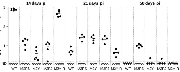
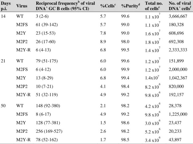
Figure 2.5. Representative staining of the GC B cell population purified by FACS ... 65 Figure 2.6. Graphical representation of limiting dilution data generated to estimate
the frequency of MuHV-4 infection in GC B cells of mice infected with WT virus at day 14 post-infection ... 67
Figure 2.7. M2 interaction motifs are important for an efficient colonization of
splenic GC B cells ... 69
Figure 2.8. Modulation of Vav activity is required for normal kinetics of latency in
the germinal centre ... 71
Figure 2.9. M2 early phenotype is independent of infective dose and mouse strain ... 73 Figure 3.1. Amino acid sequence of the M2 H-2Kd restricted CD8+ epitope ... 80
Figure 3.2. Construction and characterization of M2YF85A and M2YF85AR
viruses ... 83
Figure 3.3. M2YF85A recombinant behaves as M2FS virus during the
establishment and maintenance of latency ... 85
Figure 3.4. M2YF85A recapitulates the kinetics of M2FS latency in splenic
follicles ... 86
Figure 4.1. Construction and verification of the genomic structure of M2Y120F and
M2Y129F viruses ... 94
Figure 4.3. Normal establishment of latency in the spleen is dependent on both
phosphosites of M2 ... 96
Figure 4.4. Delayed splenic follicles colonisation by MuHV-4 viruses lacking M2
tyrosine residues ... 98
Figure 4.5. Normal kinetics of expansion of latently infected GC B cells requires
both M2 tyrosines ... 99
Figure 5.1. FACS analysis of MuHV-4-infected bone marrow-derived dendritic
cells ... 111
Figure 5.2. M2 is not required for lytic or latent MuHV-4 infection of dendritic
cells ... 114
Figure 5.3. M2 is not required for lytic or latent MuHV-4 infection of Raw264.7
macrophages ... 116
Figure 5.4. M2 is not required for lytic or latent MuHV-4 infection of peritoneal
macrophages ... 117
Figure 5.5. Mediastinal lymph node cell populations analysed in this study ... 118 Figure 5.6. M2 is not required for efficient colonization of mediastinal lymph node
dendritic cells and macrophages ... 120
Figure 7.1. Schematic representation of the construction of a recombinant shuttle
I
NDEX OFT
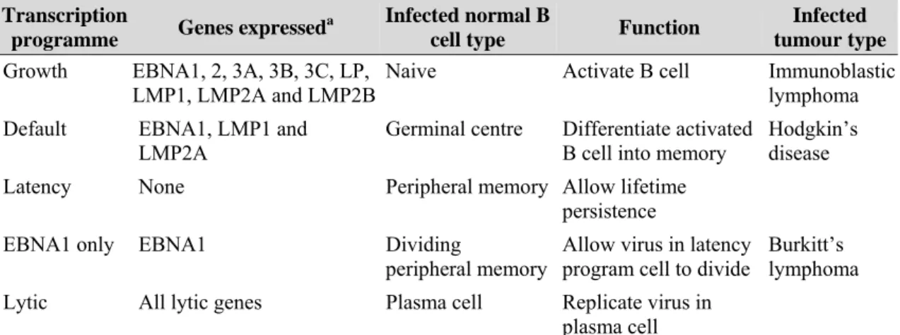
ABLESTable 1.1. Human herpesviruses ... 4
Table 1.2. Diseases linked to human gammaherpesviruses ... 8 Table 1.3. The EBV transcription programmes in normal B cells and tumours ... 20 Table 2.1. Primers and probe used to detect MuHV-4 DNA ... 66 Table 2.2. Frequency of genome-positive GC B cells ... 68 Table 4.1. Frequency of genome-positive GC B cells ... 100 Table 5.1. Frequency of MuHV-4 infection in MLN dendritic cells, macrophages
and B cells ... 119
Table 5.2. Quantification of reactivation competent virus and preformed infectious
virus in mediastinal lymph node populations at day 5 pi ... 120
Table 7.1. Primers used to amplify M2 gene ... 143 Table 7.2. Primers used to detect M2 transcripts ... 144 Table 7.3. PCR reactions and primers used for analysing genome integrity of the
recombinant viruses in the HindIII-E region ... 144
C
HAPTER1
I
NTRODUCTIONHerpesviruses are highly disseminated in nature. A long history of co-evolution with their hosts has placed these viruses amongst the most accomplished and ubiquitous pathogens. The main biological feature shared by all herpesviruses is their ability to persist for life in normal healthy individuals by establishing a latent infection, from which they periodically reactivate to disseminate to new hosts. During latency, herpesviruses persist in a non-infectious form, unnoticed by the immune system and causing minimal damage to their hosts.
In the case of gammaherpesviruses, latent infection is established preferentially in B cells and is associated with several neoplastic diseases, making the control of these viruses an important clinical goal. B cells compose a very dynamic population that is, at specific stages of differentiation, prone to clonal expansion. However, usually these cells are short lived and have the tendency to enter apoptosis unless specific survival signals are provided. Therefore, to establish latency in B cells, gammaherpesviruses must express proteins that interfere with normal host cell developmental pathways in order to induce cell survival and proliferation.
Understanding the mechanisms developed by gammaherpesviruses to subvert B cell function in its own profit will provide useful information about viral pathogenesis and facilitate the development of therapeutic strategies against these viruses. Moreover, studying virus/B cell interaction will increase the knowledge on essential processes of host cell biology and immune system.
1.1. H
ERPESVIRUS1.1.1. General Properties
Herpesvirus taxonomy has recently been updated with the creation of the order
Herpesvirales that includes three families: Herpes-, Alloherpes- and Malacoherpesviridae. Mammalian herpesviruses belong to the Herpesviridae family,
herpesviruses have been classified as Alloherpesviridae, while a single invertebrate virus constitutes the family Malacoherpesviridae (Davison et al., 2009).
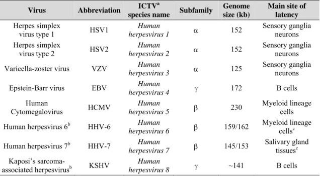
Herpesviruses are highly disseminated in nature, with a large spectrum of animals harbouring at least one of these viruses and the most studied hosts being infected by several distinct herpesviruses (Davison, 2002). The family Herpesviridae is the largest and includes over 120 herpesviruses of which eight have been isolated from humans (Table 1.1) (Davison et al., 2009). Furthermore, herpesvirus are ubiquitous in the general population with most adults harbouring herpes simplex 1, varicella-zoster virus, human herpesviruses 6 and 7, and Epstein-Barr virus (Cohen, 2003, Davison & Clements, 2005).
Table 1.1. Human herpesviruses (adapted from Davison & Clements, 2005).
Virus Abbreviation ICTV
a
species name Subfamily
Genome size (kb)
Main site of latency
Herpes simplex
virus type 1 HSV1 herpesvirus 1 Human α 152
Sensory ganglia neurons Herpes simplex virus type 2 HSV2 Human herpesvirus 2 α 152 Sensory ganglia neurons
Varicella-zoster virus VZV Human
herpesvirus 3 α 125
Sensory ganglia neurons
Epstein-Barr virus EBV Human
herpesvirus 4 γ 172 B cells
Human
Cytomegalovirus HCMV herpesvirus 5 Human β 230
Myeloid lineage cells
Human herpesvirus 6b HHV-6 Human
herpesvirus 6 β 159/162
Myeloid lineage
cellsc
Human herpesvirus 7b HHV-7 Human
herpesvirus 7 β 145/153
Salivary gland
tissuesc
Kaposi’s
sarcoma-associated herpesvirusb KSHV Human
herpesvirus 8 γ ~141 B cells
aInternational Committee on Taxonomy of Viruses
bMore than one strain has been sequenced.
cThe site of latent infection has not been completely clarified
Herpesviruses are remarkably well adapted to their hosts, a characteristic that is probably the outcome of a very long co-evolutionary history (Davison, 2002). In fact, host-specific occurrence of herpesviruses and molecular phylogenetic studies suggest that herpesvirus speciation occurred at approximately the same time as did host speciation. As a result of this adaptation, herpesvirus infection of a natural immunocompetent host is rarely fatal, therefore promoting the transmission of the viruses. Each herpesvirus is usually restricted in infection to a single species and
spreads from host to host by direct contact or by respiratory route (Davison, 2002, Davison & Clements, 2005, Roizman & Pellett, 2001).
Herpesviruses are a very heterogeneous group, with widely differing biological properties, therefore the primary means of identifying a herpesvirus has been that of virion structure (Davison et al., 2009) (refer to Fig. 1.1 for details on virion architecture). These viruses are among the largest and most complex, with a genetic material composed of a linear, double stranded DNA molecule that varies in length from 125 to 290 kb, and encodes between 70 (smallest genome) and 200 (biggest genome) genes (Roizman & Pellett, 2001).
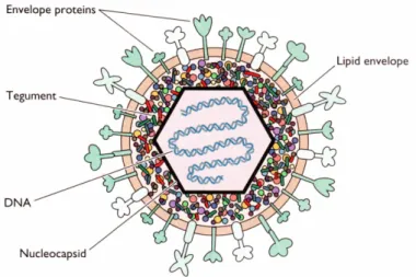
Figure 1.1. Schematic representation of a herpesvirion. Herpesvirions have a unique four-layered
structure: a core containing the genetic material in the form of a torus is enclosed by an icosadeltahedral capsid containing 162 capsomers and 100 nm diameter with a hole running down the long axis. The nucleocapsid is surrounded by the tegument, an amorphous–like structure with variable size containing several viral-coded proteins, itself enveloped by a lipid bi-layer, derived from the host cell, with numerous glycoprotein spikes projecting from its surface. Herpesvirions can vary in size from 120 to nearly 200 nm, in part due to changes in the thickness of the tegument (figure extracted from Flint et al., 2004).
The most remarkable biological characteristic shared by all herpesvirus is the ability to establish a latent infection that ensures the maintenance of their genetic material for the life of the host. Most herpesvirus infections are not apparent, but if the host’s immune defences are compromised, effects can be devastating (Davison & Clements, 2005, Flint
et al., 2004).
All herpesviruses have the capacity to establish productive infection as well as to undergo latent infection. Upon transmission to a naive host, the virus first establishes a productive acute infection at the site of infection, usually the epithelium of a mucosal surface. During the lytic phase of a herpesvirus life cycle, there is a temporal order of
viral gene expression and the viral genome is replicated several times, leading to the production of infectious virion progeny and the death of the infected cell. This primary infection is rapidly resolved by host immune defences and results in effective immunity against reinfection (Cohen, 2003, Flint et al., 2004).
The lytic infection is followed by a latent infection that lasts throughout the life of the host and is maintained in a specific set of cells. The cell type targeted is herpesvirus specific and the cell carrying the latent virus is usually different from those that harbour the productive infection. During a latent infection, usually asymptomatic, no infectious virus particles are detected in the host and the viral genome is maintained, as a circular episome, in the nucleus of the infected cell. Latency is characterized by sporadic reactivation episodes that are critical for the virus, ensuring the transmission of infectious virions to new hosts, as well as reinfection and establishment of latent infection in more cells of the same individual, thereby establishing a reservoir of infection for life. Reactivation may be spontaneous, or follow trauma, stress or other insults and is usually asymptomatic or has milder symptoms than the original acute infection. During latent infection, the expression of viral proteins is severely reduced to those required for maintenance of viral genome, manipulation of host cell function and evasion of the host immune system. Such strategies prevent or delay the elimination of the latently infected cell. By expressing few proteins during latency, herpesviruses limit the amount of foreign antigen produced, thereby reducing the chance of being detected by the host immune system (Cohen, 2003, Flint et al., 2004, Roizman & Pellett, 2001). The family Herpesviridae is further subdivided into three subfamilies, on the basis of preferential site of latency, among other biological characteristics, and DNA sequence homology: Alphaherpesvirinae, Betaherpesvirinae and Gammaherpesvirinae (refer to Table 1.1 for human herpesvirus subfamily classification). Alfaherpesviruses maintain latency mainly in neurons of the sensory ganglia, betaherpesviruses are found latent preferentially in hematopoietic stem cells and cells of the myeloid lineage and, in gammaherpesviruses, latency is usually established in either B or T cells and is frequently associated with lymphoid tissue (Davison, 2002, Davison et al., 2009, Roizman & Pellett, 2001).
Latency can be established in very diverse cellular environments depending upon the subfamily considered, which implies that many of the viral and cellular mechanisms
regulating latency are unique. Given that the work presented in this thesis focuses on the molecular pathogenesis of gammaherpesvirus latent infection, only this subfamily will be discussed in the following sections.
1.1.2. The subfamily Gammaherpesvirinae
Members of the subfamily Gammaherpesvirinae are widespread in nature. On the basis of DNA homology, this subfamily has recently been divided into four genera: the formerly assigned Lymphocryptovirus (or gamma1 group) and Rhadinovirus (or gamma2 herpesvirus) genera, and the recently created Macavirus and Percavirus genera. The genus Lymphocryptovirus infects only primates, while viruses of the genus
Rhadinovirus have been found in rodents, leporids, bovines and a variety of primates.
The lineage containing bovine, caprine, ovine and suid herpesviruses forms the genus
Macavirus. Finally, the genus Percavirus is the largest, including equine and various
carnivore viruses, and the only one to harbour non-mammalian herpesviruses, namely avian and reptilian viruses. Species in both of these last two genera were formerly assigned to the genus Rhadinovirus, which became more tightly demarcated (Davison et
al., 2009, McGeoch et al., 2006).
Gammaherpesviruses are host-range specific. All members of this subfamily are capable of replicating in vitro in lymphoblastoid cells and some also cause lytic infections in fibroblasts and epithelial cells. The gamma subfamily of herpesviruses is associated with the lymphoid tissue and latency is established mainly in either B or T cells (Davison & Clements, 2005).
A striking property of all members of this subfamily is the ability to drive the proliferation of infected lymphocytes during the establishment and maintenance of a large reservoir of latent viral genomes (Stevenson, 2004). Gammaherpesvirus latency-associated lymphoproliferation is central to host colonization but also underlies the vast majority of diseases associated with gammaherpesvirus. In fact, a hallmark of gammaherpesvirus is their ability to induce neoplasia in natural or experimental hosts, long after the initial infection has occurred (Barozzi et al., 2007, Damania, 2004). Two gammaherpesviruses that infect humans have been identified (Table 1.1), the
Lymphocryptovirus Epstein-Barr virus (EBV) (Epstein et al., 1964) and the Rhadinovirus Kaposi’s sarcoma-associated herpesvirus (KSHV) (Chang et al., 1994).
Both human gammaherpesviruses establish latent infections mainly in B lymphocytes and are associated with a number of lymphoproliferative diseases of B and/or T cells, and with different types of malignancies, mainly in lymphoid cells (Table 1.2), which makes understanding and controlling EBV and KSHV major goals of gammaherpesvirus research.
Table 1.2. Diseases linked to human gammaherpesviruses (adapted from Damania, 2004 and Cohen,
2003)
Gammaherpesvirus Associated diseases
Non-malignant Malignant
Epstein-Barr virus Infectious mononucleosis Burkitt’s lymphoma
Hairy leukoplakia of the tongue Hodgkin’s lymphoma
Post-transplant and AIDS-related lymphomas
X-linked lymphoproliferative syndrome
T-cell lymphomas
Nasopharyngeal carcinoma
Gastric carcinoma
Kaposi’s
sarcoma-associated herpesvirus Multicentric Castleman’s disease Kaposi’s Primary effusion lymphomas sarcoma
1.1.3. Gammaherpesvirus latency in B cells
B cells are a very dynamic population and at specific stages of their development, when appropriate signals are provided, have the ability to proliferate (reviewed in section 1.4). However, these cells are usually short lived and, without the correct survival and proliferation signals, are prone to die by apoptosis. Therefore, to persist successfully in B cells, gammaherpesviruses must exploit multiple cellular processes that ensure B cell survival and drive its proliferation. Gammaherpesviruses achieve this by expressing proteins that inhibit apoptotic pathways, manipulate cellular signal transduction pathways and drive cell cycle progression (Brinkmann & Schulz, 2006, Rajcani & Kudelova, 2003).
Because B cells are induced to proliferate, during latency gammaherpesvirus must also express proteins that enable the replication and segregation of the viral episomes into the daughter cells (Rajcani & Kudelova, 2003)
Additionally, viruses must deal with inherently hostile environment constantly presented in the host. Infection induces both innate and adaptive immune system
responses, in particular T cell responses (Redpath et al., 2001). Thus, to successfully persist in immunocompetent hosts, besides severely reducing gene expression during latency, gammaherpesviruses have developed sophisticated immune evasion strategies. Viral immunomodulators interfere with both innate and adaptive immune responses and many of them are analogues of cellular proteins, suggesting that they have been pirated from the host during viral evolution (Alcami & Koszinowski, 2000, Coscoy, 2007, Redpath et al., 2001, Stevenson, 2004, Vossen et al., 2002, Wiertz et al., 2007).
Gammaherpesvirus latency is therefore a dynamic and constantly changing process. Having gammaherpesviruses and hosts a very long co-evolutionary history, the virus-host balance presumably reflects a stable equilibrium of immune pressure versus modulation. Any changes in this balance can result in disease (Jerome, 2008, Redpath et
al., 2001).
B cells constitute a very complex population that is in constant renewal and change, therefore, to better understand the molecular mechanisms involved in gammaherpesvirus latent infection, it is first important to address some aspects of B cell development and biology.
1.2. T
HEB
CELLB cells, or B lymphocytes, are a highly dynamic and heterogeneous population, harbouring several distinct subpopulations. A striking property of B cells is their ability to mount a specific immune response against virtually any foreign antigen. This is possible because each individual B cell matures expressing a cell surface antigen receptor of a single specificity, generated by random recombination of variable receptor gene fragments. The resulting population of B lymphocytes collectively expresses an extensive repertoire of receptors with highly diverse antigen-binding sites. The function of the B cell antigen receptor, or B cell receptor (BCR), is to recognize and bind antigen in its native form, present outside the cells of the body, thus transmitting a signal that causes B cell activation. Structurally, the BCR is a membrane-bound form of the antibody (immunoglobulin, Ig) that the B cell secretes after activation and differentiation to plasma cell. The main effector function of B cells in the adaptive immunity is the secretion of antibodies. Antibodies bind pathogens or their toxic
products in the extracellular spaces of the body and engage specific immune mechanisms (Janeway et al., 2005).
1.2.1. The development and survival of B cells
B cells originate and develop primarily in the bone marrow, from a common lymphoid progenitor (Fig. 1.2). During the early stages of development, progenitor B cells undergo a programmed series of immunoglobulin gene rearrangement and assemble the B cell receptor that will determine the specificity of the individual lymphocyte. This phase is independent of antigen but is dependent on interactions with bone marrow stromal cells that provide signals through secreted growth factors and cell-surface molecules that bind to receptors on the B lymphocyte precursor cell (Fig. 1.2, first panel). If gene rearrangement is successful, a complete immunoglobulin B cell receptor is formed and the developing B cell is defined as an immature B cell. If a productive rearrangement is not made, the developing B cell dies (Janeway et al., 2005).
The expression of a B cell receptor on the surface of the immature B lymphocyte is a hallmark in its development, because it can now detect ligands that bind to this receptor. From this stage, all the steps in B cell development are regulated by signals received through its antigen receptor. Initially the BCR is tested for its antigen-recognition properties against molecules in the immediate environment (Fig. 1.2, second panel). B cells whose receptors bind strongly to self antigens undergo receptor editing so that the self-reactive receptor specificity is deleted, or die by apoptosis (clonal deletion). This process is termed negative selection and induces the establishment of immunological tolerance to ubiquitous self antigens. Developing B lymphocytes whose receptors do not interact with self antigens, or bind them weakly, are allowed to survive and leave the bone marrow, and are transported in the blood to the peripheral lymphoid tissues, namely the spleen and lymph nodes (Janeway et al., 2005).
The newly formed (NF) B cells are immature. They express high levels of IgM but little IgD, and need further interaction with their self ligands and neighbouring cells in the tissue to mature and survive. In the periphery, immature B cells suffer two distinct selection processes. They undergo a new step of negative selection, or peripheral tolerance, in which self-reactive cells that encounter autoantigens for the first time are eliminated or inactivated. In addition, B cells in the periphery also need to receive positive signals through their BCR in order to survive and further differentiate into
mature B cells. This process in known as positive selection and occurs in distinct areas of the secondary lymphoid organs, the lymphoid follicles, to which B cells are directed by the action of chemokines. The limited number of lymphoid follicles cannot accommodate high numbers of immature B cells released into the periphery each day and so there is continual competition for entry. If newly produced immature B cells fail to enter a follicle, their passage through the periphery is halted and they die. B cells that survive both selection processes mature to express IgD as well as IgM, and are defined as a follicular (Fo) B cell (Fig. 1.2, third panel) (Janeway et al., 2005).
Mature B cells that have not encountered their specific antigen are referred to as naive B cells and circulate continually from the blood into the peripheral lymphoid tissues, where they receive survival or tonic signals in the lymphoid follicles. They can now be activated by encounter with specific foreign antigen in a secondary lymphoid organ and differentiate to become an effector antibody-secreting plasma cell or a memory cell (Fig. 1.2, fourth and fifth panels; section 1.2.2) (Janeway et al., 2005).
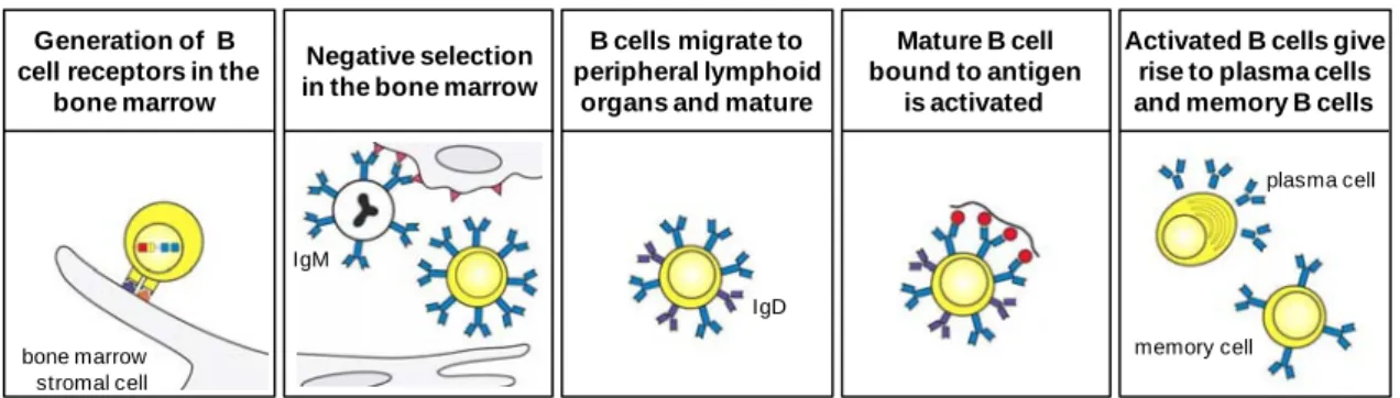
Figure 1.2. The B cell life history. B cells are generated in the bone marrow from a lymphoid
progenitor. During the initial phase of development, B cell progenitors rearrange their immunoglobulin genes, in an antigen-independent but bone marrow stomal cell-dependent process, giving rise to an immature B cell that expresses an antigen receptor in the form of IgM (first panel). This cell can now interact with antigens in the environment and undergoes the first part of its maturation process, negative selection in the bone marrow. During negative selection, immature B cells that are strongly stimulated by antigen die or are inactivated, therefore removing self-reactive B cells from the repertoire (second panel). In the third stage of the development, surviving B cells emerge into the periphery and home to secondary lymphoid organs were they suffer a second selection process (both positive and negative) and mature to express IgD as well as IgM (third panel). Mature B cells are activated by specific foreign antigen in a secondary lymphoid organ (forth panel). Activated B cells proliferate and differentiate into antibody-secreting plasma cells or long-lived memory cells (fifth panel) (adapted from Janeway et al., 2005).
Generation of B cell receptors in the
bone marrow
Negative selection in the bone marrow
B cells migrate to peripheral lymphoid
organs and mature
Activated B cells give rise to plasma cells and memory B cells Mature B cell bound to antigen is activated bone marrow stromal cell IgM IgD plasma cell memory cell
Lymphocyte maturation and survival is a highly dynamic and competitive process that is regulated by signals received through the antigen receptor. Of the about 2x10⁷ IgM+ cells that are produced daily in the bone marrow, only 10% migrate to the periphery and of these only a third enter the mature B cell pool (Chung et al., 2003). In the absence of specific antigen stimulation, mature naive B cells survive for 6-8 weeks in mice.
1.2.2. The activation and differentiation of B cells
Antigens are presented to naive recirculating B lymphocytes as they migrate through the lymphoid tissue (Batista & Harwood, 2009). Upon recognizing a specific antigen on an activated antigen-presenting cell, usually a dendritic cell or a macrophage, the B cell migrates to the T cell zone of the lymphoid tissue where it becomes fully activated by interaction with antigen presenting cells and helper T cells (Th) (MacLennan, 1994). To become fully activated, the B cell requires two distinct signals: the first main signal is delivered by the BCR following cognate antigen stimulation while the second complementary signal is delivered by armed helper cells that recognize the same epitope (linked recognition). Helper T cells are a subset of CD4+ T lymphocytes mainly involved in providing activation and differentiation signals to B cells. Th cells require prior activation by professional antigen-presenting cells that display the same antigen, this way differentiating into armed Th cells. B cells can receive help from armed Th cells after the antigen bound by the BCR is internalized, processed and returned to the cell surface as peptides bound to major histocompatibility complex (MHC) class II molecules. The MHC:peptide complex is recognized by the T cell receptor (TCR) of the armed Th cell that binds the peptide originating a B:T cell conjugate. Formation of this conjugate induces the full activation of the B cell (Klein & Dalla-Favera, 2008, Mills & Cambier, 2003).
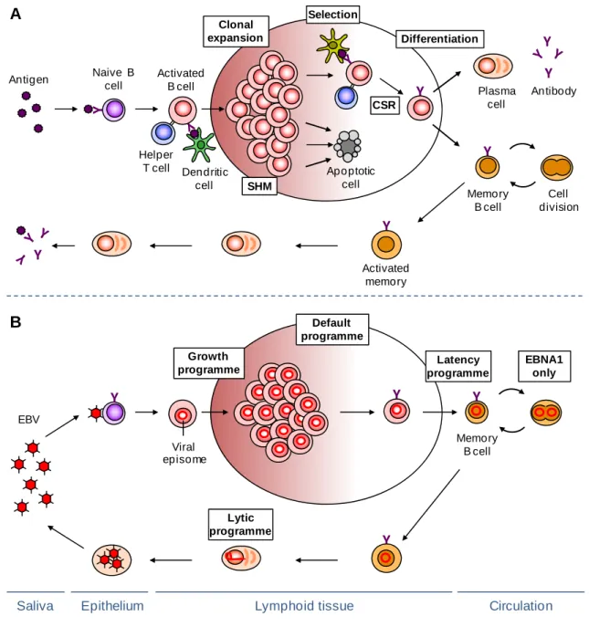
The fully activated B lymphoblast can move to the lymphoid follicle, where it starts to proliferate and differentiate into centroblasts, thereby establishing a germinal centre (GC) reaction (Fig. 1.3, panel A). The centroblasts undergo clonal expansion in the dark zone of the germinal centre where they also activate the process of somatic hypermutation of the immunoglobulin genes, thereby creating antibody variants, some with improved affinity for the antigen. Centroblasts then differentiate into resting centrocytes and move to the light zone, where the modified antigen receptor is selected for improved affinity to the immunizing antigen, with help from follicular dendritic
cells and helper T cells. Germinal centre B cells with the highest affinity for the antigen receive survival signals from T helper cells and follicular dendritic cells and undergo repeated rounds of proliferation, mutation and selection. The remaining cells that do not receive the survival signals undergo apoptosis by default and are removed (Allen et al., 2007a, Hauser et al., 2007, Klein & Dalla-Favera, 2008). Recent techniques that allow the visualization of immune cells in intact living tissue have shown that compartmentalization of the GC is often less well defined than this classical dark-zone light-zone pattern, with the movement between the light and the dark zone not always following the same direction and cell proliferation, as well as apoptosis, being sometimes detected in both zones (Allen et al., 2007b, Hauser et al., 2007, Schwickert
et al., 2007). The trafficking between the dark and light zones appears to be mediated by
a chemokine gradient, presumably established by stromal cells in the respective zones (Allen et al., 2004).
A subset of centrocytes undergoes class switch recombination, replacing the originally expressed immunoglobulin heavy-chain constant region genes from IgD and IgM to either IgG, IgA or IgE (Fig. 1.3, panel A). Isotype switching alters the effector function of an antibody without changing its specificity for the antigen (Klein & Dalla-Favera, 2008). Finally, selected GC B cells differentiate into either plasma cells or memory B cells and leave the germinal centre. Signals from helper T cells and cytokines are important for this differentiation step.
Plasma cells constitute the effector cells, secreting high affinity antibodies, but have only a limited life-span and most of them eventually undergo apoptosis. Memory B cells persist after antigen has been eliminated and are the basis of immunological memory, ensuring a more rapid and effective response on a second encounter with a pathogen and thereby usually providing lasting protective immunity.
1.2.3. Molecular events underlying B cell activation
B cell signalling and activation is initiated in response to specific antigen recognition by the B cell receptor. The BCR is a multiprotein structure composed by an antigen binding subunit, the membrane immunoglobulin, and a signalling subunit, a disulfide-linked heterodimer of the Igα and Igβ proteins, each containing a single immunoreceptor tyrosine-based activation motif (ITAM) within their cytoplasmic tail that initiates signal transduction (Dal Porto et al., 2004). Engagement of BCR by
antigen induces BCR aggregation in specialized membrane microdomains termed lipid rafts. These domains contain increased concentrations of Src-family kinases such as Lyn, Blk and Fyn, that phosphorylate the tyrosine residues of the ITAMs (Pierce, 2002). Phosphorylation of the ITAMs results in the activation of Syk kinase which, in turn, initiates the coordinated assembly of a signalosome, composed of adaptor proteins such as B cell linker (Blnk) and a variety of intracellular signalling molecules including Vav, phosphoinositide 3-kinase (PI3K), phospholipase C-γ2 (PLCγ2) and Bruton’s tyrosine kinase (Btk) (Dal Porto et al., 2004, Harwood & Batista, 2008).
Generation of the phosphoinositide PIP3 by PI3K following BCR aggregation allows the
recruitment and activation of effector proteins containing pleckstrin-homology (PH) domains. One of such proteins is the serine/threonine kinase Akt, which is activated by phosphorylation of key serine and threonine sites and translocates to the cytoplasm and nucleus where it interacts with proteins and transcription factors that control apoptosis and glycogen metabolism, thus promoting cell survival. It also activates nuclear factor-kappa B (NF-κB), which in turn upregulates genes involved in B cell development and proliferation (Dal Porto et al., 2004)
Recruitment of PLCγ2 to the plasma membrane-associated signalosome results in its phosphorylation and activation. Activated PLCγ2 cleaves membrane-associated phosphoinositide PIP2 into second messengers IP3 and DAG. DAG represents a
classical activator of conventional protein kinase C (PKC) isotypes that regulate the mitogen-activated protein kinase (MAPK) family, which, in turn, phosphorylates different sets of transcription factors including c-Jun and p38. IP3 generation causes the
mobilization of Ca2+ from intra and extracellular stores inducing the activation of transcription factors such as NF-κB and NFAT (nuclear factor of activated T cells) by atypical PKCs and Ca+2 calmodulin, respectively. (Dal Porto et al., 2004, Scharenberg
et al., 2007).
The Vav family of proteins, consisting of three isoforms – Vav1, Vav2 and Vav3 – also has a crucial role in B cell signalling. These proteins function as guanine nucleotides exchange factors (GEFs) for the Rho/Rac proteins, promoting, in a phosphorylation-dependent manner, the GDP/GTP exchange, thereby accelerating the transition of those GTPases from their inactive (GDP-bound) to active (GTP-bound) states (Bustelo, 2000). Following BCR stimulation, Vav proteins are activated by phosphorylation and
activate Rac1. Rac1 stimulates several effector proteins such as PIP5 kinase (PIP5K),
p21 activated kinase (Pak1) and c-Jun NH2-terminal kinase (JNK), thus promoting extensive changes in intracellular pathways related to cytoskeletal change, mitogenesis, and cell survival (Etienne-Manneville & Hall, 2002). In addition to the activation of Rho/Rac GTPases, Vav proteins play a role in the activation of other signalling pathways, such as PLCγ2 and PI3K pathways, modulate the activity of the transcription factors NF-AT and NF-κB and increase Ca2+ levels. (Bustelo, 2000, Turner &
Billadeau, 2002). Analyses of Vav-deficient mice have shown that Vav proteins play a key role in B lymphocyte development and proliferation. In Vav1 and Vav2 double knock-out mice, immature B cells fail to develop efficiently into mature B cells, although they are present in normal numbers within the bone marrow. Furthermore, these Vav1 and -2 null B cells also display impaired proliferation in response to BCR stimulation (Tedford et al., 2001).
Ras GTPases are activated in response to BCR crosslinking, promoting the activation of the extracellular signal-regulated (ERK) MAPKs through stimulation of Raf1 serine/threonine kinase. ERK proteins, in turn, modulate the activity of different transcription factors, altering the pattern of gene expression and promoting cell survival and proliferation. Vav proteins have also been implicated in the activation of Ras by direct activation of the GDP/GTP exchange factor RasGRP1 (Caloca et al., 2003). In summary, B cell activation triggers several interconnecting signalling pathways which modulate a variety of cellular processes, including reorganization of the cytoskeleton and regulation of gene expression to induce cell survival and proliferation. Furthermore, after triggering the signalling cascade, the ligand-BCR complex is internalized by the cell, processed within specific endosomal compartments and presented in complex with MHC class II molecules to recruit specific T cell help. This leads to full B cell activation through secreted cytokines and cell-cell interactions mediated by receptor pairs such as CD40-CD154. Thus, B cell activation is dependent on the intricate organization of both intracellular signalling pathways and intercellular communication (Dal Porto et al., 2004, Harwood & Batista, 2008).
1.3. E
PSTEIN-B
ARRV
IRUSEpstein-Barr virus, or EBV, was discovered more than 40 years ago in cultured tumour cells from patients with Burkitt’s lymphoma (Epstein et al., 1964). If the success of a pathogen is defined by the number and extent of hosts it infects, EBV is the most successful human pathogen because it is present in all populations, infecting more than 95% of human beings for life (Kutok & Wang, 2006). Primary EBV infection generally occurs in the first decade of life and is usually asymptomatic. However, if the infection is acquired during adolescence or later, it can result in a self-limiting lymphoproliferative disease known as infectious mononucleosis (Henle et al., 1968, Kutok & Wang, 2006). After primary infection, which is rapidly controlled by the host’s immune system, the virus usually persists as a harmless passenger residing in a latent form in B cells. Each person carries approximately 5x10⁵ infected B cells (Khan
et al., 1996). A low level of active virus replication continues asymptomatically in EBV
carriers, leading to virus secretion into the saliva and allowing transmission of EBV from one human to another through the oral route (Kutok & Wang, 2006).
Despite the benign and uneventful nature of EBV infection in the majority of humans, the diseases caused by this virus (Table 1.2) indicate that the situation is finely balanced. In X-linked proliferative disease, persistence infection is not established because mutations in a single gene cause acute EBV infection to become a fatal disease. Furthermore, immunological disturbance can lead to deregulation and EBV-driven tumours in otherwise healthy carriers of the virus. Individuals that are immunosupressed due to organ transplantation or due to infection by HIV are at risk for EBV lymphomas that are aggressive and often fatal. EBV is also associated with malignancies in immunocompetent individuals, such as Burkitt’s and Hodgkin’s lymphomas (Kutok & Wang, 2006, Thorley-Lawson, 2005).
EBV is the best studied gammaherpesvirus and a model of EBV latent infection has been proposed. The model is based mainly on the studies of Thorley-Lawson and colleagues which have isolated distinct human B cell subsets by flow cytometry and analysed the presence and frequency of EBV-infected cells by limiting dilution polymerase chain reaction (PCR) and the expression pattern of EBV-encoded genes by reverse transcription of RNA followed by PCR (reviewed in Thorley-Lawson, 2001, 2004, 2005, 2008 and Küppers, 2003). This model, described in the following section,