Vaccine-induced protection against
Leishmania amazonensis
is obtained in the absence of IL-12/23p40
Mayra Xiomara Hern´andez S.
a, Thales Augusto Barc¸ante
b, Luciano Vilela
c,
Wagner Luiz Tafuri
d, Lu´ıs Carlos Crocco Afonso
e, Leda Quercia Vieira
a,∗ aDepartamento de Bioqu´ımica e Imunologia, Instituto de Ciˆencias Biol´ogicas/ICB, Universidade Federal de Minas Gerais,31270-901 Belo Horizonte, MG, Brazil
bDepartamento de Parasitologia, Instituto de Ciˆencias Biol´ogicas/ICB, Universidade Federal de Minas Gerais,
31270-901 Belo Horizonte, MG, Brazil
cCentro de Pesquisas, Biomm S/A, 39400-307 Montes Claros, MG, Brazil
dDepartamento de Patologia, Instituto de Ciˆencias Biol´ogicas/ICB, Universidade Federal de Minas Gerais,
31270-901 Belo Horizonte, MG, Brazil
eDepartamento de Ciˆencias Biol´ogicas, Instituto de Ciˆencias Exatas e Biol´ogicas and N´ucleo de Pesquisa em Ciˆencias Biol´ogicas,
Universidade Federal de Ouro Preto, 35400-000 Ouro Preto, MG, Brazil
Received 14 January 2005; received in revised form 1 December 2005; accepted 16 December 2005 Available online 13 January 2006
Abstract
Protozoa of the genusLeishmaniaare intracellular parasites of macrophages and may cause diverse clinical forms of leishmaniasis, including cutaneous, diffuse cutaneous, mucocutaneous and visceral leishmaniasis. Infection with L. majorin mice indicates that a protective immune response is achieved when Th1 cells are developed. Thus, adoptive or vaccine-induced protection against leishmaniasis is largely dependent on cell-mediated immunity and IFN-␥production. Induction of a Th1 response is dependent on the presence of IL-12 whilst lymphocytes are
activated. This study was aimed at evaluating the role of IL-12 during infection withL. amazonensisand after vaccination with Leishvacin®(killed
Leishmania amazonensispromastigotes), since the role of this cytokine in vaccine-induced immunity with this preparation in experimental models or in humans is not yet elucidated. Hence, C57BL/6 interleukin-12-deficient mice (IL-12p40−/−) and wild-type controls (wt) were infected with
L. amazonensisand the course of infection, parasite burden and cytokine production were compared. IL-12p40−/−mice were more susceptible
toL. amazonensisthan wt: lesions and parasite burden were larger in IL-12p40−/−when compared to wt. Interestingly, IL-4 was not produced
in the absence of IL-12 in response to infection withL. amazonensis.To evaluate the role of IL-12 in the vaccine-induced immunity against
L. amazonensisinfection, IL-12p40−/−wt mice were vaccinated in the base of the tail and subsequently challenged with
L. amazonensisin the footpads. Surprisingly, vaccinated IL-12p40−/−mice developed smaller lesions and had fewer parasites in footpads than non-vaccinated controls.
Lymph node and spleen cells from vaccinated IL-12p40−/−mice did not produce high levels of
IFN-␥in response do in vitro stimulus with antigen.
Hence, partial protection against infection withL. amazonensiscould be obtained in the absence of functional IL-12 and a typical Th1 response. © 2006 Elsevier B.V. All rights reserved.
Keywords: Leishmania; Leishmaniasis; Protozoa; Parasite; Vaccine; Adjuvant; IL-12
1. Introduction
Leishmaniasis are parasitic infections of animals and humans caused by different species of a protozoan of the genus
Leishma-∗Corresponding author at: Departamento de Bioqu´ımica e Imunologia, ICB, Universidade Federal de Minas Gerais, CP 486, 30161-970 Belo Horizonte, MG, Brazil. Tel.: +55 31 3499 2656; fax: +55 31 3499 2614.
E-mail address:lqvieira@icb.ufmg.br (L.Q. Vieira).
nia. The clinical manifestations of the diseases are determined by the species ofLeishmaniathat infects the host and by the immune response of the host to parasite[1].Leishmania amazonensis, a
member of theLeishmania mexicanacomplex, has been isolated
from patients with diverse clinical forms of the disease in South American countries, including cutaneous leishmaniasis, dif-fuse cutaneous leishmaniasis (DCL) and visceral leishmaniasis
[2].
A clear paradigm has been established for the role of cytokines in resistance and susceptibility during experimental
infection with another parasite of the genus,L. major, in inbred mice: IL-4 production by BALB/c mice leads to susceptibility,
while IL-12-dependent IFN-␥production by most mouse strains
leads to macrophage activation and control of parasite growth
[3]. However, there is evidence that the host immune response toL. amazonensisinfection is different from that directed toL. major. C3H, C57BL/6 and C57BL/10 mice, which are resistant toL. major,develop chronic lesions with persistent parasitism
when infected withL. amazonensis[4–7]. The maintenance of
chronic lesions is independent of the expression of IL-4 and a corresponding Th2 response[4,5,7].L. amazonensistriggers early production of IL-12 and IFN-␥in C57BL/6 mice similarly
toL. major[8], but infection of C3H mice results in production
of low levels of IL-12 and IFN-␥by antigen-specific CD4+ T
cells[5]. In fact, chronic infection byL. amazonensisin C3H and C57BL/6 mice persists even after administration of exogenous IL-12[5]or IFN-␥[9]. The inability of IL-12 to drive an effective
cell-mediated immune response duringL. amazonensisinfection
suggests that the parasite can evoke a potent immunomodulatory mechanism to evade widespread parasite killing and promote a chronic infection. However, lesion development and parasite
burden have been shown to be exacerbated by CD4+ T cells
[10,11], demonstrating that T cells are activated duringL. ama-zonensisinfection and that they contribute significantly to the immuno-pathology of the chronic disease[10]. In addition, sub-optimal IFN-␥production seems to favor parasite growth[10].
However, the mechanisms responsible for susceptibility of mice to this New World parasite species remain obscure. The impor-tance of understanding the factors involved in the maintenance of this chronic disease is highlighted by the fact that chronic cuta-neous leishmaniasis in humans is often correlated with a poor
T-cell-mediated immune response[12]or a mixed T-cell response
[13].
Adoptive or vaccine-induced protection against
Leishma-niais largely dependent on cell-mediated immunity, Th1
lym-phocytes and IFN-␥ production[14]. Different antigen
prepa-rations, including defined and recombinant antigens, have been demonstrated to induce this type of beneficial response
in experimental models [15–18]. During the last 20 years,
first generation vaccines composed of killed Leishmania
pro-mastigotes and manufactured according to Mayrink et al.[17]
have been subjected to clinical trials in Brazil[15,17,19–22]. For the sake of standardization, a more simplified version of the vaccine was produced by a former licensed Brazil-ian biotechnology company, Biobr´as (Montes Claros, MG),
using only one Leishmania strain (L. amazonensis strain
IFLA/BR/1967/PH8), selected among the five present in the for-mer vaccine.
Since Leishvacin is protective against infection withL. ama-zonensisin association with Corynebacterium parvum in the murine model, we chose this as our model vaccine to evaluate the role of IL-12 in the vaccine-induced protection. C57BL/6 interleukin-12-deficient mice (IL-12p40−/−) and wild-type
con-trols (wt) were used. Our results show that IL-12 p40 is important in the late control of lesions caused byL amazonensisand that, in its absence, partial resistance to infection is obtained in vac-cinated mice.
2. Materials and methods
2.1. Animals
Female C57BL/6 and BALB/c mice (4–6 weeks old) were obtained from CEBIO (Centro de Bioterismo do Instituto de Ciˆencias Biol´ogicas, UFMG, Belo Horizonte, MG, Brazil). Matrices of mice deficient in the production of the p40 chain
of IL-12 by homologous recombination (IL-12p40−/−) in the
C57BL/6 mice were kindly provided by Dr. Luiz Vicente Rizzo, Department of Immunology, University of S˜ao Paulo (S˜ao Paulo, Brazil), and bred in the Gnotobiology and Immunology Labo-ratory of the Instituto de Ciˆencias Biol´ogicas. During the exper-iments mice were kept in an animal facility with controlled environmental conditions and environmental barriers. Animals were fed a commercial diet for rodents (Labina—Purina SP, Brazil) ad libitum.
2.2. Parasites and antigens
L. amazonensis (IFLA/BR/1967/PH8 strain) andL. major
(WHO MHOM/IL/80/Friedlin) promastigotes were grown to stationary phase (5-day-old culture) at 27◦C in Grace’s insect
medium (GIBCO Laboratories, Grand Island, NY) with 20% fetal bovine serum (FBS, HyClone Laboratories, Inc., Logan,
Utah), 2 mM l-glutamine, 100 U of penicillin-G-potassium
and 100g of streptomycin sulfate per ml. Leishmania
anti-gen was obtained from stationary-phase promastigotes washed four times in 0.1 M phosphate-buffered saline pH 7.3 (PBS)
and adjusted to a concentration of 108organisms/ml.
Par-asite suspensions were submitted to four cycles of
freez-ing at −70◦C followed by thawing at 37◦C. Antigens were
stored at −70◦C and thawed immediately before use in cell
cultures.
2.3. Vaccine
Vaccine was produced and provided by Biobr´as (Montes
Claros, Brazil). The vaccine strain ofL. amazonensis was the
same used for infections (IFLA/BR/1967/PH8).
2.4. Vaccination and infection
C57BL/6 wild-type (wt) and IL12p40−/− C57BL/6 mice
were vaccinated according to Costa et al. [16]. Vaccine was
administered subcutaneously in 0.15 ml at the base of the tail. Each animal received two inoculations at an interval of 7 days (unless otherwise stated), each dose containing 100 mg of
vac-cine protein plus 250 mg of Corynebacterium parvum
(Labo-rat´orio de Extratos Alergˆenicos Ltda, Rio de Janeiro, RJ, Brazil). Twenty-eight days after the second dose, animals received a fur-ther 10 mg of vaccine, without adjuvant. Control groups in this
study were unvaccinated C57BL/6 and IL12p40−/− mice and
mice injected with C. parvumand saline (no antigen). Seven
In one experiment, mice were challenged 21 days after the last booster. Lesion size was measured during the course of infection with a dial micrometer and expressed as the difference in size between the infected footpad and the contralateral uninfected footpad.
2.5. Cell culture and parasite quantification
Spleen and lymph node (popliteal and inguinal) single cell suspensions were obtained as previously described[8]and cul-tured at 5×10⁶ml−1 in the presence or absence of antigen
preparation for 72 h. Supernatants were collected and used for cytokine assays. Parasites were quantified by limiting dilution,
as previously described[24]. The footpads were homogenized
using a glass tissue grinder in sterile PBS. Tissue debris was
removed by centrifugation at 150×g and cells were
concen-trated by centrifugation at 2000×g. Pellets were resuspended in 500l of Grace’s supplemented culture medium (see above).
220l were plated onto culture plates and diluted in log-fold
serial dilutions in supplemented Grace’s insect tissue culture medium starting with a 1:10 dilution. Each sample was plated in duplicates and read 15 days after the beginning of the culture. Pipette tips were discarded after each dilution to avoid carrying adhered parasites from one well to another. Results are expressed as the negative log of the titer (i.e., the dilution corresponding to the last positive well) adjusted per microgram of tissue.
2.6. Cytokine assays
IL-4 and IFN-␥were assayed by two-site ELISA as described
[8]. IFN-␥ in the supernatants from spleen or lymph node
cell cultures was assayed by two site ELISA using rat
anti-IFN-␥monoclonal antibody (mAb) R46A2 and polyclonal
rab-bit serum specific for the cytokine. ELISA for IFN-␥ had
a sensitivity of 16 pg/ml. The assay for IL-4 was performed using 11B11 mAb for coating and biotinilated BVD6 mAb as detection of bound IL-4. ELISA for IL-4 had a sensitivity of 15 pg/ml.
2.7. IgE and antigen-specific IgG1 and IgG2a ELISAs
Total serum immunoglobulin E (IgE) was quantitated by ELISA. The plates were coated with mouse anti-IgE (clone 2363, Southern Biotechnology, Birmingham, AL, USA) at a 1:500 dilution overnight at 4◦C. Sera were diluted 1:20. After
1 h incubation at 37◦C, plates were washed. After washing,
wells were incubated with biotinylated murine IgE anti-body (clone R35-72, Southern Biotechnology) at a 1:500 dilu-tion, washed as above, developed using horseradish peroxidase-streptavidin (Sigma, St. Louis, MO, USA). A positive control was performed using serum from BALB/c mice rendered aller-gic to ovalbumin (kindly provided by Janaina S. Saldanha and Dr. Denise Carmona, Departamento de Patologia/ICB-UFMG, Belo Horizonte). In order to detect antigen-specific IgG1 and IgG2a, ELISA for specific IgG1/IgG2 antibodies was optimised regarding antigen concentrations, sera and conjugate dilutions.
L. amazonensis antigen preparation was derived from in vitro
promastigote cultures (10g protein/ml) was diluted in 0.1 M
sodium carbonate buffer (pH 9) and 100l per well were used
to coat flat bottom 96-well plates overnight at 4◦C. Plates were
blocked for 1h at 37◦C with PBS containing 1% bovine serum
albumin (BSA, Sigma). Mouse sera were diluted 1:50 with PBS containing 1% BSA and serially diluted in the plate (1:3 dilu-tions) After 2 h incubation at 37◦C, plates were washed five
times with PBS containing 0.05% Tween 20 (Sigma, St. Louis, USA). Wells were incubated with goat anti-mouse IgG1 or IgG2a (Southern Biotechnology) at a 1:4000 dilution washed and then incubated with rat anti-goat horseradish peroxidase-labeled antibody (Southern Biotechnology) at a 1:2000 dilution. Absorbance values were read at 492 nm in a Spectra Max Plus reader (Spectra Max Plus reader Molecular Devices Corpora-tion, Sunnyvale, CA, USA).
2.8. ELISPOT assays
IL-4 and IFN-␥-producing cells were determined by
ELISPOT assay. MultiScreen-HA Cellulose Ester filtration 96-well plates (Millopore Corp., Bedford, MA, USA) were coated with 0.5g/50l/well of anti-IL-4 (clone BVD4-1D11,
PharMingen, San Diego, CA, USA) or 0.5g/50l/well of
anti-IFN-␥(clone R4-6A2, PharMingen) in PBS overnight at 4◦C.
The plates were washed twice with PBS and blocked using 5% FBS in DMEM (Dulbecco’s minimal essential medium con-taining 2 mMl-glutamine, 100 U of penicillin 6-potassium/ml,
100g of streptomycin/ml, 25 mM HEPES) for 2 h at 37◦C.
The plates were washed with PBS. Single cell suspension of the popliteal lymph node and spleen cells the infected mice were obtained 5 and 8 weeks post-infection as indicated below. The cells were disassociated with a tissue homogenizer and were resuspended in complete tissue culture medium DMEM containing 10% FBS and were adjusted to a concentration of 3×10⁵/200 and 1×10⁶/200
l/well in 96-well plates. The plate
was incubated with or without 100l ofL. amazonensisantigen
(1 mg/ml) for 20 h at 37◦C in a humidified chamber
contain-ing 5% CO2. The wells were washed four times each with
0.01% Tween 20 in PBS and twice with PBS and overlaid with
0.025g/50l/well of biotinylated anti-IL-4 (clone
BVD6-24G2, PharMingen) or anti-IFN-␥ (clone XMG1.2,
PharMin-gen) for 2 h at room temperature. Subsequently, the plates were washed, treated with 1:2000 dilution of Streptavidin-conjugated alkaline phosphatase (PharMingen) for 1 h at room tempera-ture and washed six times with 0.01% Tween-PBS and twice
with PBS. The IL-4 or IFN-␥ secreting cells was visualized
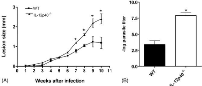
Fig. 1. Course of infection withL. amazonensisin C57BL/6 (wt) and IL-12p40−/−mice. Mice were injected in the left hind footpad with 104stationary-phase promastigotes. (A) Lesion sizes were measured weekly. Each point represents the mean difference in size±standard deviation of the mean between infected and uninfected footpads for five mice per group at each time point. (B) Parasite quantification in footpad lesions after 10 weeks of infection. Each bar represents mean±standard deviation of the mean for five animals per group. The asterisk indicates a statistically significant difference (p≤0.05) compared with the other group.
2.9. Histopathology
At the indicated time periods, foot tissues were collected, fixed in a formalin solution and embedded in paraffin. Sections were stained with hematoxylin and eosin. Sections were pho-tographed using an Olympus photomicroscope equipped with an Olympus exposure control unit (Olympus Cop., New Hyde Park, NY).
2.10. Reproducibility and statistical analysis
Experiments were performed at least three times. Means were considered statistically different whenp≤0.05 by Student’st -test.
3. Results
3.1. Course of infection with L. amazonensis in IL-12p40−/−and wt C57BL/6 mice
IL-12p40−/− and wt mice were infected with 104 L.
ama-zonensis stationary-phase promastigotes, and lesion
progres-sion was monitored (Fig. 1A). Lesions in infected wt mice
were at their peak size by 8 weeks and remained constant. IL-12p40−/− infected mice exhibited quite a distinct pattern of
disease. Although lesion sizes could remain similar to the wt group for 6 weeks (Fig. 1A), or even up to 10 weeks (data not shown), at later time points IL-12p40−/−mice developed larger
lesions, which progressively increased in size and did not show signs of healing for up to 14 weeks (one experiment performed,
data not shown). Parasite burden in IL-12p40−/− mice at 10
weeks of infection was significantly larger (about 104 times) than parasite numbers in lesions form wt mice (Fig. 1B). These results show that IL-12p40−/− mice are highly susceptible to
infection withL. amazonensis.
3.2. Cytokine production by C57BL/6 and IL-12p40−/−
mice following infection with L amazonensis
IL-12 is a crucial cytokine for the differentiation of the Th1 subset of helper cells and mice have been shown to default to a Th2 response in the absence of this cytokine[6,14,25,26]. In order to determine if the same default response would be found in L. amazonensis-infected mice in the absence of IL-12, we
Table 1
In vitro cytokine production by spleen and lymph node cells from na¨ıve (c) and vaccinated (v) wt and IL12p40−/−mice infected for 10 weeks withL. amazonensis
IFN-␥(ng/ml) IL-4 (ng/ml)
Bkga LAb Bkg LA
Spleen C57BL/6 c 6.1±5.1 9.1±7.5c 0.02±0.01 0.02±0.01
C57BL/6 v 20.3±12.2 51.5±22.7d 0.04±0.01 0.04±0.01
IL12p40−/−c 0.05±0.1 0.2±0.2 0.01±0.0 0.01±0.0
IL12p40−/−v 0.4±0.5 0.7±0.7e 0.01±0.01 0.02±0.01
Lymph node C57BL/6 c 10.5±7.6 43.2±20c 0.02±0.01 0.01±0.0
C57BL/6 v 61.3±18.5 140.2±13d 0.04±0.02 n.d.
IL12p40−/−c n.d.f 0.9±1 0.1±0.0 0.1±0.0
IL12p40−/−v 1.8±1.3 2.1±1.6e 0.1±0.0 0.1±0.0
aNo antigen added to the culture.
b L. amazonensisfreeze-thawed antigen added to the culture. cP< 0.05 wt c vs. IL12p40−/−c by Student’st-test. d P< 0.05 wt v vs. wt c by Student’st-test. eP< 0.05 wt v vs. IL12p40−/−v by Student’st-test. fNot detected. Limits of detection were: 0.016 ng/ml for
Fig. 2. IL-4-producing spleen and lymph node cells in control and vaccinated C57BL/6 and IL-12p40−/−mice after infection withL. amazonensisfor 5 and 8 weeks. IL-4 secreting cells were measured by ELISPOT assay. BALB/c mice infected withL. major(BALB/c FN) andL. amazonensis(BALB/c PH8) were used as positive controls. Spleen cells and popliteal lymph node cells draining the site of infection were incubated over wells (seeded with 5×10⁵and 1×10⁶cells) that had been precoated with mAbs against IL-4, as described in Section2. Pictures of IL-4 ELISPOT membranes from the ELISPOT assay are shown at the bottom. Each bar represents mean±standard deviation of the mean for five animals per group. The asterisks indicate a statistically significant difference (p≤0.05) compared with the other group. Bkg, no antigen added to the culture, LA,L. amazonensisfreeze-thawed antigen added to the culture.
determined the cytokine production by spleen and lymph node cells from wt and IL-12p40−/− mice in vitro. Results from a
representative experiment are shown inTable 1. Cells from wt
mice responded toL. amazonensiswith IFN-␥production, and,
quite expectedly, cell cultures from IL-12p40−/−mice produced
low levels of this cytokine both in the absence and in the pres-ence of antigenic stimulus. These results were confirmed by
ELISPOT: there were no detectable IFN-␥-producing cells in
spleens and lymph nodes of IL-12p40−/−mice infected withL. amazonensis,nor in BALB/c mice infected withL. majororL.
amazonensis(Table 2). Wild-type C57BL/6 mice showed sig-nificant numbers of IFN-␥producing cells when infected withL. amazonensis. Surprisingly, however, IL-4 production was either undetectable or just above the detection limit of the assay in cell culture supernatants from both IL-12p40−/− and wt mice
of mice (Table 1). These results were confirmed by ELISPOT
assays. As shown inFig. 2, IL-12p40−/−mice did not show an
increase in the number of IL-4-secreting cells at 5 or 8 weeks after infection. In addition, we did not find a significant inver-sion of the IgG1/IgG2a ratio, nor a significant increase in the
Table 2
Mean IFN-␥ELISPOT response by spleen and LN cells in vaccinated and control C57BL/6 and IL-12p40−/−mice after infection withL. amazonensis
IFN-␥secreting cells (cells/106)a
Spleen Lymph node
Bkgb LAc Bkg LA
BALB/c PH8d 0.66±0.40 0.86±0.50 8.2±1.9 17.5±7.5
BALB/c FN 0.66±0.60 0±0 7.0±1.6 4.1±1.6
BALB/c ni 1±0.22 0±0 0±0 15±2
C57BL/6 v 8 weeks 5.1±0.82 34.8±13.44 469±23.12* 1028±30.41*
C57BL/6 c 8 weeks 1.6±0.3 3.8±2.2 8.6±2.89 17±2
C57BL/6 ni 0±0 4±0.2 2±1 2±1
IL-12p40−/−v 5 weeks 0±0 0±0 0±0 0±0
IL-12p40−/−c 5 weeks 0±0 0±0 0±0 0±0
IL-12p40−/−v 8 weeks 0±0 0±0 0±0 0±0
IL-12p40−/−c 8 weeks 0±0 0±0 0±0 0±0
IL-12p40−/−ni 0±0 0±0 0±0 0±0
aSpleen cells and poplitean LN cells draining the site of infection were incubated over wells (seeded with 5×10⁵and 1×10⁶cells) that had been precoated with mAbs against IFN-␥. After undisturbed incubation for 20 h, cells were washed and the captured cytokine developed using a second mAb and colorimetric reagent.
b No antigen added to the culture.
cL. amazonensisfreeze-thawed antigen added to the culture.
d BABL/c PH8: BALB/c mice infected withL. amazonensisfor 8 weeks; BALB/c FN: BALB/c mice infected withL. majorfor 8 weeks, ni: non-infected mice, v: vaccinated, c: infected non-vaccinated.
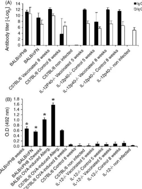
Fig. 3. Antibody isotypes in control and vaccinated C57BL/6 and IL-12p40−/− mice after infection withL. amazonensisfor 5 and 8 weeks. Uninfected mice are shown for comparison. Results for BALB/c mice infected withL. amazonensis
(BALB/c PH8) orL. major(BALB/c FN) for 8 weeks are also shown. (A) IgG1 and IgG2a were measured by incubating serially diluted sera (1:3) over wells precoated withL. amazonensisantigen, as described in Section2. Results are expressed as the log3of the antibody titer. (B) Total serum IgE was quantitated using a sandwich ELISA, as described in Section2. Results are expressed as mean of the optical density (OD). BALB/c OVA-induced allergy mice were used as a positive control. Each bar represents mean±standard deviation of the mean for five animals per group. The asterisks indicate a statistically significant difference (p≤0.05) compared with the other group.
IgE titers in IL-12p40−/−mice (Fig. 3). BALB/c mice infected
withL. majoror rendered allergic to ovalbumin were used as positive controls of a typical Th2 response[3,27,28].
Hence, IL-12p40−/−mice did not default to a Th2 response
when infected withL. amazonensis.
3.3. Vaccination of IL-12p40−/−mice against L.
amazonensis
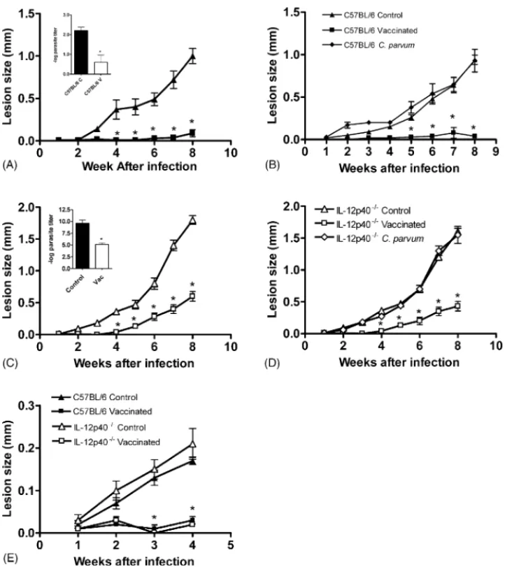
IL-12 has been shown to be an adjuvant for vaccination againstL. major[14]. Hence, we decided to investigate if IL-12 was also essential for vaccination againstL. amazonensis.A pro-tocol that was previously shown to protect C57BL/6 mice against infection with this parasite was used[16]and, as can be seen in
Fig. 4, vaccinated wt C57BL/6 mice showed smaller lesions and smaller parasite numbers than non-vaccinated controls (Fig. 4A and insert). In order to confirm thatLeishmaniaantigens were important for the vaccination protocol, we performed infections
in mice in whichC. parvumalone was injected along with saline.
C. parvum alone did not confer protection in C57BL/6 mice (Fig. 4B), in accordance with previously published[29–31]. We then proceeded to investigate the importance of IL-12 in the
immunization againstL. amazonensis.IL-12p40−/−mice were
vaccinated using the same protocol used for wt mice. Surpris-ingly, IL-12p40−/−were protected by the vaccination protocol
for at least 4 weeks, when lesions started to grow, but were still smaller than lesions in non-vaccinated mice (Fig. 4C and D). Comparison of lesions from vaccinated and non-vaccinated IL12p40−/− mice showed a statistically significant difference
from 4 to 8 weeks post-infection (Fig. 4C and D). Moreover,
in the absence of functional IL-12, vaccinated mice were able to control parasite growth more efficiently: 10,000 times fewer parasites were found in their lesions (insertFig. 4C).C. parvum
alone did not protect IL-12p40−/−against infection withL.
ama-zonensis(Fig. 4D). Moreover, IL-12-p40−/− mice (as well as wt) challenged 21 days after the last boost of antigen were still protected, showing that, at least for this time period, the effi-cacy of our vaccination protocol was lasting (Fig. 4E). Hence, in the absence of IL-12, mice could be protected by
immuniza-tion withL. amazonensisantigens in Leishvacin in association
withC. parvum.
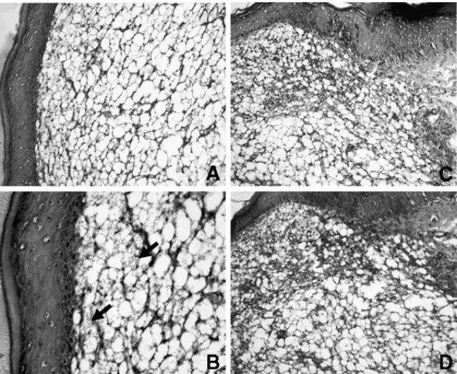
Histological analysis of the infected footpad from
IL-12p40−/−non-vaccinated mice shows an extensive vacuolated
area and parasitized macrophages (Fig. 5). These areas were
smaller in vaccinated IL-12p40−/−mice (compareFig. 5B and
D).
3.4. Cytokine production in vaccinated mice
Lymph node and spleen cell cultures from wt C57BL/6
vaccinated mice presented higher levels of IFN-␥ when
com-pared to non-vaccinated mice (Table 1) and higher numbers
of IFN-␥-producing cells (Table 2). Cells from IL-12p40−/−
mice produced practically undetectable levels of IFN-␥in vitro,
regardless of vaccination. IL-4 was not detected in supernatants from lymph node or spleen cell cultures (Table 1). In addition, no significant numbers of IL-4-producing cells were found in vaccinated wt or IL-12p40−/−(Fig. 2). The absence of
signifi-cant IL-4 production was further confirmed by IgG1 and IgG2a levels, as well as by IgE levels in sera from infected mice (Fig. 3).
4. Discussion
IL-12 is a heterodimeric pro-inflammatory cytokine that
induces production of IFN-␥ by NK cells and T cells [32].
IL-12 is critical for promoting the differentiation of naive T cells into the Th1 subset and bridges innate and adaptive immu-nity[33–35]. The development of a Th1 response is crucial for the protection of the host against many pathogens, includingL. major[36–40]. Mice that are genetically deficient for the expres-sion of IL-12 (IL-12p40−/−) are susceptible to infection withL.
Fig. 4. Course of infection withL. amazonensisin vaccinated and control C57BL/6 and IL-12p40−/−mice.C. parvumwas used as an adjuvant. Challenge was performed by injecting the left hind footpad with 104stationary-phase promastigotes. Lesion sizes were measured weekly. Each point represents the mean difference in size±standard deviation of the mean between infected and uninfected footpads for five mice per group at each time point. (A and C) C57BL/6 mice vaccinated and non-vaccinated controls. (B and D) IL-12p40−/−mice vaccinated and non-vaccinated controls. Inserts: parasite quantitation in footpad lesions 8 weeks after infection. (E) C56BL/6 and IL-12p40−/−mice vaccinated and non-vaccinated controls, challenged 21 days after the last boost of antigen. Each bar represents mean
±standard deviation of the mean for five animals per group. The asterisks indicate a statistically significant difference (p≤0.05) compared with the other group.
the maintenance of aLeishmania-specific Th1 response. Suscep-tibility in these animals was associated with a loss of the Th1
response and the development of a Th2 response[42,43].
A less clear role for the development of a Th1 response in pro-tection is found during infection withLeishmaniabelonging to the mexicana complex. In one report, infection of IL-12p40−/− mice withL. mexicana,had the same outcome as in the wild-type controls[44]. In another study, IL-12 was critical for long-term stabilization of lesions caused byL. mexicana,but insufficient to totally cure the infection, in contrast with L. major infec-tion [26]. The early control of L. mexicana infection appears
to be, thus, independent of IL-12 [26]. Here, we show that
IL-12p40−/−mice are more susceptible to infection withL.
ama-zonensis. Similar to the study by Torrentera et al.[26], IL-12 p40 seems to be more critical to long-term stabilization of lesions.
However, contrary to what was found duringL. majorinfection
[41,42], IL-12p40−/− mice do not default to a Th2 response,
as demonstrated by IL-4 production in vitro, by the number of IL-4-producing cells detected ex vivo and by low IgE levels. In our study, IgG1 levels did not correlate with IL-4 production. Hence, in the absence of functional IL-12, the higher susceptibil-ity toL. amazonensisseems to be due to a lack of an efficient Th1 response rather than a Th2 response. Indeed, this was previously suggested by Afonso and Scott[4], who found that
susceptibil-ity toL. amazonensisin the wt C57BL/10 mouse was not due to
a Th2 response, but to a lack of an efficient Th1 response.
While infection withL. amazonensisinduced IL-12 and
IFN-␥production in early stages of infection[8], infected mice fail
to express a functional IL-12R[5]. This fact could explain the failure to produce sufficient IFN-␥ to control parasites to an
extent that would allow lesion healing[4,5,7]. It is possible that another factor that is essential for control of parasite growth is missing inL. amazonensis-infected mice. In addition, it has been
amastig-Fig. 5. Representative histology of foot tissues from control and vaccinated IL-12p40−/−mice infected withL. amazonensis.Parafin-embedded sections were prepared and stained with hematoxilyn and eosin. (A) A section from control IL-12p40−/−mouse 8 weeks post-infection, showing a portion of an extensive vacuolated area (magnification 200×). (B) Numerous vacuolated and parasitized macrophages (arrows) (magnification 400×). (C) A section from IL-12p40−/−mice vaccinated at 8 weeks after infection, showing a portion of an extensive inflammatory area and the destruction of normal structures (magnification 200×). (D) A section from IL-12p40−/−mice vaccinated showing a limited vacuolated area compared with that in B (magnification 400×).
otes in macrophages[45]. According to this latter report, IFN-␥
would be, in fact, favoring the maintenance ofL. amazonensis
in the host at the later stages of infection. However, the protec-tive vaccination protocol used in our study induced higher levels
of IFN-␥production in wt C57BL/6 mice after challenge with
L. amazonensis, when compared to non-vaccinated mice. It is possible, also, that other factors, such as TNF-␣, are being
pro-duced at higher levels in vaccinated mice. This report did not investigate these other possibilities.
The higher IFN-␥production found in wt mice correlated
with less severe disease: there was a significant decrease in the number of parasites and lesions were almost negligible. This suggests to us that the decrease in lesion size may be the result of lower parasite numbers as well as a decrease in the inflam-matory response. Remarkably, histological aspects of lesions
from vaccinated and non-vaccinated IL-12p40−/− mice were
very similar, albeit lesions in vaccinated mice were consider-ably smaller than non-vaccinated mice.
IL-12 is an effective adjuvant for the initiation of protective cell-mediated immunity againstL. major, since treatment with recombinant IL-12 and parasite antigens promoted the develop-ment ofLeishmania-specific CD4+Th1 cells and the protection of a susceptible mouse strain[14,38,40]. Moreover, IL-12 was successfully used as an adjuvant for vaccination againstL. ama-zonensis[46,47]. Surprisingly, the early control ofL.
amazonen-sisconferred by vaccination with Leishvacin®andC. parvum
shown here is independent of IL-12. In addition, spleen and lymph node cells from mice that were protected against infection did not produce high levels of IFN-␥in culture upon
stimula-tion with parasite antigens. IL-12 promotes the differentiastimula-tion of CD4+T cells into the Th1, IFN-␥-producing subtype. CD8+T
cells are also able to synthesize IFN-␥but, although some
evi-dence for a protective role played by these cells has already been accumulated, this role has been far less characterized [48,49]. However, it is thought that CD8+T cells are also dependent on
IL-12 for the production of IFN-␥[50]. Another possible
expla-nation for the results described here is that even in the absence of IL-12 enoughLeishmania-specificTh1 cells developed in the
vaccinated animals. Results from Jankovic et al.[51]
demon-strate that repeated stimulation of IL-12 deficient mice induced low levels of IFN-␥-producing- CD4+T cells. It is possible that
the repeated inoculation of antigens in our vaccination proto-col was sufficient to induce the development of a number of Th1 cells capable of partially controlling the infection. How-ever, these cells were not detected in ELISPOT assays. Hence, the mechanism for resistance to infection after vaccination in IL-12p40−/−mice is still obscure, and is under investigation in our laboratory.
Here, we used vaccinated mice to study the mechanisms
of resistance against L. amazonensis. Although in wild-type
mice vaccination conferred protection, as measured by the par-asite burden and the size of lesions, and this protection cor-related with higher levels of IFN-␥, the absence of IL-12 p40
did not prevent the vaccination protocol from protecting mice.
Hence, although IL-12p40−/− mice were more susceptible to
Acknowledgements
This work was supported by FAPEMIG grant number REC 32011/99. MXHS and TAB are fellows of CAPES. LQV, WLT and LCCA are fellows of CNPq. The authors are indebted to Biobr´as S.A. for providing Leishvacin, to Antˆonio Mesquita Vaz for animal care, to Dr. Josely Lannes Vieira and Josu´e da Costa Lima Junior (FioCruz, Rio de Janeiro, Brazil) for the use of the Immunospot image analyzer, Dr. Denise Carmona Cara for the sera from allergic mice and Dr. Ricardo T. Gazzinelli for the reagents for the ELISPOT assays.
References
[1] Neva F, Sacks D, Leishmaniasis. Trop Geogr Med 1990:296–308. [2] Almeida RP, Barral-Netto M, De Jesus AM, De Freitas LA, Carvalho
EM, Barral A. Biological behavior of Leishmania amazonensis iso-lated from humans with cutaneous, mucosal, or visceral leishmaniasis in BALB/C mice. Am J Trop Med Hyg 1996;54(2):178–84.
[3] Sacks D, Noben-Trauth N. The immunology of susceptibility and resistance to Leishmania major in mice. Nat Rev Immunol 2002;2(11):845–58.
[4] Afonso LC, Scott P. Immune responses associated with susceptibil-ity of C57BL/10 mice to Leishmania amazonensis. Infect Immun 1993;61(7):2952–9.
[5] Jones DE, Buxbaum LU, Scott P. 4-independent inhibition of IL-12 responsiveness duringLeishmania amazonensisinfection. J Immunol 2000;165(1):364–72.
[6] Jones DE, Ackermann MR, Wille U, Hunter CA, Scott P. Early enhanced Th1 response after Leishmania amazonensis infection of C57BL/6 interleukin-10-deficient mice does not lead to resolution of infection. Infect Immun 2002;70(4):2151–8.
[7] Soong L, Xu JC, Grewal IS, Kima P, Sun J, Longley BJ, et al. Disruption of CD40-CD40 ligand interactions results in an enhanced susceptibility toLeishmania amazonensisinfection. Immunity 1996;4(3):263–73. [8] Oliveira MA, Santiago HC, Lisboa CR, Ceravollo IP, Trinchieri G,
Gazz-inelli RT, et al. Leishmania sp: comparative study with Toxoplasma gondii and Trypanosoma cruziin their ability to initialize IL-12 and IFN-gamma synthesis. Exp Parasitol 2000;95(2):96–105.
[9] Barral-Netto M, Von Sohsten RL, Teixeira M, dos Santos WL, Pom-peu ML, Moreira RA, et al. In vivo protective effect of the lectin from
Canavalia brasiliensis on BALB/c mice infected byLeishmania ama-zonensis. Acta Trop 1996;60(4):237–50.
[10] Soong L, Chang CH, Sun J, Longley Jr BJ, Ruddle NH, Flavell RA, et al. Role of CD4+ T cells in pathogenesis associated with Leishmania amazonensisinfection. J Immunol 1997;158(11):5374–83.
[11] Terabe M, Kuramochi T, Ito M, Hatabu T, Sanjoba C, Chang KP, et al. CD4+ cells are indispensable for ulcer development in murine cutaneous leishmaniasis. Infect Immun 2000;68(8):4574–7.
[12] Silveira FT, Lainson R, Shaw JJ, De Souza AA, Ishikawa EA, Braga RR. Cutaneous leishmaniasis due toLeishmania (Leishmania) amazo-nensis in Amazonian Brazil, and the significance of a negative Mon-tenegro skin-test in human infections. Trans Roy Soc Trop Med Hyg 1991;85(6):735–8.
[13] Pirmez C, Yamamura M, Uyemura K, Paes-Oliveira M, Conceicao-Silva F, Modlin RL. Cytokine patterns in the pathogenesis of human leishma-niasis. J Clin Invest 1993;91(4):1390–5.
[14] Afonso LC, Scharton TM, Vieira LQ, Wysocka M, Trinchieri G, Scott P. The adjuvant effect of interleukin-12 in a vaccine againstLeishmania major. Science 1994;263(5144):235–7.
[15] Antunes CM, Mayrink W, Magalhaes PA, Costa CA, Melo MN, Dias M, et al. Controlled field trials of a vaccine against New World cutaneous leishmaniasis. Int J Epidemiol 1986;15(4):572–80.
[16] Costa CA, Afonso LCC, Toledo VPCP, Tavares CAP, Genaro O, Mayrink W. Evaluation of an industrialized non-living
promastig-ote vaccine against cutaneous leishmaniasis. Parassitologia 1992;34: 45–51.
[17] Mayrink W, Da Costa CA, Magalhaes PA, Melo MN, Dias M, Lima AO, et al. A field trial of a vaccine against American dermal leishmaniasis. Trans Roy Soc Trop Med Hyg 1979;73(4):385–7.
[18] Scott P, Pearce E, Natovitz P, Sher A. Vaccination against cutaneous leishmaniasis in a murine model. II. Immunologic properties of protec-tive and nonprotecprotec-tive subfractions of soluble promastigote extract. J Immunol 1987;139(9):3118–25.
[19] De Luca PM, Mayrink W, Alves CR, Coutinho SG, Oliveira MP, Bertho AL, et al. Evaluation of the stability and immunogenicity of autoclaved and nonautoclaved preparations of a vaccine against American tegumen-tary leishmaniasis. Vaccine 1999;17(9/10):1179–85.
[20] Marzochi KB, Marzochi MA, Silva AF, Grativol N, Duarte R, Confort EM, et al. Phase 1 study of an inactivated vaccine against American tegumentary leishmaniasis in normal volunteers in Brazil. Mem Inst Oswaldo Cruz 1998;93(2):205–12.
[21] Mayrink W, Williams P, Da Costa CA, Magalhaes PA, Melo MN, Dias M, et al. An experimental vaccine against American dermal leishmani-asis: experience in the State of Espirito Santo, Brazil. Ann Trop Med Parasitol 1985;79(3):259–69.
[22] Mendonca SC, De Luca PM, Mayrink W, Restom TG, Conceicao-Silva F, Da Cruz AM, et al. Characterization of human T lymphocyte-mediated immune responses induced by a vaccine against Ameri-can tegumentary leishmaniasis. Am J Trop Med Hyg 1995;53(2):195– 201.
[23] Courret N, Prina E, Mougneau E, Saraiva EM, Sacks DL, Glaichen-haus N, et al. Presentation of theLeishmaniaantigen LACK by infected macrophages is dependent upon the virulence of the phagocytosed par-asites. Eur J Immunol 1999;29(3):762–73.
[24] Vieira LQ, Goldschmidt M, Nashleanas M, Pfeffer K, Mak T, Scott P. Mice lacking the TNF receptor p55 fail to resolve lesions caused by infection withLeishmania major, but control parasite replication. J Immunol 1996;157(2):827–35.
[25] Heinzel FP, Rerko RM, Ahmed F, Pearlman E. Endogenous IL-12 is required for control of Th2 cytokine responses capable of exacerbat-ing leishmaniasis in normally resistant mice. J Immunol 1995;155(2): 730–9.
[26] Torrentera FA, Glaichenhaus N, Laman JD, Carlier Y. T-cell responses to immunodominant LACK antigen do not play a critical role in deter-mining susceptibility of BALB/c mice toLeishmania mexicana. Infect Immun 2001;69(1):617–21.
[27] Lu XZ, Liu XM, Guo YC, Yang XG. Assessment of the BALB/c mice as a suitable animal model for the investigation of food allergy. Wei Sheng Yan Jiu 2005;34(2):211–3.
[28] Zhao Y, van Hasselt CA, Woo KS, Wong YO, Liang CY, Leung PC. Establishment of a modified intranasally ovabumin induced ani-mal model of allergic rhinitis. Zhonghua Er Bi Yan Hou Tou Jing Wai Ke Za Zhi 2005;40(3):176–80.
[29] Cardoso SR, da Silva JC, da Costa RT, Mayrink W, Melo MN, Michalick MS, et al. Identification and purification of immunogenic proteins from nonliving promastigote polyvalentLeishmaniavaccine (Leishvacin). Rev Soc Bras Med Trop 2003;36(2):193–9.
[30] Colmenares M, Kima PE, Samoff E, Soong L, Mahon-Pratt D. Perforin and gamma interferon are critical CD8+ T-cell-mediated responses in vaccine-induced immunity against Leishmania amazonensis infection. Infect Immun 2003;71(6):3172–82.
[31] Kar S, Metz C, Mahon-Pratt D. CD4+ T cells play a dominant role in protection against New World leishmaniasis induced by vac-cination with the P-4 amastigote antigen. Infect Immun 2005;73(6): 3823–7.
[32] Gately MK, Renzetti LM, Magram J, Stern AS, Adorini L, Gubler U, et al. The interleukin-12/interleukin-12-receptor system: role in nor-mal and pathologic immune responses. Annu Rev Immunol 1998;16: 495–521.
[34] Manetti R, Parronchi P, Giudizi MG, Piccinni MP, Maggi E, Trinchieri G, et al. Natural killer cell stimulatory factor (interleukin 12 [IL-12]) induces T helper type 1 (Th1)-specific immune responses and inhibits the development of IL-4-producing Th cells. J Exp Med 1993;177(4):1199–204.
[35] Manetti R, Gerosa F, Giudizi MG, Biagiotti R, Parronchi P, Piccinni MP, et al. Interleukin 12 induces stable priming for interferon gamma (IFN-gamma) production during differentiation of human T helper (Th) cells and transient IFN-gamma production in established Th2 cell clones. J Exp Med 1994;179(4):1273–83.
[36] Belosevic M, Finbloom DS, Van Der Meide PH, Slayter MV, Nacy CA. Administration of monoclonal anti-IFN-gamma antibodies in vivo abro-gates natural resistance of C3H/HeN mice to infection withLeishmania major. J Immunol 1989;143(1):266–74.
[37] Decken K, Kohler G, Palmer-Lehmann K, Wunderlin A, Mattner F, Magram J, et al. Interleukin-12 is essential for a protective Th1 response in mice infected withCryptococcus neoformans. Infect Immun 1998;66(10):4994–5000.
[38] Heinzel FP, Schoenhaut DS, Rerko RM, Rosser LE, Gately MK. Recom-binant interleukin 12 cures mice infected withLeishmania major. J Exp Med 1993;177(5):1505–9.
[39] Scott P. IFN-gamma modulates the early development of Th1 and Th2 responses in a murine model of cutaneous leishmaniasis. J Immunol 1991;147(9):3149–55.
[40] Sypek JP, Chung CL, Mayor SE, Subramanyam JM, Goldman SJ, Sieburth DS, et al. Resolution of cutaneous leishmaniasis: interleukin 12 initiates a protective T helper type 1 immune response. J Exp Med 1993;177(6):1797–802.
[41] Mattner F, Magram J, Ferrante J, Launois P, Di Padova K, Behin R, et al. Genetically resistant mice lacking interleukin-12 are susceptible to infection with Leishmania major and mount a polarized Th2 cell response. Eur J Immunol 1996;26(7):1553–9.
[42] Park AY, Hondowicz BD, Scott P. IL-12 is required to main-tain a Th1 response during Leishmania major infection. J Immunol 2000;165(2):896–902.
[43] Scott P, Artis D, Uzonna J, Zaph C. The development of effector and memory T cells in cutaneous leishmaniasis: the implications for vaccine development. Immunol Rev 2004;201(1):318–38.
[44] Buxbaum LU, Uzonna JE, Goldschmidt MH, Scott P. Control of New World cutaneous leishmaniasis is IL-12 independent but STAT4 depen-dent. Eur J Immunol 2002;32(11):3206–15.
[45] Qi H, Ji J, Wanasen N, Soong L. Enhanced replication of Leishma-nia amazonensis amastigotes in gamma interferon-stimulated murine macrophages: implications for the pathogenesis of cutaneous leishmani-asis. Infect Immun 2004;72(2):988–95.
[46] Coelho EAF, Tavares CAP, Carvalho FAA, Chaves KF, Teixeira KN, Rodrigues RC, et al. Immune responses induced by the Leishmania
(Leishmania)donovani A2 antigen, but not by the LACK antigen, are protective against experimentalLeishmania(Leishmania) amazonensis
infection. Infect Immun 2003;71(7):3988–94.
[47] Kenney RT, Sacks DL, Sypek JP, Vilela L, Gam AA, Evans-Davis K. Protective immunity using recombinant human IL-12 and alum as adjuvants in a primate model of cutaneous leishmaniasis. J Immunol 1999;163(8):4481–8.
[48] Belkaid Y, Von SE, Mendez S, Lira R, Caler E, Bertholet S, et al. CD8+ T cells are required for primary immunity in C57BL/6 mice following low-dose, intradermal challenge with Leishmania major. J Immunol 2002;168(8):3992–4000.
[49] Chan MM. T cell response in murine Leishmania mexicana amazo-nensis infection: production of interferon-gamma by CD8+ cells. Eur J Immunol 1993;23(5):1181–4.
[50] Chang J, Cho JH, Lee SW, Choi SY, Ha SJ, Sung YC. IL-12 prim-ing durprim-ing in vitro antigenic stimulation changes properties of CD8 T cells and increases generation of effector and memory cells. J Immunol 2004;172(5):2818–26.