CD8
+
T-Cells Expressing Interferon Gamma or Perforin
Play Antagonistic Roles in Heart Injury in Experimental
Trypanosoma Cruzi
-Elicited Cardiomyopathy
Jaline Coutinho Silverio1., Isabela Resende Pereira1., Ma´rcio da Costa Cipitelli1
, Natha´lia Ferreira Vinagre1, Maurı´cio Martins Rodrigues2, Ricardo Tostes Gazzinelli3,4, Joseli Lannes-Vieira1*
1Laborato´rio de Biologia das Interac¸o˜es, Instituto Oswaldo Cruz, Fiocruz, Rio de Janeiro, Brazil,2Departamento de Microbiologia, Imunologia e Parasitologia, UNIFESP, Sa˜o Paulo, Brazil,3Laborato´rio de Imunoparasitologia, Instituto Rene Rachou, Fiocruz, Minas Gerais, Brazil,4Departamento de Imunologia e Bioquı´mica, ICB, UFMG, Minas Gerais, Brazil
Abstract
In Chagas disease, CD8+T-cells are critical for the control ofTrypanosoma cruziduring acute infection. Conversely, CD8+
T-cell accumulation in the myocardium during chronic infection may cause tissue injury leading to chronic chagasic cardiomyopathy (CCC). Here we explored the role of CD8+T-cells inT. cruzi-elicited heart injury in C57BL/6 mice infected
with the Colombian strain. Cardiomyocyte lesion evaluated by creatine kinase-MB isoenzyme activity levels in the serum and electrical abnormalities revealed by electrocardiogram were not associated with the intensity of heart parasitism and myocarditis in the chronic infection. Further, there was no association between heart injury and systemic anti-T. cruziCD8+
T-cell capacity to produce interferon-gamma (IFNc) and to perform specific cytotoxicity. Heart injury, however, paralleled accumulation of anti-T. cruzicells in the cardiac tissue. InT. cruziinfection, most of the CD8+T-cells segregated into IFNc+
perforin (Pfn)negor IFNcnegPfn+cell populations. Colonization of the cardiac tissue by
anti-T. cruziCD8+Pfn+cells paralleled
the worsening of CCC. The adoptive cell transfer toT. cruzi-infected cd82/2recipients showed that the CD8+cells from
infected ifnc2/2pfn+/+ donors migrate towards the cardiac tissue to a greater extent and caused a more severe
cardiomyocyte lesion than CD8+cells fromifnc+/+pfn2/2donors. Moreover, the reconstitution of naı¨ve cd82/2mice with CD8+cells from naı¨ve ifnc+/+pfn2/2 donors ameliorated T. cruzi-elicited heart injury paralleled IFNc+cells accumulation,
whereas reconstitution with CD8+cells from naı¨veifnc2/2pfn+/+donors led to an aggravation of the cardiomyocyte lesion,
which was associated with the accumulation of Pfn+cells in the cardiac tissue. Our data support a possible antagonist effect
of CD8+Pfn+and CD8+IFNc+cells during CCC. CD8+IFNc+cells may exert a beneficial role, whereas CD8+Pfn+may play a
detrimental role inT. cruzi-elicited heart injury.
Citation:Silverio JC, Pereira IR, Cipitelli MdC, Vinagre NF, Rodrigues MM, et al. (2012) CD8+ T-Cells Expressing Interferon Gamma or Perforin Play
Antagonistic Roles in Heart Injury in Experimental Trypanosoma Cruzi-Elicited Cardiomyopathy. PLoS Pathog 8(4): e1002645. doi:10.1371/journal. ppat.1002645
Editor:David L. Sacks, National Institutes of Health, United States of America
ReceivedOctober 6, 2011;AcceptedMarch 1, 2012;PublishedApril 19, 2012
Copyright:ß2012 Silverio et al. This is an open-access article distributed under the terms of the Creative Commons Attribution License, which permits unrestricted use, distribution, and reproduction in any medium, provided the original author and source are credited.
Funding:The work performed in J.L.-V.’s laboratory is supported by grants from FAPERJ (Grant#APQ1- E-26/111.756/2008 and CNE/E-26/101.549/2010) and the Brazilian Research Council/CNPq (Grants#471518/2006-9-Universal and#302534/2008-3, National Institute for Science and Technology – INCT/CNPq). The funders had no role in study design, data collection and analysis, decision to publish, or preparation of the manuscript.
Competing Interests:The authors have declared that no competing interests exist. * E-mail: [email protected]
.These authors contributed equally to this work.
Introduction
Trypanosoma cruzi is the intracellular protozoan that causes American trypanosomiasis, which is also known as Chagas disease. In Latin America, 8–15 million people are estimated to currently be infected with this organism [1]. While 50% ofT. cruzi-infected individuals suffer from an indeterminate form of the disease, the other 50% develop the digestive or the mild-to-severe cardiac form of Chagas disease between 10 and 30 years post-infection [2]. There is an emerging consensus that the pathology of chronic chagasic cardiomyopathy (CCC) is associated with parasite persistence and an imbalanced host immune response that favors chronic heart inflammation [3]. However, the cellular mechanisms leading to tissue damage in CCC are unknown.
CD8+
T-cells are crucial for T. cruzi dissemination control
during the acute infection phase [4]. Based on the predominance
of CD8+
T-cells in the cardiac inflammatory infiltrates of CCC patients [5] and chronically infected mice [6], the participation of a portion of these heart-invading cells in the immunopathology has been proposed [7]. CCC is absent or is less severe in patients with a significantly higher frequency of circulating interferon-gamma (IFNc)-producing CD8+
T-cells that are specific forT. cruzi[8,9]. In addition, there is a correlation between the number of IFNc -producing cells and the lack ofT. cruziantigens in the heart lesions of CCC patients [10]. Adopting experimental murine models, we
confirmed the presence of IFNcmRNA and protein [6,11,12] in
the cardiac tissue at the different stages of T. cruzi infection. However, there is no clear association between CD8-enriched myocarditis, which occurs in an IFNc-containing milieu [12], and
heart injury. Conversely, infiltrating CD8+ cells expressing
patients [13], suggesting a role for cytolytic CD8+T-cells (CTL) in
cardiomyocyte lesion. Corroborating these data, the deficiency of perforin (Pfn), a component of the CTL machinery [14], resulted in less severe cardiomyocyte lesion and electrical abnormalities during chronicT. cruziinfection [7].
CD8+T-cells mediate protection against infection through the
secretion of cytokines, such as IFNc and tumor necrosis factor
(TNF), and through CTL activity via the release of cytotoxic granules containing granzymes, granulysins and Pfn [14]. In
humans, CD8+T-cells are functionally segregated into
inflamma-tory (IFNc+
Pfnneg) and cytotoxic (IFNcnegPfn+
) effectors, which may influence the outcome of an infectious process [15].
Therefore, we investigated whether CD8+
T-cell effector activities
are segregated into distinct CD8+ populations of inflammatory
(IFNc+
) and cytotoxic cells (Pfn+
) during a T. cruzi infection.
Furthermore, it is reasonable to propose that the functional
segregation of CD8+
T-cells in vivo has distinct implications for parasite control and the immunopathology of chronic
cardiomy-opathy. Therefore, adopting a murine model of chronicT.
cruzi-elicited CD8-enriched myocarditis, we analyzed CD8+
T-cells to clarify whether they are multifunctional (IFNc+
Pfn+
) or segregated into inflammatory (IFNc+) and cytotoxic (Pfn+) cells. In addition,
we examined the ability of CD8+
T-cells that selectively express
IFNc or Pfn to colonize the cardiac tissue and investigated
whether they play a role in parasitism control or cardiac tissue injury duringT. cruziinfection.
Results
Evolution ofT. cruzi-elicited cardiomyocyte lesion and electrical abnormalities were not associated with the intensities of parasitism and CD8-enriched myocarditis
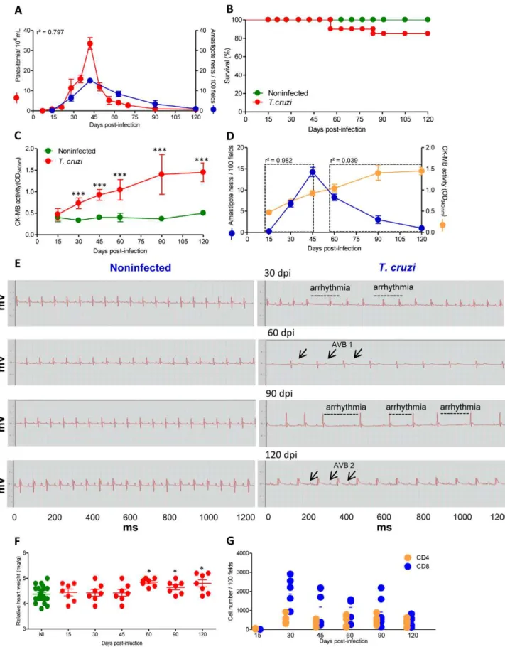
Initially, we investigated whether there was an association between the evolution of heart injury and parasite load inT. cruzi-infected C57BL/6 mice. The first peripheral blood circulating parasites were detected at 14 dpi, marking the onset of the acute infection phase. The peak of parasitemia was observed between 42 and 45 dpi and trypomastigotes were rarely found in the blood at
90 dpi, characterizing the onset of the chronic phase of infection (Figure 1A). With respect to cardiac parasitism, the first amastigote forms were detected at 15 dpi, and were frequently observed inside of myocytes from 30 dpi. The peak of heart parasitism coincided with the peak of parasitemia (42 dpi). After 60 dpi, the heart parasitism decreased and the parasite
pseudo-cysts were barely detectable at 90 and 120 dpi (Figure 1A),
although small pseudocysts and a few spots of parasite antigens were detected by IHS in every heart tissue section of all infected mice at every time point evaluated (data not shown). Therefore, in this model ofT. cruzi infection, there was a correlation between parasitemia and heart parasitism during the acute and chronic phases of infection (r2= 0.797,p,0.001). Approximately 80% of the infected mice survived the acute infection and developed the
chronic infection (Figure 1B). To explore the heart injury, we
evaluated creatine kinase cardiac isoenzyme MB (CK-MB) activity levels in serum, a marker of myocardial cell damage [16,17], and examined ECG registers looking for electrical abnormalities
[7,18]. At 30 dpi, increased CK-MB activity was observed inT.
cruzi-infected C57BL/6 mice when compared with their nonin-fected counterparts (Figure 1C). The CK-MB activity reached its highest levels at 90 dpi and remained high during chronic infection, which is suggestive of continuous cardiomyocyte injury (Figure 1C). Although during the acute infection there is a parallelism between cardiac parasitism and CK-MB activity in the serum (15–45 dpi, r2= 0.982, p,0.001), no association between the intensity of cardiac parasitism and the evolution of cardiomyocyte lesion was noticed after parasite control or in
chronic T. cruzi infection (60–120 dpi, r2= 0.0399, p.0.05)
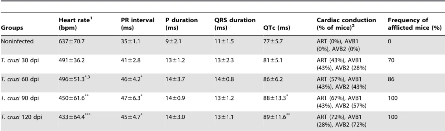
(Figure 1D). Compared with sex- and age-matched noninfected controls, in infected mice, bradycardia was the first significant ECG alteration and was firstly detected at 30 dpi. During the course of the infection, the mice presented significantly higher bradycardia, PR intervals and prolonged QTc intervals when
compared with noninfected controls (Figure 1E and Table 1).
All (100%) of the infected mice presented ECG abnormalities at 90 dpi (Table 1) and featured a delay in the conduction of electric impulses together with arrhythmia and first- and second-degree atrioventricular blocks (AVB1 and AVB2). Again, there was no relationship between the intensity of tissue parasitism and the evolution of electrical abnormalities. Interestingly, the CK-MB activity in the serum and the electrical alterations were paralleled by heart enlargement during the chronic infection phase (Figure 1F). Therefore, we evaluated whether heart injury was associated with the establishment and the intensity of CD8-enriched myocarditis. A kinetic study of heart colonization by
inflammatory CD4+
and CD8+
cells, the major components of
chagasic myocarditis [5,6], in T. cruzi-infected C57BL/6 mice
revealed that rare mononuclear cells were detected in the cardiac tissue at 15 dpi. CD8-enriched inflammation occurred at 30 dpi, after which a significant contraction of inflammation was observed; however, myocarditis that was mainly composed of
CD8+
T-cells persisted during the chronic phase (Figure 1G).
Once again, there was no correlation between CK-MB activity levels in the serum and the intensity of inflammation (r2= 0.038, p.0.05). Flow cytometry analysis of mononuclear cells harvested
from cardiac tissue ofT. cruzi-infected C57BL/6 mice confirmed
the predominance of CD8+
T-cells (Figure S1A). A small part of
the TCR+
cells were CD8negCD4+
T-cells. Also, some
CD8negNK1.1+
cells were detected (Figure S1B). Further,
CD8+ cells were mainly TCR+ cells at 40 dpi (Figure S1C).
CD8+T-cells-enriched myocarditis persisted at 90 dpi (R1 gate:
47.6–73.6% TCRab+
: 17.3–19.4% TCR TCRab+
CD4+
vs.28.2–
51% TCR TCRab+
CD8+
, two independent experiments) when
Author Summary
Chagas disease, a neglected tropical disease that is caused by Trypanosoma cruzi, afflicts between 8 and 15 million people in Latin America. Anti-parasite immunity allows for acute phase survival; however, approximately 30% of patients present chronic chagasic cardiomyopathy (CCC) with parasite persistence and CD8-enriched myocarditis at 10 to 30 years post-infection. The comprehension of the pathogenesis of Chagas’ heart disease may open a new avenue of therapy for CCC. Here, we explored the role of CD8+ T-cells in heart injury in C57BL/6 mice that were
infected with the Colombian strain ofT. cruzi. In infected mice, most of the CD8+T-cells were segregated into CD8+
interferon-gamma (IFNc)+perforin (Pfn)negand CD8+IFNcneg
Pfn+ cell populations. Importantly, the enrichment of the
chronic myocarditis in anti-parasite CD8+Pfn+ cells
paral-leled the worsening of CCC. CD8+cells from infectedifn
c2/2 pfn+/+ donors migrated towards the cardiac tissue to a greater extent than did CD8+ cells from ifnc+/+pfn2/2 donors. Moreover, accumulation of IFNc+cells in the cardiac
tissue ameliorated cardiomyocyte lesion, whereas enrich-ment in CD8+Pfn+cells aggravated cardiomyocyte injury.
Therefore, our data suggest that CD8+IFNc+ cells are
Figure 1. C57BL/6 mice infected with the Colombian strain ofT. cruzidevelop chronic cardiomyopathy.Mice were infected with 100 bt of the Colombian strain ofT. cruziand parasitological and clinical parameters were evaluated. (A) Kinetics of parasitemia and cardiac parasitism. (B) Survival rate. (C) CK-MB activity levels in the serum of noninfected andT. cruzi-infected mice. (D) Positive correlation between cardiac tissue parasitism and CK-MB activity levels during the acute phase (r2= 0.982) but not during the chronic phase (r2= 0.039) of infection. (E) Representative
ECG register segments of 1200 ms of sex- and age-matched noninfected (NI) controls andT. cruzi-infected C57BL/6 mice at 30, 45, 60, 90 and 120 dpi, showing arrhythmia and first and second degree atrioventricular block (AVB1, AVB2; arrows) in infected mice. (F) Relative heart weight (mg/g) of
CD8+
the highest level of CK-MB activity in the serum, demarking the cardiomyocyte lesion (Figure 1C), was detected.
Therefore, the present findings show that chronic cardiomyop-athy in C57BL/6 mice is not associated with the intensity of heart parasitism or inflammation, even though cardiomyocyte lesion and electrical abnormalities occurred in the persistence of the parasite and the CD8-enriched inflammation in this tissue. Collectively, these data show that this model reproduces several features of the CCC that has been described in patients [2] and it is an appropriate model for this study.
Chronic heart injury was not related to the systemic immune response but paralleled the accumulation of anti-T. cruziVNHRFTLV ASP2 effector CD8+T-cells in the cardiac tissue
Next, we investigated whether the induction of the anti-T. cruzi
CD8-mediated immune response involving IFNc production and
the cytotoxic machinery was related to heart injury. In spleen ofT. cruzi-infected mice, the ex vivoH-2Kb-restricted anti-VNHRFTLV ASP2 peptide [19,20] IFNc-secreting cells (Figure 2A) andin vivo cytotoxic CD8+
T-cells (Figure 2B) were first detected at 15 dpi. Both of these CD8+T-cell effector activities rose quickly, reaching a
maximum at the parasitemia peak (45 dpi) and remaining high until
60 dpi. Both of the anti-VNHRFTLV CD8+
T-cell effector activities declined slowly thereafter, but they persisted after parasite control
and remained detectable at 120 dpi (Figure 2A and 2B). Thus,
during the acute phase (from 15 to 45 dpi) the increase of both CD8+
T-cell effector activities (inflammatory and cytolytic) in the spleen paralleled the parasitic load in the blood and in cardiac tissues. At 60 dpi, after the parasitemia and heart parasitism were under control (Figure 2A and 2B), there was contraction of these CD8+
T-cell effector activities, as expected. However, anti-VNHRFTLV
CD8+
T-cell effector activities (inflammatory and cytolytic) persisted
during the chronic infection and were associated with an increased spleen weight and cellularity (Figure S2A and S2B), although these effector activities were not proportional to the parasite load (Figure 2A and 2B). Moreover, during the chronic infection, no relationship was observed between the intensity of the systemic
anti-VNHRFTLV CD8+
T-cell effector activities and cardiomyocyte lesion, as evidenced by the observation that the highest CK-MB activity level in the serum was detected at 90 dpi, which follows the
contraction of CD8+
T-cell effector activities (inflammatory and
cytolytic) (Figure 2A and 2B). Importantly, using the major
histocompatibility complex class I (MHC I) multimer H-2Kb/
VNHRFTLV, a critical reagent for proper visualization of specific
CD8+T-cells [21], we revealed the presence of anti-VNHRFTLV
ASP2 peptide CD8+
T-cells in spleen and cardiac tissue of the
Colombian-infected C57BL/6 mice (Figure 2C). The analysis of
CD8+ H-2Kb
/VNHRFTLV+ T-cells in spleen (Figure 2D)
confirmed the kinetic of the frequency of anti-VNHRFTLV CD8+
T-cells in this tissue detected byex vivoELISpot andin vivocytolytic assay. Further, the frequency of blood circulating CD8+
H-2Kb/ VNHRFTLV+T-cells reflected the high frequency of these cells in
the spleen in the acute infection and the contraction of these populations in the chronic infection (Figure 2D). Conversely, there
was an enrichment in CD8+
H-2Kb/VNHRFTLV+
T-cells among heart infiltrating inflammatory cells in the acute infection that
persisted during the chronic phase of infection (Figure 2D),
paralleling the persisting myocardial cell injury (Figure 1C).
Heart injury is associated with the enrichment of Pfn+ cells in the cardiac tissue during the chronicT. cruzi infection
As described above, IHS and flow cytometry analysis showed that CD8-enriched myocarditis was established early during theT. cruziinfection. Initially, we adopted an in situ IHS approach to
noninfected (NI, pool of three age-matched controls per analyzed point) andT. cruzi-infected C57BL/6 mice at 15, 30, 45, 60, 90 and 120 dpi. (G) Numbers of CD4+and CD8+cells in the cardiac tissue during acute and chronicT. cruziinfection were counted after immunohistochemistry staining. Each circle represents an individual mouse. These data represent three independent experiments.*,p,0.05,**,p,0.01, and***,p,0.001 comparing NI controls andT. cruzi-infected mice.
doi:10.1371/journal.ppat.1002645.g001
Table 1.Electrocardiograph parameters of C57BL/6 mice infected with the ColombianT. cruzistrain.
Groups
Heart rate1
(bpm)
PR interval (ms)
P duration (ms)
QRS duration
(ms) QTc (ms)
Cardiac conduction (% of mice)2
Frequency of afflicted mice (%)
Noninfected 637670.7 3561.1 962.1 1161.5 7765.7 ART (0%), AVB1
(0%), AVB2 (0%)
0
T. cruzi30 dpi 491636.2 4162.8 1361.2 1362.3 8165.1 ART (43%), AVB1
(43%), AVB2 (28%) 70
T. cruzi60 dpi 496651.3*,3 4664.2* 1463.7 1460.8 8666.2 ART (57%), AVB1
(43%), AVB2 (43%) 86
T. cruzi90 dpi 450661.6** 47
66.3* 14
60.9 1361.2 88613.3* ART (67%), AVB1
(43%), AVB2 (57%)
100
T. cruzi120 dpi 433664.4*** 4564.7* 1463.0 1361.1 89611.6** ART (72%), AVB1
(28%), AVB2 (72%)
100
1ECG parameters were evaluated using the following standard criteria: (i) heart rate (monitored by beats per minute (bpm), and (ii) the variation of the P wave and PR,
QRS and corrected QT intervals (QTc), all measured in milliseconds (ms). ART, arrhythmia; AVB1, first-degree atrioventricular block; AVB2, second-degree atrioventricular block.
2This data represent three independent experiments, with 7–8 infected mice/group. Three sex- and age-matched noninfected controls were analyzed in all experimental
time points. The results were pooled in one representative group of 12 noninfected controls per experiment.
3Significant differences
*,p,0.05; **,p,0.01;
CD8+
investigate whether inflammatory (IFNc+
) and cytotoxic (Pfn+
) cells differentially colonize the cardiac tissue at different time points duringT. cruziinfection. We evaluated the number of Pfn+
and IFNc+
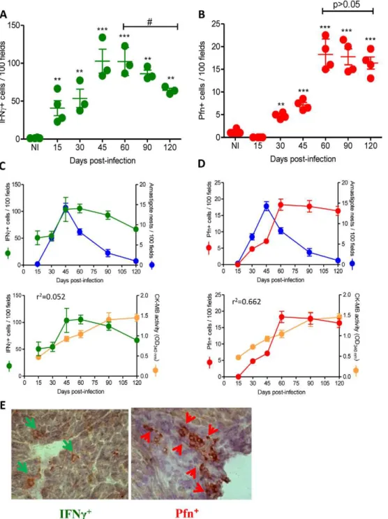
cells within the myocardium of C57BL/6 mice at 15, 30, 60, 90 and 120 dpi and revealed that IFNc+
cells are detected
at 15 dpi (Figure 3A), preceding the appearance of Pfn+
cells, which were first detected at 30 dpi (Figure 3B). Furthermore, the
highest number of IFNc+ cells was observed at 45 dpi, which
coincides with the heart parasitism peak (Figure 3C), whereas the number of Pfn+cells peaked at 60 dpi (Figure 3D). At 120 dpi,
there was a significant (p,0.05) reduction in the number of IFNc+
cells infiltrating the cardiac tissue, whereas the Pfn+
cell number
remained high (Figure 3A and 3B). Therefore, the ratio of
IFNc+
to Pfn+
cells decreased and resulted in a relative enrichment of Pfn+
cells among the inflammatory cells that invaded the cardiac tissue as the infection approached the chronic phase (Table 2). Next, we determined whether the control of the parasite and the cardiomyocyte lesion were in any way related to
the presence of IFNc+
and Pfn+
cells in the cardiac tissue in response toT. cruziinfection. The analysis of the IHS data showed that the heart parasitism was under control only after 60 dpi when both Pfn+
and IFNc+
cells were detected in high numbers in the
cardiac tissue (Figure 3C and 3D). Importantly, in the chronic
infection cardiomyocyte lesion was not related to the presence of IFNc+
cells (r2= 0.052), but correlated with the persistence of high
number of Pfn+
cells (r2= 0.662) invading the cardiac tissue (Figure 3C and 3D). Interestingly, the distribution of Pfn+
cells in the cardiac tissue was more focal, whereas the IFNc+
cells were
spread throughout the myocardium (Figure 3E). Taken together,
these data suggest a role for the infiltrating inflammatory Pfn+cells
in heart injury duringT. cruziinfection.
Segregation of CD8+T-cells into IFNc+Pfnnegand IFNcnegPfn+populations inT. cruziinfection
The kinetics of cardiac tissue colonization by IFNc+
and Pfn+
cells suggest that the inflammatory and cytotoxic effector activities were segregated into different cell populations. Therefore, we
investigated whether CD8+ T-cells express IFNc and Pfn in a
multifunctional or segregated manner during T. cruzi infection.
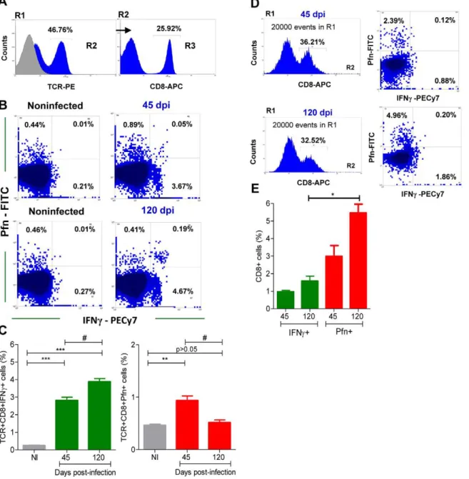
Peripheral blood CD8+ T-cells (Figure 4A), which potentially
migrate to the cardiac tissue, from acute and chronicallyT.
cruzi-infected C57BL/6 mice were assessed for intracellular IFNcand
Pfn expression. In comparison with noninfected mice, there was a
significant increase in the frequency of CD8+
T-cells expressing
IFNc and Pfn in the peripheral blood of T. cruzi-infected mice
(Figure 4B and 4 C). Indeed, most of the CD8+ T-cells
segregated into CD8+IFNc+Pfnneg
(CD8+IFNc+) and CD8+IFNcneg
Pfn+
(CD8+
Pfn+
) cell populations (Figure 4B). In the circulating
blood, there was a predominance of CD8+
IFNc+
cells in
comparison with CD8+
Pfn+
cells during the acute (45 dpi) and
chronic (120 dpi) phases of the infection (Figure 4B and 4C).
CD8+IFNc+Pfn+cells were barely detected in noninfected andT.
cruzi-infected mice, although a careful analysis revealed an upregulation of this rare CD8+
T-cell population during the chronic infection (from 0.01–0.05% in noninfected to 0.19–0.34% in chronically infected mice). Interestingly, during the chronic
infection there was an accumulation of CD8+
IFNc+
cells in the peripheral blood, whereas a decrease in the frequency of CD8+Pfn+
cells was detected (Figure 4B and 4C). Collectively, these data
suggest that in the peripheral blood most of the CD8+
T-cell effectors potentially able to perform inflammatory and cytotoxic
activities are phenotypically segregated, respectively, into
CD8+
IFNc+
and CD8+
Pfn+
T-cells during the acute and chronic phases ofT. cruziinfection. Importantly, this segregation of CD8+
T-cells into CD8+IFNc+and CD8+Pfn+populations was also detected
in the inflammatory cells infiltrating the cardiac tissue at 45 and 120 dpi (Figure 4D). In addition, the cardiac tissue infiltrating
CD8+
IFNc+
cells were IFNcdull
(MFI = 33.4–38.66) when
com-pared with the CD8+
IFNc+
cells in peripheral blood
(MFI = 144.12–166.81). Moreover, in the cardiac tissue there was
prevalence of CD8+
Pfn+
cells in the acute and, particularly, in the chronicT. cruziinfection (Figure 4D and 4E).
Considering the recent finding showing a role for interleukin (IL)-10 inT. cruzi-triggered myocarditis [22], we studied whether or not CD8+IFNc+cells coexpress IL-10 duringT. cruziinfection.
The analysis of CD8+T-cells in the peripheral blood revealed that
most of the IFNc+
cells were CD8+
IFNc+
IL-10+
in the acute and in the chronic infection (Figure S3A) and accumulated in blood in the chronic phase of infection (Figure S3B). Further, there was a significant frequency of CD8+
IFNc+
IL-10negT-cells accumulating
in the peripheral blood in the chronic infection (Figure S3C).
More important, in the heart tissue most of the CD8+IFNcdull
cells do not express IL-10 (Figure S3D) or express this cytokine in a very transient manner and were not detected in our experimental approach.
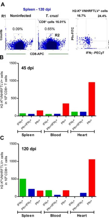
Differential compartmentalization of anti-T. cruziCD8+ T-cells expressing IFNcand Pfn
The accumulation of CD8+IFNc+cells in the peripheral blood
and the enrichment in CD8+Pfn+cells in the cardiac tissue during
the chronicT. cruziinfection led us to investigate whether or not there was distinct distribution of the CD8+
H-2Kb/VNHRFTLV+
T-cells expressing IFNc and Pfn in different immune
compart-ments of an infected mice. Indeed, in the spleen there was a
predominance of H-2Kb/VNHRFTLV+
IFNc+
cells, followed by
double-positive IFNc+Pfn+ cells and Pfn+ cells at 120 dpi
(Figure 5A). Therefore, we analyzed the presence of CD8+
H-2Kb/VNHRFTLV+
cells expressing IFNcand Pfn in the spleen, peripheral blood and cardiac tissue during the acute and chronic infection of C57BL/6 mice. In the acute infection (45 dpi), there Figure 2. CD8+T-cells recognizing the VNHRFTLV ASP2
T. cruzipeptide are enriched in the cardiac tissue.C57BL/6 mice were infected with 100 bt of the Colombian strain of T. cruzi and the anti-parasite immune response in the spleen and cardiac tissue was assessed. (A) Representative spots formed after stimulation of spleen cells from noninfected (NI) andT. cruzi-infected mice at 45 and 120 dpi with H-2Kb-resctricted VNHRFTLV peptide. The number of CD8+
IFNc+
as determined byex vivoELISpot were analyzed and compared with parasitemia and CK-MB activity levels in the serum. (B) Representative histogram profiles ofin vivocytotoxicity assay showing the specific lysis of H-2Kb-resctricted VNHRFTLV
peptide-labeled CFSEhighcells from NI controls andT. cruzi-infected C57BL/6 mice at 45 and 120 dpi. The frequencies ofin vivospecific lysis of with
H-2Kb-resctricted VNHRFTLV peptide-labeled target cells inT. cruzi-infected C57BL/6 mice at 15, 30, 45, 60, 90 and 120 dpi were analyzed and
compared with parasitemia and CK-MB activity levels in the serum. The peak of parasitemia and the maximum CK-MB activity are highlighted (dotted line). Each circle and vertical lines represent the mean6standard deviation (SD) of the studied group (5–7 animals/time point). These data represent three independent experiments. (C) Frequency of double-positive CD8+
H-2Kb/VNHRFTLV+
cells [R1 (SSCxFSC) gated] of spleen and heart of NI andT. cruzi-infected C57BL/6 mice at 40 dpi. (D) Frequencies of double-positive CD8+H-2Kb/VNHRFTLV+cells [R1 (SSCxFSC) gated] of spleen, peripheral blood and heart of NI andT. cruzi-infected C57BL/6 mice at 30, 45 and 120 dpi. Representative flow ctometry profiles and mean6SD of two or three animals per group. Bars represent the mean of two or three pools of 5 mice per pool.
was predominance of H-2Kb/VNHRFTLV+
Pfn+
cells in the
spleen, peripheral blood and cardiac tissue inflammatory CD8+
T-cells (Figure 5B). Moreover, in the chronic phase of infection
(120 dpi) the accumulation of CD8+
H-2Kb/VNHRFTLV+
IFNc+
cells in spleen was confirmed. Anti-T. cruzi IFNc+
Pfn+
cells were also retained in the spleen at 120 dpi (Figure 5C). In
contrast, similar frequencies of segregated CD8+
H-2Kb/
VNHRFTLV+
IFNc+
or Pfn+
cells were detected in peripheral
blood, while the enrichment in CD8+
H-2Kb/VNHRFTLV+
Pfn+
cells among heart infiltrating inflammatory cells persisted at
120 dpi (Figure 5C), paralleling the myocardial cell injury
(Figure 1C).
Figure 3. IFNc+and Pfn+cells differentially invade the cardiac tissue ofT. cruzi-infected C57BL/6 mice.Mice were infected with 100 bt of
the Colombian strain ofT. cruziand the presence of IFNc+and Pfn+cells in the cardiac tissue was evaluated by IHS. (A) Numbers of IFNc+cells infiltrating the cardiac tissue at 15, 30, 45, 60, 90 and 120 dpi. (B) Numbers of Pfn+
cells infiltrating the cardiac tissue at 15, 30, 45, 60, 90 and 120 dpi. (C) Relationship between IFNc+
cells infiltrating the cardiac tissue and heart parasitism or CK-MB activity in serum. (D) Relationship between Pfn+ cells infiltrating the cardiac tissue and heart parasitism or CK-MB activity in the serum. (E) Immunohistochemistry staining of IFNc+(green arrows) and Pfn+ (red arrows) cells infiltrating the cardiac tissue at 60 dpi. In (A) and (B), each circle represents an individual mouse. In (C) and (D), each circle represents the mean6SD of the studied group (5–7 animals/time point). These data represent three independent experiments.*,p
,0.05;**,p ,0.01; and***,p
,0.001, comparing noninfected (NI) controls andT. cruzi-infected mice.#
,p,0.05, comparingT. cruzi-infected mice at 60 and 120 dpi. doi:10.1371/journal.ppat.1002645.g003
CD8+
CD8+IFN
c+and CD8+Pfn+cells differentially express CCR5 and LFA-1
Our findings showed differential accumulation of CD8+
IFNc+
cells in the peripheral blood and CD8+
Pfn+
T-cells in the cardiac tissue during the chronicT. cruziinfection (Figure 4). Based on these findings and on previous data demonstrating that peripheral blood
CD8+
CCR5+
LFA-1+
cells drastically increase during T. cruzi
infection [12], we next examined whether CCR5 and LFA-1, molecules essential for the migration of inflammatory cells towards
the cardiac tissue during T. cruzi infection [11,12,23], were
differentially expressed by the CD8+IFNc+ and CD8+Pfn+ cells.
For that, we analyzed the frequencies of CCR5+LFA-1+cells among
CD8+IFNc+(Figure S4) and CD8+Pfn+peripheral blood cells. A
low proportion of peripheral blood CD8+IFNc+ cells were
CCR5+LFA-1+, whereas a high proportion of CD8+Pfn+cells were
CCR5+
LFA-1+
during the acute (45 dpi) and chronic (120 dpi) phases of infection (Table 3). Therefore, although present at a lower
frequency than CD8+
IFNc+
cells in peripheral blood, a higher proportion of CD8+
Pfn+
cells coexpress CCR5 and LFA-1, and they
are potentially more prone to migrate than CD8+
IFNc+
cells.
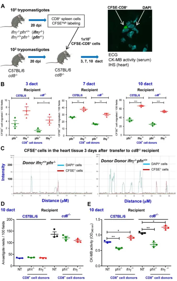
Distinct migratory behavior of CD8+cells fromifnc+/+ pfn2/2 andifnc2/2pfn+/+ mice towards cardiac tissue duringT. cruziinfection
To investigate whether CD8+
IFNc+
and CD8+
Pfn+
cells exhibit a distinct migratory behavior with a differential potential to colonize the cardiac tissue, a series of adoptive cell transfer experiments was performed. In brief, ifnc+/+pfn2/2 and ifnc2/2pfn+/+
mice were infected and their spleens were removed at 20 dpi (100% of the animals were alive at this time point). TheT. cruzi-infectedifnc+/+
pfn2/2andifnc2/2pfn+/+
mice presented increased spleen cellularity,
similar to infected C57BL/6 mice (Figure S5A). These infected
ifnc+/+pfn2/2andifnc2/2pfn+/+mice presented similar frequency of
TCR+cells, but infectedifnc+/+pfn2/2mice had higher frequency of CD8+T-cells (Figure S5A). The CD8+T-cells from infectedifnc+/+
pfn2/2 mice expressed IFNc, whereas CD8+T-cells from infected
ifnc2/2pfn+/+mice expressed Pfn (Figure S5B). Further, CD8+
T-cells from infected ifnc+/+pfn2/2 and ifnc2/2pfn+/+ mice were
potentially functional as they expressed activation markers as IL-10, TNF and CD107a (Figure S5B). Therefore, single-cell suspensions of CD8-enriched cells (from infectedifnc2/2pfn+/+
orifnc+/+
pfn2/2 donors) were prepared using magnetic beads, labeled with CFSE and intravenously transferred intocd82/2mice and C57BL/6 mice that had been infected withT. cruzi20 days before. At 3, 7 and 10 days after the cell transfer (dact) (i.e., 23, 27 and 30 dpi; all of the recipient
mice were alive) the hearts were removed from the cd82/2 and
C57BL/6 recipient mice and the CFSE+
CD8+
T-cells were examined and counted under fluorescence and confocal microscopes (Figure 6A). Our data indicate that more CFSE+
CD8+
cells
accumulated in the cardiac tissue of the cd82/2 mice that had
received CFSE+CD8-enriched cells from ifnc2/2pfn+/+ donors
compared withcd82/2mice that had received CFSE+CD8-enriched
cells from ifnc+/+pfn2/2 donors at all of the assessed time points (Figure 6B). Although the immunocompetent C57BL/6 recipient mice that had received CD8+
cells fromifnc+/+
pfn2/2orifnc2/2pfn+/+
donors accumulated more CFSE+
CD8+
cells among the inflammatory cells infiltrating the cardiac tissue compared with the cd82/2 recipients, the preferential accumulation of CFSE+CD8+cells from
ifnc2/2pfn+/+donors was reproduced (Figure 6B). The prevalence of
CFSE+CD8+cells fromifnc+/+pfn2/2donors in the cardiac tissue of recipient mice supports distinct migratory behavior of these cells rather than differential cell proliferation. Indeed, the similar intensities of fluorescence detected in the CFSE+
CD8+
cells fromifnc+/+
pfn2/2 andifnc2/2pfn+/+
donors infiltrating the cardiac tissues of recipients at 3 dact supports that, at least in the initial days after cell transfer, there was no differential activation of the transferred CFSE+CD8+ cells
(Figure 6C). Moreover, splenocytes from T. cruzi-infected mice, independently of the mouse lineage, were unresponsive to in vitro
activation with anti-CD3/anti-CD28 stimuli (Figure S5C), while
splenocytes isolated from naı¨ve ifnc+/+
pfn2/2mice were more
responsive to anti-CD3/anti-CD28 stimuli (Figure S5C), particularly the CD8+T-cells (Figure S5D). Interestingly, similar to what was
detected inT. cruzi-infected C57BL/6 mice, the CFSE+CD8+cells
fromifnc+/+
pfn2/2donors were diffusely spread in the myocardium tissue, while the CFSE+
CD8+
cells fromifnc2/2pfn+/+
donors were focally localized among inflammatory cells of the recipient (Figure 6C). Thus, CD8+
IFNcneg
(potentially Pfn+
) cells migrated and accumulated in the cardiac tissue more readily than the CD8+Pfnneg
(potentially IFNc+) cells duringT. cruziinfection.
Next, we determined whether the differential colonization of the cardiac tissue ofT. cruzi-infected C57BL/6 andcd82/2recipient mice by CD8+ T-cells from ifnc2/2pfn+/+ and ifnc+/+pfn2/2 donors had a differential impact on parasite dissemination control and cardiomyocyte lesion. At 10 dact, similar cardiac tissue parasitism was observed in recipient mice that had received
CFSE+
CD8+
cells from ifnc+/+
pfn2/2 or ifnc2/2pfn+/+
donors (Figure 6D). In C57BL/6 and cd82/2 recipient mice, the
CFSE+
CD8+
cells from ifnc+/+pfn2/2 donors had a beneficial impact on cardiomyocyte lesion as evidenced by a decrease in CK-MB activity levels in the serum compared with non-transferred
(NT) mice. In contrast, the CFSE+
CD8+
cells fromifnc2/2pfn+/+
donors had a detrimental impact as evidenced by the increase in CK-MB activity levels in the serum compared with their respective Table 2.Number of IFNc+and Pfn+cells in heart tissue sections ofT. cruzi-infected C57BL/6 mice.
dpi1 IFNc+
cells /100 fields2 Pfn+
cells /100 fields Ratio of IFNc+/Pfn+
cells3
15 50628 0 50:0
30 54617 561 11:1
45 103645 761 14:1
60 105616 1863 5:1
90 92615 1863 5:1
120 6667 1763 4:1
1dpi, days post-infection with 100 bt of the ColombianT. cruzistrain.
2The number of positive cells for each parameter was counted in 100 microscopic fields per section. Three sections were counted per each analyzed animal. 3Ratio of IFNc+
/Pfn+
NT counterparts (Figure 6E). Therefore, these data circumstan-tially support that CD8+
IFNcneg
(potentially Pfn+
) cells play a non-beneficial role in heart injury duringT. cruziinfection.
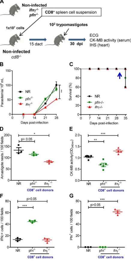
Colonization of the cardiac tissue with CD8+Pfn+T-cells was correlated with cardiomyocyte lesion
Based on the data presented above, we speculate that
CD8+
IFNc+
and CD8+
Pfn+
T-cells play distinct roles in the T. cruzi-elicited cardiomyopathy. To assess this theory, we selectively
reconstituted the CD8 compartment ofcd82/2mice. Briefly, the spleens were removed from noninfected naı¨veifnc2/2pfn+/+
(with the potential to generate Pfn+
cells) and ifnc+/+
pfn2/2 (with the potential to generate IFNc+cells) mice and single-cell suspensions
of CD8-enriched T-cells were transferred to noninfected naı¨ve recipientcd82/2mice (Figure 7A). The analysis of the peripheral blood of naı¨ve recipients showed the presence of circulating CD8+
cells (2 to 6.5% of peripheral blood cells) in cd82/2 mice
reconstituted with CD8+
cells from naı¨veifnc2/2pfn+/+
andifnc+/+
Figure 4. Segregation of CD8+T-cells into IFN
c+and Pfn+populations inT. cruziinfection.C57BL/6 mice were infected with 100 blood
trypomastigotes of the Colombian strain ofT. cruziand the expression of IFNc+and Pfn+by CD8+cells in the peripheral blood and cardiac tissue was evaluated. (A) Representative histograms of flow cytometry analysis of peripheral blood CD8+
T-cells [R1 (SSCxFSC)/R2 (TCR)/R3 (CD8) gated] that were analyzed for IFNcand Pfn expression inT. cruzi-infected mice (120 dpi). (B) Representative dot plots of flow cytometry analysis of peripheral blood CD8+
T-cells (R1/R2/R3 gated) expressing IFNcand Pfn at 45 and 120 dpi. (C) Frequency of double-stained CD8+ IFNc+
and CD8+ Pfn+
peripheral blood T-cells cells (R1/R2/R3 gated) at 45 and 120 dpi. (D) Representative histograms and dot plots of flow cytometry analysis of heart infiltrating CD8+
cells [R1 (SSCxFSC)/R2 (CD8) gated] expressing IFNcand Pfn at 45 and 120 dpi. (E) Frequency of double-stained CD8+ IFNc+
and CD8+ Pfn+
of heart infiltrating cells (R1/R2 gated) at 45 and 120 dpi. In (C) Bars represent the mean6SD of five to eight mice per group. In (E) Bars represent the mean of two or three pools of 5 mice per group. These data represent two or three independent experiments.*,p,0.05;**,p,0.01; and***,p,0.001, comparing noninfected (NI) controls andT. cruzi-infected mice.#P
,0.05, comparingT. cruzi-infected mice at 45 and 120 dpi. doi:10.1371/journal.ppat.1002645.g004
CD8+
pfn2/2donors in all analyzed recipient mice at 15 days after cell
transfer (Figure S6), at which time non-reconstituted (NR) and
CD8-reconstituted mice were infected withT. cruzi(Figure 7A). Parasitemia was monitored every two days and heart parasitism and injury were evaluated at 30 dpi (at which time all recipient
mice from every experimental group were alive). As expected, when compared with NRT. cruzi-infectedcd82/2mice at 28 dpi,
the reconstitution of the CD8 compartment with CD8+
cells from ifnc2/2pfn+/+
or ifnc+/+
pfn2/2 donors prior to infection did not
worsen parasitemia. Furthermore, cd82/2 mice that had been
reconstituted with CD8+
T-cells from ifnc2/2pfn+/+
donors
presented reduced parasitemia when compared with NRcd82/2
mice (Figure 7B). All of the animals in every experimental group survived until 34 dpi. At 35 dpi, the first deaths were recorded in
all groups (Figure 7C). In independent experiments, cardiac
tissue parasitism was analyzed at 30 dpi when 100% of the mice in all the experimental groups were alive, revealing that the non-reconstitutedcd82/2mice presented high parasitism, as expected (Figure 7D). The reconstitution of thecd82/2 mice with CD8+
cells fromifnc+/+pfn2/2donors neither ameliorated nor aggravat-ed heart parasitism, whereas the reconstitution of thecd82/2mice
with CD8+
cells from ifnc2/2pfn+/+
donors significantly reduced heart parasitism (Figure 7D). To evaluate the participation of the distinct CD8+
cells populations in heart injury, CK-MB activity levels in the serum and the ECG registers were evaluated.
Compared with the NRcd82/2animals,cd82/2 mice that were
reconstituted with CD8+
cells fromifnc+/+pfn2/2donors exhibited a significant decrease in CK-MB activity levels in the serum at 10
dact (Figure 7E). In accordance with these findings, the ECG
registers revealed that the reconstitution ofcd82/2 recipients of CD8+T-cells fromifnc+/+pfn2/2donors was beneficial, reducing the frequency of afflicted mice and the severity of the electrical abnormalities (Table 4). On the other hand, thecd82/2mice that
had been reconstituted with CD8+
cells fromifnc2/2pfn+/+
donors exhibited an aggravation of myocardial cell lesion when compared
with NR cd82/2 mice at 10 dact (Figure 7E). Further, the
electrical abnormalities observed incd82/2 mice that had been
reconstituted with CD8+
T-cells from ifnc2/2pfn+/+
donors prior
to infection were similar to those detected in NR cd82/2 mice
(Table 4).
Interestingly, IHS analysis showed that the reconstitution of cd82/2mice with CD8+
cells from ifnc+/+
pfn2/2donors led to a
significant accumulation of IFNc+
cells in the cardiac tissue (Figure 7F); however, a low number of IFNc+
cells was also
detected in the cardiac tissue of cd82/2 mice that were
non-reconstituted or non-reconstituted with CD8+
T-cells fromifnc2/2pfn+/+
donors, supporting the existence of non-CD8 IFNcproducers in the
cd82/2 mice (probably NK or CD4+ cells). Importantly, in the
cd82/2 mice that were reconstituted with CD8+ T-cells from
ifnc2/2pfn+/+donors, Pfn+cells accumulated in the cardiac tissue
(Figure 7G). No Pfn+
cells were detected in the cardiac tissue of
non-reconstituted or cd82/2 mice that were reconstituted with
CD8+
cells fromifnc+/+
pfn2/2donors (Figure 7G), supporting that all Pfn+
cells detected in the cardiac tissue were CD8+
T-cells. Because reductions in cardiomyocyte lesion and electrical Figure 5. Differential compartmentalization of anti-T. cruzi
CD8+T-cells expressing IFN
cand Pfn.C57BL/6 mice were infected
with 100 bt of the Colombian strain ofT. cruziand presence of CD8+ H-2Kb /VNHRFTLV+ T-cells expressing IFNc+ and Pfn+ in the spleen, peripheral blood and in the cardiac tissue was evaluated. (A) Representative dot plots of double-positive CD8+H-2Kb/VNHRFTLV+ T-cells (R1 gated) in the spleen of NI andT. cruzi-infected C57BL/6 mice at 120 dpi. Frequencies of CD8+H-2Kb/VNHRFTLV+T-cells (R2 gated) expressing IFNc, Pfn or coexpressing IFNc and Pfn in the spleen, peripheral blood and cardiac tissue of NI andT. cruzi-infected C57BL/6 mice at 45 (B) and 120 dpi (C). Bars represent the means of two or three pools of 5 mice per group. These data represent two independent experiments.
doi:10.1371/journal.ppat.1002645.g005
Table 3.Frequency of CD8+IFNc+and CD8+Pfn+coexpressing
CCR5 and LFA-1 among peripheral blood cells during acute and chronicT. cruziinfection.
Groups
CD8+
IFNc+CCR5+LFA-1+
(%)
CD8+
Pfn+
CCR5+
LFA-1+
(%)
Noninfected 1.0060.27 1.0160.64
42 dpi (acute) 34.5268.98 53.1265.64
120 dpi (chronic) 1.0160.64 6.3060.38
Figure 6. CD8+cells fromifn
c2/2pfn+/+andifnc+/+pfn2/2infected mice differentially migrate to the cardiac tissue.(A) Experimental scheme showing the infection of donors and recipients mice byT. cruzi, isolation of CD8+
cells fromifnc2/2pfn+/+
andifnc+/+pfn2/2infected donors
(20 dpi) by magnetic beads, CFSEhighlabeling, adoptive transfer of CFSE+CD8+cells to C57BL/6 andcd82/2infected recipients (20 dpi) and analysis at
3, 7 and 10 days after cell transfer (dact). The colonization of the cardiac tissue by CFSE+
cells, heart parasitism and CK-MB activity levels in the serum were evaluated. (B) Detection of CFSE+CD8+cells in cardiac tissue sections at 3, 7 and 10 dact toT. cruzi-infected C57BL/6 andcd82/2recipients. (C)
Graphics showing the confocal analysis of the intensity of fluorescence of CFSE+ CD8+
cells present in the myocardium of cardiac tissue sections at 3 CD8+
alterations were observed, the data support the finding that CD8+
IFNcproducers colonizing the cardiac tissue play a beneficial role by decreasing heart injury duringT. cruziinfection. Conversely, the aggravation of cardiomyocyte lesion revealed by increased CK-MB activity levels in the serum detected incd82/2mice that had been
reconstituted with CD8+
T-cells from ifnc2/2pfn+/+
donors (Figure 7E) was associated with an accumulation of Pfn+
cells in the cardiac tissue (Figure 7G), reinforcing that CD8+
Pfn+
cells play a detrimental role in heart injury duringT. cruziinfection.
Discussion
In Chagas disease, CD8+
T-cells are crucial for T. cruzi
dissemination control during the acute infection phase. In the
chronic infection the predominance of CD8+
T-cells among the cells infiltrating the cardiac tissue raised the suspicion that these cells are involved in heart injury. In this study, we adopted a
murine model of T. cruzi-elicited chronic cardiomyopathy to
examine the participation of CD8+T-cells that express IFNcand
Pfn in parasite control and heart injury. There was no association of the intensity of heart parasitism and CD8-enriched myocarditis with cardiomyocyte lesion and electrical abnormalities during the chronic infection. Furthermore, the expansion and contraction of
anti-VNHRFTLV CD8+
T-cell effector activities (IFNc-producers and CTL) in the periphery (spleen and blood) were associated with the parasite load but not with heart injury. The presence, however,
of anti-VNHRFTLV CD8+
effector T-cells in the cardiac tissue paralleled the tissue damage and electrical abnormalities. The accumulation of IFNc+
cells preceded the entry of Pfn+
cells into the cardiac tissue during acute infection. Interestingly, in the chronic infection, a decrease in IFNc+
and a relative enrichment in Pfn+
cells invading the cardiac tissue paralleled the worsening of heart injury. In fact, there was prevalent accumulation of
CD8+
Pfn+
cells in the cardiac tissue in T. cruzi infection.
Moreover, there is a differential accumulation of CD8+
H-2Kb/
VNHRFTLV+
Pfn+
T-cells in the cardiac tissue during the chronic infection. Altogether, these findings led to the idea that CD8+Pfn+
and CD8+IFNc+exhibit a distinct migratory behavior.
Corrob-orating this theory, CD8+cells fromT. cruzi-infectedifnc2/2pfn+/+
donors (Pfn+) migrate towards the cardiac tissue more so than do
CD8+ cells from ifnc+/+pfn2/2 donors (IFNc+). Moreover, we
demonstrate that the colonization of cardiac tissue ofcd82/2mice
by CD8+
Pfn+
cells aggravated cardiomyocyte injury, whereas
CD8+
IFNc+
cells ameliorated cardiomyocyte injury and electrical
abnormalities, thus supporting a differential role for CD8+
Pfn+
and CD8+
IFNc+
T-cells inT. cruzi-elicited cardiomyopathy. Following infection of C57BL/6 mice with the Colombian strain of T. cruzi, parasite load was controlled and most of the animals survived the acute phase of infection and developed a chronic infection. In these animals, myocardial cell damage and electrical abnormalities, such as arrhythmias and prolonged QTc interval, are similar to those described in CCC patients [2,24] and other murine models ofT. cruziinfection [12,20]. Moreover, inT. cruzi-infected C57BL/6 mice, chronic CD8-enriched myocarditis was associated with cardiomegaly. Taken together, these data validate this model for study of the immunopathology of Chagas’ heart disease.
Our first goal was to determine whether the development of chronic cardiomyopathy, characterized by the presence of cardiomyocyte lesion and electrical abnormalities, was associated with the intensity of the parasite load in cardiac tissue and/or CD8-enriched myocarditis. During the acute infection phase of
C57BL/6 with the Colombian strain ofT. cruzi, cardiomyocyte
lesion and electrical alterations were related to heart parasitism and inflammation. This finding partially corroborated previous data using a model of acute infection. Briefly, they infected Swiss
mice with the Y strain of T. cruzi and observed a positive
correlation between the intensity of heart inflammation, but not heart parasitism, and CK-MB activity levels in plasma [16]. Therefore, in C57BL/6 mice acutely infected with the Colombian strain ofT. cruzi, heart injury may be the consequence of the direct destruction of cardiomyocytes by intense parasitism and/or anti-T. cruzi effector immunity. However, as infection progresses, heart parasitism and CD8-enriched myocarditis decreases, coinciding with the establishment of anti-T. cruzispecific CD8+T-cell effector
activities (inflammatory and cytotoxic) and parasitemia control, whereas myocardial cell damage and electrical abnormalities are aggravated. These data confirm the lack of an association between the intensity of heart parasitism and myocarditis with the evolution
of heart injury in the chronic phase of T. cruzi infection, as
previously seen in cardiopatic chagasic patients [2,5,25] and
C3H/He mice infected with the Colombian strain ofT. cruzi[12].
Therefore, our data reinforce the concept that long-term heart inflammation, rather than its intensity, is crucial for the generation ofT. cruzi-elicited heart injury.
In spite of their importance for host resistance in T. cruzi
infection [4], CD8+
T-cells gained particular attention as the major component of myocarditis in acute [26] and chronic [6] experimental infection, and in chronic chagasic patients [5,13]. Therefore, we compared the kinetics of the generation of
inflammatory (IFNc producers) and cytotoxic CD8+
T-cell effectors specific for the immunodominant VNHRFTLV ASP2
epitope, which is linked to protective immunity againstT. cruzi
[27,28] and is a prototype for general CD8+
T-cell activation, with the kinetics of the appearance of cardiomyocyte lesions and
electrical abnormalities. The H-2Kb-restricted anti-VNHRFTLV
cytotoxic and IFNc-secreting CD8+
T-cells were first detected before the peak of parasitemia at 15 dpi and reached a maximum level at the peak of parasitemia and heart parasitism (42–45 dpi). This finding contrasts with previous data showing that the peak of
anti-VNHRFTLV cytotoxic and IFNc-secreting CD8+ T-cells
occurred after the peak of parasitemia (between 14 and 24 dpi in the studied strains), implying that there is a requirement for rounds of parasite multiplication to trigger CD8+T-cell effector activities
[29]. In light of these data, it appears that duringT. cruziinfection,
specific CD8+
T-cell effectors (IFNc producers and cytolytic
effectors) require between 15 and 20 dpi to proliferate and differentiate independent of the mouse lineage, parasite strain (virulence and inoculum size) and parasite load requirements. As
expected, after parasite control (60 dpi) CD8+
T-cell effectors (IFNcproducers and cytolytic effectors) decreased but persisted at detectable levels up to 120 dpi. Importantly, in the periphery the
maximum anti-VNHRFTLV ASP2 epitope CD8+
T-cell effector
dact toT. cruzi-infectedcd82/2recipients. DAPI was used to reveal the nucleus of the cells of the recipient mice. (D) Number of amastigote nests in
100 microscopic fields of cardiac tissue from C57BL/6 andcd82/2infected mice that were non-transferred (NT) or transferred with CD8+cells from
ifnc2/2pfn+/+
andifnc+/+pfn2/2donors, at 10 dact. (E) Cardiomyocyte lesions were evaluated at 10 dact by measuring the CK-MB activity in serum
samples from C57BL/6 andcd82/2infected mice that were non-transferred (NT) or transferred with CD8+
cells fromifnd2/2pfn+/+
andifnc+/+pfn2/2
donors. Each symbol represents an individual mouse. These data represent three independent experiments.*,p
,0.05;**,p
,0.01; and***,p ,0.001, comparing C57BL/6 andcd82/2infected mice that were non-transferred and transferred with CD8+
cells fromifnc2/2pfn+/+
andifnc+/+pfn2/2donors.
Figure 7. Distinct migratory behavior and effector function of CD8+IFN
c+and CD8+Pfn+T-cells inT. cruziinfection.(A) Experimental
scheme of CD8+
cell isolation from noninfected naı¨veifnc2/2pfn+/+
andifnc+/+pfn2/2donors,in vivoreconstitution of the CD8+
cell compartment of naı¨vecd82/2mice, infection with 100 bt of the Colombian strain at 15 days after cell transfer (dact) and analysis at 30 days post-infection. Parasitemia,
survival rate, cardiac parasitism and CK-MB activity levels in the serum and colonization of the cardiac tissue by IFNc+ and Pfn+
cells were evaluated. (B) Parasitemia and (C) survival curve ofcd82/2mice non-reconstituted (NR) or reconstituted with CD8+cells fromifnc+/+pfn2/2andifnc2/2pfn+/+ donors. In independent experiments, the animals were analyzed at 30 dpi when 100% of the mice in all the experimental groups were alive (arrow). (D) Number of amastigote nests in 100 microscopic fields of cardiac tissue sections. (E) Cardiomyocyte lesion evaluated by CK-MB activity in serum samples. The number of (F) IFNc+
and (G) Pfn+
cells in 100 microscopic fields of cardiac tissue sections from mice non-reconstituted (NR) or reconstituted with CD8+
cells fromifnc+/+pfn2/2 andifnc2/2pfn+/+
donors, at 30 dpi. Each symbol represents an individual mouse. These data CD8+
cytotoxic and inflammatory activities was achieved at the parasitemia peak (42–45 dpi) and was probably stimulated by the proportional release of parasite antigens. Importantly, it was not kinetically related to the more severe heart injury that was observed at a later time (90 dpi). Therefore, protective CD8+
T-cell effectors controlling T. cruzi growth in the periphery and
detrimental myocarditis appear to be dissociated facets of the host immune response that is triggered by the invading organism [12,23]. If so, one may be able to stimulate anti-parasite protective immunity and selectively downregulate the detrimental inflamma-tion colonizing the cardiac tissue. Therefore, an understanding of the functional role played by the cells invading the cardiac tissue is required. Our next goal was to establish the kinetics of the heart
colonization by inflammatory cells during T. cruzi infection to
determine the association with heart injury. CD8-enriched myocarditis was established during acute infection and although inflammation decreased as a whole, CD8+T-cells persisted as the
predominant cell population invading the cardiac tissue during the chronic phase of infection, corroborating findings observed in
patients [5,13] and C3H/He mice infected with the ColombianT.
cruzistrain [6]. Previous data demonstrated that IFNcis crucial for regulation of the entry of anti-parasite protective cells into the cardiac tissue [11], and a Pfn deficiency results in less severe cardiomyocyte lesion and electrical abnormalities during chronic T. cruzi infection [7]. Therefore, we investigated whether IFNc+
and Pfn+
cells colonize the cardiac tissue simultaneously or in a
dissociated manner during T. cruzi infection, and examined the
possible implications of this scenario in chronic cardiomyopathy. The entry of IFNc+
cells into the cardiac tissue preceded the influx of Pfn+
cells, suggesting that inflammatory and cytotoxic effector activities are performed by distinct cell populations during a T. cruzi infection. Indeed, most of the CD8+
T-cells that were detected in the peripheral blood of acute and chronicallyT.
cruzi-infected mice were segregated into CD8+
IFNc+
and CD8+
Pfn+
cell populations. Importantly, theseex vivostained cells reflect the
status ofin vivoactivated cells that are present in peripheral blood and migrate to tissues that are targeted byT. cruzi, e.g., the cardiac tissue. Furthermore, this segregation is a long-term feature as opposed to a transient characteristic of distinct inflammatory
(IFNc producers) and cytotoxic (Pfn-expressing) CD8+ T-cell
effectors in acute and chronic T. cruzi infection. In fact, in the cardiac tissue the accumulation of segregated CD8+Pfn+T-cells
observed in the acute infection persisted in the chronic phase of
infection. Moreover, the CD8+IFNc+ T-cells detected in the
cardiac tissue are IFNcdull. Thus, this pale expression of IFNcby the inflammatory cells invading the cardiac tissue, that confirmed previous data [30], may be related to continuousin situantigenic stimulation.
The possibility of coexpression of IFNc and Pfn by a single
CD8+
T-cell under antigen re-stimulation conditions cannot be ruled out. Indeed, there was a low but consistent increased frequency of CD8+T-cells in the peripheral bloodT. cruzi-infected
mice (from 0.01–0.05% in noninfected to 0.19–0.34% in chronically infected mice). Further, anti-T. cruziCD8+IFNc+Pfn+
T-cells were retained in the spleen during chronic infection. In this
context, a recent report showed that following the in vitro
restimulation with human immunodeficiency virus antigens, peripheral blood CD8+T-cells from elite controllers presented a
higher frequency of IFNc+Pfn+ responders than CD8+ T-cells
from chronic progressors [31]. Therefore, it remains to be determined whether the segregation of effector activities in CD8+IFNc+ and CD8+Pfn+ T-cells is a T. cruzi-driven feature
that contributes to parasite escape and persistence in chronic infection. Furthermore, if this is the case, it is reasonable to
suppose that the stimulation of anti-T. cruzi immunity during
chronic infection, for example, through immunotherapeutic vaccination, would upregulate multifunctional CD8+IFNc+Pfn+
T-cells.
A predominance of CD8+
IFNc+
cells compared with CD8+
Pfn+
T-cells was consistently detected in the peripheral blood of
represent three independent experiments. *, p,0.05; **, p,0.01; and ***, p,0.001, comparing cd82/2 infected mice non-reconstituted and
reconstituted with CD8+cells fromifnc2/2pfn+/+andifnc+/+pfn2/2donors.
doi:10.1371/journal.ppat.1002645.g007
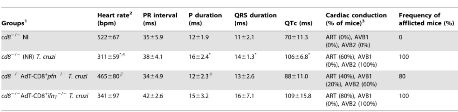
Table 4.Electrocardiograph parameters ofcd82/2mice adoptively transferred with CD8+cell fromifnc2/2pfn+/+orifnc+/+pfn2/2 and infected with the ColombianT. cruzistrain.
Groups1
Heart rate2
(bpm)
PR interval (ms)
P duration (ms)
QRS duration
(ms) QTc (ms)
Cardiac conduction (% of mice)3
Frequency of afflicted mice (%)
cd82/2NI 522
667 3565.9 1261.9 1162.1 70611.3 ART (0%), AVB1 (0%), AVB2 (0%)
0
cd82/2(NR)T. cruzi 311659*,4 3864.1 1662.4* 1461.3* 10666.8* ART (60%), AVB1
(0%), AVB2 (100%)
100
cd82/2AdT-CD8+
pfn2/2T. cruzi 465680#
3464.9 1262.3#
1362.6 88611.0 ART (40%), AVB1 (20%), AVB2 (60%)
80
cd82/2AdT-CD8+
ifnc2/2T. cruzi 341
697 4262.6 1563.2 1667.1 109615.8 ART (80%), AVB1 (0%), AVB2 (100%)
100
1
cd82/2mice were: NI, non-infected;T. cruzi, infected with 100 bt forms of the ColombianT. cruzistrain 15 days after adoptive cell transfer (AdT) of CD8-enriched cells
($98%), obtained from IFNc-deficient (ifnc2/2pfn+/+
) or Pfn-deficient (ifnc+/+
pfn2/2) mice.
2ECG parameters were evaluated using the following standard criteria: (i) heart rate (monitored by beats per minute (bpm), and (ii) the variation of the P wave and PR,
QRS and corrected QT intervals (QTc), all measured in milliseconds (ms). ART, arrhythmia; AVB1, first-degree atrioventricular block; AVB2, second-degree atrioventricular block.
3This table represented accumulated data from three independent experiments, with 3 to 5 mice/group in each experiment. 4Significant differences:
*,p,0.05 - between the values forcd82/2noninfected andT. cruzi-infected mice; #
,p,0.05 - between the values forcd82/2non-reconstituted andcd82/2reconstituted and infected withT. cruzi.