IMPACT OF INFANT FEEDING ON THE DEVELOPMENT OF PRETERM
GUT MICROBIOTA
JULIANA PEREIRA MORAIS
A dissertation submitted in partial fulfillment of the requirements for the Degree of Masters in
Biomedical Research
Dissertação para obtenção do grau de Mestre em
Investigação Biomédica
at NOVA Medical School | Faculdade de Ciências Médicas of Universidade NOVA de Lisboa
IMPACT OF INFANT FEEDING ON THE DEVELOPMENT OF PRETERM GUT
MICROBIOTA
Juliana Pereira Morais
Supervisor: Ana Faria,
Assistant Professor
Faculdade de Ciências Médicas|NOVA Medical School Universidade NOVA de Lisboa
Co-Supervisor: Cláudia Marques, Invited Assistant Professor
NOVA Medical School|Faculdade de Ciências Médicas Universidade NOVA de Lisboa
A dissertation submitted in partial fulfillment of the requirements for the Degree of Masters in
Biomedical Research
Dissertação para obtenção do grau de Mestre em Investigação Biomédica
The experimental work was performed at the Nova Medical School|Faculdade de Ciências Médicas, Universidade de Lisboa, under the supervison of Professor Ana Faria and Professor Cláudia Marques. The close collaboration with different units, namely Maternidade Dr. Alfredo da Costa and Centro de Investigação em Tecnologias e Serviços de Saúde (CINTESIS) was crucial for the success of this research. The FEEDMI trial was supported by Milupa DN-ELN2017 grant awarded by the Sociedade Portuguesa de Neonatologia and ERDF through the operation POCI-01-0145-FEDER-007746 funded by the Programa Operacional Competitividade e Internacionalização – COMPETE2020 and by National Funds through FCT - CINTESIS, R&D Unit (UID/IC/4255/2013).
The candidate states that actively contributed to the experimental works as well as in the interpretation and discussion of the results presented in this dissertation. The candidate also actively contributed to the writing of the published paper presented in this thesis:
Morais J, Marques C, Teixeira D, Durão C, Faria A, Brito S, Cardoso M, Macedo I, Tomé, T, Calhau C.: FEEDMI: A Study Protocol to Determine the Influence of Infant-Feeding on Very-Preterm-Infant’s Gut Microbiota. Neonaotlogy 2019;27:1–6.
ACKNOWLEDGMENTS
My first acknowledgments go to the parents of the preterm infants who participate in this research, to the nursing team of NICU and of the Human Milk Bank. To the Portuguese Neonatal Society, for the financial support through a grant. To Sara, Dra. Manuela, Doutor Israel and Dra. Teresa Tomé, my sincere thanks. To Professora Doutora Conceição Calhau, an example of wisdom and determination. Thank you for letting me join the group during these (almost) three years. You have given me the opportunity to learn and better understand the relationship between nutrition and gut microbiota of infants prematurely delivered (and thousands of other things).
To Professora Doutora Ana Faria and Professora Doutora Cláudia Marques for the brilliant orientation during this thesis. I will always be grateful to your support, professionalism and expertise.
To Professor Doutor Diogo and Professora Doutora Diana, thank so much for all the comprehensive advice and kind supervision. It is a great honor to work with you.
To Inês, this adventure started with you and I always carry you in my heart. Thank you for your friendship. To Shamila, Catarina e Inês C., thank you for your smiles and supportive words.
To Professor Doutor Paulo Pereira, director of the NOVA Biomedical Research master programme, and to all
my colleagues from the 1st edition of NBR. I learned a lot from all of you.
To Professora Doutora Maria de Lurdes Dapkevicius, thank you for inspiring me to do science. It all started with you.
To João Rodrigues, my friend and colleague, an example of courage and struggle. Thank you for tour wise and comforting words.
To all my crazy and lovely friends, from Viana to Azores, you make me a happier person. Specially to Pedro, my everyday friend.
To João, thank you for your friendship, support and love.
Last but not least, actually, the most important: my mother Leonor, my father Manuel and my brother Diogo. There are not enough words to express all my gratitude. Thank you for encouraging me to follow my heart and trust my gut.
ABBREVIATIONS
BMI Body Mass Index
DHM Donor Human Milk
FFQ Food Frequency Questionnaire
FISH Fluorescence in situ Hybridization
FOS Fructooligosaccharides
GOS Galatooligosaccharides
GWG Gestational Weight Gain
HM Human Milk
HMO Human Milk Oligosaccharides
HMP Human Microbiome Project
LPS Lipopolysaccharide
MAC Maternidade Dr. Alfredo da Costa
MD Mediterranean Diet
MetaHIT Metagenomics Of The Human Intestinal Tract
MOM Mothers’ Own Milk
NEC Necrotizing Enterocolitis
NICU Neonatal Intensive Care Unit
NMS|FCM NOVA Medical School|Faculdade De Ciências Médicas, Universidade NOVA de Lisboa
PCR Polymerase Chain Reaction
SCFA Short-Chain Fatty Acids
TLR Toll-Like Receptor
ABSTRACT
Background: Preterm infants are especially vulnerable to dysbiosis since their early gut microbiota is less abundant and diverse. When the first microbial colonizers reach infants’ gut remains an open question. It is assumed that maternal microbiota can influence the infants’ gut colonization, making it a critical player in the offspring’s immune and endocrine systems, as well as in metabolic health. Infant feeding has been reported as a major factor influencing the gut microbiota. Thus, studying the preterm infant gut microbiota is a research priority to complement nutritional neonatal care.
Objective: The aim of this study was to evaluate the influence of different types of infant-feeding on the gut microbiota preterm infants. In addition, it was evaluated the preterm infants’ meconium colonization and the influence of vertical microbiota transmission.
Methodology: The FEEDMI Trial is an observational longitudinal study that included very preterm infants (≤ 32 weeks of gestational age), hospitalized in the neonatal intensive care unit of Maternidade Dr. Alfredo da Costa. A total of four meconium and fecal samples from preterm infants were collected. Mothers were also asked to collect their fecal samples. Bacterial DNA present was extracted from samples and specific bacterial groups were quantified by RT-PCR.
Results: In total, 453 fecal samples were processed from 117 preterm infants and their mothers. 88% of meconium samples were colonized. Proteobacteria and Firmicutes were the most abundant phyla during the
first 26th postnatal days of infants. Meconium microbiota of preterm infants born between 28 and 32 weeks
gestation showed stronger correlations with their mothers’ microbiota, as well as infants born by cesarean. Maternal factors significantly influenced the offspring’s microbiota, specially the pre-gestational body mass index. Mode of delivery had a limited impact on infants’ meconium with C-section promoting a greater amount of E. coli. Infant feeding takes time to influence the gut microbiota of preterm infants. When adjusted for gestational age, antibiotherapy and maternal diet, mothers’ own milk (MOM) promoted a healthier gut microbiota with higher levels of total bacteria and Bifidobacterium compared to donor human milk (DHM) and formula. Nevertheless, these differences were lower in DHM than formula fed infants. It was also observed lower levels of Firmicutes in infants fed with formula after adjusting for the same factors.
Conclusions: The findings of this thesis suggest that infants’ meconium may have bacterial DNA prior to birth and maternal factors may have a central role in this process. Furthermore, this thesis highlights the importance of human milk on gut microbiota composition of infants prematurely-delivered with MOM promoting higher levels of total bacteria and Bifidobacterium, which may be translated in future healthier outcomes.
RESUMO
Introdução: Os bebés prematuros são especialmente vulneráveis a disbiose intestinal, uma vez que o seu microbiota é pouco abundante e diverso. O momento em que os primeiros microrganismos colonizam o intestino do recém-nascido é uma questão em aberto. Sabe-se que o microbiota materno pode influenciar a colonização do intestino dos bebés, tendo um papel crucial no desenvolvimento dos sistemas imunitário e endócrino, assim como na saúde metabólica dos mesmos. A alimentação tem sido descrita como um dos fatores mais importantes que influencia o microbiota da criança. Neste sentido, estudar o microbiota intestinal de bebés prematuros é uma prioridade para complementar os cuidados alimentares neonatais.
Objetivo: O objetivo deste estudo foi avaliar a influência dos diferentes tipos de alimentação infantil no microbiota intestinal de bebés prematuros. Mais ainda, foi analisada a colonização microbiana do mecónio destes bebés e a influência da transmissão vertical.
Metodologia: O FEEDMI é um estudo observacional e longitudinal que inclui bebés muito prematuros (≤ 32 semanas de gestação) hospitalizados nos cuidados intensivos neonatais da Maternidade Dr. Alfredo da Costa. Foram recolhidas quatro amostras de mecónio e fezes de bebés prematuros. Às mães também foi pedido que fizessem uma recolha das suas fezes e que respondessem a um questionário de frequência alimentar. O ADN bacteriano foi extraído das amostras e grupos específicos de bactérias foram quantificados por RT-PCR. Resultados: No total, foram processadas 453 amostras fecais de 117 bebés prematuros e das suas mães. 88% das amostras de mecónio estavam colonizadas. Proteobacteria e Firmicutes foram os filos mais abundantes durante os primeiros 26 dias. O microbiota do mecónio de bebés nascidos entre as 28 e 32 mostrou ter correlações mais fortes com o microbiota das suas mães, assim com nos bebés nascidos por cesariana. A via de parto teve um efeito reduzido no mecónio dos bebés, sendo que a cesariana promoveu quantidades mais elevadas de E. coli. A influência da alimentação infantil no microbiota dos bebés prematuros não é imediata. Quando ajustado para idade gestacional, antibioterapia e dieta materna, o leite da própria mãe (LPM) promoveu um microbiota intestinal mais saudável com quantidades mais elevadas de bactérias totais e
Bifidobacterium, quando comparado com o leite de dadora (LD) e formula. Contudo, estas diferenças foram
inferiores nos prematuros alimentados com LD do que com formula. Também foram observados quantidades inferiores de Firmicutes nos bebés alimentados com fórmula, quando ajustado para os mesmos fatores. Conclusão: Os resultados desta tese sugerem que o mecónio poderá ter ADN bacteriano antes do nascimento e fatores maternos podem ter um papel central neste processo. Mais ainda, esta tese evidência a importância do LPM na composição do microbiota intestinal de bebés nascidos prematuramente.
Palavras-chave: bebés prematuros, fatores maternos, formula, leite da própria mãe, leite de dadora,
COTENTS ACKNOWLEDGMENTS ... iv ABBREVIATIONS ... v ABSTRACT ... vi RESUMO ... vii COTENTS ... ix TABLE LIST ... xi
FIGURE LIST ... xii
INTRODUCTION ... 13
Studying the human gut microbiota ... 14
The gut microbiota is dynamic ... 15
The gut microbiota is complex ... 15
Microbial colonization of the infant intestine ... 17
Maternal influence in offspring microbiota ... 19
Mode of delivery ... 22
The preterm infant’s microbiota ... 22
The impact of the infant feeding ... 25
AIMS ... 29
METHODOLOGY ... 30
Study Design ... 30
Participants Recruitment... 30
Sample Collection ... 30
Clinical Data Collection ... 31
Microbiota Analysis ... 31
Quality Control Analysis ... 31
Maternal Mediterranean Diet Adherence Score ... 32
Statistical Analysis ... 33
RESULTS ... 34
Clinical characterization of the preterm infants and their mothers ... 34
Characterization of the preterm infants’ and mothers’ microbiota... 36
Mother-to-infant bacterial transmission ... 39
Rupture of membranes and maternal antepartum antibiotics exposure ... 41
Maternal diet ... 41
Maternal pre-gestational BMI and weight gain during pregnancy ... 42
Gestational Diabetes ... 43
Establishment of the intestinal microbiota of preterm infants until 26th of life ... 44
Infant feeding ... 44
DISCUSSION ... 48
CONCLUSION ... 56
TABLE LIST
Table 1 Culture independent techniques for microbiome analysis and their interaction with the host by answering these essential questions: Who is there?; What pathways are activated?; What proteins
are being produced?; How do they interact with the host?...15
Table 2 Principal bacteria groups in preterm infants...23
Table 3 Energy, macronutrient and total bacterial content composition of breast milk from mothers who delivered term and preterm; DHM and preterm infants’ formula. ...26
Table 4 Primers sequences used for gut microbiota analysis...32
Table 5 Description of food groups and respective portion sizes used to assess the MD adherence score…..33
Table 6 Clinical characteristics of preterm infants...35
Table 7 Preterm infants’ mothers’ characteristics………...35
Table 8 Influence of fetal membranes ruptured exposure on the mothers’ microbiota...38
Table 9 Fetal membranes rupture and antepartum antibiotic exposure...38
Table 10 Influence of antepartum antibiotic exposure on the mothers’ microbiota...39
Table 11 Association between Mediterranean Diet Score and meconium microbes………..42
Table 12 Gestational diabetes by maternal pre-gestational BMI...43
Table 13 Clinical data of preterm infants receiving different types of infant feeding...44
FIGURE LIST
Figure 1 Bacterial phyla composition at different body human sites……….13
Figure 2 Contribution of both host-endogenous and host-exogenous factors on the gut microbiota…………...16
Figure 3 Microbiota colonization of infants – two different hypothesis………..19
Figure 4 Maternal diet can alter the structure and function of the gut microbiota………...21
Figure 5 Gut microbiota of preterm infants.……….………..…24
Figure 6 Study timeline……….……….30
Figure 7 Flowchart identifying study population……….……….34
Figure 8 Specific bacterial levels groups in meconium and fecal sample of preterm infants and in their mothers……….……….…..37
Figure 9 Scatterplots showing the association between mother microbiota and their infants’ meconium…...40
Figure 10 Influence of fetal membrane rupture on premature meconium microbiota………41
Figure 11 Influence of maternal MD adherence on premature meconium microbiota………..41
Figure 12 Influence of maternal pre-gestational body mass index on premature meconium microbiota……….43
Figure 13 Influence of feeding types on preterm infants’ microbiota during the first 26 days of life………44
Figure 14 Influence of enteral feed on the preterm infant meconium microbiota……….46
Figure 15 Preterm infant microbiota at 10th postnatal day according to feeding type……….46
Figure 16 Preterm infant microbiota at 18th postnatal day according to feeding type………...47
Figure 17 Preterm infants’ microbiota at 26th postnatal day according to feeding type……….47
Figure 18 Simplified mechanism by which maternal factors may alter microbiota, inflammation and glucose metabolism.……….………..………51
INTRODUCTION
In the XVII century, Antonie van Leewenhoek described the rod, sphere, and spiral forms and movement of bacteria in his oral and fecal samples [1]. It was the first description of bacteria. The culture-based techniques allowed the identification and classification of several microorganisms in the human body, initially associated with infections and diseases. Nowadays, it is known that these microorganisms form a complex symbiotic relationship with their host. This bacterial network, together with non-bacterial members – viruses, fungi and archaea, forms a complex and dynamic ecosystem designed as microbiota [2]. Advances of the non-cultured techniques made possible the characterization and the better understanding of the encoded genes of microbes (microbiome) [2] and their role in the human health.
In the human body bacterial cells outnumber our own somatic and germ cells with an estimated count of
3.8×10¹³ and 3.0×10¹³ cells (1:1 ratio), respectively [3]. However, when bacterial cells are compared to
nucleated human cells (0.3×10¹³, non-nucleated red blood cells is not included in the calculation) the ratio will
be about 10:1 [3]. Although the number of these human-associated microbes is very variable between individuals [4], the human microbiota is composed by four dominant bacterial phyla: Firmicutes, Bacteroidetes,
Proteobacteria and Actinobacteria (Figure 1) [5].
The microbes are present throughout the human body (Figure 1), mainly in the external and internal surfaces, such as skin, oral and nasal mucosa, conjunctiva, genital and urinary tracts [6], and even in mammary glands [7] and placenta [8]. But it is in the colon that inhabits the major bacterial community – the gut microbiota, representing one-half (0.2 kg) of the overall mass of the colon content [3].
Experimental and large population-scale projects focused on the potential association between gut microbiome and the human health and disease, such as Metagenomics of the Human Intestinal Tract (MetaHIT) [9] and Human Microbiome Project (HMP) [10], are essential for the understanding of this complex microbial ecosystem.
Studying the human gut microbiota
After the first observations of human-associated bacteria, Pasteur, Metchnikoff, Koch, Escherich, Nissle and other reference scientists, made great contributions in the microbiology field. However, it was only in 1940s with the development of methods to culture anaerobic organisms that the fascinating human-microorganism world has been gaining exponential interest. Nevertheless, it is not possible to culture the majority of bacterial species. Actually, it is estimated that only 1% of environmental bacterial are cultivated [11]. Later, the use of germ-free animals to study the influence of the gut microbiota on the host and, more recently, the development of sequencing-based approaches (non-cultured methods) provide detailed answers about the diversity, richness, composition and distribution of these microbes.
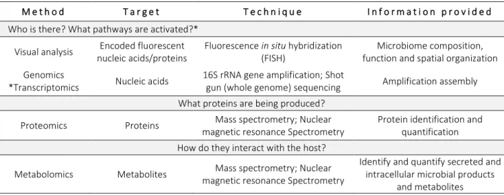
Since 1990s, it is possible to assess bacterial diversity by sequencing a small subunit ribosomal RNA gene (16S rRNA) [12]. The 16S rRNA gene is a highly conserved phylogenetic marker present in the major bacterial groups presents in the human gut [12]. Using the properly primers for amplification of single or multiple regions of the 16S rRNA by Polymerase Chain Reaction (PCR), the marker gene sequencing is a good method to determine microbial phylogenies of a sample [13]. The introduction of Next Generation Sequencing (NGS) replaced the 16S rRNA gene-based microbial profiling analysis using primers. Sequencing of the gut microbiota not only revealed new microbial species, but also microbial metabolic activities and how these correlate with human health and disease [14]. Table 1 show these and other “omics” techniques that can be used to detected and classify microbes, their genes, their products and clarify their functions.
More recently, synthetic microbial communities are being developed [11]. Using mathematical models and engineered microbes in artificial environments it is possible to study microbial interactions with each other and their responses to environmental factors [11].
Table 1 - Culture independent techniques for microbiome analysis and their interaction with the host by answering these essential questions: Who is there?; What pathways are activated?; What proteins are being produced?; How do they interact with the host?. Adapted from Ilhan, 2016 [15] and Méndez-García et al, 2018 [16].
M e t h o d T a r g e t T e c h n i q u e I n f o r m a t i o n p r o v i d e d
Who is there? What pathways are activated?*
Visual analysis Encoded fluorescent
nucleic acids/proteins
Fluorescence in situ hybridization (FISH)
Microbiome composition, function and spatial organization Genomics
*Transcriptomics Nucleic acids
16S rRNA gene amplification; Shot
gun (whole genome) sequencing Amplification assembly
What proteins are being produced?
Proteomics Proteins Mass spectrometry; Nuclear
magnetic resonance Spectrometry
Protein identification and quantification How do they interact with the host?
Metabolomics Metabolites Mass spectrometry; Nuclear
magnetic resonance Spectrometry
Identify and quantify secreted and intracellular microbial products
and metabolites
The identification of over 1000 bacterial species prove that the human gut microbiota is a dynamic and complex ecosystem [17].
The gut microbiota is dynamic
Contrasting to human genome, the microbiome is plastic. During the course of life the richness and diversity of bacterial composition change significantly [18]. The neonatal intestine is colonized by facultative and aerotolerant bacteria [Proteobacteria (Enterobacteria)] that will reduce oxygen content in the intestine to promote the anaerobic bacteria colonization [Actinobacteria (Bifidobacterium) and Bacteroidetes (Bacteroides)] [19]. At three years of age it is established the adult-like composition [20]. Since the very beginning, the architecture and functionally of the gut microbiota is highly dependent of host-endogenous and host-exogenous factors providing a very individual and unique microbial composition as a “microbiome fingerprint” [21]. Some of these factors include gestational age, mode of delivery, diet, exercise, antibiotic
exposure, fasting, culture traditions, geography and genetic susceptibility (Figure 2) [22]. From all, diet is
considered the most determinant factor for the gut microbiota [23].
The gut microbiota is complex
The human health success depends on these large and diverse host-bacteria community [2]. Actually, there are numerous physical and molecular mechanisms that allowed the symbiotic relationship between host and commensal microorganisms [24]. A successful commensal gut bacterial composition provide essential enzymes that digest the polysaccharides and peptides that are indigestible to human cells [24]. The role of the
gut microbiota in metabolism of dietary components through bacterial fermentation process results in the production of important and beneficial metabolites [25]. However, if there is inadequate intake of fermentable dietary components (such as fermentable fiber) the bacteria use alternative energy sources resulting in the production of metabolites that can be detrimental to human health [25].
Under the influence of the factors mentioned above these metabolites, such as short-chain fatty acids (SCFAs), trimethylamine N-oxide (TMAO), bile acids, gut hormones and others, may contribute to the regulation of human metabolism and immune system development and function and, consequently, to the host health or disease (Figure 2) [22,25]. Indeed, an imbalance in the composition, stability and resilience of gut microbiota (dysbiosis) is associated to the etiology and development of many diseases involving not only the gastrointestinal tract, but also other distal organs (Figure 2) [26].
Figure 2 - Contribution of both host-endogenous and host-exogenous factors on the gut microbiota. Different bacteria produce different metabolites that can influence the disease development in the intestine and in other distal organs, through the regulation of host metabolism and immune system. SCFAs, short-chain fatty acids; TMAO, trimethylamine N-oxide; BCAA, branched-chain amino acid; IPA, indole propionic acid; NAFLD, nonalcoholic fatty liver disease; NASH, nonalcoholic steatohepatitis; IBD, inflammatory bowel disease. The image is an adaptation of Cani et al, 2019 [23] and Levy et al, 2017 [27].
It is imperative to understand how microbiota can be modulated with a more personalized medicine and nutrition treatments in order to prevent or delay the onset of the pathologies mentioned above. Identify
windows of opportunity for microbiota modulation can “revolutionize our approach to healthcare” [27]. In fact, a special focus is being given to pregnancy and preterm birth [27].
Microbial colonization of the infant intestine
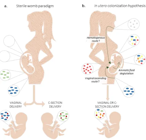
The moment when the first microbial colonization occurs is an open question. Actually, the colonization process is a dogma between the “sterile womb” and “in utero colonization” hypotheses for more than 100 years.
Traditionally, it is accepted that vertical (maternal) and horizontal (from other persons and environment) transmission of microorganisms occurs only at birth, through vaginal canal and/or through contact with the mother’s skin microbiota immediately after birth (Figure 3a). Most studies that mentioned the uterus bacterial-free used cell cultured and/or microscopy methods – methods that, although still valid, have several limitations. These studies, many from the XX century, have shown negative results for the detection of aerobic and anaerobic bacteria in amniotic fluid [28–31] and meconium (the newborn’s first intestinal discharge comprising ingested or secreted material by the gastrointestinal tract during fetal life [32]) [33–35] of healthy pregnancies. However, there is no study with all samples sterile. The presence of bacteria in the amniotic fluid and placenta was limited to complications during pregnancy, such as premature labor, premature rupture of membranes and neonatal sepsis [36].
Later, Steel et al. (2005) studied fetal membranes of term and preterm deliveries [37]. The membrane samples were collected and placed in fixative within 30 min for FISH. Bacterial DNA was detected in term deliveries with (46% positive samples, n = 26) or without labor (73% positive samples, n = 26), preterm deliveries with labor (92% positive samples, n = 13) or without labor (83% positive samples, n = 12), and preterm delivered with prolonged premature rupture (81% positive samples, n = 22) [37]. In the same year (2005), Jiménez et al. (2005) reported the presence of bacteria in umbilical cord blood healthy neonates [38]. Later, two independent studies conducted by Aagaard and Mysorekar, detected bacterial content in placenta samples (n = 320 and n = 195, respectively) of women who gave birth prematurely with and without infection and also in women who had term healthy pregnancy [8,39]. However, the evidence is unclear. Lauder et al. (2016) did not found any difference between placental samples and negative controls [40]. A recent work “confidently” detected bacterial rRNA in placentas of 13 from 16 spontaneous preterm births and in 18 of 22 term unlabored cesareans, with no significant differences between preterm and term deliveries [41].
Bacteria communities were also detected in amniotic fluid [42,43]. However, a more recent study with 24 uncomplicated term pregnancies concluded that bacterial microbiota of amniotic fluid was indistinguishable from contaminated controls [44]. Interestingly, an Australian group wrote a critical comment about the
methodology used in that study [45]. The concerns raised were: sample processing, qPCR approach, and results analysis questioning the conclusions made [45].
Despite the growing evidence in favor of the hypothesis of colonization in utero, there is still no consensus among peers and several aspects have to be taken into consideration. A recent and exhaustive review pointed out several methodological limitations to studies suggesting that the microbial colonization occurs in the prenatal period, namely: (i) molecular techniques with an insufficient detection limit to analyze small microbial populations; (ii) lack of appropriate negative controls; and, (iii) lack of sterility in sample collection in clinical/hospital settings [36]. Another aspect to be considered is the contamination of samples by DNA extraction kits due to the ‘kitome’ present in the reagents and other components of the kits [46].
Although the placenta protects the fetus from bacterial infections, it is also the maternal-fetal communication organ (and not a barrier) providing oxygen, nutrients and, perhaps, bacteria or bacteria residue resulting from the action of antimicrobial peptides and immunoglobulins expressed in the placenta [36]. The in utero colonization hypothesis became stronger when animals studies suggested the maternal-fetal transfer of microbes. Using labeled Enterococcus faecium isolated from breast milk of healthy woman, pregnant mice were orally inoculated and then delivered the pups by C-section [38]. The labeled strain was detected in amniotic fluid [38] of these animals and in the pups’ meconium [47]. External factors, such as maternal stress also influenced the gut microbiota of infant monkeys, reducing the content of Lactobacillus and
Bifidobacterium [48]. In fact, the maternal transmission of microbes is a universally shared phenomena in the
animal kingdom [49].
In humans, the studies are scare and the evidence is less conclusive [50]. Among 12 vaginally delivery mother-infants, 11 presented monophyletic bifidobacterial strains indicating a vertical transmission of bacteria [51]. Rautava et al. (2012) administered probiotic supplementation to mothers during pregnancy and the
Lactobacillus group were detected in all placentas specimens [52]. In addition, Toll-like receptor (TLR)-related
gene expression was associated with bacterial DNA in amniotic fluid and placenta [52], indicating that maternal-fetal transfer of bacteria could influence the immune development of the offspring [50]. Recently, it was reported a highly shared microbial population in a large cohort of mother and their infants (n = 415) [53]. Mechanism by which maternal bacteria pass to the fetus are not well understood [50]. However, two hypotheses are being considered (Figure 3b).
Figure 3 - Microbiota colonization of infants – two different hypothesis. a. Sterile womb paradigm defends that placenta and fetus are sterile and that gut microbiota is acquired during and after birth. Mode of delivery has been associated to transmission of specific bacteria: vaginal delivered promote a vagina-like microbiota in newborn and C-section leads to a bacterial colonization that resemble maternal skin microbiota b. The “in utero colonization hypothesis” suggests that bacterial colonization occurs prior to birth and it is influenced by maternal microbiota (oral cavity, gut and vagina). Two mechanism have been proposed to explain the mother-infant bacterial transmission: (i) the hematogenous bacterial route of bacteria from gastrointestinal tract (oral cavity and gut); and, (ii) ascension of bacteria from vaginal microbiota. Both routes argue that bacteria go into blood circulation and incorporated into the placenta decidua and, consequently, the developing fetus via amniotic fluid and cord blood [55]. The image is an adaptation of Perez-Muñoz et al, 2017 [37] and Milani et al, 2017 [55].
Maternal influence in offspring microbiota
Maternal factors can disrupt the normal infants’ intestine colonization that play a critical role in the maturation of immune, endocrine and metabolic pathways and in development of disease later in life [54]. The prenatal nutrition and lifestyle are crucial for intrauterine fetal programming [55] and maternal microbial transmission seems to interact with biological systems being crucial for fetal health [50]. And even if this in utero colonization occurs only under certain circumstances (subclinical conditions), it is important to understand how, in order to optimize all mother, fetus and infants’ health relationships. When mothers are obese, have (gestational) diabetes, increased insulin resistance and increased gestational weight gain (GWG) have an imbalanced and an unhealthy microbiota (dysbiosis) that is directly transmitted to the offspring [56]. As review
by Milani (2015), alterations in the endothelial integrity of placenta may lead to an permeable barrier and, consequently, the passage of bacteria and endotoxins, as lipopolysaccharide (LPS), into the cord blood and
amnion [57]. Since fetal intestine is highly sensitivity to inflammatory mediators such as LPS, it is suggested
that maternal microbes may trigger intestinal inflammation in utero [58].
During pregnancy the total gut bacteria abundance was reported to decrease, as well the bacterial diversity between the first and third trimesters and differences were observed between normal weight and overweight women [59,60]. To study the impact of microbial transmission, gut microbiota samples from women in the third trimester were transferred to gnotobiotic mice and it was shown that mice gained more weight, became more insulin resistant and had higher levels of inflammatory markers [59]. Furthermore, it was found that children’s microbiota were most similar to their mothers’ microbiota at first trimester [59]. This was the first study that used high-throughput sequencing to suggest that mother-to-child microbial transmission plays a major role, and care should be taken not only during pregnancy but also before conception. Even if the fetus is developed in a sterile environmental, it has been realized that metabolites and others molecular products of maternal gut microbiota are part of placental molecular exchange [61]. Therefore, maternal diet exposure before and during pregnancy and breastfeeding can influence the gut microbiota of their infants, with long-lasting effects [56].
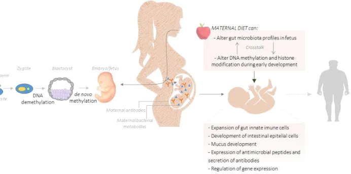
Maternal microbiota metabolites of dietary components can reach the fetus via placenta or, after birth, through breast milk [61]. These molecules, or even maternal bacterial fragments, act as signals for the expansion of innate immune cells, development of intestinal epithelial cells and mucus, expression of antimicrobial peptides and secretion of antibodies into the intestinal lumen in offspring (Figure 4) [61]. In line with this, malnutrition (that includes deficiencies, excesses or imbalances in nutrients and/or energy intake) during pregnancy impairs fetal immune system through direct (for example, prolonged vitamin A deficiency leads to an impaired production of B lymphocytes [62]) and indirect (maternal immunosuppression decreased the transfer of immunoglobulins for fetus increasing the chance for infection [61]) mechanisms.
Fetal exposure to excess blood lipids, particularly saturated fatty acids, can activate proinflammatory pathways, which could impact substrate metabolism and affect organ development and the response to the postnatal environmental factors, since the epigenetic regulation of gene expression is characterized by covalent modifications to DNA and chromatin that alter gene expression independent of gene sequence [63]. Furthermore, a prospective large cohort of 66 000 pregnant women reported that women adhering to a ‘prudent’ (characterized for high intake of vegetables, fruit, berries, nuts, whole grains, poultry and water as beverage) or a ‘traditional’ (rich in boiled potatoes, vegetables, lean fish and fish products, low fat milk and rice pudding) dietary patterns were at lower risk of preterm delivery compared with women with a ‘Western’ diet (salty and sweet snacks, white bread, desserts and processed meat products) [64].
Figure 4 - Maternal diet can alter the structure and function of the gut microbiota leading to an epigenetic reprogramming processes (DNA methylation and histone modification) during early embryogenesis. In addition, it has been suggested that exist bacteria in fallopian tube and in endometrium that play a key role during conception and, posteriorly, in the embryo/fetus development [67]. However, even considering that the intrauterine environment is sterile, it is known that maternal microbiota metabolize dietary components of the diet and produce molecular signals that can reach the offspring during in utero development via the placenta. These metabolites, as well as maternal antibodies, influence the fetus innate immune system, namely in the expansion of gut innate immune cells, development of intestinal epithelial cells, mucus production, expression of antimicrobial peptides and secretion of antibodies. An unbalanced maternal diet can result in changes on adipogenesis and metabolism leading to a higher susceptibility to obesity and obesity-related metabolic diseases in adult life. The image is an adaptation of Macpherson et al, 2017 [63] and Li, 2018 [68].
Mediterranean Diet (MD) – characterized by high consumption of vegetables, fruit, pulses, cereals and fish; using olive oil as preferential fat source (high monounsaturated:saturated fat ratio); moderate alcoholic beverages consumption; and, low intake of meat and meat products, milk and dairy products [65] – has been associated with higher birth weight and lower risk of premature birth [66]. Moreover, MD adherence during pregnancy seems to have a protective role in fetus against the development of Metabolic Syndrome throughout life [67]. Even the studies in pregnant women is scare due to ethical reasons. To the best of our knowledge, there are no works studying the influence of MD adhesion in maternal-fetal microbial transfer. One the other hand, in vivo studies with high-fat diet during pregnancy (contrarily to what is encouraged in the MD) showed that female mice fed with a high-fat diet before and during pregnancy led to an increased risk of offspring obesity [68]. The impaired gut barrier integrity, increased circulation levels of LPS, increased placenta hypoxia and impaired placenta vascularization found in the high-fat group may be the reason of this association [68].
Mode of delivery
Regardless of when the first microbial colonization occurs, it is known that is during delivery that takes place the greatest microbial colonization of the newborn. While infants born by vaginal delivered receive a microbiota similar to the maternal vagina (through the passage on cervix and vagina), C-section delivered infants are enriched in skin microbiota, hospital staff and environment [69] (Figure 3). Infants born by vaginal delivery showed more Lactobacillus and Prevotella [70]. On the other hand, C-section delivered infants were associated with lower biodiversity, a delay in colonization by beneficial bacteria and higher colonized by
Staphylococcus, but less colonized by Enterococcus [71].
Despite some contradictory works, it is assumed that the mode of delivery may play a decisive role in the development and growth of the newborns, since the bacteria present in fetal gastrointestinal tract can influence the development the immune system and therefore have relevant health consequences [72]. As mentioned by Dominguez-Bello, “Epidemiological studies, although not showing causality, have reported associations between C-section delivery and an increased risk of obesity, asthma, allergies and immune deficiencies” [73]. So, to minimalize the disruption of vertical transmission of microbiota provoked by C-section, it was developed a technique to “restore” the microbiota of these infants. A sterile gauze was pass first in the mother’s vagina after the surgery and, immediately after, the same gauze was applied in the newborn (mouth, face and in rest of the body) [73]. Despite these procedure restored the infant’s gut, oral and skin microbiota, it is important to ensure the costs and potential risks [74]. More recently, it was reported that the mode of delivery had a temporary and little effect on infants gut microbiota and the gestational age seems to be the main driver [75].
The preterm infant’s microbiota
A healthy full-term vaginal delivered and exclusively breast-fed infant has been considered to be the standard for a healthy infant microbiota [76]. On the other hand, preterm infants born by C-section, fed with formula and exposed to antibiotics have abnormal patterns of colonization with lower abundance of “healthy” bacteria as Lactobacillus and Bifidobacterium [58]. Despite the very high variability between preterm infants [77], their microbiota composition is significantly different from that of the full-term infants [78]. Table 2 summarizes the principal bacteria groups in preterm infants.
Table 2 - Principal bacteria groups in preterm infants. Adapted from Underwood et al, 2017 [79].
Phylum Class Family Genus
Proteobacteria γ-Proteobacteria Enterobacteriaceae
Pseudomonadaceae Moraxellaceae Klebsiella -/▫ Escherichia -/▫ Proteus -/▫ Serratia -/▫ Enterobacter -/▫ Cronobacter -/▫ Pseudomonas -/▫ Acinetobacter -/▫ Firmicutes Bacilli Clostridia Negativicutes Staphylococcaceae Streptococcaceae Enterococcaceae Lactobacillaceae Clostridiaceae Veillonellaceae Staphylococcus +/▫ Streptococcus +/▫ Enterococcus +/▫ Lactobacillus +/▫ Clostridium +/• Veillonella -/•
Bacteroidetes Bacteroidetes Bacteroidaceae Bacteroides -/•
Actinobacteria Actinobacteria Bifidobacteriaceae Propionibactebacteriales
Bifidobacterium +/• Propionibacterium +/•
- ,Gram-negative bacteria; +, Gram-positive bacteria; ▫, facultative anaerobic; •, strict anaerobic
Preterm infants – defined as an infant born before 37 weeks of pregnancy, that includes very preterm infant born between 28 and 32 weeks and extremely preterm born before 28 weeks [80] – born to soon due to different reasons. Cervical and vascular disorders, uterine overdistension, breakdown of maternal-fetal tolerance and environmental factors as maternal age at pregnancy, maternal chronic diseases, maternal nutritional status and infection are some of the factors that can lead to premature labor [80,81].
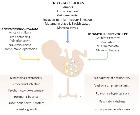
The preterm infant presents an immature intestinal microbiota with a marked vulnerability to dysbiosis, with a change in abundance, diversity and progressive acquisition of bacteria. If, on the one hand, premature infants present an immature gut, immune system and metabolism, they also require long hospitalization leading to a colonization mainly by bacteria from the neonatal intensive care unit (NICU) and invasive procedures as intravenous access, parenteral feeding and mechanical ventilation [82]. Premature infants are exposed to endogenous factors, environmental and maternal and postnatal therapeutic conditions that constitute an imminent trigger for infection and, consequently, dysbiosis (Figure 5).
An inflammatory status in preterm infants – which can be triggered even in utero – can affect all organs(Figure
5). Late-onset sepsis and necrotizing enterocolitis (NEC), a very common cause of morbidity and mortality in preterm infants, were associated with microbiomes dominated by Proteobacteria and Firmicutes [71] – the dominant phyla of the preterm infant microbiota [77]. As reviewed by Staude et al (2018), an imbalanced gut microbiota in preterm infants, could contribute to impaired somatic growth and psychomotor development, bronchopulmonary dysplasia, retinopathy of prematurity, cardiovascular diseases and behavioral and stress responses (Figure 5) [83].
Figure 5 - Gut microbiota of preterm infants. Environmental and endogenous factors, as well as pre- and postnatal therapeutic interventions can determine the composition of gut microbiota of preterm infants. The impact of gut microbiota in NEC, nosocomial infection, psychomotor development, hormonal balance, autonomic nervous system and somatic growth have been reported. On the other hand, the association of gut microbiota and others short and long term-morbidities is unclear. Adapted from Staude et al, 2018 [85].
To combat this high susceptibility to infection and pathological bacteria in preterm infants, antibiotherapy is used, in most of the cases, either ante- and postpartum. However, data show that concerns should be taken when administering antibiotics due life threatening morbidities [58]. One study with 4039 preterm infants from 19 neonatal centers, showed that empirical and prolonged antibiotic therapy (≥ 5 days) was associated with increased odds of death and NEC [84]. In line with these, another study found that administration of antibiotics for 5-7 days on the first postnatal week lead to more cases of NEC, sepsis and death [85]. These preterm infants presented lower bacterial diversity and an increased abundance of Enterobacter [85]. One study that gathered 10 premature infants, found that only one single infant was dominated by Bifidobacterium, since it was also the only baby that did not receive antibiotic treatment during the first four weeks of life [77]. Perinatal antibiotic exposure also affected “strongly” the initial microbiota establishment in the preterm infants [76]. In addition, a recent study that analyzed 436 mother-child pairs followed until 7 years of age, found that children born from mothers who were given antibiotics during the second or third trimester of pregnancy had an 84% higher risk of obesity at age 7, but the use of antibiotics in the first trimester had no effect [86].
Microbiota in early life is crucial to a healthy maturation of the immune system and other organs [83]. It is essential to identify strategies to establish a healthy early microbiota in preterm infants, since it provides antigenic stimulus for the adequate development and maturation of the immune system, intestine and even distal organs [69]. For instance, providing breast milk to preterm infants during hospitalization led them to developed a normal microbiota resembling that of term infants [87].
The impact of the infant feeding
In premature infants, especially in very and extremely preterm infants, an appropriated nutrition is essential to decrease the risk of adverse health outcomes and to improve cognition in adulthood [88]. According to international guidelines [88,89], which are contemplated in national recommendations [90], mothers’ own milk (MOM) is always the first choice to fed preterm infants. However, MOM is not always available and sometimes it is insufficient. In that cases pasteurized donor human milk (DHM) should be administrated [91]. When Human Milk (HM – that comprises MOM and DHM) is unavailable, bovine-based preterm formulas (designed as formula hereinafter) should be used.
Breast milk composition changes through the course of gestation and lactation, and even with the time of the day [89]. Despite the protein, fat and carbohydrates source, HM is a “carrier of biochemical messages” that modulate the growth, development and the immune system of the newborn [92]. The composition of the breast milk of mothers that delivered preterm infants is significantly different that mothers that delivered at term, namely in protein and fat contents that are higher in preterm breast milk (Table 3) [93]. The most abundant proteins present in the HM are casein, α-lactalbumin, lactoferrin, secretory immunoglobulin IgA, lysozyme, serum albumin and antimicrobial peptides [92,93]. HM also contains other bioactive compounds
such as essential fatty acids, enzymes, growth factors and hormones that have immune-related functions
[89,92,93]. The main source of carbohydrates in HM is lactose and is the least variable macronutrients [93]. HM also contained oligosaccharides – HMOs – that are glycosylated compounds synthesized in mammary gland by glycosyltransferases. HMOs are remarkable components of breast milk due to their prebiotic effect stimulating the growth of beneficial bacteria [93].
Furthermore, breast milk is a source of probiotics providing commensal bacteria to the infant. The literature
suggests that breast milk still contains 106 bacteria cells per mL, with a dominance of Staphylococcus,
Streptococcus (both bellowing to Firmicutes phylum) and Pseudomonas (Proteobacteria phylum) [94]. Two
potential mechanisms can explain the presence of bacteria in breast milk. In a nutshell, mothers’ skin microbiota and infants’ oral can reach breastmilk through retrograde flux [95]. In addition, it has been demonstrated that dendritic cells (and macrophages) can pass intestinal epithelium through the expression of tight-junctions proteins to take up commensal bacteria and, via lymph/blood circulation, reach the mammary
gland [96]. This process showed to occur during late pregnancy and lactation in mice [97]. This may be the reason why in premature MOM the percentage of bacteria present in milk samples was lower [98]. Despite the lower total bacterial content in preterm breast milk (n = 19) compared to term breast milk (n = 13), no significantly difference were observed (Table 3) [99]. Staphylococcus, Streptococcus, Lactobacillus,
Enterococcus and Enterobacteria were the main genera isolated in milk from preterm gestations with Bifidobacterium concentration significantly decreased compared to term milk [98,99].
Table 3 - Energy, macronutrient and total bacterial content composition of breast milk from mothers who delivered term and preterm; DHM and preterm infants formula [93].
MOM term MOM preterm DHM Formulaa
Energy (kcal/dL) 65 – 70 78 50 – 115 80
Protein (g/dL) 0.9 – 1.2 2.2 0.6 – 1.4 2.6
Fat (g/dL) 3.2 – 3.6 4.4 1.8 – 8.9 3.9
Lactose (g/dL) 6.7 – 7.8 7.6 6.4 - 7.6 5.6
HMO (g/dL) 0.817 [100] 0.857 [100] 0.8 [101] 0.8b
Bacterial content 5.37 Log (gene copies ml–1) [99] 5.00 Log (gene copies ml–1) [99] 102CFU/mL [102] n.a.
a The composition of the formula described above refers to the commercial formula Aptamil Prematil, Milupa Danone®,
which was used in the preterm infants included in this study; b The total amount oligosaccharides present in formulas
is the sum of GOS (0.72 g/dL) and FOS (0.08 g/dL); n.a. – information not available.
MOM composition is also influenced by protein intake, parity, return of menstruation and nursing frequency [93]. Maternal diet also influence breast milk composition. A very recent study demonstrated the adherence to MD lead to a 10-fold higher Lactobacillus abundance in mammary glands of female monkeys compared to the Western diet group [7]. Likewise, maternal body mass index (BMI) showed to have influence: breast milk from obese mothers (n = 10) had more bacterial content, but it was less diverse when compared to normal weight mothers (n = 8). More, these mothers had lower content of Lactobacillus in colostrum and
Bifidobacterium at 6 months [103].
Despite the rapidly increase of human milk banks, DHM is not available to all preterm infants. In Portugal, for example, there is only one human milk bank. Typically, DHM is provided by mothers who delivered at term that have excess milk production. Thus, DHM is a term and late lactation milk and require additional protein and fat acids supplementation. Moreover, DHM is submitted to Holder pasteurization (62.5°C for 30 minutes) to avoid transmission of infectious agents. Due to these reasons and to the handling and storage procedures, the DHM presents some differences in their composition in relation to preterm MOM (Table 3) [104,105].
Some compounds, such as protein, lactose, long-chain polyunsaturated fatty acids, vitamins (A, D, E, B12, B9)
and some growth factors are preserved [91]. On the other hand, bioactive compounds of breastmilk are affected. B and T lymphocytes, macrophages, neutrophils, as well as lipoprotein lipase and IgM are inactivated.
The lactoferrin concentration decreased 50-75%, as well 24-74% of lysozyme and 20-30% of IgA [91]. Pasteurization procedure does not decreases the HMOs content (Table 3) [106]. However, the content of HMOs in DHM presented significantly lower amount of total HMOs in comparison to MOM [101]. The heat
treatment leads to the inactivation of viruses and kills 99% of bacteria. However, Cacho et al. (2017) found 102
CFU/mL of bacteria in 44% of DHM [102]. The most abundant genera identified were Acinetobacter,
Enterobacteriaceae and Serratia (all bellowing to Proteobacteria phylum). These authors used a small amount
of MOM to inoculate the pasteurized DHM, making possible to personalize DHM with MOM microbiota [102]. This could be a promising and innovative method to provide beneficial bacteria to preterm infants.
Contrarily to HM, formulas have a very standardized composition (Table 3). Preterm formulas contain all essential nutrients to provide a good growth and development to infants [88]. Although formula offers a similar percentage of total calories from fat in relation to MOM, the composition of specific fatty acids can be very different, as well the bioactive compounds [107]. Synthetic HMOs (namely, galatooligosaccharides – GOS and fructooligosaccharides - FOS) are added to formula. However, these molecules are structurally different from that naturally present in HM [108]. These differences could influence the immunogenic development of preterm infants [108]. Regarding the bacterial content, during preparation, powdered formulas may be contaminated [109]. In NICU, single doses of sterile liquid infant formula should be used [109].
It is possible to understand why the type of infant-feeding influence in different ways the development of the preterm infants’ health outcomes and their gut microbiota. There are several studies showing short and long term benefits associated with HM intake in preterm infants: lower incidence of NEC, late-onset sepsis and retinopathy of prematurity; better neurological development promoting a significantly intelligence quotient in later years; lower risk of hypertension and atherosclerosis in adulthood [88]. Moreover, in preterm infants the intake of both MOM and DHM were associated with better feeding tolerance, shorter duration of hospital stay and reduced hospital costs [110].
Due to prebiotic and probiotic properties, preterm infants fed with MOM showed a greater initial diversity of gut microbiota compared infants fed non-MOM [111,112]. An effect that was maintained until 30 postnatal days [112]. On the other hand, in DHM and formula-fed infants the rate of diversity was slow over in time and at 30 postnatal days the diversity index was lower [113]. A recent study did not found differences in microbial diversity and richness between the feeding types [114]. However, significantly differences were found in bacterial profile [114]. Despite Bifidobacteriales showed to be higher in infants fed with MOM compared to DHM and formula infants [115], DHM showed closer microbial profiles to MOM than formulas [114]. Microbiota profile of breast-fed infant’s changes to a formula-fed infant profile, with significant increase in the count of Enterococci and Enterobacteriaceae, and the appearance of Bacteroides and Clostridium [115]. Although, no significant differences were observed between MOM and formula-fed preterm infants at fourth
week of life [115]. MOM was reported to mitigate the effect of gestational age (gut immaturity) [111]. In fact, preterm infants fed with HM could develop a microbiota similar to the term infants, independently of being MOM or DHM [75].
The available data support that maternal microbiota as well as newborns postnatal microbiota contribute to a healthy or disease status of preterm infants with short- and long-term consequences. Exposure to a diversity of commensal species regulate local microbial growth, modulate the morphology and function of enterocytes, reduce activation of proinflammatory cascade and affect gene expression [116].
AIMS
The goal of this observational study was to evaluate the impact of different types of infant feeding (MOM, DHM and formula) on the gut microbiota of preterm infants hospitalized in NICU of Maternidade Alfredo da Costa (MAC). Furthermore, the influence of mode of delivery and mother’s diet on vertical microbiota transmission were also evaluated.
Specifically, the aims of this study were: - to characterize the preterm gut microbiota; - to characterize the maternal gut microbiota;
- to evaluate the impact of maternal gut microbiota, antepartum factors (dietary pattern, pre-gestational body mass index, antibiotic therapy) and perinatal factors (gestational age and mode of delivery) on the bacterial colonization of preterm infants;
METHODOLOGY
This study protocol was approved by the Ethics Committee of Centro Hospitalar Universitário de Lisboa Central (Ref. 443/2017) and by the Ethics Committee of NOVA Medical School|Faculdade de Ciências Médicas, Universidade NOVA de Lisboa (NMS|FCM). The study was conducted in accordance to the ethical principles expressed in the Declaration of Helsinki, the Portuguese law and Good Clinical Practice guidelines.
Study Design
The FEEDMI Study was an observational longitudinal study, conducted at the NICU of MAC and NMS|FCM. The study is registered in ClinicalTrials.gov platform, with the registration number NCT03663556. The detailed study protocol was already published [117]. An overview of the study design is described in Figure 6.
Figure 6 - Study timeline. After obtaining informed consent from legal representatives, infants will be enrolled in the study. The first stool sample will be collected within the first 24 hours after birth, followed by three subsequent collections every 7 days.
Participants Recruitment
Very preterm infants (< 32 weeks gestational age) hospitalized in the NICU of MAC were recruited within the first 24 hours after birth. Inclusion criteria are displayed in Figure 6. Written informed consents were obtained for each preterm infant after explaining the entire study protocol to their legal representatives.
Sample Collection
Meconium, the newborn’s first intestinal discharge, and the 3 additional fecal samples of preterm infants were collected by the nursing team of MAC’s Neonatology Unit. Fecal samples were collected weekly from diapers into sterile tubes. Mothers were also asked to collect their own fecal samples with an appropriate stool collection kit (EasySampler®).
Inclusion criteria
- Admission at NICU in less than 24 hours
- Gestational age < 32 weeks
- Absence of congenital malformations or metabolic diseases
- Newborns’ parents have to be Caucasian
Obtaining Informed Consent
1st Harvest 2nd Harvest 3rd Harvest 4th Harvest
7 days Birth of the
premature infant
Clinical Data Collection
Detailed clinical data were collected during the preterm infant enrollment in the study through medical records. Personal clinical data include sociodemographic information and clinical intrapartum and postpartum outcomes such as newborn’s somatometry evolution, antibiotic exposure and its duration, number of total days of hospitalization, and other outcomes related to the preterm clinical evolution. Additionally, type of infant-feeding (MOM, DHM and formula) was recorded daily to select the most representative (> 50 %) type of infant-feeding received during the 7 days prior to each fecal sample collection.
Microbiota Analysis
Fecal sample collection is a non-invasive procedure commonly used to assess the intestinal microbiota composition. Bearing in mind meconium’s tar-like texture and samples’ low bacterial amount [118], DNA were extracted and purified from stool samples using NZY Tissue gDNA Isolation Kit (nzytech, Lisbon, Portugal), as previously described [119].
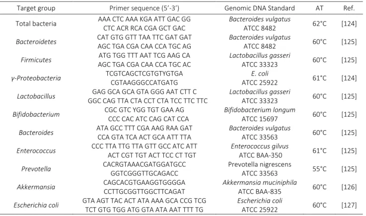
Different bacterial populations were analyzed by quantitative real-time PCR using LightCycler instrument (Roche Applied Science, Indianapolis, ID, USA). Specific microorganisms were assessed based on previous studies regarding preterm gut microbiota composition [111,120–122]. Two phyla (Bacteroidetes and
Firmicutes), one class (γ-Proteobacteria), four genera (Lactobacillus, Bifidobacterium, Bacteroides and Enterococcus) and one specie (Escherichia coli) were analyzed of preterm infants’ samples. In mothers’
samples, the same bacteria groups were analyzed in addition to Prevotella and Akkermansia. Primer sequences used to target bacterial 16S rRNA genes were described in Table 4. Results on microbiota are expressed as log10 16S rDNA gene copies/10ng of DNA.
Quality Control Analysis
Increasing evidence has been suggested that DNA extraction kits and other laboratory reagents are commonly contaminated [123]. This contamination could have a critical impact on results, especially in samples containing low microbial biomass [123], such as preterm infants. For control purposes, a fecal collection was simulated: an empty tube (that same tubes used for collecting meconium and feces samples) was opened inside the infants’ incubator and the spatula was passed through diaper; the tube was stored under the same conditions as the others; in the lab, it was added 200 mL of ultrapure water into the tube; and DNA was extracted. In addition, DNA amplification was performed in duplicated and samples with lower levels than negative controls (from extraction and PCR procedures) were discarded.
Table 4 - Primers sequences used for gut microbiota analysis. AT, annealing temperature.
Target group Primer sequence (5’-3’) Genomic DNA Standard AT Ref.
Total bacteria AAA CTC AAA KGA ATT GAC GG
CTC ACR RCA CGA GCT GAC
Bacteroides vulgatus
ATCC 8482 62°C [124]
Bacteroidetes CAT GTG GTT TAA TTC GAT GAT
AGC TGA CGA CAA CCA TGC AG
Bacteroides vulgatus
ATCC 8482 60°C [125]
Firmicutes ATG TGG TTT AAT TCG AAG CA
AGC TGA CGA CAA CCA TGC AC
Lactobacillus gasseri ATCC 33323 60°C [125] γ-Proteobacteria TCGTCAGCTCGTGTYGTGA CGTAAGGGCCATGATG E. coli ATCC 25922 61°C [124]
Lactobacillus GAG GCA GCA GTA GGG AAT CTT C
GGC CAG TTA CTA CCT CTA TCC TTC TTC
Lactobacillus gasseri
ATCC 33323 60°C [125]
Bifidobacterium CGC GTC YGG TGT GAA AG
CCC CAC ATC CAG CAT CCA
Bifidobacterium longum
ATCC 15697 60°C [125]
Bacteroides ATA GCC TTT CGA AAG RAA GAT
CCA GTA TCA ACT GCA ATT TTA
Bacteroides vulgatus
ATCC 33563 60°C [125]
Enterococcus CCC TTA TTG TTA GTT GCC ATC ATT
ACT CGT TGT ACT TCC CT TGT Enterococcus gilvus ATCC BAA-350 61°C [125] Prevotella CACRGTAAACGATGGATGCC GGTCGGGTTGCAGACC Prevotella nigrescens ATCC 33563 55°C [125] Akkermansia CAGCACGTGAAGGTGGGGA CCTTGCGGTTGGCTTCAGAT Akkermansia muciniphila ATCC BAA-835 60°C [126]
Escherichia coli GTA AGT TAC ACT ATA AAA GCA CCG TCG
TCT GTG TGG ATG GTA ATA AAT TTT TG
Escherichia coli
ATCC 25922 60°C [127]
Maternal Mediterranean Diet Adherence Score
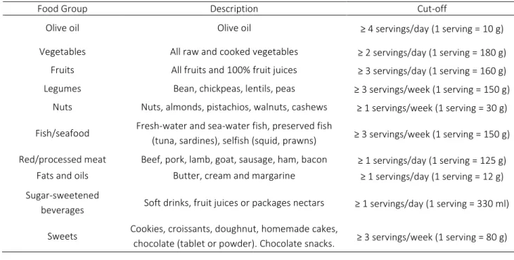
Mothers were requested to completed a semi-quantitative Food Frequency Questionnaire (FFQ), previously validated for the Portuguese population [128,129]. In the current study, the FFQ with 86 items was used to assess food consumption during pregnancy. Response options are in a 9-point frequency scale, ranging from ‘never’ to ‘≥ 6 times per day’. Using portion sizes and frequency of consumption, daily portions (g/day) were computed for each FFQ item. Data from FFQ was used to calculate the MD adherence score using 13 items of the original questionnaire [130]. The following items were classified with 1 point if the answer to the question was “YES”: olive oil as main culinary added fat/oil; olive oil ≥ 4 daily tablespoons; vegetables (including vegetable soup) ≥ 2 daily serving; fruit ≥ 3 daily servings; legumes ≥ 3 weekly servings; fish/seafood ≥ 3 weekly servings; tree nuts ≥ 1 weekly serving; and more poultry than red meat. If the answer was “NO”, the item was classified with 0 points. On the other hand, the following items were classified with 1 point if the answer to the question was “NO”: red/processed meat ≥ 1 daily serving; butter, cream and margarine ≥ 1 daily serving; sugar-sweetened beverages ≥ 1 daily serving; sweets and confectionary ≥ 3 weekly servings; and any consumption of wine. If the answer was “YES”, the item was classified with 0 points. The MD adherence score ranged, therefore, from 0 to 13 points, where higher scores (≥ 10 points) reflect better adherence to the MD. Table 5 summarizes the portion sizes used in each item.
Table 5 - Description of the food groups and respective portion sizes used to assess the MD adherence score.
Food Group Description Cut-off
Olive oil Olive oil ≥ 4 servings/day (1 serving = 10 g)
Vegetables All raw and cooked vegetables ≥ 2 servings/day (1 serving = 180 g)
Fruits All fruits and 100% fruit juices ≥ 3 servings/day (1 serving = 160 g)
Legumes Bean, chickpeas, lentils, peas ≥ 3 servings/week (1 serving = 150 g)
Nuts Nuts, almonds, pistachios, walnuts, cashews ≥ 1 servings/week (1 serving = 30 g)
Fish/seafood Fresh-water and sea-water fish, preserved fish
(tuna, sardines), selfish (squid, prawns) ≥ 3 servings/week (1 serving = 150 g)
Red/processed meat Beef, pork, lamb, goat, sausage, ham, bacon ≥ 1 servings/day (1 serving = 125 g)
Fats and oils Butter, cream and margarine ≥ 1 servings/day (1 serving = 12 g)
Sugar-sweetened
beverages Soft drinks, fruit juices or packages nectars ≥ 1 servings/day (1 serving = 330 ml)
Sweets Cookies, croissants, doughnut, homemade cakes,
chocolate (tablet or powder). Chocolate snacks. ≥ 3 servings/week (1 serving = 80 g)
Statistical Analysis
Statistical analysis was performed by SPSS software, version 25 (IBM SPSS Statistics corporation, Chicago, IL, USA). The normality of the data, at each sampling point, was checked using the Kolmogorov-Smirnov test. Comparisons between groups were performed using t-test, Mann-Whitney test or Fisher’s exact test, as appropriated depending on value distribution. In order to evaluate the impact of different BMI categories and types of feeding on the evolution of gut microbiota composition, a non-parametric test (Kruskal-Wallis) was used. Univariate and multivariate linear regression analysis adjusted for gestational age, mode of delivery, gestational diabetes and antepartum antibiotic therapy were performed to investigate the association between mean Mediterranean Score (independent) and the microbes’ abundance in meconium samples (dependent variables). In addition, multivariate linear regression analysis adjusted for gestational age, infants’ antibiotherapy and mean Mediterranean Score were fitted to study the association between infant feeding
(independent) and the microbiota composition of preterm infants at 26th postnatal day (dependent variables).
Data are expressed as mean ± standard deviation (SD). The differences were considered statistically significant when p < 0.05.
RESULTS
Clinical characterization of the preterm infants and their mothers
A total of 159 preterm infants were recruited consecutively from the NICU between May 2017 and April 2019. Forty-two were excluded: 14 for not meeting the inclusion criteria, 13 due to early death, 7 for having nonconformities in their fecal samples, 4 for early discharge, 3 were transferred to another hospital, and 1 for unknown perinatal factors. The remaining 117 preterm infants include 22 pairs of twins and one set of triples. The respective mothers of the eligible preterm infants (n = 93) were also enrolled in this study (Figure 7).
Figure 7 - Flowchart identifying study population
Preterm infants were born at gestational age between 25 and 31 weeks with birthweights ranging from 455 to 2020 g. The majority of the infants were male (56%) and delivered by cesarean-section (C-section). Clinical data of preterm infants is reported in Table 6.
Enteral feeding was introduced between the first and sixth postnatal day and the exclusive enteral feeding was achieved at 15 ± 8 postnatal days. Preterm infants were fed with their MOM, DHM and/or infant formula. Considering the most predominant type of infant feeding the one that represented more than 50% of the total feedings during the study period, 64.7% of the infants were fed with MOM. Twenty infants were fed predominantly with DHM and 13 with infant formula. In some cases, infants received mixed feeding of MOM plus DHM (design as HM, n = 6) or MOM plus formula (n = 1). Due to specific clinical conditions, a preterm infant was fed mainly by parenteral feeding, reaching exclusive enteral feeding at 48 postnatal day.
Preterm Recruited n = 159
Preterm infants assessed for eligibility n = 117
Excluded n = 42
Preterm infants’ mothers n = 93
64 fecal samples 59 FFQ 389 fecal
![Figure 1 - Bacterial phyla composition at body human sites. Adapted from Dethlefsen et al, 2007 [5] and Blum, 2017 [6]](https://thumb-eu.123doks.com/thumbv2/123dok_br/15149253.1012673/14.892.168.711.652.1131/figure-bacterial-phyla-composition-human-sites-adapted-dethlefsen.webp)

![Table 2 - Principal bacteria groups in preterm infants. Adapted from Underwood et al, 2017 [79]](https://thumb-eu.123doks.com/thumbv2/123dok_br/15149253.1012673/24.892.111.779.176.493/table-principal-bacteria-groups-preterm-infants-adapted-underwood.webp)
![Table 3 - Energy, macronutrient and total bacterial content composition of breast milk from mothers who delivered term and preterm; DHM and preterm infants formula [93]](https://thumb-eu.123doks.com/thumbv2/123dok_br/15149253.1012673/27.892.92.809.406.590/energy-macronutrient-bacterial-content-composition-mothers-delivered-preterm.webp)