Cell-laden
micropatterns
using
self-assembled
cell-ECM microtissues
in soft pectin
hydrogels, for skin
regeneration
Fábio Jorge OliveiraRangel
Mestrado em Biologia Molecular e Celular
Departamento de Biologia 2014-2015
Orientador
Pedro Granja, Ph.D, Professor Auxiliar, FEUP
Coorientador
Todas as correções determinadas pelo júri, e só essas, foram efetuadas.
O Presidente do Júri,
Dissertação de candidatura ao grau de Mestre em Biologia Celular e Molecular submetida à Faculdade de Ciências da Universidade do Porto.
O presente trabalho foi desenvolvido sob a orientação científica do Professor Doutor Pedro Granja, com co-orientação pela Doutora Aureliana Sousa, no INEB (Instituto Nacional de Engenharia Biomédica), I3S (Instituto de Investigação e Inovação em Saúde).
Dissertation for applying to a Master’s Degree in Cell and Molecular Biology, submitted to the Faculty of Sciences of the University of Porto.
The present work was developed under the scientific supervision of Professor Bruno Silva-Santos, co-supervised by Doutora Auraliana Filipa and was done at INEB (Instituto Nacional de Engenharia Biomédica), I3S (Instituto de Investigação e Inovação em Saúde
Aknowledgements
First of all, I would like to thank Professor Doutor Pedro Granja. His passion for this work drew me to the field with the feeling that I always belonged there. Thank you for pushing me to do my best.
To the person that contributed the most to my growth, Doutora Aureliana Sousa, thank you for all the knowledge you passed on to me on both the scientifical and personal fields. Thank you for allowing me to fall on my face.
To my collegues in INEB, who welcomed me into the family and helped me whenever I needed. To my friends, Helena Brigas and Miguel Rocha, who stayed with me and shared their joys and sorows. To Romeu Catarino, Daniela Gonzáles, André Resende, Bárbara Andrade, João
Teixeira, André Silva, André Resende e Paulo Neves, who got me out of my house so I can clear my head.
Finally, and foremost, to my family. Thanks to my parents Fatima and Jorge Rangel to always supported me at the economic, emotional and intelectual levels.To my girlfriend, Marta Monteiro who, even when away, gave me strenght to carry on. To my sister Catarina and Daniel who bugged me when I needed to. To Sr. José and Dona Isabel who offered me home and distraction when I needed.
Abstract
Advanced skin regeneration therapies can combine biomaterials, cells, growth factors and advanced biomanufacturing techniques for the fabrication of constructs that will ultimately mimic native skin anatomy. Regardless of the specific tissue-engineering approach for in vitro artificial skin substitute production, to engineer functional skin, the formation of an efficient vascular network is required.Aiming to develop a strategy to improve constructs microvascularization with fibroblasts support endothelial cells in the formation of self-assembled vascular structures, this study allowed the dissection of human umbilical vein endothelial cells (HUVEC) and neonatal human dermal fibroblasts (NHDFs) behavior in a 3D microenvironment. We addressed for the first time the effect of several culture parameters on cells behavior when embedded on RGD-grafted soft pectin hydrogels. Conditions such as media composition, cell density, cell type to type ratio and polymer concentration were optimized on standard 2D culture conditions. The results obtained allowed us to choose the best conditions to proceed into a 3D experimental setup.
A 3:1 ratio of M199 to DMEM media was selected for HUVEC:NHDFs co-cultures and we also determined that low HUVEC to NHDFs ratios, in 2D environments led to NHDFs spreading in detriment of HUVEC proliferation while higher ratios sustained a controlled environment where HUVECs were able to grow and assemble in spider web-like structures. In a three dimensional context, Cell behavior parameters displayed better outcomes for lower hydrogel formulations (1.5% w/v) and higher cell densities
(1.5x10⁷ cells.mL-1).. Fibroblasts formed spheroidss and contracted the matrix, while maintaining the metabolic activity, in a matrix and cell density-dependent way, with 1.5% (w/v) pectin hydrogels embedded with 1x10⁷cells.mL-1 demonstrating microtissues formation.
Based on combination of NHDFs and HUVECs, a cocuture systems were developed in soft pectin hydrogel matrices. Within these, HUVEC survival was increased, and fibroiblast spheroids formation was observed. Although further investigation is needed, we developed a a three-dimensional co-culture system in RGD-grafted soft pectin hydrogel in which fibroblasts support endothelial cells, and established this techniques as a promising strategy for in vitro microvascularization towards skin regeneration therapies.
Resumo
As terapias avançadas de regeneração da pele combinam biomateriais, células, fatores de crescimento e técnicas avançadas de biofabrico de estruturas que, em última análise, visam mimetizar a anatomia da pele. Independentemente da abordagem in vitro usada em engenharia de tecidos para regeneração de pele artificial, para produzir uma pele funcional, é necessária a formação de uma rede vascular eficiente.
Com o objetivo de desenvolver uma estratégia para melhorar a microvascularização in
vitro, este estudo visou dissecar o comportamento de células endoteliais da veia
umbilical humana (HUVECs) e fibroblastos dérmicos humanos neonatais (HDFns) num microambiente 3D. Abordamos, pela primeira vez, os efeitos de vários parâmetros de cultura no comportamento das células de em cultura em matrizes macias de hidrogéis de pectina modificados com RGD. Condições como a composição do meio, a densidade celular e a proporção entre os tipos de células foram optimizadas em condições de cultura 2D padrão. Os resultados obtidos permitiram-nos escolher as melhores condições para proceder às experiencias em ambientes 3D.
Um meio composto por um rácio de 3:1 de M199 para DMEM, foi selecionado para a a cocultura de HUVEC:HDFns. Determinamos também que, em condições de cultura 2D, um baixo rácio de HUVEC para HDFns levou à proliferação de HDFns em detrimento do crescimento das HUVEC enquanto rácios mais elevados sustentaram um ambiente onde as HUVECs foram capazes de crescer e estabelecer estruturas numa formação semelhante a teias de aranha. Os parâmetros de comportamento celular sobre os quais nos debruçamos exibiram melhores resultados para formulações de hidrogéis com concentrações de pectina menores (1.5% w/v) e concentrações altas de células (1.5x10⁷ celulas.mL-1). Os fibroblastos, demonstraram-se capazes de formar esferóides e contrair a matriz, mantendo a atividade metabólica, de uma forma dependente da densidade celular e da matriz, verificando-se que, aquando do aprisionamento de 1x10⁷ celulas.mL-1 em hidrogéis de pectina com uma concentração de 1.5% (w/v), ocorreu a formação de microtecidos.
Com base na combinação de NHDFs e HUVECs, foram desenvolvidos dois sistemas de cocultura em hidrogéis de pectina macia. Nestes sistemas, a sobrevivência das HUVECs foi aumentada e a formação de esferóides foi observada nos fibroblastos. Embora seja necessária uma investigação mais aprofundada, desenvolvemos um sistema de cocultura tridimensional em hidrogéis macios de pectina transformada com RGD no qual os fibroblastos suportam as células endoteliais. A técnica neste trabalho
estabelecida apresenta-se assim como uma estratégia promissora para a a microvascularização in vitro tendo em vista terapias de regeneração da pele.
Table of contents
Aknowledgements... ii
Abstract ... iii
Resumo ...iv
List of Abbreviations ... xii
1. Introduction ... 1
1.1. Skin ... 2
1.1.1 Skin lesions and regenerative medicine ... 3
1.2. Vascularization ... 6 1.2.1 Endothelial cells... 7 1.2.2 Vascularization strategies ... 8 1.3. Extracellular matrix ... 11 1.3.1 Hydrogels ... 12 1.4. Main Goals... 17
2. Materials and Methods ... 18
2.1. Cell Culture ... 19
2.1.1 Routine passaging ... 19
2.1.2 Cell thawing ... 20
2.1.3. Co-culture media selection ... 20
2.1.4. HUVECs and FBs density optimization ... 21
2.2. Pectin hydrogel... 21
2.2.1. Pectin purification ... 21
2.2.2. Carbodiimide RGD-grafting ... 22
2.3. 3D in vitro cell characterization ... 23
2.3.1 Characterization of HUVECs and FBs monocultures behavior within 3D RGD-grafted soft pectin hydrogels ... 23
2.3.2. HUVEC and Fibroblasts 3D monocultures performance under different culture media ... 24
2.3.3. 3D HUVEC:FB co-culture in soft pectin hydrogels ... 24
2.4. Phenotype characterization ... 26
2.4.1. Cell metabolic activity ... 26
2.4.3 2D co-culture readouts... 27
2.4.4. HUVECs and FBs 3D monocultures and co-culture morphology and spatial distribution ... 28
2.5. Data treatment ... 29
2.5.1. Statistical analysis ... 29
2.5.2. Image treatment ... 30
3. Results ... 31
3.1 Preparation of 3D biofuncional RGD-grafted pectin ... 32
3.2. Determination of 2D optimal HUVEC/FB culture media composition ... 33
3.3. Determination of 2D optimal in vitro HUVEC/FB ratio ... 36
3.4. Analysis of HUVEC and FB monocultures’ behavior in 3D-culture ... 40
3.4.1 HUVEC behavioral analysis on 3D soft pectin hydrogels... 41
3.4.2. FBs behavioral analysis on 3D soft pectin hydrogels FBs ... 42
3.5 HUVEC:FB co-culture establishment in 3D soft pectin hydrogels ... 53
3.5.1. Characterization of the influence of M 3:1 supplementation on HUVECs or FB monocultures in 3D soft pectin hydrogels ... 53
3.5.2. Characterization of HUVEC:FB co-culture behavior in a 3D soft pectin hydrogel ... 54
3.5.3 Micropatterning ... 57
4. Discussion ... 59
4.1. 2D characterization of an HUVEC:FB co-culture ... 60
4.1.1. Characterization of HUVEC and FB 2D monocultures under under different supplementation conditions ... 61
4.1.2. Characterization of HUVEC:FB co-culture behavior in a 2D environment, under different seeding ratios ... 62
4.2 HUVEC and FB monocultures’ behaviour in 3D-culture ... 64
4.3 HUVEC:FB co-culture establishment in 3D soft pectin hydrogels ... 70
4.4 3D HUVEC:FB co-culture spatial patterning: Microinjected HUVEC-laden soft pectin on a FB-ladden soft pectin bed ... 73
5. Conclusions and Future Remarks ... 75
6. References ... 78
7. Annexes ... 97
Figure 1. A schematic of the structure of skin. Image from Naturally Healthy Skin
(http://www.naturallyhealthyskin.org/anatomy-of-the-skin/the-dermis/dermis-anatomy-of-the-skin/ ... 2
Figure 2. Chronological representation of the phases of wound healing. Adapted from
Häggström et al., 2010. ... 4
Figure 3. Schematic representation of the dynamics of a co-culture system. Adapted from
Battiston et al., 2014 ... 10
Figure 4. Representation of pectin structure. Adapted from Munarin et al., 2012 ... 15 Figure 5. Schematic representation of an “egg box” structure formation in the presence of
Ca2+. Adapted from Coimbra et al., 2011 ... 16
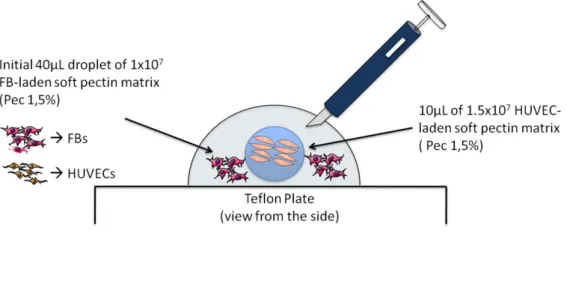
Figure 6. Schematic representation of the 3D HUVEC:FB co-culture spatial patterning
embedding process. ... 26
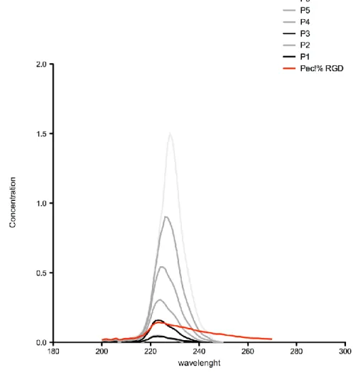
Figure 7. UV spectra of RGD-pectin, soluble RGD peptide and serial dilutions of RGD in a 1%
pectin solution ... 32
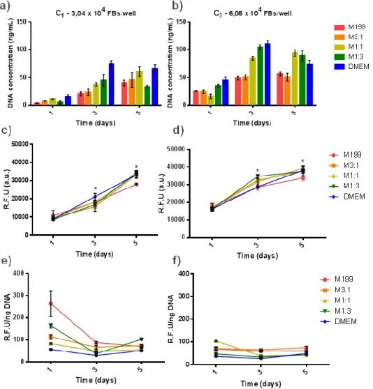
Figure 8. Effect of cell density and medium composition on metabolic activity and
proliferation of HUVECs in 2D during 5 days in culture a) and b) total dsDNA (PicoGreen assay), c) and d) metabolic activity (resazurin assay) and e) and f) metabolic activity per nanogram of dsDNA of HUVECs. * denotes statistically significant differences (p < 0.05). ... 34
Figure 9. Effect of cell density and medium composition on metabolic activity and
proliferation of FBs in 2D within a 5 days culture period. a) and b) total dsDNA (PicoGreen assay), c) and d) metabolic activity (resazurin assay) and e) and f) metabolic activity per nanogram of dsDNA of NHDFs. * denotes statistically significant differences (p < 0.05). ... 35
Figure 10. Effect of cell density and cell ratio on metabolic activity and proliferation of
HUVEC:FB co-culture in 2D within a 5 days culture period. a) and b) total dsDNA (PicoGreen assay), c) and d) metabolic activity (resazurin assay) and e) and f) metabolic activity per nanogram of dsDNA of HUVEC: * denotes statistically significant differences (p < 0.05). ... 37
Figure 11. Pictures of HUVEC:FB co-cultures prepared at 6.08x10⁴ cells/well at the different
ratios of 1:1, 2:1, 3:1 and 5:1. Images were obtained at the first and last day of 5-days. Scale bars, 200 µm... 38
Figure 12. Pictures of HUVEC:FB co-cultures prepared at 6.08x10⁴ cells/well at the different
ratios of 1:1, 2:1, 3:1 and 5:1. Images were obtained at the first and last day of a 5. Scale bars, 200 µm... 39
Figure 13. Effect of initial cell entrapment density and pectin concentration on metabolic
activity and proliferation of HUVEC in a 3D soft pectin hydrogel within a 6 days culture period. a) and b) total dsDNA (PicoGreen assay), c) and d) metabolic activity (resazurin assay) and e) and f) metabolic activity per nanogram of dsDNA of HUVEC * denotes
Figure 14. Effect of initial cell entrapping density and pectin concentration on the 3D
HUVECs’ spatial distribution within a 6 days culture period on a soft pectin hydrogel.
HUVECs were stained for F-actin (Green) and nuclei (Blue). Scale bars, 100 µm. ... 43
Figure 15. Effect of initial cell entrapping density and pectin concentration on the 3D
HUVECs’ conformation within a 6 days culture period on a soft pectin hydrogel. HUVECs were stained for F-actin (Green) and nuclei (Blue). Scale bars, 100 µm... 44
Figure 16. Effect of initial cell entrapping density and pectin concentration on metabolic
activity and proliferation of NHDFs in a 3D soft pectin hydrogel within a 6 days culture period. a) and b) total dsDNA (PicoGreen assay), c) and d) metabolic activity (resazurin assay) and e) and f) metabolic activity per nanogram of dsDNA of NHDFs * denotes
statistically significant differences (p < 0.05) . ... 45
Figure 17. Effect of the initial entrapping density on a 1.5% pectin 3D hydrogel on FBs’
spatial distribution within a 6 days culture period. FBs were stained for F-actin (Green) and nuclei (Blue). Scale bars, 100 µm. ... 46
Figure 18. Effect of the initial entrapping density on a 1.5% pectin 3D hydrogel on FBs’
conformation within a 6 days culture period. FBs were stained for F-actin (Green) and nuclei (Blue). Scale bars, 100 µm. ... 47
Figure 19. Effect of the initial entrapping density on a 2.5% pectin 3D hydrogel on FBs’
spatial distribution within a 6 days culture period. FBs were stained for F-actin (Green) and nuclei (Blue). Scale bars, 100 µm. ... 48
Figure 20. Effect of the initial entrapping density on a 2.5% pectin 3D hydrogel on FBs’
conformation within a 6 days culture period. FBs were stained for F-actin (Green) and nuclei (Blue). Scale bars, 100 µm. ... 49
Figure 21. Effect of Initial cell entrapping and pectin concentration over FBs spheroid size. a)
and b) spheroids average size throughout the 6 days of culture. c).and d) relative frequency of spheroid size at day 6. * denotes statistically significant differences (p < 0.05) ) between different entrapping densities on different pectin concentrations. ... 50
Figure 22. Effect of pectin concentration over the ability of FBs to contract the matrix.
Macroscopic differences of 1.5% and 2.5% pectin hydrogels seeded with 1x10⁷ FBs.mL-1. Images were obtained at the first and last day of a 6-days culture using an inverted microscope using a magnification of 16.3 xs. a) and b) correspond to 1.5% (w/v) pectin hydrogels at day 1 and 6 respectively. c) and d) correspond to 2.5% (w/v) pectin hydrogels at day 1 and 6 respectively. e) represents the relative size of the pectin matrices when
compared to day 1 ... 51
Figure 23. Effect medium composition on metabolic activity and proliferation of HUVECs and
FBs in a 3D pectin hydrogel within a 6 days culture period. a) total dsDNA (PicoGreen assay), b) metabolic activity (resazurin assay) and c) metabolic activity per nanogram of dsDNA. * denotes statistically significant differences (p < 0.05). ... 54
Figure 24. Co-culture of HUVECs and FBs in a ratio of 3:1 (HUVEC:FB) in a 3D pectin hydrogel
within a 6 days culture period. a) total dsDNA (PicoGreen assay), b) metabolic activity (resazurin assay) and c) metabolic activity per nanogram of dsDNA ... 55
Figure 25. Effect of cell type to type 3:1 ratio on HUVEC:FB Co-culture cell morphology and
spatial distribution for a 6 days culture period. Cells were stained against vWF (Red) and α-SMA (Gray), for F-actin (Green) and nuclei (Blue). Scale bars, 100 µm ... 56
Figure 26. Effect of cell type to type 1:3 ratio on HUVEC:FB co-culture cell morphology and
spatial distribution for a 6 days culture period. Cells were stained against vWF (Red) and α-SMA (Gray), for F-actin (Green) and nuclei (Blue). Scale bars, 100 µm ... 58
Figure 27. Effect of the initial seeding density on a 2.5% pectin 3D hydrogel on FBs’ spatial
distibution within a 6 days culture period. Pectin surface view. FBs were stained for F-actin with Alexa Fluor 488 phalloidin (Green) and nuclei were counterstained with DAPI (Blue). Scale bars, 100 µm. ... 98
List of Tables
Table 1. Effect of initial cell entrapping densities and pectin concentration over FBs
spheroid size and number. Area means and standard deviation is presented in micrometers. N stands for total number of spheroids. * denotes statistically significant differences (p < 0.05) between the same entrapping density on different pectin concentrations. α denotes statistically significant differences (p < 0.05) between different entrapping densities on different pectin concentrations. ... 51
Table 2. Effect of pectin concentration over the ability of FBs to contract the matrix.
Macroscopic differences of 1.5% and 2.5% pectin hydrogels entrapped with 1x10⁷FB.mL-1. Areas means and standard deviation is measured in milimeters ... 52
List of Abbreviations
2D – Two-dimensional3D – Three-dimensional
bFGF - Basic fibroblast growth factor Ca2+ - Calcium ions
CaCO3 – Calcium carbonate
CD31 - Cluster of differentiation 31 DAPI - 4',6-diamidino-2-phenylindole DM - Degree of methylation
DMEM - Dulbecco’s Modified Eagle Medium DNA – Deoxyribonucleic acid
dsDNA – Double-stranded Deoxyribonucleic acid ECGS - Endothelial cell growth supplement ECM - Extracellular matrix
ECs - Endothelial cells
EDC - (N-(3-dimethylaminopropyl)-N’-ethylcarbodiimide) EDTA - Ethylenediaminetetraacetic acid
Em - Emission Ex - Exitation
FBS - Fetal bovine serum FBs - Fibroblasts
FN - Fibronectin
G4RGDSP - (Glycine)4-Arginine-Glycine-Aspartic acid-Serine-Proline
GAGs - Glycosaminoglycans
GalA - (1–4)-linked-α-D-galacturonic acid GDL- D-glucono-d-lactone
NHDFs - Neonatal human dermal fibroblasts HGA - Homogalacturonan
HM - High methoxyl
HUVECs - Human umbilical vein endothelial cells LM - Low methoxyl
MES - 2-(N-morpholino) ethanesulfonic acid buffer PBS - Phosphate-buffered saline
PCL - Poly(e-carpolactone) PEG - Poly(ethylene glycol)
Pen - Penicillin
PFA – Paraformaldehyde PGA - Poly(glycolic acid)
pHEMA - Poly(2-hydroxyethyl methacrylate) PLA - Poly(lactic acid)
PLGA - Poly(lactic-co-glycolic acid) PMMA - Poly(methyl methacrylate)
PVGLIG - Proline-valine-glycine-leucine-isoleucine-glycine RFUs - Relative fluorescence units
RGD - Arginine-Glycine-Aspartic acid RG-I - Rhamnogalacturonan-I RG-II - Rhamnogalacturonan-II RPM – Rotations per minute RT – Room temperature Strep - Streptomycin sulfo-NHS - N-hydroxy-sulfosuccinimide TBS - Tris-buffered saline TE – Tris-EDTA buffer UV –Ultraviolet
VE-cadherin (CD144) – Vascular endothelial cadherin VEGF - Vascular endothelial growth factor
VSMCs - Vascular smooth muscle cells vWF - von Willebrand factor
1. Introduction
1.1. Skin
Skin is the largest organ of the human body, representing roughly one tenth of the body mass (Metcalfe & Ferguson 2006; Groeber et al., 2011) performing very important functions besides its obvious aesthetical function. Skin performs several functions: acts as a protective barrier, preventing dehydration, limiting organism invasion by potentially noxious agents (e.g. toxins, virus, UV radiation) also by impermeabilizing the body, helps in the thermoregulation of the body, works as a cushion, among others (Metcalfe & Ferguson 2007; Yildirimer et al., 2012; Pereira et al., 2013).
Figure 1. A schematic of the structure of skin. Image from Naturally Healthy Skin (http://www.naturallyhealthyskin.org/anatomy-of-the-skin/the-dermis/dermis-anatomy-of-the-skin/
The skin is composed of three layers: epidermis, dermis and hypodermis (Figure 1) (hypodermis (Groeber et al., 2011; Pereira et al., 2013). The epidermis is thin and totally cellular, mainly composed of keratinocytes but also containing other cell types, such as Langerhans cells and melanocytes. Due to the constant exposure, homeostasis is achieved by constant substitution of the environment-exposed cells by cell migration from the basal layers, which, in turn, are composed of epidermal stem cells able of self-renewal and repair (Alonso et al., 2003; Chunmeng & Tianmin, 2004; Metcalfe & Ferguson 2007; Pereira et al., 2013). In addition, the skin appendages (e.g hair, nails, sweat glands and sebaceous glands) are derived from and linked to the
epidermal layer presenting however deep projections into the dermal layer (Martin, 1997). Situated directly below the epidermis is the dermis. This layer constitutes the bulk of the skin, providing support and nourishment. It contains vascularized extracellular matrix (ECM) rich in collagen, elastin and glycosaminoglycans (GAGs), being responsible for the elasticity and mechanical integrity (Jones et al., 2002; Metcalfe & Ferguson 2007; Groeber et al., 2011). These properties are modulated by fibroblasts, the main cell type in the dermal layer and the the main source of ECM (Berthod et al., 2006). Furthermore, fibroblasts also produce remodeling enzymes, such as proteases and collagenases, playing an important role in wound healing (Ratner et al., 2004). Present in this layer, but in lesser amounts, are also endothelial cells and smooth muscle cells, composing a vascular system, mast cells, which are part of the immune system being responsible for the early recognition of pathogens and cutaneous sensory nerves that pass through dermis into the epidermal layer (Metcalfe & Ferguson 2007; Urb & Sheppard 2012; Pereira et al., 2013). The third layer, the hypodermis, is a well vascularized area mostly composed of adipose tissue, contributing for the mechanical and thermoregulatory properties of the skin as well as acting as an energy source (Metcalfe & Ferguson 2007; Yildirimer et al., 2012; Pereira et al., 2013).
1.1.1 Skin lesions and regenerative medicine
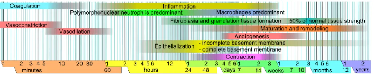
Skin lesions, whether caused by physical/chemical factors (e.g. burns, lacerations, ulcers, acute wounds, surgery, among others) or by chronicle diseases are fairly common (Martin, 1997; Groeber et al., 2011). Upon injury that leads to the disruption of the structure and function of natural tissue, under certain physiological circumstances, skin displays a complex and continuous natural process, overlapping events of hemostasis, inflammation, migration, proliferation and differentiation. These occur due to a constant environmental change that exposes cells to complex molecular patterns which sets off a series of metabolic cascades, propelling the wound through the phases of healing, overlapping events of hemostasis, inflammation, migration, proliferation and differentiation (Figure 2) (Mutsaers et al., 1997; Martin, 1997; Guo & DiPietro, 2010; Häggström et al., 2010).
Figure 2. Chronological representation of the phases of wound healing. Adapted from Häggström et al., 2010.
However, depending on the lesion extent, wound environmental exposure can pose a high infection risk that can lead to deeper skin damage, tissue necrosis or, ultimately, death. As such, skin lesions must be treated as a critical issue in healthcare (Zöller et al., 2014). In current medical treatments, clinical strategies rely on the use of closure materials that may act solely as a barrier while natural wound healing occurs or actively contribute for the restoration of the epidermal function while becoming incorporated into the healing wound. Nowadays, it is possible to find several solutions for skin wound treatment (Guo & DiPietro, 2010). Depending on the wound type, depth, extension and the patient, several strategies can be applied. For superficial lesions (mainly affecting the epidermis), creams and ointments are used for disinfection, cleaning, debridement or to help the wound healing process. Although still used, due to their properties their limited permanency in the human body, these solutions have been substituted for more advanced strategies (Boateng et al., 2008). Wound dressings have been widely used due to their low cost and effectiveness. This medical strategy consists in the application of natural or synthetic material over the wound protecting it from the environment. Traditional wound dressings (e.g. bandages, cotton wool, lint and gauzes), covered the wound, keeping the wound dry and preventing the entry of pathogens into the wound (Boateng et al., 2008; Pereira et al., 2013).
Nowadays, accompanying the evolution in the science and technological fields, wound dressings present more advanced solutions for wound healing. Obtained from natural or synthetic sources, modern wound dressings are available as films, foams or gels (Boateng et al., 2008). Based on the concept of creating an optimal environment, which includes an exudation control allowing a moist, non-detrimental environment, effective oxygen circulation aiding the regeneration process, good adhesion to the lesion surface and low pathogen penetration, while minimizing maceration and scar formation, (Stephen-Haynes et al., 2014), several modern wound dressings were developed, as reviewed by Pereira et al. (2013). Moreover, some dressings can even act as drug delivery systems, incorporating the therapeutic agent releasing it in the wound bed (Elsner & Zilberman, 2010; Pereira et al., 2013; Boateng et al., 2015; Momoh et al., 2015). However, due to the complexity of the healing process and the wide variety of
skin wounds existent, no single dress is able to fulfill the requirements for full skin recovery. Notwithstanding the importance of the referred methods for skin regeneration therapies, in cases of severe lesions in the dermis or hypodermis, a complex treatment is required. At the present day, autografts, surgical reconstruction using the patient own skin, are the ―gold standard‖ procedure (Goldberg, 1992; MacNeil, 2007). This strategy however presents limitations depending on the lesion extension and due to the creation of additional surgical sites (Goldberg, 1992). Another solution is the use of allografts, surgical reconstruction using another patient skin. This, however, can pose complication at both ethical and medical levels, as another patient is exposed to a risk situation while also subjecting the wounded patient to a graft that can potentially carry a disease or suffer immunological rejection (Goldberg, 1992).
A potential solution to this problem is to approach this from a tissue engineering–based standpoint for de novo organogenesis, using biomaterial scaffolds and a person’s own cells to grow or fabricate skin substitutes (Cuono et al., 1986; Zöller et al., 2014). To date, there are several clinically available skin substitutes, with these being divided into epidermal, dermal, and dermo-epidermal tissue-engineered constructs. As mimicking the extracellular matrix (ECM) structural integrity and function is of key importance, several strategies are revolving around collagen-based matrices (Boyd et al., 2007; Johnen et al., 2008; Cen et al., 2008). Other skin substitute biomaterials used as matrices are chitosan (Mao et al., 2003; Mohd et al., 2013), hyaluronic acid (Park et al., 2004; Wang et al., 2006), among others. Despite recent developments wound healing, the techniques and biomaterials available present significant limitation for skin regeneration. To our knowledge, at the present time, there are no models of skin substitutes that fulfill all the criteria, replicating the anatomical and physiological requirements for biological stability at epidermal and/or dermal. Additionally, available skin substitutes suffer from poor integration, scarring and lack of differentiated structures (e.g. hair and sebaceous glands), contrasting with the aesthetics of uninjured skin (Boateng et al., 2008). Advanced skin regeneration therapies already combine biomaterials, cells, growth factors and advanced biomanufacturing techniques for the fabrication of constructs that mimic skin anatomy. Recently, several methods have been developed to spatially encode local properties to 3D materials-based culture systems. These biofabrication techniques are capable of constructing micropatterned materials, with a high degree of control, by finely tuning and defining material geometries, localization of biomolecular cues, and other mechanical properties, enabling a precise control over the bulk material properties (Nichol & Khademhosseini, 2009; Nikkhah et al., 2012; Pataky et al., 2012; Culver et al., 2012). These are designated bottom up approaches and consist on the formulation of tissue building
blocks with specific microarchitectural features for modular assembly, in an attempt to replicate the heterogeneous nature of endogenous tissues and organs. Another used approach to is to use tissue engineering strategies typically that employs a ―top-down‖ These consist on seeding cells into biomaterial matrices capable of recreating biomimetic structures, exploiting the innate abilities of cells to sense their local environment through cell–cell and cell–extracellular matrix (ECM), self-assembling into complex networks (Dean et al., 2007; Seidlits et al., 2011; Maia et al., 2014). This strategy relies on the ability of the cells to reconstruct the intricate microarchitectural and functional features of natural microenvironments to achieve the desired biological effect. However, for a given skin substitute to attach promptly, a vascularized wound bed is required. Deep wounds that affect the dermal layer constitute a problem. If the skin substitute surpasses a certain thickness nutrient diffusion is limited and the vascularization process is too slow, resulting in necrosis and graft loss. As such, any tissue-engineering constructs that aims to mimic natural tissues and, ultimately, organs, must ideally conjugate all the key components – cells, extracellular matrix (ECM), and vasculature – in precise geometries (Auger et al., 2013; Battiston et al, 2014).
1.2. Vascularization
From the various obstacles for tissue engineered skin substitutes, the inability of the grafts to acquire proper vascularization has been proposed as the most likely reason for deleterious effect on epidermal survival human tissue-engineered skin constructs. The inability to properly assemble a vascular structure within the graft, leads to necrosis at the tissue core, and poor survival due to ischemic injury (Rivron et al., 2008; Auger et al., 2013). Regardless of the specific tissue-engineering approach to create artificial skin any construct that involves living cells needs to fulfill the conditions in which cells are able survive and redeem their biological functions. Reconstructed tissues need to be able to access to oxygen and nutrients, as well as elimination of carbon dioxide and other cellular waste products (Folkman & Hochberg, 1973; Novosel et al., 2011; Auger et al., 2013). It is, therefore, paramount, for the successful transplantation of human tissue-engineered constructs, the formation of a vascular network. At both the stage of in vitro growth and assemble and after the patient implantation of the graft (Rivron et al., 2008; Novosel et al., 2011; Auger et al., 2013).
1.2.1 Endothelial cells
Blood vessels are a multi-cellular system composed of vascular smooth muscle cells (VSMCs), fibroblasts and endothelial cells (ECs) (Ratner et al., 2004). Vascular networks, ranging from large sized vessels, as are arteries and veins, to the micro-sized vasculature networks formed within organs, are lined with a single layer of endothelial cells (ECs), on which one part of the surface defines the lumen while the other is in contact with a highly specialized EC, the basement membrane. In order to sustain their tubular architecture and allow a contractile behavior in these structures, ECs are enveloped by mural cells (e.g. pericytes, VSMCs). EC formation occurs mainly through mesodermal precursor’s differentiation of hemangioblasts and/or angioblasts, a critical process in embryogenesis and tumor formation (Augustin et al., 1994; Mani et al., 2008). These cells form a barrier that, due to their capacity of extravasation and high surface-to-volume ratio are capable of actively transport small molecules, macromolecules and hormones, while also performing multiple functions depending on the location and size of the blood vessel that they are lining (Ruoslahti & Rajotte, 2000, Bouis et al., 2001; Pinkney et al., 1997). As such, ECs play an important role mediating many physiological functions such as hemostasis maintenance, vasomotor tone, blood cell trafficking, permeability, proliferation, survival, and innate and adaptive immunity (Aird, 2007).
There are two processes from which neovascularization can take place: angiogenesis, a process through which new blood vessels are formed from preexisting ones, and vasculogenesis, the generation of a new vascular network from endothelial progenitor cells (EPCs) in the absence of preexisting blood vessels (Luttun et al., 2002). These capillary generation events involve a complex sequence of events, which cell adhesion, migration, alignment, protease secretion, and tubule formation. Throughout these, ECs must be exposed to growth factors interaction and mechanical cues as well as cell-cell and cell-EMC interactions all of which must be precisely timed and with the correct concentrations (Yamamoto et al., 2003; Lokmic et al., 2008; Arnaoutova et al., 2009). ECs can be isolated from different endothelium. With proper specific medium supplementation, several ECs population like human umbilical vein endothelial cells (HUVECs) or human dermal microvascular endothelial cells (HDMECs) can be isolated and cultured in vitro. Although they possess several common characteristics like cell-cell contact inhibition when confluent, similar morphology and identical expression of cellular markers, choosing the source of ECs is of critical issue. Due to the endothelium
heterogeneity, site specific properties of the ECs could be translated in vitro, originating different outcomes when exposed to the same factors. Throughout the years, the phenotypic heterogeneity of the endothelium has been characterized and described recurring lectin staining, immunohistochemistry, in situ hybridization and real-time intravital microscopy, being, nowadays possible to select the most appropriate EC type for each design (Boius, 2001; Aird, 2003; Aird, 2012).
Among these, human umbilical vein endothelial cells (HUVECs) have been of critical importance, largely contributing for scientific knowledge breakthroughs in molecular medicine providing insights over ECs embryogenesis, angiogenesis, vasculogenesis and pathology, at both cellular and molecular levels (Nakatsu et al., 2003; Poliseno et al., 2006; Anand et al., 2010). HUVECs are easily available, free from any pathological process and they are physiologically more relevant than many established cell lines (Cooper & Sefton, 2011).Initial passages of these cells, maintain nearly all of the features of native vascular endothelial cells expressing several endothelial cell specific markers such as: von Willebrand factor a large adhesive glycoprotein that, in the blood, serves as a stabilizing factor for Factor VIII (Zanetta et al., 2000); platelet endothelial cell adhesion molecule-1 (PECAM or CD31), an endothelial specific adhesion molecule (Goldberger et al., 1994); VE-cadherin (CD144), a cadherin expressed in the tight junctions (Esser et al., 1998); and specific signaling pathways receptors markers for vascular endothelial growth factor (VEGF) and fibroblast growth factor (FGF) (Esser et al., 1998; Salcedo et al., 1999). HUVECs have an average life span of 10 serial passages, time after which the cells enter senescence, tending to stop proliferation, form giant multicellular aggregates and dye (Jaffe et al.,1973). Although recovered from a major vessel, HUVECs have been proven capable of forming microvascular structures (Kenneth et al., 2006; Sorrell et al., 2007; Zheng et al., 2012). All summed up, HUVECs 3D culture presents itself as a promising strategy for in vitro microvasculature formation and characterization.
To recover functional endothelial cell self-assembled into microvascular structures could presents itself as a major advance in biomanufacturing techniques forthcoming the construction of functional grafts for patient transplantation.
1.2.2 Vascularization strategies
New vessel formation is essential for wound healing. As such, to culture cells under 3D conditions using a material that can mimic the ECM, and recapitulate some key aspects of the native cellular microenvironment is paramount. Although several 2D
strategies were conducted, in 1983, Montesano et al. (1983) evidenced the importance of culture ECs in a three dimensional environment. Bidimensional environments fail to mimic several cues necessary for the creation of a specific cellular organization. Nowadays, 3D cell cultures stand as essential models for the study of cell biology, as well as support matrices that can incorporate mechanical and biochemical stimuli directly conveyed by the ECM. As such, in vitro 3D microvascularization is highly dependent on the composition and properties of biomaterial matrix along with the presence of precisely timed delivery of angiogenic growth factors (Montesano et al., 1983; Nakatsu et al., 2003; Sieminski et al., 2004; Ghajar et al., 2008), being necessary for any attempt that intends to mimic this process, a fine tune of the conditions to which ECs will be exposed. Since the perception that angiogenesis could be achieved, several in vitro (Folkman & Haudenschild, 1980), several studies attempted to mimic the natural conditions necessary for this process to occur. Although some single component matrices (e.g. collagen, Matrigel and fibrin), when coupled with specialized growth factors, were able to support tube formation (Montesano et al., 1983; Montesano et al., 1986; Chalupowicz et al., 1995; Bach et al., 1998; Dai et al., 2004; Kleinman & Martin, 2005), attempts to monoculturing ECs on biomaterial matrices for microvasculature formation has not been an effective strategy. In monocultures, ECs seem unable to survive and proliferate and, subsequently, self- assembly into tube-like structures is not archived (Janvier et al., 1997). In addition, it is important, that newly formed structures mature and form stable structures. This implies that the interconnected capillary structures are self-sustained after the initial conditions are not present, which is hard to achieve in monoculture as capillary structural sustainability is dependent on the formation of highly specific bonds between ECs and ECM as well as the envelopment of these cells by mural cells (Ribatti et al., 2011). Although during angiogenesis ECs migrate and make sprouts without mural cells’ perivascular cells (PCs) are among the first cells responsible for the invasion of newly vascularized tissues, determining the location of sprout formation and guiding newly formed vessels by interaction with EC via paracrine communication (Ribatti et al., 2011).
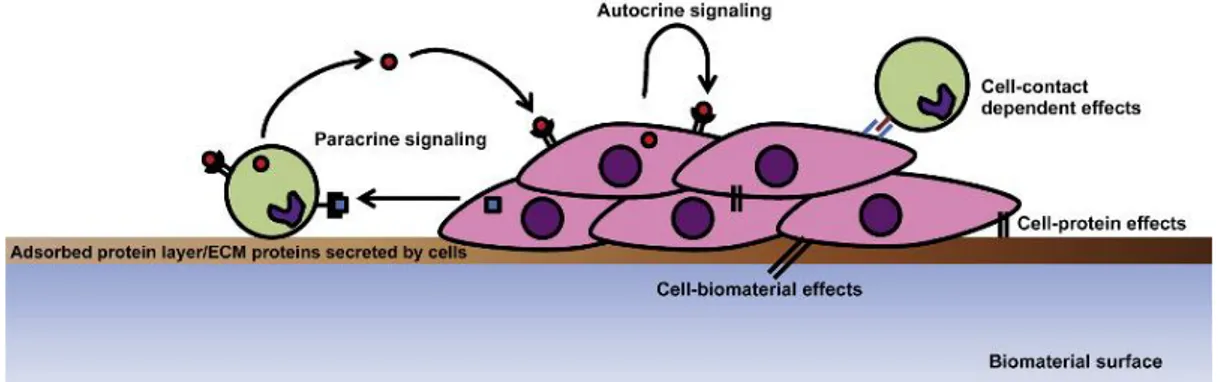
Figure 3. Schematic representation of the dynamics of a co-culture system. Adapted from Battiston et al., 2014 To reproduce this complexity, co-culture systems can be developed to mimic the natural conditions. These involve the culture of two or more types of cells within the same matrix (Battiston et al., 2014). This strategy takes advantage of both the natural cell-ECM interaction and the natural crosstalk between cells, through soluble factors and/or cell-cell interaction and cell-cell contact (Figure 3) (Seghezzi et al., 1998; Grinnel et al., 2000; Saito et al., 2005; Wenger et al., 2005). Co-culture systems are often used with the intent of using one cell type to provide a desired stimulus to a second cell type, presenting a natural, cost-effective strategy for tissue regeneration. This strategy as proven itself effective for ECs tube-like structures formation, as coculturing ECs with fibroblasts (Wenger et al., 2005; Sorrell et al., 2005; Li et al., 2013; Guerreiro et al., 2014; Costa-Almeida et al, 2015), osteoblasts (Hoffman et al., 2008; Grellier et al., 2009; Ghanaati et al., 2011), mesenchymal cells (Wu et al., 2007; Kolbe et al., 2011) and smooth muscle cells (Melero-Martin et al., 2007; Foubert et al., 2008) provides the necessary stimulation for increased ECs survival, proliferation and capillary-like structures assembly that resemble the normal ECs alignment. Human fibroblasts are abundant in the dermis, being the main source of ECM components (e.g. collagen, fibronectin and proteoglycans) and, therefore, modulating mechanical extracellular microenvironment which is critical for vasculogenesis (Berthod et al., 2006). Furthermore, these cells are strongly related to angiogenesis as they infer over the EC behavior through fibroblast-derived proteins (e.g. fibroblast growth factor-2 (FGF-2) and vascular endothelial growth factor (VEGF), the latter a key modulator of normal vessel generation (Seghezzi et al., 1998; Saito et al., 2005), cell-cell dynamics (Wenger et al., 2005) and mechanical extracellular microenvironment contraction (Grinnel et al., 2000), all of which are necessary to modulate EC sprouting and the expansion of capillary-like network (Neufeld et al., 1999; Velazquez et al., 2002; Yamamoto et al., 2003).
However, to build a co-culture system, the physico-chemical properties must be carefully considered as biomaterial will serve as support in the initial stages of the
culture. Matrix dimensionality plays a key role in cell signaling event, affecting, in particular, the way cells experience mechanical stresses and strains (Cukierman et al., 2001; Cukierman et al., 2002; Reilly et al., 2010.), which was proven to have a direct effect on cells’ self-patterning (Sieminski et al., 2004; Palama et al., 2012). To engineer a functional tissue, compliant hydrogel matrices with a storage modulus, G’ inferior to 1000 Pa (hereafter designated as soft matrices), facilitate different cellular activities, including spreading, proliferation and migration (Bott et al., 2010; Ehrbar et al., 2011; Maia et al., 2014). As Reinhart-King et al. (2011) described endothelial cells communicate through mechanical signals in a stiffness-dependent manner, reacting to strains created by the traction stresses of neighboring cells. In addition, Bott et al. (2010) demonstrated that softer hydrogels matrixes increase fibroblasts spreading and proliferation.
All together, decreasing the substrate stiffness and, therefore, creating a more compliant matrix, while coculturing EC with fibroblasts, presents itself as a promising strategy for the self-assembly of endothelial cells into network-like structures.
1.3. Extracellular matrix
Multicellular organisms are governed by cohesion mechanisms. Among these mechanisms, the matrix adhesiveness is known to be a potent modulator of the architecture and organization of the tissue, playing a key role in cell survival, proliferation, migration and differentiation (Wang et al., 2010; Bowers et al., 2010). The extracellular matrix (ECM) consists in network of proteins and proteoglycans secreted locally and assembled into an organized meshwork. Among the macromolecules that compose the ECM, special attention is given to glycosaminoglycans (GAGs), negatively charged unbranched polysaccharide chains composed of repeating disaccharide units, collagens, which are fibrous proteins, and fibronectin (FN), a glycoprotein (Labat-Robert et al., 1990; Bowers et al., 2010). Different types of collagen provide unique properties the ECM, modeling tensile strength and fibril formation. As such, alterations in the biochemical composition of collagens impose different mechanical properties to the microenvironment (Daley et al, 2008). On the other hand, FN plays crucial role in cell-matrix interactions, serving as a substrate for different adhesion molecules, namely integrins (Romer et al., 2006; Daley et al, 2008). More precisely, FN has been shown to interact with αvβ3 through a small sequence of amino acids, Arginine-Glycine-Aspartate or RGD, mediating cell survival, migration and invasion (Stupack et al., 2003; Yu et al., 2009). This ECM-integrin interaction plays a
key role in cellular fate, providing not only anchorage, but also information concerning their microenvironment (Stupack et al., 2003). Variations in the relative amounts of these macromolecules, coupled with modifications in their organization, provide different patterns of cell adhesion to matrix and growth behavior, leading to in situ specific cellular response (Discher et al., 2005; Engler et al., 2006; Li et al., 2010). It is therefore imperative for progress in developmental biology, regenerative medicine, and tissue engineering to provide to the cells the matrix cues necessary for a driven response to the desired effect.
1.3.1 Hydrogels
Biomaterials play a critical role in tissue engineering as they can modulate cell response via different material properties such as surface chemistry and topography, spatial patterning, roughness, mechanical compliance, porosity, isotropy, surface wettability, among others (Ratner et al., 1996; Battiston et al., 2014). Cell-biomaterial interactions affect cell-cell interactions in 3D culture systems, promoting unique behaviors upon interaction with different biomaterials. An ideal biomaterial should able to mimic functionality and complexity of native tissues, providing biospecific cellular adhesion and the subsequent control of cellular functions. Three-dimensional (3D) hydrogels matrixes offer an exciting possibility, capturing many important features of the ECM (Pereira et al., 2013; Drury et al., 2003). Hydrogel matrices are water-swollen crosslinked polymeric networks. These provide a highly hydrated and mechanically compliant environment, permeable to oxygen, nutrients, wastes and water-soluble metabolites (Tibbitt et al., 2009). The microenvironment profile, however, is not only dependent on the biomaterial’s properties. By altering the crosslinking reaction scheme, which can be achieved by physical or chemical methods, the gelation reaction kinetics can be tuned and the subsequent hydrogels properties, altered (Yu & Ding, 2008; Neves et al., 2015). Moreover, hydrogels can often be formed under mild conditions, creating the adequate conditions for cytocompatible cell entrapment (Drury et al., 2003). Their delivery can be performed in a minimally invasive manner as several hydrogel matrices can be prepared from soluble precursor’s solutions that crosslink in situ (Hall, 2007). As such, hydrogels have been proposed for a myriad functions in the field of tissue engineering, ranging from as space cling agents (Yao & Swords, 2001; Drury & Mooney, 2003; Koran et al., 2007), drug/bioactive molecule delivery (Ribera et al., 2004; Green et al., 2006; Qiu & Kinam, 2012), cell/tissue delivery vehicles (Bidarra et al, 2011; Munarin et al., 2012; Fonseca et al., 2013;
Bidarra et al., 2014) and 3D cellular microenvironments (Seidlits et al., 2011; Fonseca et al., 2011; Neves et al., 2015).
As previously described, hydrogels can be adjusted to fit the demands of each construct. By tuning the biochemical and viscoelastic profile of the hydrogels, it is possible to effectively modulate the process of mechanosensing, promoting, for example, the proliferation and spreading of fibroblasts and favoring endothelial cells network assembly and tubulogenesis (Grinnell & Petroll, 2010; Bott et al., 2010; Bidarra et al., 2011). Naturally derived polymers include components of the extracellular matrix (e.g. collagen, fibronectin, and fibrinogen) or present a chemical structure similar to natural glycosaminoglycans (GAGs) (e.g. alginate, hyaluronic acid, chitosan). Due to this, natural polymers present intrinsic advantages over synthetic ones (e.g. Poly(ethylene glycol) (PEG), Poly(glycolic acid) (PGA), Poly(lactic acid) (PLA); Poly(lactic-co-glycolic acid) (PLGA), Poly(methyl methacrylate) (PMMA), Poly(e-carpolactone) (PCL) (Hoffman, 2012; DeVolder & Kong, 2012). Although some contain cellular binding domains due to their derivation from natural sources, thus allowing cell adhesion, others constitute permissive hydrogels. Notwithstanding that the latter provides a 3D environment for cell culturing, it lacks the ability to promote the specific cell-matrix interactions necessary for cell adhesion and the subsequent physiologic events of anchorage-dependent cells (Munarin et al, 2011). This occurs due to the presence of negatively charged carboxyl groups. To overcome this problem non-adhesive hydrogels can be modified to have a bioactive role by grafting a small oligopeptide sequence that is known to be present in FN, namely, RGD (Stupack et al., 2003; Yu et al., 2009). Incorporating this cell-adhesive peptide (RGD) into the non-adhesive polymer has been shown to significantly improve cell adhesion, growth and differentiation (Rowley & Mooney, 1999; Rowley et al., 2002; Grellier et al., 2009; Bidarra et al., 2011). Furthermore, hydrogels can also be modified with protease-sensitive peptides (e.g. PVGLIG). This allows the matrixes to mimic two key features of the natural ECM: cell-matrix adhesion and cell-driven matrix proteolytic degradation (Raeber et al., 2005; Fonseca et al., 2011).
Three dimensional matrices for cell culture are no longer thought only a structural support to maintain tissue and organ configuration. Nowadays, it is widely accepted that the highly dynamic interactions between cells and the ECM are of key importance in the cellular fate (Berrier & Yamada, 2007). As such, the success of matrices in these roles hinges on finding an appropriate material to address the variables inherent to the desired application. Different biomaterials should be explored to develop new approaches for tissue regeneration therapies, thus providing an insight on the best possible design for each situation.
1.3.1.1.
Pectin
A multitude of natural biomaterials has been explored to form hydrogels. Natural polymers possess highly organized structure, being are frequently used in tissue engineering applications as they are either components of or have macromolecular properties similar to the natural ECM (Drury et al., 2003). Due to their tunable characteristics, by grafting of the desired peptides into the biopolymer structure or by controlling their viscoelastic profile through the control of the gelation kinetics (e.g. variations in pH, gelation time, and crosslinking divalent cation), specific tissue engineering matrices can be constructed. Among these, pectin, a complex structural polysaccharide present in the cell walls of higher plants, stand out as an attractive cell carrier. Pectin is a biocompatible anionic polysaccharide that constitutes 30% of the cell wall of plants (Harholt et al., 2010) widely used as thickener, gelling agent, stabilizer, and emulsifier in several food products (Tho et al., 2003). As depicted by Munarin et al. (2012), pectin is mainly extracted from waste products of juice, apples and cider industries through chemical or enzymatic methods. Due to the number of sources and extraction processes that pectin can be obtained from, a wide range of pectin degrees of esterification can be obtained. As such, each batch must be thoroughly characterized for an adequate microenvironment construction and results interpretation (Munarin et al., 1012). Furthermore, the interest in pectin as spread into the pharmaceutical and medical fields (Maxwell et al., 2012) as it has been reported to have multiple positive effects on human health, including lowering cholesterol and serum glucose levels (Mohnen et al., 2008) reducing cancer (Jackson et al., 2007) and stimulating the immune response (Inngjerdingen et al., 2007).
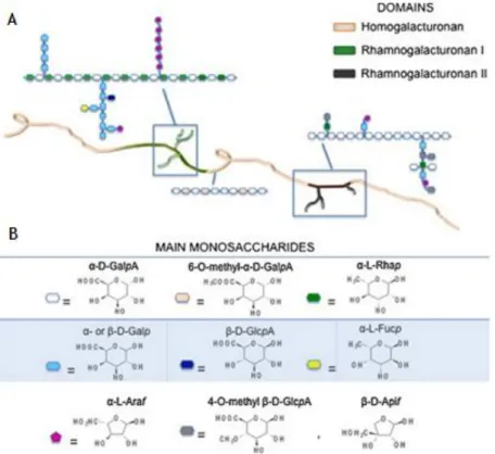
Figure 4. Representation of pectin structure. Adapted from Munarin et al., 2012
Pectin is composed of at least three polysaccharide domains: homogalacturonan (HGA), which is the major component, rhamnogalacturonan-I (RG-I) and rhamnogalacturonan-II (RG-II) (Jarvis, 1984; Mohnen, 2008; Yapo, 2011), forming a branched macromolecule with high molecular weight. The current model proposed, consists of a linear backbone of unbranched HGA residues (―smooth region‖) alternately linked to branched RG-I residues (―smooth region-hair region‖) (Figure 4). HGA, the major component of pectin polysaccharides (~65%) (Mohen et al., 2008) ,is mainly composed of a homopolymer of (1–4)-linked-α-D-galacturonic acid (GalA) units (Ridley et al., 2001). These units can be partially methyl-esterified on the carboxyl group and sometimes partially acetyl-esterified on the secondary hydroxyls. Based on the ratio of methyl-esterified residues (6-O-methyl-α-D-GalA) HGA backbone to the total carboxylic acid units in their salt form, which defines the degree of methylation (DM), pectins can be classified into two categories: low methoxyl pectins (LM, DE < 50%) or high methoxyl pectins (HM, DE > 50%) (Durand, 1990). These methylation
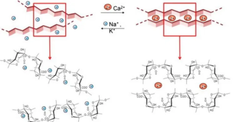
Figure 5. Schematic representation of an ―egg box‖ structure formation in the presence of Ca2+. Adapted from Coimbra et al., 2011
differences provide different properties to the pectin, significantly affecting the properties of the formed gels.
LM pectins, in the presence of strong, positive, divalent metal ions, such as Ca2+ ions, establish strong bonds between the carboxyl groups of the HGA pectin backbone leading to the formation of an ―egg box‖ structure. This mechanism involves side-by-side associations of specific sequences of GalA monomer in parallel or adjacent chains linked through electrostatic and ionic bonding of carboxyl groups using the divalent ions, forming a flexible network of polymer chains that can swell but does not dissolve in water (Pérez et al., 2001; Fang et al., 2008) (Figure 5). Furthermore, van der Waals interactions and hydrogen bonds are established within the polymer, stabilizing the egg-boxes formed between neighbored chains (Braccini et al., 1999; Fraeye et al., 2010).The promising aspects of pectin gels for biomedical applications, namely, its easily tunable physical properties, high water content and ability to homogeneously immobilize cells, genes, proteins, drugs or growth factors (Munarin et al., 2010 a; Munarin et al., 2010 b; Munarin et al., 2011; Munarin et al., 2012; Neves e al., 2015), led to a renewed interest on this polymer. Moreover, this biopolymer’s solubility can be controlled by quickly displacing the Ca2+ ions by monovalent counterions such as Na+ or K+ (Munarin et al., 2011). Pectin fulfils all of the requirements for hydrogel formation, presenting itself as particularly appealing biomimetic systems providing an adequate microenvironment by simulating the ECM-cell dynamics. Nonetheless, as other natural polysaccharides (e.g. alginate), due to the presence of negatively charged carboxyl groups, pectin presents a hydrophobic nature, resisting to protein adsorption and cell adhesion. To bypass this issue, RGD-containing oligopeptides must be grafted into the pectin backbone, granting the minimal peptide sequence required for the adhesion of integrins to the ECM components (Pierschbacher & Ruoslahti, 1987; Ruoslahti & Pierschbacher, 1987; Yamada, 1997; Giancotti & Ruoslahti, 1999). As for other polymers (e.g. alginate (Bidarra et al, 2011; Fonseca et al., 2011; Fonseca et al., 2013; Bidarra et al., 2014)) RGD-containing
pectin gels present a higher cytocompatibility, cell adhesion and proliferation, improving al the subsequent cellular functions (Munarin et al., 2011; Munarin et al., 2012; Neves e al., 2015). Furthermore, in addition to the structural resemblance between pectin and alginate, allowing into present the same numerous benefits of alginate, pectin stands out as it presents an interesting degradation profile under simulated physiological conditions (Munarin et al., 2012). Finally, more recently, our group explored the potential of the pectin hydrogels crosslinking by internal ionotropic gelation using the slow-gelling calcium carbonate/D-glucono-d-lactone (CaCO3/GDL) system. Neves e al.,
(2015) addressed, for the first time, the use of in situ-forming pectin hydrogels as skin cell carriers for tissue engineering, providing an ionotropic internal gelation scheme suitable for in situ gelling systems.
Although much still remains to be elucidated about this polymer as a biomaterial, the studies found about the easy tunability of this biomaterial for tissue regeneration (Morra et al., 2004; Bussy et al., 2008; Nagel et al., 2008; Munarin et al., 2011; Munarin et al., 2012; Neves et al., 2015), evidence the promising capabilities of pectin hydrogels as a powerful material system for cell delivery, tissue engineering and regenerative medicine applications.
1.4. Main Goals
The incorporation of microvascular networks within the in vitro tissue-engineered skin before its transplantation into a patient would be a major contribution, surpassing the need of relying only on the host’s system ability to promote vascularization. Although encouraging developments have been made in the field (Rivron et al., 2008; Place et al., 2009), in vitro vascularization remains a challenge. In this work, we intend to use a combined approach using the tunable characteristics of soft pectin hydrogel, cells and growth factors to mimic the natural mechanisms involved in the formation of a microvascular network. We aim to construct a three-dimensional, RGD-grafted, soft pectin hydrogel in which fibroblasts support endothelial cells in the formation of self-assembled vascular structures for skin regeneration therapies, while also providing new insights on the biomimetic properties of soft pectin hydrogel’s for future tissue-engineering strategies.
2. Materials and Methods
2.1. Cell Culture
2.1.1 Routine maintenance
Commercial human umbilical vein endothelial cells (HUVECs) (LONZA) and neonatal human dermal fibroblasts (NHDFs, hereafter referred as FBs) (Corriel Institute) were used. HUVECs were cultured in T75 culture flasks, coated with 0.2% (w/v) gelatin from porcine skin (30 minutes at 37 °C, Fluka), with M199 medium (Sigma) supplemented with 10% v/v of inactivated fetal bovine serum (FBS), 1% of antibiotic solution composed of penicillin and streptomycin (Pen/Strep, Gibco) and 0.1 mg.mL-1 of heparin (Sigma-Aldrich) with every-other-day medium exchange. Fibroblasts were cultured in T75 culture flasks with Dulbecco’s Modified Eagle Medium (DMEM; Gibco) supplemented with 10% (v/v) non heat inactivated FBS (Gibco), 1% (v/v) 1% pen/strep (Gibco) and 1% of antimycotic Amphotericin B solution (Sigma) with no medium changes necessary. The cells were incubated at 37 °C, under a humidified atmosphere of 5% v/v CO2 in air. Entrapped cells in pectin discs, when in monoculture, were also
cultured in the same conditions, with the media being renewed every three days. After reaching confluence, the cells were trypsinized. For HUVEC trypsinization, the culture medium was removed and the T75s were washed with 5 mL of PBS (NaCl 137 mM, KCl 2.7 mM, NaHPO4.2H2O 10 mM, KH2PO4 1.8 mM, pH 7.4). The HUVECs were
incubated with 2 mL of Trypsin/EDTA in PBS (Trypsin 0.05 % w/v, Sigma; EDTA 0.5 mM, Sigma; pH 7.5) for 5 minutes at 37 °C. The T75s were gently tapped to loosen the cells and 2 mL of M199 were added to inactivate the enzyme. The cells were recovered into a single T75, resuspended to avoid aggregates and 10 µL of the solution were loaded into a Neubauer chamber, where the cells were counted under a microscope. HUVECs where seeded in 0.2% (w/v) gelatin-coated T75 at a density of 6x10⁵ cells/T75 and supplemented with 12 mL M199 with 0.03 mg.mL-1 of ECGS. For FBs trypsinization the culture medium was removed and the T75s were washed with 5 mL of PBS. The cells were incubated with 1 mL of Trypsin/EDTA in PBS (Trypsin 0.25% w/v, Sigma; EDTA 2.21 mM, Sigma; pH 7.5) for 5 minutes at 37 °C, after which the flasks were gently tapped to loosen the cells. Neutralization of the trypsin was archived by adding 1 mL of DMEM to each T75 and cells were recovered to a single flask. FBs were resuspended, and cells were counted under the microscope using a Neubauer chamber. FBs were seeded at 5 x 10⁵ cells/T75 and supplemented with 8 mL of DMEM. Both cell types were incubated in the previously described conditions.
For each experiment, HUVECs were used at passages 6-10 and fibroblasts were used at passages 5-10.
2.1.2 Cell thawing
HUVECs and FBs cryovials containing 1x10⁶ cells.mL-1 in 10% v/v DMSO in medium, stored in liquid nitrogen, were thawed by immediately placing them in a 37 °C water bath for 1 minute. To the cryovials containing HUVECs or FBs, 1 mL of, respectively, M199 or DMEM was added and a mild up and down was carried out to resuspend the cells. HUVECs were seeded at 6x10⁵ cells/T75 and on supplemented with 12 mL of M199 with 0.03 mg.mL-1 of endothelial cell growth supplement (ECGS, Corning), whereas FBs were seeded at 5x10⁵ cells/T75 and supplemented with 8 mL of DMEM. The cells were incubated at 37 °C under a humidified atmosphere of 5% v/v CO2 in air.
Each medium was the next day, for DMSO removal.
2.1.3. Co-culture media selection
In order to select a medium that provides a performance close to the ideal for both cell types, HUVECs and FBs behavior was evaluated in several media, including M199, DMEM (both supplemented as previously described) and a combination of the two in three different ratios of M199:DMEM: 3:1 (M3:1), 1:1 (M1:1) and 1:3 (M1:3). Monocultures of both HUVECs and FBs were carried out on 12-well plates with seeding densities of 3.0x10⁴ (D1) and 6.1x10⁴ (D2), which corresponds, respectively, to
the relative seeding density per surface area of a T75 and twice as much cells per surface area. For each medium composition, HUVECs were seeded on 0.2% (w/v) gelatin-coated 12-well plates, whereas 1 mL FBs were seeded in uncoated 12-well plates. For HUVECs, each medium composition was supplemented with 0.03 mg.mL-1 of ECGS. To evaluate the effect of the different media on cell behavior, three time points were selected (24h, 72h and 120h) and metabolic activity and total double-stranded DNA quantification assays were carried out. Three replicates were conducted for each time point.
2.1.4. HUVECs and FBs density optimization
FBs and HUVECs co-cultures were established with four different cell ratios of 1:1 (R 1:1), 2:1 (R 2:1), 3:1 (R 3:1) and 5:1 (R 5:1) (HUVECs:FBs), at the two seeding densities of D1 and D2. Cells were obtained from T75s cultures following the previously
described trypsinization methods for each cell type (Section 2.1.1.). After cell count, the different cell ratios were established. Cells were seeded on 0.2% (w/v) gelatin-coated 12-well plates with M 3:1 supplemented with 0.03 mg.mL-1 of ECGS. To evaluate the effect of the different ratios on cell behavior, at 24h, 72h and 120h, metabolic activity and total double-stranded DNA quantification assays were carried out. Three replicates were conducted for each time point. At each time point, cells were fixed with 1 mL of 4% (v/v) paraformaldehyde (Merk) in PBS for 20 minutes at room temperature (RT) for phenotype characterization.
2.2. Pectin hydrogel
2.2.1. Pectin purification
Low methoxyl (LM) citrus pectin (Classic CU701), 86% and a DM of 37%, kindly provided by Herbstreith & Fox (Neuenbürg, Germany), hereafter known as RawPec, was purified based on the protocol as described in Neves et al. (2015). A 1% (w/v) RawPec solution was prepared in ultrapure water (18 MU, Milli-Q UltraPure Water System, Millipore). Following complete pectin dissolution, the pH of the solution was measured and adjusted to 6. The 1% (w/v) RawPec solution was submitted to a sequential filtration through decreasing pore diameter filters, namely, 0.80 µm, 0.45 µm, and 0.22 µm filter membranes (mixed cellulose esters, MCE, Millipore). After filtration, activated charcoal (Norit, Sigma-Aldrich, 2% (w/w) were added to the solution, which was stirred for 1 hour at RT. The suspension was centrifuged for 1h at 27 000 rcf at RT. The supernatant was carefully recovered, and submitted to a new centrifugation with the same parameters, to remove the activated charcoal. The supernatant was recovered and submitted to filtration through a 0.22 µm filter membranes. Pectin was then lyophilized and stored at -20 °C until further use.
2.2.2. Carbodiimide RGD-grafting
To surpass the cell-anchorage difficulties imposed by the hydrophobic nature of the hydrogels, biofuncional chemically modified pectin has to be obtained. Pectin was covalently modified with the oligopeptide (Glycine)4-Arginine-Glycine-Aspartic
acid-Serine-Proline (G4RGDSP) (GenScript), using aqueous carbodiimide chemistry (Rowley et al. 1998), based on the methods previously described for pectin (Munarin et al. 2011; Munarin et al. 2012; Neves et al. 2015). To minimize carbodiimide chemistry side reactions and provide maximum reaction efficiency, a purified pectin solution (1% (w/v)) was prepared in 2-(N-morpholino) ethanesulfonic (MES) acid buffer (0.1 M MES buffering salt, Sigma, and 0.3 M NaCl), with the pH adjusted to 6.5 using 1 M NaOH at RT, overnight. The solution was divided in two in order to obtain RGD-grafted purified pectin (RGDPec) and a control of unmodified pectin (BLKPec). The covalent pectin-RGD bond is then achieved by adding, water-soluble carbodiimide, 1-ethyl-(dimethylaminopropyl) carbodiimide (EDC), which is used to form amide linkages between amine containing molecules and the carboxylate moieties on the polymer backbone, N-hydroxy-sulfosuccinimide (sulfo-NHS), a co-reactant which stabilizes the reactive EDC-intermediate form against a competing hydrolysis reaction and G4RGDSP, our RGD-containing oligopeptide. To increase efficiency of the amide bond formation, by minimizing COOH on the RGD reaction, these components were added quickly and following the order: sulfo-NHS (N-Hydroxysulfosuccinimide) (Pierce Chemical, 27.40 mg) and EDC (N-(3-dimethylaminopropyl)-N’-ethylcarbodiimide) (Sigma, 48.42 mg), at a molar ratio of 1:2, followed by addition of the oligopeptide (G4RGDSP) (16.70 mg) to the RGDPec, and both solutions were allowed to react for 20 h under constant stirring. The reactions were quenched with hydroxylamine hydrochloride (Sigma, 18 mg per gram of pectin), and dialyzed against decreasing concentrations of NaCl (30 g, 25 g, 20 g, 15 g, 10 g, 5 g) in ultrapure water for the first 2 days and against ultrapure water with 0 g of NaCl on the last day. The membranes stayed in each solution at least 4 hours, remaining at least one night in the ultrapure water. Both RGDPec and BLKPec were treated with activated charcoal (Norit, Sigma-Aldrich, 2% (w/w)) for 1h, at RT with stirring. The suspension was centrifuged for 1h at 27 000 rcf at RT. The supernatant was carefully recovered and submitted to filtration through a 0.22 µm filter membranes. Pectin was then lyophilized and stored at -20 °C until further use.
The success of the immobilization was further confirmed via UV spectra analysis of RGD-pectin. The amount of covalently modified peptide was estimated by successive