Mesenchymal Stem Cells
Jingyu Wang1,2, Jinxiu Zhang3, Xiaoli Zhang1,2, Hao Zhou1,2*
1State Key Laboratory of Medicinal Chemical Biology, Nankai University, Tianjin, China,2College of Life Sciences, Nankai University, Tianjin, China,3College of Life Science, Hebei Normal University, Shijiazhuang, Hebei, China
Abstract
Hydrogels are widely used as scaffolds in tissue engineering because they can provide excellent environments for bioactive components including growth factors and cells. We reported in this study on a physical hydrogel formed by a specific protein-peptide interaction, which could be used for the three dimensional (3D) cell culture of murine mesenchymal stem cells (mMSC). The mMSC kept dividing during the 7-day culture period and the metabolic-active cell number at day 7 was 359% more than that at day 1. This kind of physical hydrogel could be converted to a homogeneous solution by firstly adding an equal volume of culture medium and then pipeting for several times. Therefore, mMSC post culture could be easily separated from cell-gel constructs. We believed that the protein-based hydrogel system in this study could be developed into a promising scaffold forin vitroexpansion of stem cells and cell therapy. This work would be in the general interests of researchers in the fields of biomaterials and supramolecular chemistry.
Citation:Wang J, Zhang J, Zhang X, Zhou H (2013) A Protein-Based Hydrogel forIn VitroExpansion of Mesenchymal Stem Cells. PLoS ONE 8(9): e75727. doi:10.1371/journal.pone.0075727
Editor:Irina Kerkis, Instituto Butantan, Brazil
ReceivedJune 23, 2013;AcceptedAugust 20, 2013;PublishedSeptember 19, 2013
Copyright:ß2013 Wang et al. This is an open-access article distributed under the terms of the Creative Commons Attribution License, which permits unrestricted use, distribution, and reproduction in any medium, provided the original author and source are credited.
Funding:This work is partially supported by the Fundamental Research Funds for the Central Universities (65011621) and National Natural Science Foundation of China (31100527). The funders had no role in study design, data collection and analysis, decision to publish, or preparation of the manuscript.
Competing Interests:The authors have declared that no competing interests exist.
* E-mail: [email protected]
Introduction
The generation of genetically encoded protein-based hydrogels for an array of applications, such as tissue regeneration, 3D cell culture and drug delivey, has been much anticipated[1,2,3,4,5,6]. 3D cell culture is evolving rapidly. Different from traditional 2D culture, its continuous sophistication makes us understanding of cellular microenvironment well and guides us comprehending how the basic building blocks of biological systems are integrated into the dynamic landscape of tissue physiology[7,8,9]. In order to form protein-based hydrogels, genetically encoded proteins with multiple cross-linking sites are needed[10]. These proteins can then be used to cross-link polymers or proteins via cova-lent[11,12,13,14,15,16,17,18,19] or non-covalent[20,21,22,23,24] interactions, leading to hydrogels formation. Among the non-covalent interactions, specific interactions between proteins and their cognate peptide lingands have attracted recent research interests. For example, hydrogels formed by a specific protein-peptide interaction can respond to external stimuli such as ionic strength change[22] and calcium addition[4]. Therefore, they hold big potential for controlled drug delivery, cells encapsula-tion, and cells delivery[20,21,25,26,27,28,29].
In order to produce protein-based hydrogelsviaspecific
protein-peptide interactions, obtaining proteins with multiple binding sites to their peptide ligands is the key. There are two strategies to obtain these kinds of proteins. One strategy is designing and producing recombinant proteins with tandem repeating binding domains[22,23,30], and the other one is making recombinant proteins that can form oligomeric structures[21,24]. Until now, there are only several proteins designed by these two strategies which been used for the formation of protein-based hydrogels.
Recently, Yanget al.reported a tetrameric recombinant protein
(ULD-TIP-1)-based hydrogel[24]. In their study, the addition of ULD-TIP-1 enhances the interactions between fibers which have been formed by the peptide of Nap-GFFYGGGWRESAI. In this study, we used the tetrameric recombinant protein (ULD-TIP-1) to cross-linker four-armed PEG through a specific protein-peptide interaction, thus resulting in the hydrogel formation. We also demonstrated that hydrogels in our study were well suitable for three dimensional (3D) expansion of mMSC.
Materials and Methods
Materials and general methods
Fmoc-amino acids were obtained from GL Biochem (Shanghai, China). Maleimide-end-capped four-armed poly(ethylene glycol) [company claim data: molecular weight (Mw) = 2.36104] was
obtained from Laysan Bio (Arab, AL). The Live/Dead Viability/ Cytotoxicity Kit was purchased from Invitrogen (Life Technolo-gies, USA). Low glucose Dulbecco’s Modified Eagle Medium with GlutaMAX was purchased from GIBCO (Life Technologies, USA). Murine mesenchymal stem cells (mMSC) specific fetal bovine serum (FBS) was purchased from Hyclone (Thermo Scientific, USA). Trypsin (0.25%)+EDTA and Penicillin/streptomycin were pur-chased from GIBCO (Life Technologies, USA). Cell Counting Kit-8 was purchased from Beyotime (Jiangsu, China). All the other Starting materials were obtained from Alfa Aesar (USA). Commer-cially available reagents were used without further purification, unless noted otherwise. Nanopure water was used for all experi-ments. All other chemicals were reagent grade or better.
NMR (Bruker ARX 300) using DMSO-d6 and H2O-d2 as the
solvent, respectively (Figure S2, Figure S3). HPLC was conducted at LUMTECH HPLC (Germany) system using a C18RP column
with MeOH (0.1% of TFA) and water (0.1% of TFA) as the eluents. LC-MS was conducted at the LCMS-20AD (Shimadzu) system, and rheology was performed on an AR 2000ex (TA instrument) system using a parallel plates (40 mm) at the gap of 500mm.
Protein expression and purification
To make a single-chain fusion protein of the ULD-TIP-1 from human, DNA fragments corresponding to the ULD domain of human SATB1 (residues 71–172) and human TIP-1 were amplified by polymerase chain reaction (PCR). The single open reading frame was cloned into an in-house modified version of the pET32a (Novagen) in which the S-tag and the thrombin recognition site were replaced with a sequence encoding a PreScission protease-cleavable segment (Leu-Glu-Val-Leu-Phe-Gln-Gly-Pro). The resulting protein contained a Trx-His6-tag in
its N-terminus.
BL21(DE3) CodonPlus Escherichia coli cells harboring the
expression plasmid were grown in LB medium at 37uC until the OD600reached 0.6 and then induced with 0.3 mM isopropyl-b
-D-thiogalactoside at 16uC for about 16–18 h. After being spun at 5,000 r.p.m. for 15 min, E. coli cells were re-suspended in
T50N500I5buffer (50 mM Tris-HCl pH 7.9, 500 mM NaCl and
5 mM imidazole) supplemented with 1 mM phenylmethylsulfonyl fluoride, 1mg/mL leupeptin and 1mg/mL antipain. The cells were then lysed by AH-1500 (ATS Engineering Limited). After the lysates had been centrifuged at 18,000 r.p.m. for 30 min, the supernatant was loaded onto a Ni-NTA agarose column (Qiagen) that was equilibrated with T50N500I5buffer. The Ni-NTA column
was washed with 3 column volumes of T50N500I5buffer. The
Trx-his6-tagged protein was eluted with T50N500I5 buffer containing
500 mM imidazole. The eluted proteins loaded on a HighLoad 26/60 Superdex-200 size-exclusion column (GE Healthcare) and eluted with T50N100E1D1 buffer (50 mM Tris-HCl pH 8.0,
100 mM NaCl, 1 mM EDTA and 1 mM DTT) at a flow rate of 2.5 ml/min. Each fraction of the column elute was 5 ml. The protein peak was identified by SDS-PAGE gel. After digestion with PreScission Protease to cleave the N-terminal Trx-His6-tag, the
target protein was purified on a Hiprep Q FF 16/10 anion-exchange column (GE Healthcare) by eluting with a linear gradient of NaCl up to 500 mM (10 column volumes). The final purification step was size-exclusion chromatography on a HiLoad 26/60 Superdex 200 column in 50 mM PBS pH 7.4, 100 mM NaCl and 1 mM EDTA[24]. The protein was concentrated to 40 mg/mL for preparation of hydrogel. The purified ULD-TIP1 protein was analyzed by 15% SDS-PAGE.
Synthesis and characterizations
Peptide systhesis. The peptide derivative was prepared by solid phase peptide synthesis (SPPS) using 2-chlorotrityl chloride resin and the corresponding N-Fmoc protected amino acids with side chains properly protected by a tert-butyl group or Pbf group. The first amino acid was loaded on the resin at the C-terminal with the loading efficiency about 0.6 mmol/g. 20% piperidine in anhydrous N,N9-dimethylformamide (DMF) was used during deprotection of Fmoc group. Then the next Fmoc-protected amino acid was coupled to the free amino group using O-(Benzotriazol-1-yl)-N,N,N9,N9 -tetramethyluroniumhexafluoropho-sphate (HBTU) as the coupling reagent. The growth of the peptide chain was according to the established Fmoc SPPS protocol. After the last amino acid coupling, excessive reagents were removed by a
single DMF wash for 5 minutes (5 mL per gram of resin), followed by five steps of washing using DCM for 2 min (5 mL per gram of resin). The peptide derivative was cleaved using 95% of trifluoroacetic acid with 2.5% of TIS and 2.5% of H2O for 30
minutes. 20 mL per gram of resin of ice-cold diethylether was then added to cleavage reagent. The resulting precipitate was centrifuged for 10 min at 4uC at 10,000 rpm. Afterward the supernatant was decanted and the resulting solid was dissolved in DMSO for HPLC separation.
Preparation of PEG-peptide. We designed and synthesized a small molecular peptides (CGGGRGDGWRESAI). C could reacted with maleimidated PEG as it containing a thiol[18], with GGG as a linker, and WRESAI could tightly bind to TIP-1[31]. Peptides RGD could combine with integrin of the cell surface, which played a role in cell adhesion[32]. A mixture of cysteine-end-capped peptide (CGGGRGDGWRESAI) and maleimidated PEG (molar ratios of the peptide to maleimide group was 10:1) in dimethyl formamide (DMF)was gently stirred overnight. Subse-quently, this reaction product was dialyzed against filtrated H2O(dialysis membranes MWCO 8000,14000) for 6 hours,
repeating 4 times to remove unreacted peptide and DMF. Finally, it was freeze-dried to give a flaxen solid (PEG-peptide).
Preparation of hydrogel. 20 mg/mL of PEG-peptide was dissolved in a PBS buffer solution (pH = 7.4) as a clear solution, an equal volume of PBS buffer solution (pH = 7.4) containing 40 mg/ mL of ULD-TIP-1 was then added. The gel would form instantly. And the final concentration of PEG-peptide and protein in the gel was 1 wt% and 2 wt%, respectively.
Rheology. Rheological test was done on an AR 2000ex (TA instrument) system. 40 mm parallel plates were used during the experiment at the gap of 500mm. For the dynamic time sweep, the solution of PEG-peptide was directly transferred to the rheometer and then equiv. of ULD-TIP-1 was added and it was conducted at the frequency of 10 rad/s and the strain of 1%. The gels were then characterized by the mode of dynamic strain sweep in the region of 0.1%210% at the frenquency of 10%. Dynamic frequency sweep was performed in the region of 0.1–100 rad/s at the strain of 1%.
Scanning Electron Microscopy (SEM). The silica wafer was cleaned with the assistance of sonication in ethanol for 10 minutes, and then a thin layer of the sample was cast on it and freeze-dried in a lyophilizer overnight. A layer of gold was spluttered on the sample by vacuum spray to make conduction surface. The SEM was done on a Hitachi X650 system (Japan) operating at 15 kV.
Cell culture in hydrogel
mMSC substrate contain Low glucose Dulbecco’s Modified Eagle Medium with GlutaMAX supplemented with 10% FBS (fetal bovine serum), 100 units/mL of penicillin, and 100mg/mL
the hydrogel. The 96-well plate was maintained in a 37uC/5% CO2incubator.
Live–dead assay
Viability of encapsulated cells was tested by a Live–Dead assay (Sigma-Aldrich) performed 1day, 3days, 5days, and 7days post culture. The cell–gel constructs were washed three times with DMEM medium without FBS, and then a 60mL aliquot of the assay solution containing 4mM EthD-1 (ethidium homodimer-1)
and 2 M calcein AM was pipetted onto each cell–gel construct. After 30 min incubation at room temperature, the constructs were observed using a Nikon Eclipse TE2000-U fluorescence micro-scope with excitation filters of 450–490 nm (green, Calcein AM) and 510–560 nm (red, EthE-1).
Determination of cell proliferation rate by CCK-8
To quantify cell proliferation inside the cell-gel constructs, a CCK-8 assay was performed at a series of time points. A 3D Culture standard was made by encapsulating cells into hydrogels following the above 3D-culture procedure. To perform the CCK-8 assay, each cell-gel construct was incubated with 10mL of CCK-8 agent in serum-free DMEM (300mL). The plates were then
incubated in the 5% CO2 incubator for 4 h at 37uC. The
absorbance at 450 nm was determined using the microplate reader (MultiskaniMark, Bio-Rad, USA).
Results and Discussion
Recently, Yang group had reported that a tetrameric recom-binant protein of ULD-TIP-1 from mouse could increase cross-linking points of self-assembled nanofibers, thus leading to molecular hydrogelations[24]. We opted to develop hydrogels based on ULD-TIP-1 and polymers and test whether they were suitable for 3D cell culture or not. The ULD-TIP-1 had four peptide binding sites, which might be used to cross-link polymers
for hydrogelations (Figure 1). Therefore, we expressed and purified ULD-TIP-1 (Figure S6) from human (only one residue difference to ULD-TIP-1 from mouse) to test whether it could form hydrogels or not in the presence of four armed PEG polymer terminated at peptide ligands.
Rheological measurement and SEM
We observed rapid formation of hydrogels upon mixing a protein solution and a polymer solution (Figure 2A) which showed an optical image of a gel containing 1.0 wt% of the polymer and 2.0 wt% of the protein. If fixing the concentration of the polymer to be 1.0 wt%, the minimum protein concentration needed for hydrogel formation was about 1.8 wt%. We then choose the concentration of the polymer and the protein of 1.0 and 2.0 wt%, respectively, to make hydrogels for further analysis and application (Figure 2, Figure S4 & Figure S5).
The hydrogelation process was monitored by a rheometer with the mode of dynamic time sweep. As shown in Figure 2A, a hydrogel formed rapidly within seconds upon mixing the protein and the polymer solutions. The elasticity (G9) of the gel increa-sed slowly and reached a final value of about 80 Pa at 3,600 seconds time point. The small G9 value of the resulting gel in-dicated a weak gel, which was a characteristic of physical protein hydrogels[20,21,22].
We then obtained the morphology of micro-structures in a freeze-dried gel by scanning electron microscopy (SEM). As shown in Figure 2B, we observed a 3D porous structure in the sample, which was also frequently observed in other reported freeze-dried hydrogel samples. The pore size was about 1–2mm and the pores connected with each other to form a 3D network (Figure 2B).
3D cell culture
We then tested whether the hydrogels were suitable for 3D cells culture or not. The 3D cell-gel constructs were formed by mixing two solutions in 96-well plates, one phosphate buffer saline (PBS)
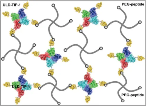
Figure 1. A cartoon representation to illustrate the formation of the hydrogels.3D networks of the hydrogels are formed by the specific protein-peptide interaction. The blue, green, red and cyan represent ULD tetramer; the yellow represent TIP-1 protein; the grey thick line represent PEG-peptide; the grey balls represent hexapeptide of WRESAI which can bind with TIP-1.
solution containing 4.0 wt% of ULD-TIP-1 and the other Dulbecco’s Modified Eagle’s Medium (DMEM) solution contain-ing 2.0 wt% of the polymer and mMSC supplemented with 10% FBS. After these components were well-mixed, the final density of cells was 2,000,000 cells/mL of gels (30mL of gel/well). The
results showed that mMSC grown very well in our ULD-TIP-1 protein-based hydrogel. Simultaneously, the mMSC could reach to a high cell density over a period of 7 days (Figure 3A–3D).
Live-dead assay
The live-dead assay was performed at different time points. As shown in Figure 3A–3D, mMSC adopted spindle or polyhedron shapes in 3D cell-gel constructs, which indirectly showed that the state of the mMSC were very good. Most of them were alive, as indicated by the cells in green color, and the dead cells were stained red (Figure 3A–3D). At the same time, the statistical data showed that cell survival rate was maintained at above 95% in the seven days of culture (Figure S7). The density of cells in cell-gel constructs kept increasing during the 7-day culture period (Figure 3, Figure 4). At day 7, we carefully took out a small piece of cell-gel construct, which was observed by the fluorescent microscopy. The top (Figure 3E) and side (Figure 3F) views of the cell-gel construct indicated that all cells were evenly distributed in it and both cells at the surface of and within the cell-gel construct were alive.
CCK-8 assay
A CCK-8 assay was then used to determine the proliferation rate of mMSC in hydrogels. Figure 4 showed the results that the number of metabolic-active mMSC in hydrogels kept increasing during the 7-day culture period. The metabolic-active cell number at day 3, day 5, and day 7 was 184%, 230% and 359% more than that at day 1, respectively (Figure 4). Our physical hydrogels could be converted to homogeneous solution by firstly adding an equal volume of aqueous solution and then pipeting for several times[33]. Therefore, the mMSC post culture could be separated from cell-gel constructs and obtained after centrifugation from the homogeneous solutions. These results indicated that our protein-based hydrogel was suitable for 3D culture of mMSC, which might be used for in vitro large scale expansion of mMSC for tissue
engineering and regenerative medicine.
Conclusions
In summary, we have constructed a protein-based hydrogel system by a specific protein-peptide interaction. The simple mixing strategy for hydrogelation was convenient and biocompat-ible to cells encapsulation, which could guarantee its future applications in 3D cell culture and controlled delivery of phar-maceutical agents. We also demonstrated that our hydrogel system
Figure 2. Rheological measurement and SEM image of the gel. A, Rheological measurement with the mode of dynamic time sweep for the sample containing 2.0 wt% of the protein and 1.0 wt% of the polymer (strain value = 1% and frequency value = 10 rad/s). The inserted is an optical image of the resulting hydrogel.B, An SEM image of the gel. The bar represents 10mm.
doi:10.1371/journal.pone.0075727.g002
Figure 3. Live-dead assay of mMSC in hydrogels a continuous seven days in culture.A, day 1; B, day 3; C, day 5; D, day 7. The living cells were stained with calcein AM (green) and the dead cells were stained with EthD-1 (red). The images of E and F showed top and side views of a 3D cell-gel construct at day 7, respectively.
could provide excellent environments for mMSC. The mMSC kept dividing in hydrogels during 7-day culture period. The cells could be separated from cell-gel constructs post culture by a simple pipetting process. These results suggested its big potential for in vitroexpansion of mMSC for further applications. We believed that
our hydrogel system could also be used for the delivery of cells for cell therapy, which would be studied in our lab in near future.
Supporting Information
Figure S1 Chemical structure of compound 1 (CGGGRGDGW-RESAI).
(TIF)
Figure S2 1H NMR of compound 1. CGGGRGDGWRESAI:
1
H NMR (300 MHz, DMSO-d6)d8.85–8.95 (t, 1H), 8.67–8.77 (t,
1H),8.15–8.35 (m, 6H), 7.88–8.14 (m, 7H), 7.55–7.61 (d, J = 7.58 Hz, 1H), 7.46–7.53 (m, 1H), 7.25–7.35 (d, 3H), 7.10 (s, 2H), 6.91–7.05 (m, 3H), 5.00 (s, 1H), 4.49–4.61 (m, 2H), 4.22–4.40 (m, 5H), 4.06–4.19 (m, 2H), 3.69–3.93 (m, 8H), 3.51–3.67 (m, 4H),
2.99–3.15 (m, 5H), 2.88–2.98 (m, 1H), 2.60–2.77 (m, 1H), 2.21– 2.33 (m, 2H), 1.83–1.97 (m, 1H), 1.61–1.83 (m, 4H), 1.30–1.61 (m, 7H), 1.13–1.25 (m, 4H), 0.76–088 (t, 6H).
(TIF)
Figure S3 1H NMR of PEG (above) and PEG-peptide (below). The hydrogen peak at 6.7 ppm disappeared completely, indicating the Michael addition reaction of maleimide with cysteine. (TIF)
Figure S4 Rheological measurement with the mode of dynamic frequency sweep at the strain of 1% for the gel containing 1 wt% of PEG-peptide and 2 wt% of protein. The solution gradually became a hydrogel with the frenquency from low to high. (TIF)
Figure S5 Rheological measurement with the mode of dynamic strain sweep at the frequency of 10 rad/s for the gel containing 1 wt% of PEG-peptide and 2 wt% of protein. The hydrogel showed weak stain dependences from 0.1% to 10%, with the G9
value of about 80 Pa. (TIF)
Figure S6 The purified ULD-TIP1 protein was analyzed by 15% SDS-PAGE. M, Marker.
(TIF)
Figure S7 The survival rate of mMSC cells were cultured in the hydrogel at different day.
(TIF)
Acknowledgments
We thank Ms An for her kind help to obtain zeta potential of self-assembled structures.
Author Contributions
Conceived and designed the experiments: JYW HZ. Performed the experiments: JYW JXZ XLZ. Analyzed the data: JYW JXZ XLZ. Contributed reagents/materials/analysis tools: JYW JXZ XLZ. Wrote the paper: HZ.
References
1. Petka WA, Harden JL, McGrath KP, Wirtz D, Tirrell DA (1998) Reversible hydrogels from self-assembling artificial proteins. Science 281: 389–392. 2. Krishna OD, Kiick KL (2010) Protein- and peptide-modified synthetic
polymeric biomaterials. Biopolymers 94: 32–48.
3. Jia X, Kiick KL (2009) Hybrid multicomponent hydrogels for tissue engineering. Macromol Biosci 9: 140–156.
4. Topp S, Prasad V, Cianci GC, Weeks ER, Gallivan JP (2006) A genetic toolbox for creating reversible Ca2+-sensitive materials. J Am Chem Soc 128: 13994– 13995.
5. Tang Y, Ghirlanda G, Petka WA, Nakajima T, DeGrado WF, et al. (2001) Fluorinated Coiled-Coil Proteins Prepared In Vivo Display Enhanced Thermal and Chemical Stability. Angew Chem Int Ed 40: 1494–1496.
6. Sui ZJ, King WJ, Murphy WL (2008) Protein-Based Hydrogels with Tunable Dynamic Responses. Adv Funct Mater 18: 1824–1831.
7. Santos E, Hernandez RM, Pedraz JL, Orive G (2012) Novel advances in the design of three-dimensional bio-scaffolds to control cell fate: translation from 2D to 3D. Trends Biotechnol 30: 331–341.
8. Zhou M, Smith AM, Das AK, Hodson NW, Collins RF, et al. (2009) Self-assembled peptide-based hydrogels as scaffolds for anchorage-dependent cells. Biomaterials 30: 2523–2530.
9. Pek YS, Wan AC, Ying JY (2010) The effect of matrix stiffness on mesenchymal stem cell differentiation in a 3D thixotropic gel. Biomaterials 31: 385–391. 10. Wang HM, Shi Y, Wang L, Yang ZM (2013) Recombinant proteins as
cross-linkers for hydrogelations. Chemical Society Reviews 42: 891–901.
11. Ehrick JD, Luckett MR, Khatwani S, Wei Y, Deo SK, et al. (2009) Glucose responsive hydrogel networks based on protein recognition. Macromol Biosci 9: 864–868.
12. Ehrick JD, Deo SK, Browning TW, Bachas LG, Madou MJ, et al. (2005) Genetically engineered protein in hydrogels tailors stimuli-responsive charac-teristics. Nat Mater 4: 298–302.
13. Esser-Kahn AP, Trang V, Francis MB (2010) Incorporation of antifreeze proteins into polymer coatings using site-selective bioconjugation. J Am Chem Soc 132: 13264–13269.
14. Esser-Kahn AP, Iavarone AT, Francis MB (2008) Metallothionein-cross-linked hydrogels for the selective removal of heavy metals from water. J Am Chem Soc 130: 15820–15822.
15. Esser-Kahn AP, Francis MB (2008) Protein-cross-linked polymeric materials through site-selective bioconjugation. Angew Chem Int Ed 47: 3751–3754. 16. Yuan W, Yang J, Kopeckova P, Kopecek J (2008) Smart hydrogels containing
adenylate kinase: translating substrate recognition into macroscopic motion. J Am Chem Soc 130: 15760–15761.
17. Murphy WL, Dillmore WS, Modica J, Mrksich M (2007) Dynamic hydrogels: translating a protein conformational change into macroscopic motion. Angew Chem Int Ed 46: 3066–3069.
18. Sui ZJ, King WJ, Murphy WL (2007) Dynamic Materials Based on a Protein Conformational Change. Adv Mater 19: 3377–3380.
19. Mosiewicz KA, Johnsson K, Lutolf MP (2010) Phosphopantetheinyl transferase-catalyzed formation of bioactive hydrogels for tissue engineering. J Am Chem Soc 132: 5972–5974.
20. Yamaguchi N, Zhang L, Chae BS, Palla CS, Furst EM, et al. (2007) Growth factor mediated assembly of cell receptor-responsive hydrogels. J Am Chem Soc 129: 3040–3041.
21. Ito F, Usui K, Kawahara D, Suenaga A, Maki T, et al. (2010) Reversible hydrogel formation driven by protein-peptide-specific interaction and chondro-cyte entrapment. Biomaterials 31: 58–66.
22. Grove TZ, Osuji CO, Forster JD, Dufresne ER, Regan L (2010) Stimuli-responsive smart gels realized via modular protein design. J Am Chem Soc 132: 14024–14026.
Figure 4. Cell proliferation rate of mMSC in gels determined by a CCK-8 assay.One asterisk (*) indicates p value smaller than 0.05 (p,0.05). Three asterisks (***) indicate p value smaller than 0.001 (p,0.001).
23. Wong Po Foo CT, Lee JS, Mulyasasmita W, Parisi-Amon A, Heilshorn SC (2009) Two-component protein-engineered physical hydrogels for cell encapsu-lation. Proc Natl Acad Sci U S A 106: 22067–22072.
24. Zhang X, Chu X, Wang L, Wang H, Liang G, et al. (2012) Rational Design of a Tetrameric Protein to Enhance Interactions between Self-Assembled Fibers Gives Molecular Hydrogels. Angewandte Chemie-International Edition 51: 4388–4392.
25. Ehrbar M, Schoenmakers R, Christen EH, Fussenegger M, Weber W (2008) Drug-sensing hydrogels for the inducible release of biopharmaceuticals. Nat Mater 7: 800–804.
26. Charati MB, Ifkovits JL, Burdick JA, Linhardt JG, Kiick KL (2009) Hydrophilic elastomeric biomaterials based on resilin-like polypeptides. Soft Matter 5: 3412– 3416.
27. Mi L, Fischer S, Chung B, Sundelacruz S, Harden JL (2006) Self-assembling protein hydrogels with modular integrin binding domains. Biomacromolecules 7: 38–47.
28. Aguado BA, Mulyasasmita W, Su J, Lampe KJ, Heilshorn SC (2012) Improving viability of stem cells during syringe needle flow through the design of hydrogel cell carriers. Tissue Eng Part A 18: 806–815.
29. Fischer SE, Liu X, Mao HQ, Harden JL (2007) Controlling cell adhesion to surfaces via associating bioactive triblock proteins. Biomaterials 28: 3325–3337. 30. Mulyasasmita W, Lee JS, Heilshorn SC (2011) Molecular-level engineering of protein physical hydrogels for predictive sol-gel phase behavior. Biomacromo-lecules 12: 3406–3411.
31. Yan X, Zhou H, Zhang J, Shi C, Xie X, et al. (2009) Molecular mechanism of inward rectifier potassium channel 2.3 regulation by tax-interacting protein-1. J Mol Biol 392: 967–976.
32. Hersel U, Dahmen C, Kessler H (2003) RGD modified polymers: biomaterials for stimulated cell adhesion and beyond. Biomaterials 24: 4385–4415. 33. Grzanka A, Grzanka D, Gagat M, Tadrowski T, Sokolowska-Wojdylo M, et al.