Universidade de Lisboa
Faculdade de Ciências
Departamento de Biologia Vegetal
The role of OsRMC gene in the JA signaling pathway: a transgenic
approach in Arabidopsis and by an Y1H assay
João Guilherme Costa de Alvarez Cortes
Dissertação
Mestrado em Biologia Celular e Biotecnologia
Universidade de Lisboa
Faculdade de Ciências
Departamento de Biologia Vegetal
The role of OsRMC gene in the JA signaling pathway: a transgenic approach in Arabidopsis and by an Y1H assay
João Guilherme Costa de Alvarez Cortes
Dissertação
Mestrado em Biologia Celular e Biotecnologia
Orientador interno: Prof. Doutora Helena Trindade (Centro de Biotecnologia Vegetal – IBB, Faculdade de Ciências da Universidade de Lisboa)
Orientador externo: Doutor Tiago Lourenço (Laboratório de Genómica de Plantas em Stress – GplantS, Instituto de Tecnologia Química e Biológica – ITQB)
i
Agradecimentos
Em primeiro lugar gostaria de agradecer á professora Margarida Oliveira por me ter aceite no seu laboratório, numa fase já tardia de aceitação de alunos para projectos de mestrado. A experiência que obti neste laboratório (GplantS) durante este ano de dissertação foi muito especial para mim, no sentido em que foi a minha primeira e verdadeira experiência pessoal e profissional do que é estar e trabalhar num laboratório.
Gostaria de agradecer à minha orientadora interna, a professora Helana Trindade, por ter aceite ser a minha orientadora interna neste projecto de mestrado. Ao meu orientador interno, Tiago Lourenço, não tenho palavras para demonstrar a paciência e confiança que ele sempre depositou em mim. Sinto que não podia ter tido melhor orientação ao longo deste ano de trabalho no laboratório GPlantS, e esta dissertação não teria sido conseguida não fosse a constante força e motivação que o Tiago me injectou com as suas chamadas de atenção nas nossas conversas particulares. Obrigado por acreditares em mim.
Um especial obrigado à Mafalda Rodrigues, estudante de doutoramento no laboratório GPlantS, pela sua ajuda e disponibilidade em todo o trabalho com Arabidopsis. A todos os membros do laboratório GPlantS um muito obrigado pela amizade desmontrada, em períodos especialmente difíceis para mim, sempre me senti aceite no laboratório e o espírito de entrea-ajuda que envolve e caracteriza o trabalho neste laboratório foi sem dúvida para mim uma agradável surpresa.
Gostaria por fim de agradecer à minha família, aos meus pais e irmãos, a qual sempre depositou confiança em mim e me deu alguns “empurrões” nos momentos mais díficieis da escrita desta dissertação.
iii
List of Abbreviations
3-AT
3-amino-1,2,4-triazole
bp
Base pairs
cDNA
Codifying DNA
CEF
Cefotaxime
Col-0
Arabidopsis thaliana Columbia ecotype
DMSO
Dimethyl sulfoxide
DNA
Deoxyribonucleic acid
EREBP
Ethylene-responsive element binding protein
g, mg, µg, ng
Gram, miligram, microgram, nanogram
JAs
Jasmonates
JA
Jasmonic acid
JAZ
Jasmonate ZIM domain
L, mL, µL
Liter, mililiter, microlitre
M, mM, µM
Molar, milimolar, micromolar
NaCl
Sodium Chloride
min
Minutes
ºC
Degree Celsius
PR
Pathogen related
PEB
Protein Extraction Buffer
PCR
Polymerase chain reaction
RT
Room temperature
RT-PCR
Semi-quantitative reverse transcriptase PCR
RMC
Root meander curling
RFP
Ref fluorescent protein
RNA
Ribonucleic acid
RNAi
RNA interference
TF
Transcription factor
Vol
Volume
Y1H
Yeast one hybrid
v
Abstract
Jasmonic acid is a plant hormone that has been involved in several biological processes such as development, biotic and abiotic stress. Previously, it was found in the host lab that a rice RNA interference (RNAi) – OsHOS1 line had up-regulation of OsRMC gene expression. In rice, this gene seems to be involved in the modulation of abiotic stress and root curling, acting as a negative regulator of the jasmonic acid signaling. OsRMC does not have a known homologue in Arabidopsis which makes this model species an important system to study the function of this gene. To achieve this goal, we overexpressed OsRMC in Arabidopsis in two different systems either in a cell suspension cultures (to study gene expression changes of JA-responsive genes) or whole plants (phenotypic changes induced by JA). We were able to obtain stable transgenic cell suspension culture overexpressing OsRMC as well for the empty vector (negative control). However, we were not able to see gene expression changes induced by JA even in untransformed cell suspension cultures for the analyzed genes (PDF1.2, VSP1 and MYC2). Regarding the transformation of whole plants, we generated three independent lines overexpressing OsRMC. These lines were characterized for transgene expression level and corresponding protein (OsRMC-RFP).
We also performed a direct yeast one hybrid (Y1H) assay to complement the work done with Arabidopsis plants and cell suspension cultures regarding JA-signaling. In this assay, the promoter sequences of three genes involved in JA-signaling from rice (JAZ-like 1, JAZ-like 2 and JAZ-like 3) were analyzed. Promoter fragments corresponding to 1000bp upstream of the ATG codon of each gene were used as bait sequences. As prey, we used two transcription factors from the EREBP subfamily, previously identified as binding to the OsRMC gene promoter. However, we couldn’t detect interaction between the two TFs analyzed and the bait sequences from the JAZ genes. As future work, these baits strains will be screened by Y1H with cDNA libraries induced by salt or JA.
As main achievements of this work, we were able to produce transgenic Arabidopsis plants overexpressing OsRMC which were fully characterized at transgene expression and protein level. These lines will be used in further studies such as to
vi
monitor expression changes in JA-responsive genes, determination of the sub-cellular localization of the OsRMC-RFP, and JA-induced phenotypic characterization.
vii
Resumo
O ácido jasmónico é uma hormona vegetal que está envolvida em vários processos biológicos tais como vias de desenvolvimento, stress biótico e abiótico. Foi descoberto anteriormente no laboratório anfitrião que uma linha de arroz de RNA de interferência (RNAi) – OsHOS1 apresentava uma expressão génica do gene OsRMC aumentada. Em arroz, este gene parece estar envolvido na modulação do stress abiótico e do encaracolamento das raizes, agindo como um regulador negativo da via de sinalização do ácido jasmónico. O gene OsRMC não tem nenhum homólogo conhecido em Arabidopsis, tornando esta espécie modelo num sistema importante para estudar a função deste gene. De modo a alcançar o objectivo principal, nós sobrexpressamos OsRMC em Arabidopsis em dois sistemas diferentes, quer em cultures de células em suspensão (para o estudo das alterações da expressão génica de genes que respondem ao ácido jasmónico) quer em plantas (alterações fenotípicas induzidas por ácido jasmónico) Fomos capazes de obter culturas estáveis transformadas de células em suspensão sobrexpressando OsRMC e também culturas transformadas com o vector vazio (controlo negativo). Contudo, não fomos capazes de vizualisar quaisquer alterações de expressão génica (para os genes analisados PDF1.2, VSP e MYC2) induzidas por ácido jasmónico mesmo em culturas de células em suspensão não transformadas. Em relação à transformação de plantas, foram geradas três linhas independentes sobreexpressando OsRMC. Estas linhas foram caracterizadas em termos de nível de expressão do transgene e da proteína correspondente (OsRMC-RFP)
Neste trabalho também se inclui um ensaio directo de yeast one hybrid (Y1H) de modo a complementar o trabalho realizado com as plantas de Arabidopsis e as culturas de células em suspensão, no que diz respeito à sinalização pelo ácido jasmónico. Neste ensaio, as sequências dos promotores de três genes de arroz envolvidos na sinalização pelo ácido jasmónico (JAZ-like1, JAZ-like2 e JAZ-like3) from analizadas. Fragmentos dos promotores, correspondendo a 1000bp a montante do codão ATG de cada gene, foram usados como sequências “isca”. Como “presas”, usamos dois factores de transcrição da subfamilia EREBP, previamente reportados com capacidade de se ligarem ao promotor do gene OsRMC. Contudo, não conseguimos detectar qualquer interacção entre os dois TFs analisados e as sequências “isca” dos genes JAZ. Como trabalho futuro, estas estirpes
viii
criadas irão ser examinadas por Y1H com bibliotecas de cDNA induzidas por sal ou ácido jasmónico.
Este trabalho tem como principais realizações a produção de plantas transgénicas de Arabidopsis sobrexpressando o gene OsRMC, as quais foram caracterizadas nos níveis de expressão do transgene e da proteína. Estas linhas serão usadas em estudos futuros de modo a monitorizar alterações de expressão de genes que respondem ao ácido jasmónico, na determinação da localização sub-celular da proteína OsRMC-RFP e na caracterização fenotípica induzida por ácido jasmónico.
1
Table of contents
Agradecimentos ... i
List of Abbreviations ... iii
Abstract ... v
Resumo ... vii
Table of contents ...1
General Introduction ... 5
General Introduction ... 7
Plant abiotic and biotic stress responses ... 7
Jasmonates ... 8
Thesis objectives ... 12
CHAPTER ONE: Preparation and analysis of Arabidopsis thaliana cell suspension cultures and whole plants overexpressing OsRMC ... 15
Introduction ... 17
JA-marker genes for expression analysis ... 17
Biological material ... 18
Arabidopsis thaliana ... 18
Cell suspension cultures ... 19
Transformation techniques ... 19
The Floral Dip Method ...20
Transformation of cell suspension cultures ...20
Material and Methods ... 21
Genetic Construct ... 21
Arabidopsis cell suspension cultures ... 21
Arabidopsis plant material... 22
Agrobacterium tumefaciens transformation ... 22
2
Arabidopsis thaliana transformation – Floral Dip Method ... 23
Seed sterilization ... 23
Plant genotyping ...24
Arabidopsis cell culture transformation ...24
Jasmonic acid assays ...24
RNA extraction ... 25
DNase treatment procedures (TURBO DNA-free Life Technologies, USA) ... 25 cDNA synthesis ...26 Protein extraction ...26 Protein quantification ... 27 Western blot ... 27 Protoplast preparation ...28 Results ... 29
Cell suspension cultures ... 29
Gene expression analysis under JA ... 30
Protoplasts... 32
Arabidopsis plants ... 33
Western Blots ... 36
Discussion ... 39
Arabidopsis cell suspension cultures ... 39
Sub-cellular localization of OsRMC ... 40
Arabidopsis plants ... 41
CHAPTER TWO: Yeast-One-Hybrid direct screening of JAZ-like promoter fragments with two EREBP transcription factors ... 43
Introduction ... 45
JAZ genes / JAZ proteins ... 46
3
Preparation of bait sequences ...47
Primer design ...47
Amplification and sequencing ... 49
Cloning of the bait sequence in the pINTHIS3NB vector ... 51
Preparation of bait strains ... 51
Growth and preparation of yeast cells for transformation (competent cells) ... 51
Titulation of bait strains ... 52
Direct Screening ... 53
Results ... 55
Primer testing ... 55
Titulation of bait strains ... 55
Direct screening analysis ... 57
Discussion ... 61
Final Conclusions / Future Work ... 63
Yeast One Hybrid ...65
Personal remarks ... 67
References ... 69 Appendix ... I
5
7
General Introduction
Plant abiotic and biotic stress responses
Terrestrial plants, as sessile organisms, are constantly exposed to a variety of environmental challenges. These varying conditions can cause stress in plants reducing its fitness. In fact, in most crops, actual yields are only 20% of attainable yields and a dominant factor limiting yield is abiotic stress such as excess or deficient water, high or low temperature, and high salinity (Boyer, 1982). Abiotic stress is defined as environmental conditions that reduce growth and yield below optimum levels (Cramer et al., 2011). Plant responses to abiotic stress are dynamic and complex; they are both elastic (reversible) and plastic (irreversible) (Cramer et al., 2011). To survive these challenges, plants have developed elaborate mechanisms to perceive external signals and to manifest adaptive responses with proper physiological changes, at cellular and molecular levels. At the molecular level, the perception of environmental stimuli and the subsequent activation of defense responses require a complex interplay of signaling cascades (Hashimoto et al., 2004). Understanding the mechanisms by which plants perceive and transduce these stress signals to initiate adaptive responses is essential for engineering stress-tolerant crop plants (Xiong et al., 2001). A large number of abiotic stress responsive genes have been reported in a variety of plants including rice and Arabidopsis. These genes induced during stress conditions may function not only in the protection of cells from stress by production of important metabolic proteins, but also in the regulation of genes, including transcription factors (TFs), for signal transduction in the stress responses. These TFs regulate expression of multiple downstream target genes under stress conditions (Todaka et al., 2012). These regulatory systems are achieved through specific cis-elements in the promoter regions of target genes, which are termed ‘regulons’. Several regulons involved in abiotic stress responses have been identified in Arabidopsis (Todaka et al., 2012).
Plant hormones constitute a battery of regulators involved in control of developmental, physiological and metabolic processes and are also likely to mediate the responses of plants to environmental stresses (Rup Kumar Kar, 2011). Beyond their role as developmental keys in a plant’s life, the action of several plant hormones is also crucial
8
in responses to biotic and abiotic challenges. The five classical plant hormones are auxins, cytokinins, gibberellins, abscisic acid and ethylene, but other important group of plant hormones are also involved in responses to abiotic and biotic stress and these include the well characterized group designated as jasmonates.
Hormones are important regulators of plant responses to abiotic stress, with the two most important being abscisic acid (ABA) and ethylene. The essential components of ABA signaling have been identified, and their mode of action was clarified (Cramer et al., 2011). Plants consistently face challenges from a wide array of biotic stresses, including insect herbivory, and fungal, bacterial and viral attacks (Dreher et al., 2007). One area in which the details of the defense responses are becoming more evident is in the plant’s production of pathogen-related (PR) proteins. PR proteins have been defined as plant proteins that are induced during pathogen infection or wounding. This term describes the collective induction of numerous plant genes that are induced in a variety of species in response to pathological attacks (Hashimoto et al., 2004). The major families of PR proteins have been grouped at least in 14 different classes, primarily on the basis of their amino acid sequence identity (Van Loon et al., 1999). The PR10 class of proteins, first identified as a major pollen allergen (Bet v1) from white birch (Breiteneder et al., 1989), are induced by pathogen attack in a wide variety of plant species, including parsley (Somssich et al., 1986), potato (Matton and Brisson, 1989), pea (Barratt and Clark, 1991), soybean (Crowell et al., 1992), asparagus (Warner et al., 1993), sorghum (Lo et al., 1999) and rice (Midoh and Iwata, 1996, McGee et al., 2001). The biotic and abiotic stress-inducible nature of the PR10 genes has been investigated including pathogen infection (Midoh and Iwata 1996, McGee et al,. 2001, Tanaka et al., 2003), salt tolerance (Moons et al., 1997), UV irradiation (Rakwal et al., 1999), and ozone stress (Agrawal et al., 2002).
Jasmonates
Among plant hormones, jasmonates (JA) are one of the best characterized and studied. The signalling pathway of JA is involved in the modulation of either biotic (Kloek et al., 2001) or abiotic (Sugano et al., 2003) stress although most of the reports implicate this hormone in the organization of the plant defense mechanism. Nevertheless, the signalling pathway of JA has been well characterized in the recent years
9
especially in dicots like Arabidopsis and tomato (Chini et al., 2007 and 2009, Staswick, 2008, Thines et al., 2007) but not in monocots and in particular rice.
Jasmonates (JAs) are fatty acid derivates that act as signaling molecules. These signaling molecules share notable structural and functional properties with prostaglandins found in animals. They are mainly derived from linolenic acid and function in normal developmental pathways but also play a crucial role in allowing plants to mount a defense to biotic challenges. JAs affect different processes such as pollen development and fruit ripening, and as such, they modulate vital physiological processes such as flower development, and fertility. Jasmonates are essential to complete the last steps of stamen development, specifically in three processes: (a) pollen maturation after the trinucleated stage to guarantee viability and fertility; (b) elongation of stamen filaments so that the anthers can reach the stigma in preparation for pollination; and (c) dehiscence of anther locules for pollen release (Acosta and Farmer, 2010). Impairments of these functions in jasmonate synthesis or perception mutants results in male sterility. Jasmonates also control seed size although the specific tissue types affected in seeds are not yet identified (Farmer and Dubugnon, 2009). Jasmonates also modulate petal size and affects vein structure. They also promote resistance to insects and pathogens, and in fact, in Arabidopsis it is estimated that 67 to 85% of genes activated by wound or insects are regulated, in terms of expression, by jasmonates (Acosta and Farmer, 2010). The genes activated in a wound response, for example, in leaf damage, fall into a series of categories including jasmonate synthesis, jasmonate signaling components, defense genes, defensins, resource allocation genes, general stress response genes, etc. The cyclopentanones JA and methyl-jasmonate (MeJA) are produced via the octadecanoid pathway, JA being the key terminal product of this pathway. The octadecanoid pathway has been extensively studied in Arabidopsis thaliana and tomato, but not as well in rice.
The JAs for which there is most evidence for biological activity are: (+)-7-iso-jasmonoyl-L-isoleucine (JA-Ile) (Fonseca et al., 2009), which is the ligand that seems to play the major role in jasmonate signaling in Arabidopsis leaves and possibly flowers; Jasmonoyl-L-tryptophan, as an auxin signaling inhibitor active in Arabidopsis roots (Staswick, 2009); and OPDA and dinor-OPDA, which have signaling properties either independent of canonical jasmonate signaling (Stintzi et al., 2001; Taki et al., 2005) or using part of the jasmonate signaling machinery (Ribot et al., 2008).
10
The analysis of mutants, mainly from Arabidopsis thaliana and tomato (Solanum lycopersicum) that are deficient in jasmonate biosynthesis or jasmonate signaling has been extremely useful for dissecting jasmonate function (Wasternack, 2010). Among these mutants, the Arabidopsis mutant coi1-1, which was already isolated in 1994, has been exceptional, because it is deficient in nearly all jasmonate responses. Jasmonate research has come full circle by the recent discovery that COI1 protein is a jasmonate receptor. Following the perception of JA a number of cellular signaling processes occur that result in the posttranslational modification of upstream regulatory proteins, transcriptional activation of JA-responsive transcription factors and downstream genes.
The first step in the JA signaling takes place in the nucleus, where JA-Ile binds to its receptor, the CORONATINE INSENSITIVE 1 (COI1) protein which forms part of an SCF ubiquitin E3 ligase (see Scheme 1). SCF complexes are a type of E3 ubiquitin ligases, multisubunit machines that specify and mediate protein ubiquitination for targeted degradation by the 26S proteosome. SCF ubiquitin E3-ligases contain a variable F-box protein that directly binds the targets of the ubiquitination complex thereby conferring its specificity. The F-box protein COI1 defines an SCF complex that is specifically involved in jasmonate signaling (Santner et al., 2007). The other components of the SCF-COI1 complex are: either ASK1 or ASK2, which serve as adaptors for SCF-COI1 to form the substrate-recognition subunit; RBX1, a RING finger protein that recruits an E2 ubiquitin conjugating enzyme to bring it close to the substrate; and the scaffolding CULLIN1 protein that holds the substrate-recognition portion of the complex at its N-terminus and RBX1 at its C-terminus. RBX1 and CULLIN1 are necessary for normal jasmonate signaling and so are other compounds known to interact with or regulate SCF complexes such as AXR1, the COP9 signalosome and SGT1b. The main targets of this SCF-COI1 complex are the Jasmonate ZIM-domain (JAZ) repressors proteins which are ubiquitinated. The destruction of JAZ proteins by the 26S proteosome releases transcription factors, and allows gene expression JA-dependent (Browse, 2009; Chung et al., 2009; Fonseca et al., 2009a). JAZ proteins lack recognizable DNA-binding domains, however, their interaction with the transcription factor MYC2 suggests that JAZ proteins control jasmonate related gene expression by preventing the function of transcriptional activators, but the exact mechanism is still not known. MYC2 is a basic helix-loop-helix (bHLH) transcription factor that activates a first wave of gene transcription upon jasmonate perception, including the JAZ genes and the putative jasmonate biosynthesis gene LOX3 (Chini et al., 2007; Pauwels et al., 2008). In Arabidopsis, JAZ proteins are
11
encoded by 12 different genes (JAZ1 through JAZ12), which are predicted to produce over 20 protein variants, including two characterized alternative splice products, JAZ10.3 and JAZ10.4 (Staswick, 2008). Most JAZ genes are rapidly induced by MYC2 upon activation of jasmonate signaling. In general, the promoters of JAZ genes contain putative MYC2 binding motifs that have been proven functional for JAZ3.
Among JAZ proteins, there are two conserved features which are the Jas motif and the ZIM domain. The Jas motif is towards the C-terminus and constitutes the binding site of COI1 in the presence of JA-Ile; therefore, the Jas motif is required for timely JAZ protein degradation upon jasmonate perception. This motif is also necessary and sufficient for the interactions of JAZ proteins with MYC2 (Chini et al., 2009). This motif might also be important for nuclear localization of JAZ proteins, even if its absence does not necessarily prevents their entry into the nucleus. The ZIM domain is located in the center of the JAZ proteins and contains a conserved TIFY motif (TIFYXG). This domain mediates homo- and heteromeric interactions between many JAZ proteins, a capability that is critical for their function as negative regulators of jasmonate signaling.
As for any other hormone, jasmonate produces strong effects in cells. It is important then to control hormone signaling after the initial response. In the case of jasmonates, one way to accomplish this is to metabolize the bioactive forms of the hormone to make them inactive. The fact that JAZ gene transcription is rapidly induced upon jasmonate signaling suggests a negative feedback loop control mechanism, whereby newly made JAZ proteins would repress again their corresponding transcription factors. Cells may rely on alternative splice variants of JAZ proteins, such as JAZ10.3 and JAZ10.4, which lack the Jas motif (Yan et al., 2007; Chung and Howe, 2009). In consequence, they are not recognized efficiently by COI1 and are not subject to effective jasmonate-induced proteosome degradation. Rapid accumulation of these variants is predicted to desensitize cells to the presence of the hormone (Acosta and Farmer, 2010).
12
Scheme 1 - An overview of cellular compartments in JA synthesis and signaling (from Acosta and Farmer, 2010)
Thesis objectives
The main goal of this work is to understand how a rice gene, OsRMC (ROOT MEANDER CURLING), may influence the JA-signaling pathway in Arabidopsis by overexpressing it and monitoring gene expression changes of JA-responsive genes either in wild-type (WT) and in transgenic cell-suspension cultures and plants.
This rice gene was shown to be involved in the modulation of abiotic stress (salinity stress; Zhang et al., 2009) and root curling (Jiang et al., 2007) mediated by JA in rice. According to homology analysis, OsRMC belongs to the cysteine-rich-repeat
receptor-13
like protein kinase subfamily (CRK, also known as DUF26 - domain unknown function 26 - subfamily receptor-like kinase) but it has no transmembrane domain or kinase domain.
Previously studies developed in the thesis host-lab, have putatively linked the function of this gene in root mechanosensing in rice through a negative regulation of the JA-signalling pathway (Lourenço, T. et al., unpublished results). The OsRMC gene expression is also up-regulated in a rice RNA interference (RNAi) line that silences the expression of an ubiquitin E3-ligase, OsHOS1 (Lourenço, T. et al. unpublished results), which suggest that its expression can be modulated through the action of the proteasome (Lourenço, T. et al. unpublished results).
The OsRMC gene does not have any known homologue in Arabidopsis. Therefore, the use of a model-plant such as Arabidopsis, with a well-characterized JA-signaling pathway, seemed appropriate to study the function of OsRMC to complement other studies being developed in the host-lab.
To further complement the work developed, we also decided to perform a Yeast-One hybrid assay using the promoter region of three rice JAZ genes. These genes have been shown to be up-regulated in the RNAi::OsHOS1 line. Therefore, and since JAZ genes are well known negative regulators of the JA-signaling pathway, we thought it could be interesting to test the binding of two EREBP TFs, previously identified as binding to the OsRMC promoter region (Serra, T. et al., unpublished results).
As main outputs of this work, we were able to generate three independent Arabidopsis transgenic lines overexpressing the OsRMC gene. These three lines were characterized in transgene expression (RT-PCR) and protein expression (Western blot). Due to the fusion construct between OsRMC and a fluorescent tag (Red Fluorescent Protein; RFP), we were also able to visualize the sub-cellular localization of OsRMC in Arabidopsis roots. We were also able to generate stable transgenic Arabidopsis cell-culture lines overexpressing OsRMC. However, this approach failed to deliver consistent results regarding JA-responsive genes expression changes. As for the Yeast-One hybrid approach, we were able to produce yeast bait-strains for the three rice JAZ genes selected and test the binding of the two EREBP TFs.
The results of this work and future perspectives will be discussed in the next chapters.
15
CHAPTER ONE: Preparation and analysis of Arabidopsis
thaliana cell suspension cultures and whole plants
17
Introduction
The main goal of this work was to overexpress the rice OsRMC gene both in Arabidopsis cell suspension cultures and in whole plants. OsRMC gene has been described in rice as being negatively involved in root curling (Jiang et al., 2007) and salinity tolerance (Zhang et al., 2008). This gene has also been showed to be involved in JA-mediated root curling in rice (Jiang et al., 2007)) but the mechanism is still unclear. Since Arabidopsis does not have a known homologue for OsRMC, we considered this model suitable for this work. Ultimately, our goal was to study the JA responses using two different approaches overexpressing OsRMC: 1) using Arabidopsis cell suspension cultures to rapidly observe JA-dependent gene expression changes in JA-marker genes such as VEGETATIVE STORAGE PROTEIN (VSP, Benedetti et al., 1995) and PLANT DEFENSIN1.2 (PDF1.2, Penninckx et al., 1996), and a gene involved in JA-signaling, MYC2, overcoming the tissue culture step and 2) using whole Arabidopsis plants to observe JA-dependent phenotypic changes. In addition, we also wanted to clarify the subcellular localization of OsRMC since the two reports describing work with this gene had contradictory results, placing this protein either in the plasma membrane or the apoplast (Jiang et al., 2007; Zhang et al., 2008).
In this chapter, all the work done with Arabidopsis thaliana cell suspension cultures and whole plants is presented. We were able to establish transformed cell suspension cultures and attempted to use them to analyze gene expression of JA-responsive genes. The Arabidopsis cell suspension cultures were also used to produce protoplasts and these were also subject to an assay involving JA. We were also able to produce three independent transgenic Arabidopsis plants which were characterized for their transgene expression and corresponding protein level. The identification of the sub-cellular localization of OsRMC-RFP protein was also attempted in the transgenic plants.
18
Vegetative storage proteins (VSP) serve as proteinaceous storage reserves. These protein have been identified in numerous plants, such as soybean (Glycine max; Wittenbach, 1983), potato (Solanum tuberosum; Mignery et al., 1984, 1988), sweet potato (Ipomoea batatas; Maeshima et al., 1985), white clover (Trifolium repens; Goulas et al., 2003), alfalfa (Medicago sativa; Meuriot et al., 2004b), and in the bark of deciduous trees such as poplar (Populus deltoides; Coleman et al., 1991) and elderberry (Sambucus nigra; Van Damme et al., 1997). Arabidopsis VSP transcripts are induced by mechanical wounding, jasmonic acid (JA), insect herbivory, and osmotic and nutritional stresses (Liu et al., 2005).
Plant defensins may be highly expressed or upregulated in plants challenged by various abiotic and biotic stresses. For example PDF1.2 is highly and constitutively expressed in the halophyte salt cress Thellungiella halophila (Taji et al., 2004). A defensin-like protein from wheat was also found to be induced during cold acclimatization (Koike et al., 2002). Induction of gene expression of plant defensins by drought or salt stresses has also been reported (Yamada et al., 1997; Maitra and Cushman, 1998) but their potential roles in abiotic stress tolerance are not known.
MYC2, a basic helix-loop-helix (bHLH) domain-containing transcription factor, participates in the jasmonate (JA) signaling pathway and is involved in the modulation of diverse JA functions. However, a comprehensive list of MYC2-dependent JA-responsive proteins has yet to be defined (Guo et al., 2012). Nonetheless, results have been published that support a positive role for MYC2 in regulating JA-mediated carbohydrate metabolism and oxidative stress tolerance.
Biological material
Arabidopsis thaliana
The virtues of Arabidopsis thaliana as an experimental model plant for genetic, biochemical, and molecular biological studies have been extensively reviewed Arabidopsis thaliana is a small dicotyledonous species, member of the mustard (Brassicaceae) family, which includes economically important plants such as cabbage,
19
turnip, broccoli, radish and canola. Although this species is not of major agronomic importance, the important advantages it presents for basic research in genetics and molecular biology makes it an important model plant worldwide, and as a fact, it was the first one to be established as such. The advantages this species brings to basic research are: (1) it has a small genome, which has already been sequenced and annotated; (2) there are extensive genetic and physical maps of all its 5 chromosomes; (3) it has a short life cycle, of approximately about 6 weeks from germination to seed maturation; (4) the plant is of small size and easily cultivated; (5) it produces numerous self progeny; (6) its genome is efficiently manipulated by genetic engineering; (7) there are already a large number of mutant lines and genomic resources available; and additionally (8) it has been elected as a flowering plant model for a long time now, leading to an endless collection of studies and information gathered for more than 40 years.
Cell suspension cultures
Suspension culture of isolated plant cells is a valuable tool for providing the material for high-throughput studies such as metabolic analyses, production of secondary plant products, and herbicide discovery (Boisson et al., 2012). Also, plant cell suspension cultures are useful tools for investigations of physiological phenomena such as cell proliferation and differentiation. In such experimental systems, the environment should be completely controlled and the population of target cells should, if possible, be homogenous (Fukuda et al., 1994). Although Arabidopsis thaliana represents a model plant, it remains somewhat recalcitrant in tissue culture (Gleddie, 1989). In addition to providing cells in quantity for biochemical and molecular analysis, it may be possible to use Arabidopsis cell suspensions for protoplast isolation, regeneration and fusion experiments since plant cell cultures are excellent sources of protoplasts (Xuan and Menczel, 1980). Techniques have been developed for establishing and maintaining cell suspensions of Arabidopsis cells in order to have efficient regeneration and transformation protocols available (Gleddie, 1989).
20
The Floral Dip Method
Whole plants were transformed with the floral dip method (Clough and Bent, 1998). The Arabidopsis floral dip transformation is notable for a number of reasons. First, it is strikingly easy to perform, no plant tissue culture is required and the procedure can be performed by nonspecialists. Generally, success rates are high: it is common that 1% of the progeny seedlings are transgenic. In Arabidopsis floral dip transformation, essentially all T1 transformants are the product of germline transformation events, meaning that the plants are uniformly transgenic in all cells. It is also intriguing that bacteria other than Agrobacterium have recently been used for floral dip transformation. Many different Agrobacterium tumefaciens strain backgrounds have been used successfully in the floral dip method. GV3101 is the most commonly used and the most suited for Arabidopsis transformation. LBA4404 is also commonly used but generally works less well than GV3101, and this aspect of LBA4404 can be exploited to reduce the proportion of transformants that carry more than one T-DNA insertion.
Transformation of cell suspension cultures
Reports of transformation of Arabidopsis cell suspension cultures date back to 1997, where a stably transformed cell suspension culture was obtained overexpressing the firefly luciferase gene and that could be readily used for protoplasting, PEG-mediated transformation and transient expression studies (Forreiter et al., 1997)
21
Material and Methods
Genetic Construct
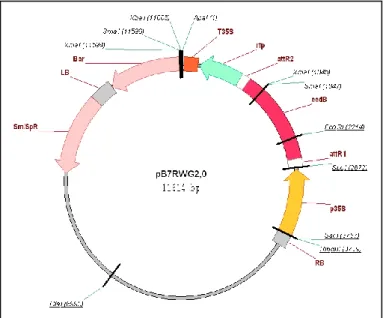
A genetic construct was made in order to overexpress OsRMC in plants and cell suspension cultures. This genetic construct was made using a GATEWAY (Life Technologies, USA)-based vector (pB7RWG2.0; VIB, University of Gent, Belgium) (Fig. 1) that allows our protein of interest to be fused in frame with the Red Fluorescence Protein (RFP) tag in the C-terminal and it has the Glufosinate-ammonium gene that confers tolerance to BASTA (Sigma-Aldrich, Germany) as the plant selective marker. The generated genetic construct was designated as pB7::OsRMC-RFP (courtesy of Tânia Serra, GPlantS). Also, the promoter present in this vector is the p35S promoter from the Cauliflower mosaic virus, which permits constitutive expression of the OsRMC-RFP transcriptional fusion.
Figure 1 – GATEWAY based vector used in the preparation of the genetic contruct
Arabidopsis cell suspension cultures
Arabidopsis thaliana cell suspension cultures (kindly provided by Prof. Laszlo Bogre, Royal Holloway, University of London, England), were used in all experiments as
22
genetic background. These cell suspension cultures were diluted on a weekly basis with fresh Arabidopsis Medium (see appendix for composition), in a laminar air flow chamber (Braun Horizontal BBH6). In brief, 10mL of a one week-old culture was diluted in 40mL of fresh Arabidopsis medium in a sterilized 250mL flask.
Arabidopsis plant material
Three week old Arabidopsis thaliana plants grown in soil (Shamrock Professional Range Specialist Pot Plant Medium, Ireland), ecotype Columbia-0 (Col-0), were used in all transformations with the floral dip method (modified from Clough and Bent, 1998). A total of two sets of transformations were performed with the floral dip method.
Agrobacterium tumefaciens transformation
Agrobacterium, strain LBA4404, was transformed with the vector pB7::OsRMC-RFP using the following protocol: 0.1-1µg of plasmid was added to frozen competent cells and the mixture was incubated for 5 minutes at 37ºC. The cells were then incubated on ice for 30 minutes and plated on LB solid medium (see appendix for composition) with the antibiotics Rifampicin and Spectinomicin at concentrations 25mg/L and 100mg/L respectively. The plates were grown on a chamber for 2-3 days at 28ºC. The colonies grown in this plate were re-streaked in a fresh LB solid medium with the same selection pressure as above. To further confirm the Agrobacterium transformation, we performed a colony-PCR to the re-streaked colonies with the appropriate primers to amplify OsRMC.
Agrobacterium tumefaciens growth
First an Agrobacterium 5mL pre-culture was made, inoculating a single colony into 5mL of LB medium supplemented with 25mg/mL of Rifampicin and 100mg/mL of Spectinomicin. The culture was grown at 28ºC with 220 rpm. From the pre-culture, a larger culture was established transferring 1-5mL of the pre-culture to 200 mL of LB
23
medium supplemented with the antibiotics Rifampicin and Spectinomicin at the same concentrations as above. The larger culture wan grown overnight at 28ºC with 220 rpm.
Arabidopsis thaliana transformation – Floral Dip Method
The Agrobacterium cultures were grown until an OD600nm of ~1 was achieved. At
this point, the bacteria were spinned down gently by centrifuging at 4.000xg for 10 minutes at room temperature. The pellet was ressuspended in a same volume of a freshly made solution composed of 5% sucrose, 10nM MgCl2 and 0.05% Silwet L-77. The
ressuspended solution was transferred to a 5L plastic beaker for the dipping phase of the transformation method. Above-ground parts of Arabidopsis thaliana plants were dipped in the Agrobacterium solutions with gentle agitation. The pots were labeled and the plants were wrapped in Saran Wrap, and laid on their sides overnight. The next day, the Saran Wrap was removed and plants were placed straight up and under standard growth conditions. In one of the transformations the whole dipping procedure was repeated 5 days after the first dipping. The plants received no more water when the siliques became mature, and the seeds were harvested.
Seed sterilization
Seeds were surface sterilized with a solution containing equal parts of double-distilled water and commercial bleach along with Tween-20 0.1% for 10 minutes at room temperature in an eppendorf tubes rotator. The next steps were performed in a horizontal laminar air flow chamber (Braun Horizontal BBH6). The seeds were washed with sterile double-distilled water for at least 7 times at room temperature. The seeds were then left in the last wash water and were stored in the dark at 4ºC for 2-3 days for stratification. After stratification, the selection of transformed seeds was made by plating them on MS Medium (see appendix for composition) supplemented with BASTA at a concentration of 20ug/L. BASTA-resistant seedlings would became visible after a week of growth at 22ºC with a 16h photoperiod.
24
Plant genotyping
Quick Plant Extract buffer was used to extract DNA from leaf of transformed Arabidopsis T0 or T1 plants. The following protocol was used: first 20μL of Quick Extract Plant Buffer (Epicenter Biotechnologies, USA) was added to more or less 1 mm2 of leaf tissue and the mixture was left to incubate for 6 minutes at 65ºC; next, the mixture was transferred to a new bath at 98ºC and let to incubate for 2 more minutes. The DNA extracted was used to perform PCR for the confirmation of insertion of the transgene. A total of 2µL of DNA extracted with the protocol mentioned above was used to perform the PCR.
Arabidopsis cell culture transformation
The cell suspension cultures were transformed using the protocol as follow: 100 µL of Agrobacterium tumefaciens LBA4404 grown to an optical density (OD600) of 0.8,
and carrying either the empty vector pB7RWG2.0, or the pB7::RMC-RFP, were added to 3 to 4 days old cell suspension cultures. After adding the Agrobacterium the cell suspension cultures were co-cultured for 3 days at 25ºC with agitation at 120 rpm. After the incubation with the Agrobacterium and on a weekly basis, the cultures were diluted and treated with Cefotaxime and BASTA at 500mg/L and 20µg/mL respectively. The concentration of Cefotaxime was gradually decreased in steps of 100mg/L each week, until 100mg/L was achieved. This antibiotic was suspended and no longer added to the cultures in the later stages after confirmation of Agrobacterium-free cultures.
Jasmonic acid assays
The wild type and transformed cell suspension cultures were used to perform assays with jasmonic-acid (JA). In these assays, 495µL of the 3-4 days old cultures were treated with 5µL of JA at concentrations ranging from 50µM to 500µM. The culture was left to incubate with the JA in dark conditions. The total volume of the treated culture were collected at different time points of 1h, 2.5h, 5h and 7h and centrifuged. The
25
supernatant was discarded and the cellular pellet was snap-frozen in liquid nitrogen and stored at -80ºC until use.
RNA extraction
For all total RNA extraction procedures the following protocol was used. This TRIzol RNA extraction Protocol uses TRIzol (Life Technologies, USA) as the main reagent.
The following protocol for total RNA extraction was used:
1 mL of TRIzol was added to 100 mg of grinded frozen samples immediately vortexed to allow the tissue to thaw in the TRIzol reagent. Then, the homogenate was passed over a qiashredder (Qiagen, USA) column by centrifuging at max speed for 30 seconds. The cleared homogenate was then centrifuged at 10.000xg for 5 minutes at room temperature. The liquid was quickly transferred to a new 2mL tube and incubated at room temperature for 5 minutes for complete dissociation of nucleoprotein complexes. After this incubation step, 200μL of chloroform was added to the solution and this was vigorously shaken for 15 seconds, and incubated at room temperature for 3 minutes. The solution was then centrifuged for 10 minutes at full speed, at room temperature. After centrifugation, different phases could be identified. The RNA, present exclusively in the clear upper-phase of the solution, was transferred to a new 2 mL tube. Isopropanol (500µL) was added to the RNA solution and incubated for 10 minutes at room temperature. Then, the solution was centrifuged for 10 minutes at 10.000xg at room temperature for precipitation of the RNA. The supernatant was discarded and the RNA-pellet was washed with 1mL of a 75% EtOH solution. Another centrifugation step of 5 minutes at 7.500xg at room temperature was performed. After discarding the supernatant the RNA pellet was dried for 10 minutes in a laminar air flow chamber The RNA was ressuspended in RNase-free water (treated with DEPC).
DNase treatment procedures (TURBO DNA-free Life Technologies,
USA)
26
First the DNase digestion reagents were added to the RNA samples: typically 0,1X volume of TURBO DNase Buffer and 0.75 µL of TURBO DNase, and the mixture was gently mixed. This mixture was incubated for 30 minutes at 37ºC. After the 30 minute incubation, 0.75 µL of TURBO DNase was again added to the RNA samples, and incubated for another 30 more minutes at 37ºC. After the second incubation: DNase Inactivation Reagent (typically 0.1 volumes) was added to the samples and mixed well. This mixture was incubated for 5 minutes at room temperature, mixing occasionally. After this incubation, the samples were centrifuged at 10.000xg for 1.5 minutes and after the RNA samples were transferred to a fresh tube. After this treatment to remove any contaminating DNA, all RNA samples were checked for their quality through gel electrophoresis and followed by approximate quantification using NanoDrop (Nanodrop 3330, ThermoScientific, USA)
cDNA synthesis
All cDNA was synthesized using a Transcriptor High Fidelity cDNA synthesis kit (Roche, Switzerland). The procedure to obtain cDNA from the RNA samples was carried exactly in the same way for all samples, and as described below.
Five hundred nanograms of total were used to produce cDNA. The first-strand using oligo-dT as primer. This mixture was incubated for 10 minutes at 65ºC in the thermocycler. After the ten minutes incubation a mixture composed of 4µL of buffer, 0.5µL of RNase inhibitor, 2µL of dNTPs, 1µL of DTT and 1.1µL of retrotranscriptase was added to each RNA sample. This mixture was then put in the thermocycler for the actual synthesis of the cDNA. The program steps for the cDNA synthesis can be viewed in the appendix:
27
We tested two protocols [Laccus Buffer and Protein Extraction Buffer (PEB) protocol] for protein extraction of the cell suspension cultures and the PEB protocol for the whole plants
The first protein extraction protocol was done with the Laccus Buffer (see appendix for composition). Briefly, 50µL of Laccus Buffer was added to each sample, and then centrifuged for 20 minutes at 10.000xg. The supernatant was passed to a new tube and stored at -20ºC until use.
The PEB protocol (see appendix for composition) allowed for a Bradford protein quantification assay and was used to extract total protein from transformed Arabidopsis seedlings. The following protocol for the total protein extraction with PEB was used: 10 days after germination seedlings were grinded in liquid nitrogen in a mortar until a fine powder was obtained. For each 100 mg of grinded material 200µL of PEB with 2x complete protease inhibitor was added and mixed well. The mixture was centrifuged at maximum speed at 4ºC for 15 minutes. The supernatant was transferred to a new 1.5mL tube and stored at -20ºC until use.
Protein quantification
Protein quantification was done using the Bradford reagent (Bio-rad, Germany). A calibration curve was made with BSA incubating the Bradford reagent for 30 minutes in BSA concentrations of 2.5, 1, 0.5, 0.25 and 0.1mg/mL. All samples were read on a plate reader with the Gen5 software, measuring absorbance at 595nm.
Western blot
Twenty micrograms of each sample total protein, mixed with Loading Buffer, were loaded per lane on a 12% SDS-PAGE and gels were blotted into PVDF membranes. OsRMC-RFP detection was carried out using a 1:1000 dilution (0.2µg/mL) of a polyclonal α-GFP antibody (200µg/mL Santa Cruz Biotechnology; USA) raised against rabbit (1:20000 dilution (0.52µg/mL), 1mg/mL from Abm Inc; Canada). Chemioluminescent
28
detection was performed with Western Lightning Plus-ECL (Perkin-Helmer, USA), according to manufacturer’s instructions. Coomassie Brilliant Blue staining was as total protein loading control.
Protoplast preparation
Protoplasts were prepared from wild type Arabidopsis cell suspension cultures, and from the transformed Arabidopsis cell suspension culture with the empty vector pB7RWG2.0 and the vector carrying the genetic construct pB7RWG::OsRMC-RFP. The following protocol was used: 3-4 days old Arabidopsis cell suspension cultures were collected by centrifuging (in a swing out rotor) in a 50 mL Falcon tube for 5 minutes at 1500rpm. After, the supernatant was discarded by decanting it. The cells were then mixed with 25mL of enzyme solution (see appendix for composition) and the falcon was filled up with B5-GM medium (see appendix for composition). The 50mL content was then split into two large Petri plates and to each of them another 25mL of B5-GM medium was added. The plates were then shaken carefully and slowly at 30-40 rpm/min for 3 hours in the dark. The protoplasts were checked in an inverted microscope to decide the time of harvest. When about 80% of the cells started to look spherical another 20-30 minutes were allowed for further shaking. After, the protoplasts were transferred to two 50mL falcon tubes. The protoplasts were then centrifuged for 5 minutes at 1500 rpm at room temperature. The supernatant was discarded by decanting it and the cellular pellet was ressuspended in 25mL of B5-GM medium. The cells were then centrifuged for 5 minutes at 1000rpm at room temperature. After, the supernatant was discarded and the cellular pellet was ressuspended in 5mL of B5 0.28M sucrose medium (see appendix for composition). The cells were transferred to 15mL falcon tubes and centrifuged for 7 minutes at 800rpm at room temperature. The floating cells were carefully transferred to a new 15mL falcon tube using a sterile plastic wide-mouth Pasteur pipette. The protoplasts were counted in an inverted microscope using a Fuchs-Rosenthal counting chamber and the concentration of protoplasts was calculated. A total of 3 different protoplasts preparation were prepared in this way, one for the wild-type culture, and one for each transformed culture. These protoplasts preparation were used to view in an inverted microscope to check for RFP localization. Also, the protoplasts were also used in a jasmonic acid assay.
29
Results
In this section of the chapter all results will be shown concerning the work done with Arabidopsis cell suspension cultures and whole plants.
Cell suspension cultures
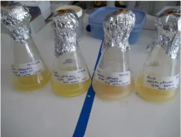
Figure 2 - Transformed Arabidopsis cell suspension cultures; the two flasks on the left correspond to the pB7RWG:: OsRMC-RFP cultures and the two on the right to the
pB7RWG2.0 cultures
The transformed (confirmed by PCR analysis in to the culture overexpressing OsRMC using specific primers; data not shown) Arabidopsis thaliana cell suspension cultures were used in a JA assay and were also used to produce protoplasts. For accurate measure, each transformed culture was kept in duplicate. These transformed cultures were used in only one JA assay, the first ones being performed with wild type cultures. The reason to start the JA assays only with the wild type cultures comes from the fact that the transformed cultures were still in the process of homogenization through rounds of dilution and treatment with BASTA, and because the JA assay still needed optimization in terms of concentration of JA to be used and times of exposure. These transformed cultures were very prone to contamination, especially the pB7RWG:: OsRMC-RFP cultures. Figure 2 shows one week old transformed cell suspension cultures,
30
where it can be seen that the growth of the cultures was not exactly the same for all cultures.
Gene expression analysis under JA
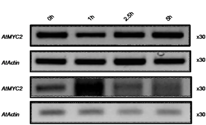
Figure 3- Transcript accumulation from 0 hours to 5 hours of AtMYC2 and AtACTIN as control; the top two panels refer to the first Jasmonic acid assay conducted
with wild type Arabidopsis cell suspension cultures; the bottom two panels refer to a repetition of the first assay; treatment - 50µM JA
Figure 3 shows results for the RT-PCRs obtained with cDNA from wild type Arabidopsis cell suspension cultures. Results for the gene MYC2 are shown at time points of 0, 1, 2.5 and 5 hours of treatment with 100µM of JA. In this assay RT-PCRs for the genes PDF1.2 and VSP were also performed. In the RT-PCRs mentioned above no bands were seen in the gels and so no results are shown for the PCRs for these genes. The RT-PCRs for the first assay showed that the gene MYC2 seems to be consistently transcribed along the 5 hours of treatment, with the same happening for the control gene Actin. This assay was repeated because no results were obtained for the genes PDF1.2 and VSP. The bottom two panels refer to the assay repetition. In this assay still no bands were visible for the genes PDF1.2 and VSP and also the results for the gene MYC2 seem consistent with the first assay, although in this case the cDNA appeared contaminated with genomic DNA.
31
Figure 4 - Transcript accumulation from 0 hours to 5 hours of AtMYC2 and AtACTIN as control; the top two squares refer to the first repetition of the Jasmonic acid
assay conducted with wild type Arabidopsis cell suspension cultures; treatment - 50µM JA
A third JA assay with wild type cultures was conducted, but in this case the treatment consisted of 100µM of JA. Still the RT-PCRs for the genes PDF1.2 and VSP resulted in no bands and the results for the MYC2 gene seem to be inconsistent with the first two assays, although in this case the gene Actin, which served as a endogenous control, did not show consistent accumulation of transcript at the different time points. This can be due to the fact that the concentration of cDNA for all time points was not homogenous.
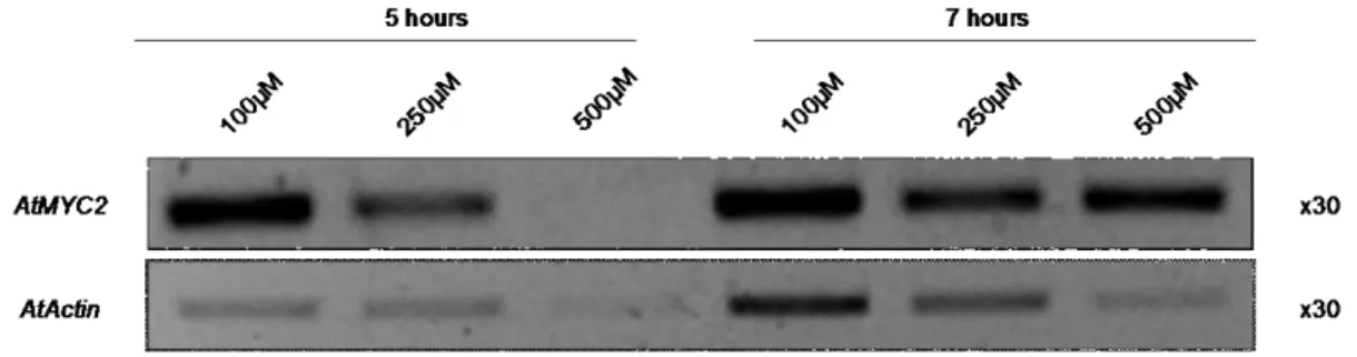
Figure 5 - semi-quantitative RT-PCR results for the gene AtMYC2; the concentrations refer to the JA used; the gene AtActin was used as a control
A fourth JA assay with wild type cell suspension cultures was performed. In this assay only time points of 5 and 7 hours were gathered but a range of concentrations of JA was used in this assay (see figure 5). Still no results for the genes PDF1.2 and VSP are shown because no bands were visible in the gels of all the RT-PCRs performed with primers for the genes mentioned above. As for the gene MYC2, there seems to be no consistent accumulation with respect to the treatment involved, either for the hours or
32
the concentration of JA used. The gene Actin still showed no consistent accumulation in the different treatments, and so this gene was discarded as a potential housekeeping gene to serve as control and another gene was selected, namely Tubulin.
Figure 6 – semi-quantitative RT-PCR results for the gene AtTubulin
In the final JA assay, RNA was extracted from the protoplasts produced from all the transformed and wild type cell suspension cultures and cDNA was produced. This gene was also not a good candidate because it showed different accumulation in the different treatments. RT-PCRs for all the JA-responsive genes analyzed in this work were also performed but showed no convincing results, and so no results are shown for this final assay.
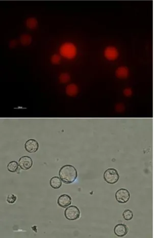
Protoplasts
Protoplasts were produced from the transformed Arabidopsis thaliana cell suspension cultures. These protoplasts were used in a JA assay but were also used to visualize the RFP protein in an inverted fluorescent microscope (Leica, Germany). The protoplasts of the pB7RWG2.0 culture confirmed that the RFP protein was being expressed (Fig. 7). No images for the protoplasts of the pB7RWG::OsRMC-RFP culture is
33
shown because there was no signal detected in the protoplasts of the mentioned culture. This negative result could be an indication of the sub-cellular localization of OsRMC.
Figure 7 - Protoplasts of pB7RWG2.0 culture. In these photos it is clear that the RFP gene is being transcribed and translated into the RFP protein. The image below is the bright field of the upper image; the upper image was gathered with a fluorescence
inverted microscope and with the filter TRITC
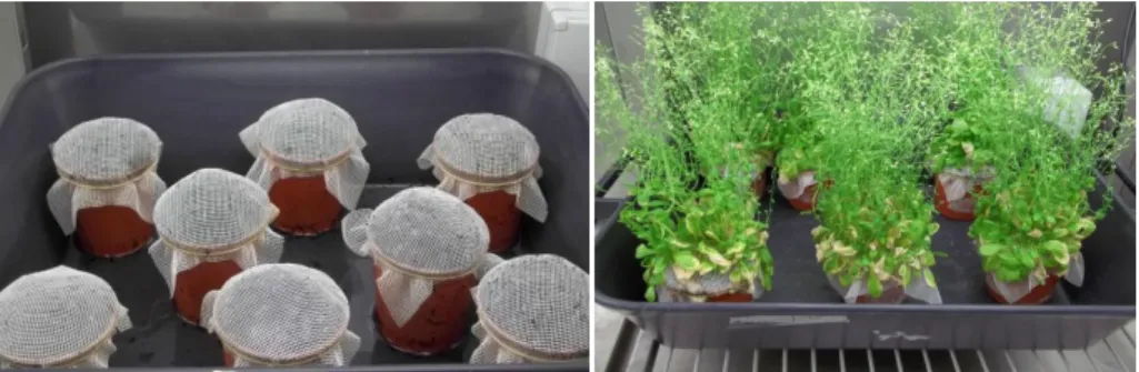
Arabidopsis plants
Arabidopsis thaliana Col-0 plants were used for the transformation with the floral dip method. In figure 8 (right panel) the plants are in their right stage to perform the floral dip.
34
Figure 8 - Pots where (left) the Arabidopsis Col-0 plants were grown and (right) at the stage of development to perform the floral dip method
In the first transformation with the floral dip method only one transformant was obtained and was designed line RMC#1. The seedling resistant to BASTA was transferred to soil (see figure 9) to produce more seeds.
Figure 9 - Transgenic line RMC#1 plant overexpressing the OsRMC gene
In order to access if line RMC#1 was truly transformed with the OsRMC gene DNA was extracted from the plant and a simple PCR was performed with primers for the OsRMC gene. In figure 10 it is shown that the DNA extracted from line RMC#1 showed some amplification for the gene.
35
Figure 10 - Genotyping of line RMC#1. Negative controls correspond to amplification from Arabidopsis col-0 genomic DNA; positive control
(pB7RWG::OsRMC-RFP) demonstrated over amplification
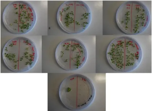
With the second floral dip method transformation two more lines overexpressing the OsRMC gene were obtained and these lines were designated line RMC#2 and RMC#3. Figure 6 shows the results for the segregation analysis for the mentioned lines. The main objective of this segregation analysis was to access if any of the sublines was homozygous for the gene. It is shown that all seeds plated in MS medium with BASTA for the lines RMC#2.5 and RMC#3.1 germinated and grow, demonstrating that these sublines are homozygous for the gene. In this segregation analysis the seeds were also plated in non-selective medium (MS medium without BASTA) to access possible problems with germination of the seeds.
In the second floral dip method transformation also two positive seedlings (resistant to BASTA) were gathered for the empty vector (pB7RWG2.0). These seedlings were also transferred to soil to produce more seed. The plants obtained from these seedlings produced very few seeds and only one of these seeds germinated again under selective medium (see figure 11 bottom). This seedling was not transferred to soil because it showed abnormal phenotype in the plate. This abnormal phenotype was mainly characterized by very short roots and may be due to the local of insertion of the construction in the plant genome.
36
Figure 11 - Segregation analysis of lines RMC#2 and RMC#3
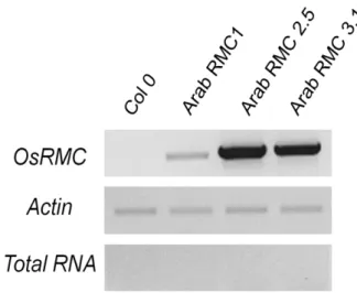
RNA was extracted from all the three obtained lines, RMC#1 and homozygous lines RMC#2.5 and RMC#3.1 and cDNA was produced from the RNA of these lines. The figure 12 shows results for the RT-PCR from the three lines, with Actin as an endogenous control and cDNA from Col-O plants as a negative control. The figure 12 shows that the three lines transcribe the gene OsRMC although the accumulation of the transcript is clearly different in the three lines, with line RMC#2.5 being the one that shows higher accumulation, and line RMC#1 showing very little accumulation. The fact that line RMC#1 shows very little accumulation of the transcript may be due to the fact that this line is an heterozygous line.
Figure 12 - Analysis of transcription of OsRMC in Col-0, RMC#1 (heterozygous) and lines RMC#2.5 and RMC#3.1 (homozygous)
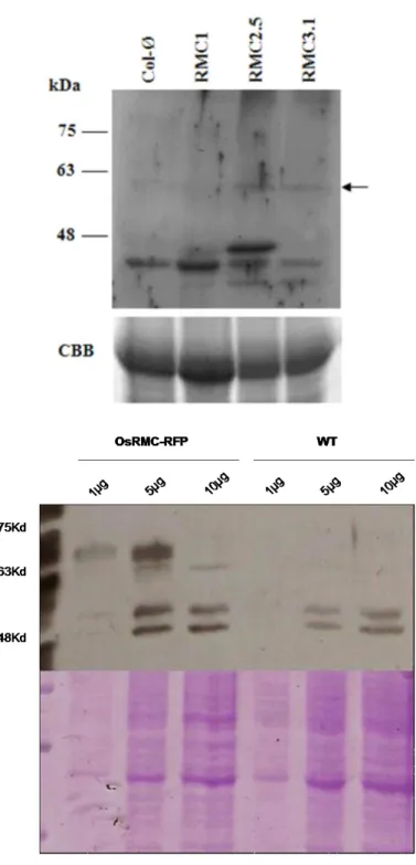
Western Blots
A western blot was also conducted with protein extracts from the transgenic lines and Col-0 in order to access if the gene was being transduced. The OsRMC gene was transcriptionally fused with the tag RFP in this construction, and so, the detection of the OsRMC gene was done with an antibody to detect the tag. Although no antibody was available in the lab specifically for the RFP gene, there was one available for the GFP gene, and since these two proteins (RFP and GFP) share considerable similarity, and
37
since the antibody used in this assay was a polyclonal one, some results could still be obtained.
Figure 13 shows bands below the 63kDa band in the protein extracts from lines RMC#2.5 and RMC#3.1 that were absent for the protein extracted from Col-O plants, showing that the fused protein RMC-RFP could still be detected in this western blot. Although we could not detect any band for the RMC#1 line this could be due to low OsRMC gene expression observed in the RT-PCR analysis (Fig. 12)
38
Figure 13 - Western Blot with anti-GFP polyclonal antibody to detect RFP; protein was extracted from transformed Arabidopsis line RMC#1, RMC#2.5, RMC#3.1 and Col-0. . The arrow on the right of the panel shows the bands corresponding to the OsRMC-RFP fusion protein (~57-60kDa). Coomassie Brilliant Blue (CBB) staining was used as loading
39
Discussion
Arabidopsis cell suspension cultures
The Jasmonic acid assays were repeated a total of three times due to lack of consistent results in the analysis of gene expression of Jasmonic acid responsive genes. These assays still needed to be optimized in order to obtain good quality cDNA for the RT-PCRs. Although much optimization will be needed in these assays in order to do a gene expression analysis much of the optimization does not concern, in my opinion, the way the assay was performed, but rather the quality of the material used in the assay.
The lack of consistent results in this part of the work may be explained by several reasons. First the transformed cell suspension cultures were not homogenously transformed, despite several weeks growing in selective medium with BASTA. The Arabidopsis cell suspension cultures were co-cultured with the Agrobacterium for 3 days, and despite this method proved successful in obtaining good transformed cultures with Arabidopsis cell suspension cultures with other work done previously in the laboratory by other colleagues, it seemed that these cultures were not efficiently transformed/homogenous to proceed with the Jasmonic-acid assays and all the work done after obtaining the samples. We cannot also discard the hypothesis of the influence of the OsRMC gene in the transformed cell cultures, as these cultures would grow poorer and were prone to contaminate more easily. It is thus tempting to speculate that this gene may have an influence in the cellular response of Arabidopsis cell suspension cultures affecting the responses to biotic stress (which may be related to JA sensitivity)
Another reason for the lack of consistent results in this part of the work was the RNA extracted from the samples. The RNA extracted from all the time points, and even in the 0h time point, was of low quality and in low concentrations. The cDNA synthesized from this RNA suffered from the same problems and so the RT-PCRs
The RT-PCRs for the genes VSP and PDF1.2 did not show any transcript accumulation in the gel. Only the gene AtMYC2 showed bands, which showed show very little differences in terms of accumulation. There seems to be a decrease in accumulation