UNIVERSIDADE DE LISBOA
FACULDADE DE CIÊNCIAS
DEPARTAMENTO DE BIOLOGIA VEGETAL
Characterization of bacteria isolated from
Portuguese traditional cheeses
Patrícia Andrea Bastião Rocha
Mestrado em Microbiologia Aplicada
Dissertação orientada por:
Teresa Maria Leitão Semedo Lemsaddek
Ana Maria Gonçalves Reis
This Dissertation was fully performed at Centre for Interdisciplinary
Research in Animal Health (CIISA), Faculty of Veterinary Medicine of the University of
Lisbon, under the direct supervision of Teresa Maria Leitão Semedo-Lemsaddek.
Professor Ana Maria Gonçalves Reis was the internal supervisor designated in the
scope of the Master in Applied Microbiology of the Faculty of Sciences of the University
of Lisbon
Agradecimentos
Em primeiro lugar, agradeço à Doutora Teresa Semedo Lemsaddek por me dar a oportunidade de realizar este trabalho, por todo o conhecimento transmitido, pela paciência, compreensão e apoio durante todos estes meses.
À minha orientadora interna, a Professora Doutora Ana Maria Gonçalves Reis, agradeço a disponibilidade, o apoio e a calma que me proporcionou principalmente durante os últimos meses de trabalho.
Agradeço a todos aqueles que me ajudaram e apoiaram na FMV: Professor Doutor António Salvador Barreto, Professora Doutora Maria João Fraqueza, Engenheira Maria José Fernandes, Maria Helena Fernandes. Também gostaria de agradecer à Esther Nataly Baptista Batista pela paciência, a ajuda incansável e as horas partilhadas de trabalho e conversa. E às colegas Joana e Margarida que ajudaram a realizar parte do trabalho.
Às minhas colegas e amigas Maria Inês Elias e Carolina Almeida pelas tardes de desabafo e partilha de aventuras, pelo apoio incondicional e pela diversão apesar de dissertações e outros dramas.
Agradeço de todo coração aos meus pais, ao meu primo Daniel e ao resto da família pelo apoio ao longo da minha vida e em todos os meus projetos. À minha “squad” que apesar da distância nunca deixaram de me apoiar e nos piores momentos senti sempre que ia conseguir porque vos tinha ao meu lado. Em especial, à Isa por nunca deixar de me apoiar, ouvir e de me oferecer a força que precisava nestes últimos meses. Obrigado a todas e a todos.
Abstract
Azeitão and Nisa cheeses are products with protected designation of origin (PDO) traditionally manufactured in Portugal with sheep’s raw milk. The predominant group of bacteria in cheeses is lactic acid bacteria (LAB) which includes genera like Lactococcus spp. and Enterococcus spp., these are the microorganisms our work will be focusing on. The fact that these bacteria have important roles both in foods and the gut of animals (including humans) makes them conductors for both positive impact, such as probiotic features, and negative impact, such as transference of virulence factors or antibiotic resistances. Our aim with this work was to analyse the diversity of LAB in Azeitão and Nisa cheeses from several origins and years of production and assess the negative traits these bacteria could have and transfer such as antibiotic resistances, hemolysis and production of gelatinase.
Enumeration of previously mentioned bacteria was performed, and results were compared to other years of production. During the years studied no unit had consistently higher CFU counts. Lactococcus spp. was the group with highest bacteria counts in the majority of units during the three years studied and Enterococcus spp. was the one with the lowest CFU. For diversity analysis, RAPD-PCR were performed in order to create dendrograms for each cheesemaking unit and bacterial group. Just like for bacterial enumeration, no specific trends were observed in the diversity values for the units throughout the studied years. Concerning 2018 cheeses, the group with the highest diversity was Enterococcus spp. even though it was also the one with less CFU. This independence between CFU counts and diversity was noted all through our work in different years, units and groups of bacteria. Identification of 2017 enterococci representatives was performed using a multiplex PCR and showed a predominance of E.
faecium, followed by E. faecalis and E. durans that were equally represented.
Pathogenic potential of representative isolates was assessed through the search of antibiotic resistances and virulence factors. Antibiotic susceptibility was studied through disc diffusion assays. Significant differences were observed in the number of resistances found in studied years for lactococci and enterococci. Between units there was also significant differences in the total number of resistances during 2016, 2017 and 2018 in enterococci. In LAB isolates resistances considered non intrinsic were found, to clindamycin and erythromycin. Furthermore, enterococci isolates resistant to teicoplanin, ciprofloxacin, tetracycline, chloramphenicol, erythromycin and vancomycin were observed. Isolates resistant to three or more antimicrobial agents were observed but these didn’t comply with the characteristics necessary to be classified as multidrug-resistant (MDR) bacteria. Virulence factors were studied and hemolysis was detected in 11% of representative isolates from 2016, 6% from 2017 and 12% in isolates from 2018 cheeses. Furthermore, only two positive results for gelatinase were found in 2017 representative isolates.
In conclusion, our results showed that no pattern of bacterial enumeration or microbiome diversity is present throughout different years of production in artisanal cheeses such as the ones studied. Moreover, although alarming resistances were found in enterococci no multi-drug resistance isolates were observed. Further work should be performed to continue the characterization of the pathogenic potential of isolates present in these cheeses.
Keywords: traditional Portuguese cheese; protected designation of origin; lactic acid bacteria;
Resumo
A fermentação tradicional de queijos é realizada em muitos países do mundo, nomeadamente Portugal, sendo que consiste no aproveitamento dos microrganismos naturalmente presentes no leite cru. Estes microrganismos encontram-se no leite devido às diferentes etapas de colheita e manuseamento durante e após esse processo. Sendo que, alteram as moléculas presentes no leite através da fermentação dando novas propriedades ao produto final que será posteriormente curado. As condições destas fases de fermentação e maturação diferem entre tipos de queijos e zonas de produção, determinando as propriedades organoléticas do produto final.
Os queijos de Azeitão e Nisa fazem parte dos 10 queijos portugueses que possuem a categoria de denominação de origem protegida (DOP). As zonas de produção dos queijos de Azeitão são Palmela, Sesimbra e Setúbal e as de Nisa são Nisa, Crato, Castelo de Vide, Marvão, Portalegre, Monforte, Arronches e Alter do Chão. Ambos são produzidos com leite de ovelha cru. No caso de Nisa o leite provém de uma raça de ovelha chamada Merina Branca e no caso de Azeitão não se especifica uma raça. O coalho vegetal usado na produção de ambos queijos é obtido de Cynara cardunculus L. Devido às diferentes fases e condições de produção o queijo de Azeitão consiste numa pasta semi-mole e amanteigada, de cor branca ou ligeiramente amarelada com um sabor ácido e salgado. O queijo de Nisa consiste numa pasta semi-dura de cor branca amarelada com um sabor ligeiramente ácido e um cheiro intenso.
As bactérias ácido lácticas (BAL) são um grupo de vários géneros que partilham características em comum como serem gram-positivas, catálase negativas, não formam esporos, anaeróbicas facultativas e terem um nível G+C baixo. Para além disto, o seu nome provém da sua capacidade para fermentar açucares, transformando-os em ácido láctico através de homo- ou heterofermentação. Este grupo é o predominante em leite cru e, portanto, em queijos produzidos de forma artesanal e essas bactérias vão desempenhar diversos papéis ao longo da fermentação e maturação deste tipo de queijo. Os géneros de BAL que predominam em comidas fermentadas como os queijos são Lactococcus, Streptococcus,
Pediococcus, Leuconostoc, Lactobacillus e Enterococcus. Tendo em conta que estas bactérias formam
parte tanto da cadeia alimentar como da microbiota de animais e seres humanos podem servir como veículo de transmissão de genes ao existir uma transferência genética entre espécies ou géneros, como descrito na literatura. A problemática desta possível transferência ocorre quando esses genes conferem resistência a antibióticos ou fatores de virulência que anteriormente essas bactérias não possuíam. Essa transferência pode ocorrer para algumas bactérias que formam parte da nossa microbiota e são responsáveis por infeções oportunistas ou para bactérias patogénicas presentes no nosso corpo devido a uma infeção. Resistências adquiridas a antibióticos têm sido estudadas e observadas em BAL, em concreto em Enterococcus spp. devido ao seu papel patogénico oportunista. Foram descritas resistências a antibióticos de diferentes classes como ß-lactâmicos, cefalosporinas, aminoglicósidos, lincosamidas e estreptograminas. Em concreto, resistências à tetraciclina e eritromicina são das mais preocupantes devido à importância destes antibióticos e porque esta resistência tem sido atribuída ao uso indevido de antibióticos em comida de animais.
Este trabalho teve como objetivo continuar o estudo de queijos de Azeitão e Nisa DOP que começou numa dissertação anterior onde se caracterizaram estes queijos pela sua microbiota e propriedades físico-químicas. Seguido deste estudo foi também analisada a diversidade dos microbiomas destes produtos como parte de outra dissertação. Assim, o presente trabalho consistiu em comparar os resultados anteriores de diversidade e caracterização microbiológica assim como completar essa análise com a procura de resistências a antibióticos e fatores de virulência.
Durante o ano de 2018 foram recolhidos queijos de seis queijarias de Azeitão e de duas queijarias de Nisa. A partir destes foram feitas contagens de unidades formadoras de colónias (UFC) e isoladas as
Lactobacillus spp.), Lactococcus spp. com meio M17 e Enterococcus spp. isoladas com meio SBA.
Depois foi extraído o ADN desses isolados para realizar a técnica de RAPD-PCR e análise de dendrogramas criados com os perfis de bandas obtidos. A partir estes dendrogramas foi analisada a semelhança entre os isolados dos distintos queijos e foram obtidos índices de diversidades dos diferentes grupos de bactérias e queijarias estudadas. Esta diversidade e as contagens de bactérias foram comparadas às obtidas noutras dissertações deste mesmo projeto nas quais se estudaram anos de produção anteriores. Partindo destes mesmos dendrogramas, foram escolhidos isolados representantes de cada queijaria e realizados testes de resistência a antibióticos assim como a presença de fatores de virulência.
Relativo aos diferentes anos de produção estudados não foi observada uma tendência clara de contagens de UFC quando comparados os anos ou queijarias, isto é, não houve queijos que tivessem consistentemente maior ou menor número de bactérias sendo que este número foi variável ao longo dos anos. Enquanto à diversidade, foram observadas poucas coincidências nas queijarias com maior ou menor índice de diversidade. No caso dos lactococos foi observado o menor índice correspondente aos anos 2018 e 2016 na mesma queijaria, A4. Nos enterococos foi também encontrada a menor diversidade na mesma queijaria, N9, nos queijos dos anos 2018 e 2016. Nas amostras de 2018 o grupo com maior diversidade foi o dos enterococos mas cabe destacar que os três grupos bacterianos tiveram índices muito parecidos. A identificação dos representantes deste grupo de bactérias dos queijos de 2017 foi realizada com uma multiplex PCR e a maioria de isolados foi identificado como E. faecium, seguido por E. faecalis y E. durans estando igualmente representados em número de isolados.
Foram encontradas diferenças significativas no número de resistências ao longo dos anos nos grupos
Lactococcus spp e Enterococcus spp. No caso deste último género, ocorreram também diferenças
significativas entre o número de isolados resistentes de diferentes queijarias durante os três anos estudados. As resistências a antibióticos encontradas no grupo de BAL foram à clindamicina e eritromicina, no caso dos lactococos as resistências que foram observadas neste trabalho são consideradas intrínsecas pelo que não haveria possibilidade de transferência dessas resistências para outras bactérias. As resistências extrínsecas encontradas nos enterococos foram à teicoplanina, ciprofloxacina, tetraciclina, cloranfenicol, eritromicina e vancomicina. Apesar de terem sido observados isolados resistentes a três antibióticos de três classes diferentes estes não cumpriam todos os requisitos de modo a serem considerados multirresistentes. No estudo de fatores de virulência foi detetada a capacidade de hemólise em 11% dos representantes de 2016, 6% nos de 2017 e 12% nos de 2018. No teste para identificar a produção de gelatinase só foram observados dois resultados positivos e esses pertenciam a isolados de queijos de 2017.
Concluindo, a diversidade do microbioma e as contagens dos queijos de Azeitão e Nisa estudados não seguiram nenhum padrão nos anos que foram comparados. O número de Enterococcus spp. resistentes diminuiu desde 2016 até ao último ano estudado neste trabalho, 2018, mas as resistências encontradas nos isolados de 2018 foram a antibióticos mais relevantes para a saúde pública como são a vancomicina, eritromicina e tetraciclina. No mesmo sentido, também não foram encontrados isolados multirresistentes e os fenótipos dos fatores de virulência estudados não foram observados em grande quantidade. Contudo, deve ser referido que o estudo destes queijos continuará e serão feitos mais testes tanto no âmbito da diversidade como do potencial patogénico dos isolados das bactérias ácido lácticas presentes.
Palavras-chave: queijo tradicional Português; denominação de origem protegida; bactérias ácido
Index
1- INTRODUCTION ... 1
1.1. Cheese characteristics and manufacture ... 1
1.1.1. Azeitão and Nisa protected designation of origin (PDO) cheeses ... 2
1.1.2. Fermentative bacteria ... 2
1.1.2.1. Fingerprinting and identification of LAB ... 4
1.2. Pathogenic potential in cheese microbiota ... 5
1.2.1. Antibiotic resistance ... 5
1.2.2. Virulence factors ... 7
1.3. Aims of the study ... 7
2- MATERIAL AND METHODS ... 8
2.1. Conventional microbiological procedures ... 8
2.2. Molecular procedures ... 9 2.2.1. DNA extraction ... 9 2.2.3. Genomic diversity ... 10 2.2.3.1. RAPD-PCR ... 10 2.2.3.2. Data analysis ... 10 2.3. Antibiotic resistance ... 11
2.4. Assessment of virulence factors ... 12
3- RESULTS AND DISCUSSION ... 12
3.1. Enumeration of bacteria ... 12
3.1.1. Lactic acid bacteria: comparison between production years and cheesemaking units 12 3.1.2. Genus and species identification ... 15
3.1.3. Other microorganisms of interest ... 16
3.2. Cheese microbial diversity ... 17
3.2.1. Comparison between production years ... 17
3.2.1.1. Lactic acid bacteria ... 17
3.2.1.2. Lactococcus spp. ... 19
3.2.1.3. Enterococcus spp. ... 21
3.2.2. Comparison between production units and groups of bacteria ... 23
3.3. Evaluation of pathogenicity potential ... 24
3.3.1. Antibiotic resistance ... 24
3.3.1.1. Lactic acid bacteria ... 25
3.3.1.2. Lactococcus spp. ... 26
3.3.1.3. Enterococcus spp. ... 28
3.3.2. Virulence factors ... 33
4- CONCLUSIONS ... 34
5- REFERENCES ... 36
APPENDIX A – DENDROGRAM OF ENTEROCOCCUS SPP. ISOLATES ... 41
APPENDIX B – DENDROGRAM OF LACTOCOCCUS SPP. ISOLATES ... 45
Tables and figures list:
Table 2.1. Growth media, incubation details and characteristic colonies to consider for enumeration. Table 2.2. PCR amplification details for enterococcal genus and species identification.
Table 2.3. Antibiotics used for disc diffusion assays.
Table 3.1. Chi-square test results of resistant isolates obtained through 2016 and 2017 LAB isolates analyzed and between cheesemaking units.
Table 3.2. Chi-square test results of resistant isolates obtained through 2016 and 2017 Lactococcus spp. isolates analyzed and between cheesemaking units.
Table 3.3. Chi-square test results of resistant isolates obtained through 2016, 2017 and 2018 in
Enterococcus spp. isolates analyzed and between cheesemaking units.
Figure 3.1. Enumeration of lactic acid bacteria. Figure 3.2. Enumeration of Lactococcus spp. Figure 3.3. Enumeration of Enterococcus spp. Figure 3.4. Enumeration of yeast and molds.
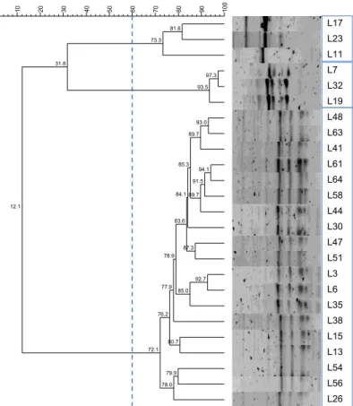
Figure 3.5. Dendrogram of LAB isolates from A4 unit (2018) with 60% similarity cut-off and formed clusters indicated.
Figure 3.6. Dendrogram of LAB isolates from A5 unit (2018) with 60% similarity cut-off and formed clusters indicated.
Figure 3.7. Dendrogram of Lactococcus spp. isolates from N10 unit (2018) with 60% similarity cut-off and formed clusters indicated.
Figure 3.8. Dendrogram of Lactococcus spp. isolates from A4 unit (2018) with 60% similarity cut-off and formed clusters indicated.
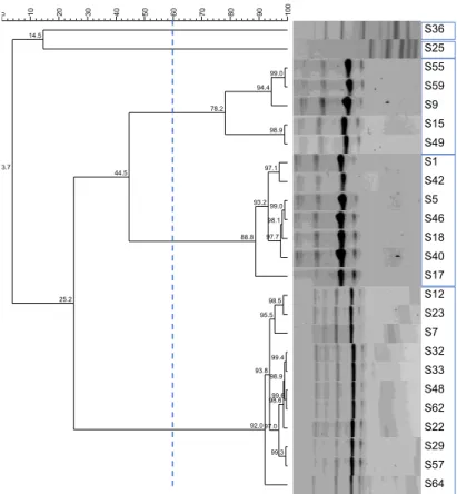
Figure 3.9. Dendrogram of Enterococcus spp. isolates from N10 unit (2018) with 60% similarity cut-off and formed clusters indicated.
Figure 3.10. Dendrogram of Enterococcus spp. isolates from N9 unit (2018) with 60% similarity cut-off and formed clusters indicated.
Figure 3.11. Lactic Acid Bacteria (LAB) antibiotic resistance frequencies in 2016 (A) and 2017 (B) for representative isolates.
Figure 3.12. Lactococcus spp. antibiotic resistance frequencies in 2016 (A) and 2017 (B) for representative isolates.
Figure 3.13. Enterococcus spp. antibiotic resistance frequencies in 2016 (A), 2017 (B) and 2018 (C) cheeses isolates, according to EUCAST breakpoints.
Figure 3.14. Enterococcus spp. resistance frequencies in 2016 (A), 2017 (B) and 2018 (C) cheese isolates, according to CLSI breakpoints.
Figure 3.15. Percentage of ß-hemolytic in representative Enterococcus spp. from 2016 (34 isolates), 2017 (31 isolates) and 2018 (58 isolates).
List of relevant abbreviations:
CFU – Colony-forming units
CLSI - Clinical Laboratory Standards Institute DNA – Deoxyribonucleic acid
EUCAST - European Committee on Antimicrobial Susceptibility Testing FDA – Food and drug administration
HE – Hektoen enteric
HGT – Horizontal gene transfer LAB – Lactic acid bacteria MDR – Multi-drug resistance MRS – Man Rogosa and Sharpe PCR – Polymerase chain reaction PDO – Protected designation of origin
RAPD – Random amplification of polymorphic DNA SBA – Slanetz and Bartley agar
TSA – Trypticase soy agar TSI – Triple sugar ion
VRE – Vancomycin-resistant enterococci XLD – Xylose lysine deoxycholate
1- Introduction
1.1. Cheese characteristics and manufacture
Fermented foods and beverages have been produced and consumed by humans since the first civilizations. In particular, cheese may be one of the most important fermented milk products and all around the world there are a lot of cheeses still made through a traditional process of fermentation and maturation. According to Codex Alimentarius (Codex standard 283, 1978) cheese is the ripened or unripened soft, semi-hard, hard, or extra-hard product that may be coated, and in which the whey protein/casein ratio doesn’t exceed that of milk. It is obtained by coagulating wholly or partly the protein of milk, skimmed milk, partly skimmed milk, cream, whey cream or buttermilk, or any combination of these materials, through the action of rennet or other suitable coagulating agents. Partially the whey resulting from the coagulation is drained, while respecting the principle that cheese-making results in a concentration of milk protein (in particular, the casein portion). Consequently, the protein content of the cheese will be distinctly higher than the protein level of the blend of the above milk materials from which the cheese was made.
Inside a healthy udder milk is sterile, but the nutrient composition of milk makes it a good growth medium for many microorganisms. Milk contamination happens from three sources: from within the udder, exterior of the udder and/or from the surface of milk handling and storage equipment (Quigley et al., 2013). The most represented group of bacteria in raw milk are lactic acid bacteria (LAB), which are bacteria that ferment lactose to lactate. Within this group the dominant genera in raw milk include
Lactococcus, Lactobacillus, Leuconostoc, Streptococcus and Enterococcus. The different microbiota
associated to the fermented raw milk to obtain cheese has a direct impact in its organoleptic properties, quality and time until spoilage (Devirgiliis, Zinno, & Perozzi, 2013; Quigley et al., 2013).
The traditional process of fermentation in cheese is essentially the exploitation of the microorganisms naturally present in raw milk, meaning that no starter microbial cultures are added. These microorganisms will break down complex molecules into simpler ones through fermentation, giving new properties to the final product that will be further enhanced with subsequent production stages, such as cheese maturation (Macori & Cotter, 2018). Differences between cheeses, Serra da Estrela and Irish cheeses, manufactured from pasteurized or raw milk have been studied because the replacement of natural microbiota for starter cultures eventually changes the sensory properties of such cheeses (Macedo, Tavares, & Malcata, 2004; Quigley et al., 2012).
Starter bacteria need a proteolytic system to hydrolyze the milk proteins to the amino acids and peptides required for bacterial growth. This proteolysis and the capacity to produce acid rapidly are important properties of these bacteria, since it will help reduce the propensity of spoilage and the development of flavor in cheese (Cogan et al., 1997).
Due to the fact that this is a traditional process of manufacture, differences between cheesemaking units are expected and are likely to be related to different methods, raw materials and handling which can affect the final concentrations or presence of certain groups of microorganisms. This will also affect the organoleptic properties of the cheeses and emphasize variation between cheeses from different units (Pintado et al., 2008). In a study with traditional raw milk Camembert cheeses (Henri-Dubernet, Desmasures, & Guéguen, 2008) variations were observed between units where lactobacilli species had a high diversity and its dynamics varied among those dairies contributing to a specific microbiota in each cheese. Differences in bacterial development was also observed in Nostrano di Primiero cheeses (Poznanski, Cavazza, Cappa, & Cocconcelli, 2004) that were manufactured with raw milk from different regions.
1.1.1. Azeitão and Nisa protected designation of origin (PDO) cheeses
In Portugal the artisanal production of regional cheeses is an important part of cultural heritage and 10 of those traditional cheeses have PDO (https://europa.eu/rapid/press-release_IP-96-153_en.htm. Consulted: August 28, 2019). In this work we will be focusing in two of those PDO cheeses: Azeitão and Nisa. Production of Azeitão cheese is restricted to counties of Palmela, Sesimbra and Setúbal whereas Nisa cheese is produced in Nisa, Crato, Castelo de Vide, Marvão, Portalegre, Monforte, Arronches and Alter do Chão.
PDO Azeitão and Nisa cheeses are obtained from sheep raw milk, in Nisa’s case the milk comes from a concrete breed of sheep called Merina Branca while in Azeitão’s cheese no breed is specified. Vegetable rennet used in both cheeses is obtained from Cynara cardunculus L.
Production method for Azeitão cheese begins by filtering the raw milk, after this the rennet and salt are added, and the milk is stored at 30ºC for 45 min. After coagulation is completed the serum excess is manually removed and a compressed bulk is obtained, which remains 20 days at 10ºC-12ºC under a relative humidity of 85% to 90%.
On the other hand, the production method for Nisa cheese consists in adding the vegetable rennet to the milk at 25ºC-28ºC during 60 min. After this some of the serum is removed and the salt is added. The maturation consists of two phases, the first one is up to 18 days long at 8ºC-10ºC with a relative humidity of 80%-90% while the second phase lasts up to 40 days at 10ºC-14ºC and a relative humidity of 85%-90%.
As a consequence of these different production methods Azeitão cheese is semi-soft and buttery with a characteristic spicy, acidic and salted flavor, while Nisa cheese is semi-hard, with a slightly acidic flavor and intense smell. Both cheeses have white or slightly yellow color (https://tradicional.dgadr.gov.pt. Consulted: June 15, 2019).
1.1.2. Fermentative bacteria
Lactic acid bacteria (LAB) constitute a group of multiple genera that share physiological features and owe their designation to the capacity to ferment sugar into lactic acid through homo- or heterofermentative metabolism. This group is characterized by being Gram-positive, catalase negative, non-spore forming, facultative anaerobic and having low G+C content. Their natural habitats are usually nutritionally rich environments, like plants and animal raw materials, fermented food products, animal skin and mucous membranes (Settanni & Moschetti, 2010). Concerning cheese production these bacteria can play different roles such as participation in the fermentation process or maturation of cheese. As mentioned before, proteolysis is very important in cheese production for final texture and flavor. Hence, LAB possess a complex proteolytic enzymatic system and plays an important role in degradation of casein and peptides producing free aminoacids that contribute directly to the basic taste of cheese and indirectly to the production of volatile aroma compounds (Herreros, Fresno, González Prieto, & Tornadijo, 2003). Apart from their role in texture, flavor and smell development these microorganisms have also a protective role in improving food safety and as probiotic bacteria that confer health benefits for humans (Settanni & Moschetti, 2010). The most relevant LAB in fermented foods belong to the genera Lactococcus, Streptococcus, Pediococcus, Leuconostoc and Lactobacillus. Likewise, several species from these groups are part of the gut microbiota of healthy humans (Devirgiliis et al., 2013).
In a review (Quigley et al., 2013), about the microbiota of raw milk it was documented that in sheep milk, which is very used throughout Europe for cheese production, LAB is the predominant group of bacteria. The genera that dominates cheeses produced with this milk are lactococci, lactobacilli and leuconostoc.
Pico cheese is a Portuguese PDO cheese (Domingos-Lopes, Stanton, Ross, Dapkevicius, & Silva, 2016), although manufactured with raw cow milk all its process is traditional and the predominant genera from LAB are the same as mentioned before. However, in this case Enterococcus genus was more dominant than in previously mentioned cheeses. Terrincho cheese is also a PDO cheese traditionally manufactured with raw sheep milk. In this type of cheese (Pintado et al., 2008)
Lactobacillus spp. and Lactococcus spp. were the predominant genera and enterococci were also found
at considerable high numbers. São Jorge is a traditional cheese from Azores island (Portugal) (Kongo, Ho, Malcata, & Wiedmann, 2007) produced with raw bovine milk in which lactobacilli and enterococci were identified as the dominant groups of bacteria found in all phases of production. Manchego cheese, for example, is produced in Spain and some of it is manufactured from raw sheep milk. It has been documented that Lactococcus spp., Lactobacillus spp. and Leuconostoc spp. are predominant while
Enterococcus spp. is also present but not as much as the aforementioned (Cabezas, Sanchez, Poveda,
Seseña, & Palop, 2007; Nieto-arribas et al., 2011).
Impact of different LAB in cheese’s microbial community has also been studied. For example, in a review about Serra da Estrela cheeses (Macedo et al., 2004) it was observed that the addition of
Lactococcus lactis and/or Lactobacillus plantarum reduces the numbers of Enterobacteriaceae but
changes the flavor of Serra cheeses reinforcing the idea that the presence and proportion of different groups of bacteria is important to the unique characteristics of each cheese.
Enterococcus spp. is the most controversial genus in LAB due to its concerns regarding food safety
and antibiotic resistances and because of this it’s also one of the most studied. High levels of these bacteria usually result from poor hygienic practices during manufacture, but it has been proven they play a major role in ripening and aroma development in many cheeses such as Manchego, Mozzarella, Kefalotyri, Serra da Estrela or Cebreiro (Franz, Holzapfel, & Stiles, 1999). Persistence of these bacteria during stressful stages like ripening can be attributed to their wide range of growth temperatures, high tolerance of heat, salt and acid (Cogan et al., 1997). Although some Enterococcus spp. are associated to human diseases they have also an important role in food safety due to their capacity to produce enterocins that inhibit other pathogenic bacteria (Moraes et al., 2012). Moreover, much like other LAB this genus has also probiotic characteristics beneficially affecting the host by improving the properties of its microbiota (Franz et al., 1999).
Fermentative bacteria present in raw milk, and subsequently in cheeses manufactured with that milk, have different roles during fermentation. Primary role of Lactotoccus spp. is the acidification of cheese through the production of L-lactate but they also contribute to proteolysis, conversion of amino acids into flavor compounds, citrate utilization and fat metabolism (Smit, Smit, & Engels, 2005).
Lactobacillus helveticus is known to have a rapid autolysis resulting in the release of intracellular
enzymes and reduction in bitterness which leads to increased and desirable flavor notes in cheese (Broadbent et al., 2011). This species is also characterized for being the most proteolytic of the LAB group and the release of free fatty acid after lipolysis introduces important flavor compounds to cheese (Hickey, Kilcawley, Beresford, & Wilkinson, 2007). In Italian and Swiss-type cheeses it was observed that a consortium between different subspecies of L. delbrueckii and other thermophilic LAB are likely involved in correct acidification and casein degradation during starter preparation and into the cheese curd (G. Giraffa et al., 2004). Furthermore, there are several other lactobacilli that increase in number during manufacture of dairy products and become dominant during the ripening of cheese (Henri-Dubernet et al., 2008). Streptococcus thermophilus is widely used as a starter culture in dairy products (Ott, Germond, & Chaintreau, 2000) being considered one of the most important bacteria in this industry. Its importance is due to the ability to rapidly decrease pH through lactate formation and the production of important metabolites. Other Streptococcus spp. have also been isolated from artisanal raw milk cheeses (De Vuyst & Tsakalidou, 2008; Lombardi et al., 2004) and studied their
technologically relevance such as ability to acidify and produce peptidases while lacking antibiotic resistance and hemolytic activity. Several Propionibacterium spp. have been isolated from different cheeses or milk (Meile, Le Blay, & Thierry, 2008), this group has been proposed as probiotics since the only isolates with human clinical relevance belong to the “acnes group” and dairy propionibacteria have a long documented history of use in foods. Leuconostoc spp. are present in milk probably due to contamination during collection or storage and processing since they have the ability to survive on surfaces and tools for long periods of time and to resist hot and cold temperatures (Hemme & Foucaud-Scheunemann, 2004). These bacteria grow poorly in milk due to a lack of proteolytic activity and require amino acids or peptides to stimulates growth provided by other microorganisms (Hemme & Foucaud-Scheunemann, 2004; Vedamuthu, 1994). Even so, genome sequencing of a strain of L.
pseudomesenteroides isolated from dairy products (Victoria, Valentin, & Renaulta, 2012) showed genes
involved in carbohydrate fermentation, protein and amino acid metabolism and a key pathway in production of aromatic compounds. Concerning Enterococcus spp. we have already commented the positive influence these bacteria have in many cheeses, they comprise a major part of the fresh cheese curd microbiota and, in some cases, they are the predominant microorganisms in the fully ripened product (Giorgio Giraffa, 2003). Furthermore, studies in raw milk cheese (Foulquié Moreno, Sarantinopoulos, Tsakalidou, & De Vuyst, 2006) have showed that this group is an important component of the natural cultures involved in fermentations and contribute to ripening, taste and flavor. This is attributed not only to their primary and secondary metabolisms, these bacteria can also produce several enzymes that interact with milk components and promote other important biochemical transformations (Giorgio Giraffa, 2003) and contribute to fermentation due to their proteolytic activity and contribution to the development of flavor compounds (Franz et al., 1999). Finally, even though they are not bacteria, fungi and yeasts also play a major role in dairy fermentations concretely in cheese, yeasts secrete enzymes that play a key role in texture and produce various aromas during ripening (Quigley et al., 2013).
1.1.2.1. Fingerprinting and identification of LAB
Polymerase chain reaction (PCR) is a method very used in molecular biology to obtain copies of a specific DNA segment. Using this method, we are able to identify isolates to a level of genus, species or sometimes even strain. Furthermore, one molecular method widely used in biology is random amplification of polymorphic DNA (RAPD) that is a type of PCR where the segments of amplified DNA are random. This method is also used to identification usually through fingerprinting where profiles from amplified segments are obtained and grouped to find similarities between isolates.
A study with Taleggio cheese (Feligini et al., 2012) used RAPD-PCR analysis to characterize LAB isolates at various stages of cheese production. RAPD was also used in a study that investigated the origin of Lactobacillus plantarum from different points in the manufacture of Roncal cheese (Oneca, Irigoyen, Ortigosa, & Torre, 2003) and results allowed to conclude that those bacteria didn’t come from the milk. In other foods such as sausages (Cocolin et al., 2004), RAPD has also been used to characterize
L. sakei isolates and study the different strains found in this food. A study with isolates from traditional
French cheeses (Cibik, Lepage, & Tailliez, 2000) used RAPD to differentiate strains of Leuconostoc isolates and allowed investigators to observe that L. mesenteroides was the dominant species present. Representative isolates from Manchego cheese (Nieto-arribas et al., 2011) were chosen after analysis of RAPD-PCR profiles and then a species-specific PCR was used to identify the different Enterococcus species present.
Through a multiplex PCR several Lactobacillus species were identified in a study (Kwon, Yang, Yeon, Kang, & Kim, 2004) that aimed to create this type of one-step method to identify the major
a RAPD analysis to detect L. paracasei in food products. Multiplex PCR have also been developed for other bacteria such as Leuconostoc species, in this study (Lee, Park, & Kim, 2000) it was possible to identify several species both from pure cultures and mixed populations. In a study with Bryndza cheese (Jurkovič et al., 2006) PCR was used to identify the Enterococcus species present and know which species dominated the different kinds of cheeses analyzed.
Overall, studies previously mentioned demonstrate the applicability of PCR-based amplification methodologies to identify and characterize cheese-related bacteria.
1.2. Pathogenic potential in cheese microbiota 1.2.1. Antibiotic resistance
The food chain has been considered as the main route of transmission of antibiotic resistant bacteria between the animal and human population (Witte, 1997). Concretely, fermented foods that aren’t submitted to heat before consumption provide a vehicle for antibiotic resistant bacteria with a direct link between the animal microbiota and the human gastrointestinal tract (Mathur & Singh, 2005).
Taking this into consideration, the main problem with foodborne bacteria is their possible role as reservoir of antibiotic resistance genes that can either present a problem if this bacteria act at some point as opportunistic pathogens or be transferred to commensal bacteria and from those to human/animal pathogens and thus impairing antibiotic treatment of common infections (Devirgiliis et al., 2013; Mathur & Singh, 2005). Studying the pathogenic potential and antibiotic susceptibility of food microbiota has become more and more relevant in recent years. However, in a review about published data of antibiotic resistance in lactobacilli and lactococci (Devirgiliis et al., 2013) it was stated that, when tested in conjugation experiments, the potential of horizontal transmission to pathogens or opportunistic pathogens was low in these genera.
Resistance to antibiotics can be intrinsic to a bacterial genus or species and lead to that microorganism capacity to survive in the presence of an antimicrobial agent due to inherent characteristics of its genome. This type of resistance is usually not relevant because it’s not horizontally transferable to other potentially pathogenic bacteria and in its original non-pathogenic bacteria it poses no risk. On the other hand, resistance to antibiotics can be acquired and spread horizontally among different bacteria. This type of resistance can arise from genome mutations or through the acquisition of additional genes coding for a resistance mechanism.
Lactobacilli isolates from Pico cheese (Domingos-Lopes et al., 2016) were found to be resistant to cephalosporins and in other traditional dairy products (Guo et al., 2017) high resistance to ciprofloxacin, gentamicin and streptomycin were found whereas resistance to ampicillin, penicillin, chloramphenicol and tetracycline were low level. LAB isolates from a traditional Turkish white cheese (Erginkaya, Turhan, & Tath, 2018) showed resistances to erythromycin, chloramphenicol, gentamicin and ciprofloxacin. A study that evaluated several European probiotic products (Temmerman, Pot, Huys, & Swings, 2003) detected resistances to kanamycin, vancomycin, tetracycline, penicillin, erythromycin and chloramphenicol in LAB isolates. Vancomycin resistance has been found in several studies with lactobacilli isolates from dairy products (Domingos-Lopes et al., 2016; Erginkaya et al., 2018; Gad, Abdel-hamid, & Farag, 2014) giving credibility to the theory that this resistance is intrinsic in this genus. However, resistance results in lactococci, lactobacilli and leuconostoc must be carefully interpreted since there is little consistency among researchers for phenotypical assays and official cut off values (M. Álvarez-Cisneros & Ponce-Alquicira, 2019).
Susceptibility to antibiotics present in food-related microorganisms has recently become more and more relevant especially concerning enterococci. As mentioned before this genus is highly widespread because of their adaptability to different environments and they’re especially important as part of the gastrointestinal tract of humans and animals. Enterococcus spp. are also the only genus of LAB known as opportunistic pathogens, being a major cause of healthcare associated infections (Russo et al., 2018). This controversial role is accentuated due to their part in humans and animal microbiota, being part of the food chain, having intrinsic and acquired resistance to different antibiotics and their possible involvement in food-borne illnesses due to the presence of virulence factors. Moreover, the use of antibiotics as growth promoters in food animals has been revealed as one of the most important factors in creating reservoirs of transferable antibiotic resistance in this group (Giorgio Giraffa, 2002).
In enterococci from artisanal Portuguese cheeses (Porto, Fujimoto, Borges, Maria, & Döering, 2016) several resistances have been described such as to erythromycin, vancomycin, teicoplanin and tetracycline. A study with isolates from Terrincho cheese (Pimentel et al., 2007) showed that only 8% had some form of resistance, specifically to tetracycline and ciprofloxacin, with an overall low resistance to the antibiotics analysed. Strains isolated from French raw milk cheeses (Bertrand, Mulin, Viel, Thouverez, & Talon, 2000) showed a high-level resistance to kanamycin and gentamicin. Isolates from Italian PDO cheeses have also been studied (Russo et al., 2018), specifically species E. faecium, E.
faecalis, E. durans and E. hirae. In this study the highest incidence of resistance was observed against
rifampicin and erythromycin followed by chloramphenicol and tetracycline while a low resistance to vancomycin was detected. Other Italian cheeses (G. Giraffa, Olivari, & Neviani, 2000) showed resistance to vancomycin in 25% of its isolates. Patterns of antibiotic resistance in enterococci from food of animal origin in Germany (Peters, Mac, & Wichmann-schauer, 2003), including cheeses, showed resistances to penicillin, tetracycline, quinupristin/dalfopristin, chloramphenicol and erythromycin. Artisanal Turkish white cheeses were also studied (Ispirli, Demirbas, & Dertli, 2017) and the only high-level resistance identified was from two E. durans isolates to vancomycin. However, enterococci antibiotic resistance profiles isolated from different foods of animal origin also from Turkey (Mus et al., 2017), including dairy products, described high resistance to tetracycline followed by quinupristin/dalfopristin, ciprofloxacin, penicillin, linezolid, ampicillin, streptomycin and gentamicin. In this study many E. faecalis isolates were resistant to one or more antibiotics and resistance to tetracycline was especially important among E. faecium isolates. Isolates from European raw milk cheeses (Teuber, Meile, & Schwarz, 1999) exhibited resistance to penicillin (18%), erythromycin (48%), gentamicin (80%), tetracycline (59%), rifampicin (7%), chloramphenicol (32%), fusidic acid (14%) and vancomycin (4%). In a study about enterococci in artisanal food (Delpech et al., 2012), including cow and goat cheeses, the most frequently detected resistance among E. faecium was tetracycline, high resistance to erythromycin and ciprofloxacin were also observed. There were also resistances to relevant classes of antibiotics such as beta-lactams, aminoglycosides and glycopeptides some of these being known as intrinsic resistances. Moreover, resistance to linezolid in some E. faecium strains was also observed.
Enterococcus spp. intrinsic resistances to several antibiotic classes have been described such as
ß-lactams, cephalosporins, aminoglycosides, lincosamides and steptogramins (Hollenbeck & Rice, 2012). Taking all this into account, from the resistances described above the relevant ones considering them as acquired resistances would be erythromycin, linezolid, chloramphenicol, tetracycline, teicoplanin, vancomycin and ciprofloxacin. Resistances to tetracycline and erythromycin have also been observed in isolates from animal facilities and in food of animal origin, moreover, resistance to tetracycline has been attributed to the overexploitation of these antibiotics in veterinary practices (Chopra & Roberts, 2001).
1.2.2. Virulence factors
Bacteria can act as reservoir for virulence factors genes just like with antibiotic resistance genes as seen before. Concretely, within the group of LAB, Enterococcus spp. has been the genus in which most studies about virulence factors have focused due to their duality in food and human infections, as we’ve commented before. As mentioned before, horizontal gene transfer is a process in which an organism transfers genetic material to another organism that’s not its offspring. This mechanism has been shown to represent a crucial factor in bacterial evolution and a major driver of adaptation in food systems (Andam, Carver, & Berthrong, 2015). Evidence of this has been provided in cheese-associated bacteria species (Bonham, Wolfe, & Dutton, 2017), many of the transferred regions are multi-gene islands and shared by numerous. HGT enhances the evolution of antibiotic resistances and virulence factors in bacteria communities due to its capacity to transfer these genes even over species and genus borders. Furthermore, transfer of virulence determinants from enterococci to other bacteria via natural conjugation has been demonstrated before (Eaton & Gasson, 2001).
Several species of enterococci have been studied for their role on human diseases although
Enterococcus faecalis has been considered responsible for 65% to 80% of all enterococcal healthcare
associated infections and E. faecium for the remainder (Jett, Huycke, & Gilmore, 1994; Malani, Kauffman, & Zervos, 2002; Murray, 1990). So, numerous studies have been done searching for virulence factors on enterococci, specifically in those isolates found in food, to have a better understanding of the possible danger of treating these bacteria as probiotic.
Gelatinase is an important virulence factor since it’s a protease involved in the hydrolysis of gelatine, casein, collagen, haemoglobin and small proteins. Production of gelatinase is usually associated with clinical isolates and its expression is important for the infectious process. A study about enterococcal isolates from food (Soares-Santos, Salvador Barreto, & Semedo-Lemsaddek, 2015) revealed that 39% of those isolates were gelatinase producers and all isolates harboured the gene responsible for that protein. Gelatinase activity in Enterococcus spp. from Pico cheese (Domingos-Lopes et al., 2016) was positive in 64% of those isolates and in isolates from Bryndza cheese (Jurkovič et al., 2006) only 4% of those showed a positive phenotype for this assay.
Hemolysin is an extracellular protein that is a toxin active against both eukaryotic and prokaryotic cells and plays an important role in enterococcal virulence. This toxin is also referred to as cytolysin because of its wide target cell range and is one of the most studied virulence traits attributed to
Enterococcus spp. (Franz et al., 2001; Semedo et al., 2003). This role in virulence has been documented
before (Ike, Hashimoto, & Clewell, 1987) since clinical isolates have been reported as hemolytic in percentages much higher than isolates from uninfected sources. In a recent study (Porto et al., 2016) production of hemolysins has been observed in all Enterococcus spp. isolates from traditional cheeses although results varied according to blood origin used on growth medium. Other study with raw milk and cheeses (Moraes et al., 2012) obtained similar results where most of the isolates presented ß-hemolysis. Contrary results have also been found in Turkish white cheeses (Ispirli et al., 2017) where no isolate exhibited ß-hemolysis. It has also been reported that there is a higher incidence of this virulence trait in E. faecalis than for E. faecium, which correlates with this species causing more enterococcal infections (Franz et al., 2001).
Taking this into account, it seems the percentage of phenotypically positives for gelatinase and hemolysin in food isolates is variable.
1.3. Aims of the study
The purpose of this study was to continue the experimental work previously performed by two other master students (Batista, 2017; Ruivo, 2018). The first one focused on the physical, chemical and
microbiological characterization of these cheeses collected during 2016. Meanwhile, the second thesis focused on microbiological characterization and microbiome diversity of 2016 and 2017 cheeses. Our work consisted on comparing the characterization of lactic acid bacteria and microbiome diversity from 2016, 2017 and 2018 cheeses. Furthermore, we extended our study to the search of pathogenic potential in Azeitão and Nisa cheeses microbiota from all the years of production previously mentioned.
Briefly, our aim was to address the diversity present in Azeitão and Nisa PDO-cheeses due to differences in manufacture (processing-units) and years of production. We also studied the antibiotic resistances present in the different groups of bacteria and virulence factors in enterococci throughout the studied years. We were able to identify new resistances found each year, how those resistances evolved, and which ones were the most worrying. The same approach was taken with virulence factors. Overall, a characterization of bacterial diversity and pathogenic potential of isolates from Azeitão and Nisa PDO-cheeses through different years of production was performed.
2- Material and methods
2.1. Conventional microbiological procedures
PDO-cheeses produced in Azeitão and Nisa were collected from different cheesemaking units, six from Azeitão and two from Nisa, and kept in sterile recipients at -20ºC until microbiological characterization. Preparation of samples was performed adding 225 ml of ISO Peptone Water (Scharlau) to 25 g of cheese in a Stomacher bag and then processed in a peristaltic blender (Stomacher Lab-Blender 400) for 90 sec. In order to have a representative sample of every analyzed cheese both the rind and the interior were part of the total 25 g used. The product of this first step is the mother solution (10-1),
subsequently used to prepare serial dilutions, which were inoculated in different growth media to quantify cheese microorganisms, as shown in Table 2.1. For further characterization, approximately 20% of Enterococcus spp., lactic acid bacteria (LAB) and Lactococcus spp. were isolated.
Microorganism Growth medium Growth
conditions Inoculation
Characteristic
colonies ISO
Enterococcus spp. Slanetz and Bartley (SBA)
37ºC±2ºC/ 44h±4h
0.1mL/
Superficial Red color
7899-2:2002 Fungi and yeast Rose Bengal
Chloramphenicol 23ºC±2ºC/ 72h 1 mL distributed in 5 dishes (0.2mL each) Filamentous for fungi and pink for yeasts
3277: 1987 Lactic Acid Bacteria Man Rogosa and
Sharpe (MRS) 30ºC/72h (anaerobiose) 0.1mL/ Superficial All 15214: 1998 Lactococcus spp. M17 30ºC/72h (anaerobiose) 0.1mL/ Superficial All
In addition to enumeration and isolation mentioned above a search for pathogenic microorganisms such as Listeria spp. and Salmonella spp. was performed according to ISO standards
11290-2:1998/Amd.1:2004 and 6579:2002 respectively. The first step was submitting the mother solution to
an enrichment at 37ºC for 24 h. Afterwards, the enriched mother solution was inoculated in Chromogenic Listeria Agar (ALOA) at 37ºC for 24 h to search for Listeria spp. Furthermore, the
enriched mother solution was inoculated in two different broths in order to search for Salmonella spp: Rappaport-Vassiliadis soya peptone (RVS) and Mueller Kauffmann Tetrathionate Novobiocin (MKTTn). After an incubation period of 24 h at 37ºC for MKTTn and 42ºC for RVS, broths were inoculated in Xylose Lysine Deoxycholate (XLD) agar and Hektoen Enteric (HE) agar at 37ºC for 24 h. When suspected colonies appeared in XLD and HE agar they were inoculated in Triple Sugar Iron (TSI) agar and if this test was positive according to ISO standards those colonies were isolated in a general growth medium such as Trypticase Soy Agar (TSA). Finally, those suspicious colonies were identified with API 20E (Biomerieux) tests.
2.2. Molecular procedures 2.2.1. DNA extraction
Genetic material from purified isolates was extracted using the boiling method (Millar, Jiru, Moore, & Earle, 2000). The first step was to suspend a colony in 50 µl of Tris-EDTA buffer with 0.1% (v/v) Tween 20 (Merck) and incubate for 10 min at 100ºC. Immediately after this, samples were put in ice for 5 to 10 min in order to cause a thermic shock. The final step was to centrifuge the samples at 13000 rpm for 2 min in a Hermle® Z233 MK-2 centrifuge (Hermle, Germany) and store the supernatant at -20ºC or directly used in PCR reactions.
2.2.2. Genus and species identification
In order to confirm genus and identify the species of suspected Enterococcus spp. a multiplex polymerase chain reaction (PCR) was performed. A set of primers, Ent1 and Ent2, were used to confirm genus (Ke et al., 1999). As to species identification, three sets of primers were used in order to identify the three most common species found in traditional cheeses as shown in Table 2 (Arias et al., 2006; Jurkovič et al., 2006). The reaction mixture had a total volume of 20 µl, containing 4 µl of Buffer 5x for Taq II polymerase (NZYtech, Portugal), 0.2 µl of primer Ent1 and 2 at 50 pmol, 0.3 µl of each of the rest primers at 50 pmol, 0.8 µl of MgCl2 at 50 mM, 0.3 µl of dNTPs at 10 mM, 1U of NZYTaq II DNA
polymerase and 1 µL of DNA.
Target bacteria Primer Sequence (5' to 3') Product (bp)
E. faecalis ddlE1 ATCAAGTACAGTTAGTCTT 941
ddlE2 ACGATTCAAAGCTAACTG
E. faecium ddlF1 GCAAGGCTTCTTAGAGA 550
ddlF2 CATCGTGTAAGCTAACTTC
E. durans mur2edF AACAGCTTACTTGACTGGACGC 177 mur2edR GTATTGGCGCTACTACCCGTATC
Enterococcus spp. ent1 TACTGACAAACCATTCATGATG 112 ent2 AACTTCGTCACCAACGCGAAC
Amplification was performed using a Doppio thermocycler (VWR, USA) and the following conditions: 95ºC for 5 min, followed by 35 cycles consisting of 95ºC for 1 min, annealing at 57ºC for 1 min, extension at 72ºC for 1 min, and a final step at 72ºC for 10 min. After this samples were stored at
4ºC until electrophoresis. Reference strains used as a positive control were Enterococcus faecalis MMH 594, E. faecium DSMZ 20477 and E. durans DSMZ 20633.
Electrophoresis was performed adding 2 µl of GelStar (stock solution 10X) fluorochrome to 8 µl of sample and applying it to 1.2% agarose gel in 0,5X TBE buffer. The conditions were 80 V for 50 min and the molecular weight marker used was NZYDNA Ladder VI also mixed with GelStar. After electrophoresis agarose gel was then imaged at ChemiDoc XRS+ with the software ImageLab.
2.2.3. Genomic diversity 2.2.3.1. RAPD-PCR
The reaction mixture had a total volume of 20 µl, containing 2 µl of Buffer 10x for Taq II polymerase Supreme (NZYtech, Portugal), 1 µl of M13 primer (5’-GAG GGT GGC GGT TCT-3’) at 50 pmol (Cocolin et al., 2004), 1.25 µl of MgCl2 at 50 mM, 0.5 µL of dNTPs at 10 mM, 1U of NZYTaq II DNA
polymerase Supreme and 2 µL of DNA.
Amplification was performed using a Doppio thermocycler (VWR, USA) and the following conditions: 94ºC for 5 min, followed by 40 cycles consisting of 94ºC for 1 min, annealing at 40ºC for 2 min, extension at 72ºC for 2 min, and a final step at 72ºC for 10 min. Once amplification was finished products were stored at 4ºC until electrophoresis.
For electrophoresis 8 µl of product were mixed with 2 µl of GelStar (stock solution 10X) fluorochrome and applied to 1.2% agarose gel with 0,5X TBE buffer. The conditions for this run were 110V for 2h 15 min. Afterwards an image of the gel was taken at ChemiDoc XRS+ with the software ImageLab.
2.2.3.2. Data analysis
In order to analyse the different profiles obtained we used the software BioNumerics (version 6.6.5, Applied Maths, Belgium). All images were normalized, Pearson correlation coefficient was calculated and dendrograms were created through unweighted pair group method with arithmetic mean (UPGMA). These dendrograms were used to choose representative isolates for subsequent analysis, as described below.
Two diversity indexes were calculated for all three groups analysed Simpson (D) (Hunter PR & Gaston MA, 1988) and Shannon-Weaver (J’) (Tramer, 1969). Simpson index (D) measures the probability of two randomly picked isolates belonging to the same genomic group or cluster, however the index used in this work is the complement (D’=1-D) that tells us the probability of randomly choosing two isolates that belong to two different clusters or groups.
𝐷 = 1 − 1
𝑁(𝑁 − 1)( 𝑛*+𝑛*− 1,
-*./
In this formula “N” is the total number of analysed isolates, “s” represents the total number of groups formed and “nj”the total number of isolates in each group.
Shannon-Weaver (J’) index allows us to assess the groups richness and the uniformity of abundance within the different formed groups. It represents the proportion between observed and maximum possible diversity.
𝐻1= 𝑁 log6𝑁 − ∑8*./𝑛*log6𝑛*
𝑁 𝐻1max = log6𝑠 𝐽1= 𝐻′ 𝐻′𝑚𝑎𝑥
In this formula, “N” represents the total number of analysed isolates, “s” the number of formed clusters, “nj” number of isolates inside every cluster and “J’” the ratio of observed diversity over
maximum possible diversity. 2.3. Antibiotic resistance
The disc diffusion technique was used to study antibiotic resistance in LAB, grown in MRS medium;
Lactococcus spp., grown in M17 medium; and Enterococcus spp., grown in Brain Heart Infusion (BHI)
medium. Briefly, each bacterial culture, collected from overnight grown plates, was suspended in 500 µl of Ringer (Oxoid, UK) until the suspension reached a concentration of 0.5 in the McFarland scale (approximately 108 CFU/ml). This bacterial suspension was distributed on a squared petri dish with a
sterile swab. A disc from each antibiotic (Oxoid, UK) was placed sufficiently separated from each other so that resulting halos could be easily read. Petri dishes were incubated at 37ºC for 24 h for Enterococcus spp. and at 30ºC with anaerobic atmosphere for 48 h for LAB and Lactococcus spp. After incubation transparent halo diameters were measured and interpreted according to Clinical Laboratory Standards Institute (CLSI, 2016) and European Committee on Antimicrobial Susceptibility Testing (EUCAST, 2019).
All antibiotics used, their class, cell target and concentrations for the three bacterial groups studied are shown in Table 3.
Using the software JASP (Version 0.10.2) chi-square statistics were performed and p-values were calculated to know if differences between the number of resistances in different years and units were significant. The chi-square formula is:
𝜒C6= (
(𝑂E− 𝐸E)6
𝐸E
Antibiotic Concentration for Enterococcus spp. Concentration for LAB Concentration for Lactococcus spp. Class Target
Gentamicin 120 10 10 Aminoglycosides Protein synthesis inhibition Kanamycin - 30 30 Streptomycin 300 10 10 Clindamycin - 2 2 Lincosamides Erythromycin 15 15 15 Macrolides Linezolid 30 - - Oxazolidinones Chloramphenicol 30 30 30 Chloramphenicol Quinupristin /Dalfopristin 15 - - Streptogramins Tetracycline 30 30 30 Tetracyclines Cell wall synthesis inhibition Teicoplanin 30 - - Glycopeptides Vancomycin 30 30 30 Amoxicillin/
Clavulanic acid 30 - - β-lactams
Ampicillin 10 10 10
Ciprofloxacin 5 - -
Quinolones synthesis DNA inhibition
Levofloxacin 5 - -
Where “c” are degrees of freedom, “O” is the observed value and “E” is the expected value. 2.4. Assessment of virulence factors
Enterococcus spp. were submitted to two tests to assess virulence potential. One of these tests was a
hemolysis assay in order to know if these isolates had hemolytic properties. Columbia Agar with 5% sheep blood plates (Frilabo, Portugal) were inoculated and then incubated at 37ºC for 48 h. Hemolytic activity was determined according to the agar color observed around the colonies: green color corresponded to ⍺-hemolytic (partial hemolysis), white to 𝛾-hemolytic (no hemolysis) and transparent to ß-hemolytic (total hemolysis).
The other test performed was for the evaluation of gelatinase production, which was performed in gelatin agar medium, prepared with 1.25 g of peptone (Scharlau, Spain), 0.75 g of yeast extract (Scharlau, Spain), 0.75 g of gelatin (Difco), 5 g of bacteriological agar (Scharlau, Spain) and 250 ml of distilled water. Isolates were incubated for 48 h at 37ºC, after incubation a saturated solution of ammonium sulfate was added covering the medium surface and after waiting 5 min results were observed. If transparent halos appeared surrounding the colonies it indicated gelatin digestion and diameters were measured.
3- Results and discussion
The study of Azeitão and Nisa PDO-cheeses started with the physico-chemical and microbiological characterization of 2016 samples from different cheesemaking units (Batista, 2017). Afterwards, this project was followed by the study and comparison of microbiome diversity of these cheeses from 2016 and 2017 produced in the same cheesemaking units (Ruivo, 2018). In the present work we compared the microbiological characterization and microbiome diversity of 2016, 2017 and 2018 cheeses from the same cheesemaking units. One unit (A6 from Azeitão) was added to this study in 2018 so data is only available from this year and can’t be compared to prior years.
We also extended the study to the evaluation of bacterial pathogenic potential studying antibiotic resistances and virulence factors such as hemolysis and gelatinase production for cheese enterococci. 3.1. Enumeration of bacteria
3.1.1. Lactic acid bacteria: comparison between production years and cheesemaking units
As part of the large group of lactic acid bacteria (LAB) there are several genera which include
Lactococcus spp. and Enterococcus spp. and so although all studied bacteria belong to LAB we will be
referring to three separated groups. LAB were isolated from MRS agar which allows lactobacilli growth although other lactic acid bacteria can also appear (Corry, Curtis, & Baird, 2003). Isolation of
Lactococcus spp. was performed using a selective growth medium for this genus, M17 agar. For Enterococcus spp. the medium used was Slanetz and Bartley agar, a selective medium for these bacteria.
Isolation and enumeration of bacteria from 2018 samples was done in the context of the present work while for 2016 and 2017 microbial isolation and enumeration was performed in the context of previous thesis as mentioned before (Batista, 2017; Ruivo, 2018).
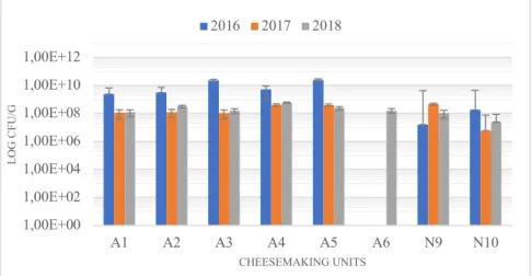
Concerning LAB we could observe, as shown in Figure 3.1., that in all units except N9 from Nisa samples from 2016 had a higher number of these bacteria while data from 2017 and 2018 years were very similar. Values of colony-forming units (CFU) observed in this work were very much alike to data previously published about LAB in Manchego cheeses (Cabezas et al., 2007) that have similar characteristics with PDO-cheeses analyzed in this work such as being manufactured with sheep milk, having PDO designation and being artisanal.
A study that compared Terrincho Portuguese PDO-cheeses (Pintado et al., 2008), which is also made with raw sheep milk, from five dairy farms showed significant differences between farms for all microbial groups studied (lactobacilli, lactococci, enterococci, staphylococci, enterobacteria, pseudomonads, yeasts and molds). In a previous study (Quigley et al., 2012) that worked with 62 artisanal Irish cheeses from cow, goat or sheep milk it was also noted how Lactobacillus populations had a significant increase, even when produced in the same farm, due to differences in ripening temperatures. One hypothesis that could explain the differences that we observed between years and units could be that temperature and humidity conditions oscillated even between established limits. It has already been suggested for artisanal raw milk cheeses like Camembert (Henri-Dubernet et al., 2008) that there is strong variability in lactobacilli not only because of the composition of raw milk but also due to the practices of each cheesemaking unit.
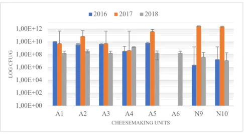
Lactococcus spp., which were isolated with M17 growth medium, CFU counts are represented in
Figure 3.2., presented similar values to LAB. This was expected since this genus is one of the predominant in lactic acid bacteria found in raw sheep milk and cheeses (Cabezas et al., 2007; Quigley et al., 2013). Furthermore, in a study with 4379 isolates from 35 different cheeses (Cogan et al., 1997) 38% of those isolates were identified as lactococci, making this group of bacteria the predominant. Higher CFU counts were observed in 2017 samples from Nisa units although this didn’t occur in 2016 and 2018.
Figure 3.1. Enumeration of lactic acid bacteria.
1,00E+00 1,00E+02 1,00E+04 1,00E+06 1,00E+08 1,00E+10 1,00E+12 A1 A2 A3 A4 A5 A6 N9 N10 LO G C FU /G CHEESEMAKING UNITS 2016 2017 2018
These differences could be due to variations on the maturation phases of 2017 Nisa cheeses as it has been observed in a study about flavor formation by lactic acid bacteria (Smit et al., 2005) that
Lactococcus spp. are predominant in starter cultures for cheese-making. Most definitely the flavor of
these cheeses would be different from other years since acidification is the primary role of this genus in cheese fermentation. In our case, the year that had higher CFU in most of cheesemaking units is 2017, unlike what we’ve seen above with LAB, where 2016 had higher values than the other two years. So, although we have similar CFU counts in LAB and Lactococcus spp. the years of production with highest counts do not match in both groups. An explanation for this could be the fact that although lactococci are part of LAB, MRS medium favors the growth of other bacteria such as Lactobacillus spp. and M17, the medium growth used to isolate Lactococcus spp., is selective for this genus. Moreover, in a study with Serra da Estrela cheese (Macedo et al., 2004) it was observed that lactobacilli survive better when their counts are similar to those of lactococci, as we see in cheeses analyzed in this work, since it seems that pH reduction caused by lactococci didn’t affect negatively lactobacilli survival.
Regarding Enterococcus spp. CFU counts, represented in Figure 3.3., high variability was observed through the three years and the different units. In half of the units the higher CFU’s were observed in 2016 samples, in one unit (N9 from Nisa) it was observed in 2017 samples and other one (A5 from Azeitão) in 2018.
Figure 3.2. Enumeration of Lactococcus spp.
1,00E+00 1,00E+02 1,00E+04 1,00E+06 1,00E+08 1,00E+10 1,00E+12 A1 A2 A3 A4 A5 A6 N9 N10 LO G C FU /G CHEESEMAKING UNITS 2016 2017 2018
Figure 3.3. Enumeration of Enterococcus spp.
1,00E+00 1,00E+02 1,00E+04 1,00E+06 1,00E+08 1,00E+10 1,00E+12 A1 A2 A3 A4 A5 A6 N9 N10 LO G C FU /G CHEESEMAKING UNITS 2016 2017 2018


