74
Andreia Sofia Ferreira Saruga
Bachelor in Biotechnology
Cyto- and Genotoxicity Assessment of
Manufactured Nanomaterials in the A549 Cell Line
A thesis submitted in conformity with the requirements for the degree Master
in Biotechnology
Supervisor: Doutora Maria João Silva, Departamento de Genética Humana do
Instituto Nacional de Saúde Doutor Ricardo Jorge, I.P.
Co-supervisor: Doutora Maria Henriqueta Louro, Departamento de Genética
Humana do Instituto Nacional de Saúde Doutor Ricardo Jorge, I.P.
Jury:
President: Professor Doutor Pedro Miguel Calado Simões
Examiner: Professor Doutor António Sebastião Rodrigues
iii
Andreia Sofia Ferreira Saruga
Bachelor in Biotechnology
Cyto- and Genotoxicity Assessment of
Manufactured Nanomaterials in the A549 Cell Line
A thesis submitted in conformity with the requirements for the degree Master
in Biotechnology
v
Cyto- and Genotoxicity Assessment of Manufactured Nanomaterials in A549 the Cell Line
Copyright © Andreia Sofia Ferreira Saruga, Faculdade de Ciências e Tecnologia, Universidade Nova de Lisboa.
A Faculdade de Ciências e Tecnologia e a Universidade Nova de Lisboa têm o direito, perpétuo e sem limites geográficos, de arquivar e publicar esta dissertação através de exemplares impressos reproduzidos em papel ou de forma digital, ou por qualquer outro meio conhecido ou que venha a ser inventado, e de a divulgar através de repositórios científicos e de admitir a sua cópia e distribuição com objetivos educacionais ou de investigação, não comerciais, desde que seja dado crédito ao autor e editor.
Copyright © Andreia Sofia Ferreira Saruga, Faculty of Sciences and Technology, New University of Lisbon.
vii
ACKNOWLEDGMENTS
First of all, I must thank my family, especially my parents, because without them, without their support this journey could not be possible to realize.
I want to thank my supervisors, Drª Maria João Silva for had accepted me in their research group, for all the scientific knowledge transmitted and for revision of this thesis; Drª Henriqueta Louro, for all the help in the lab work, during the processing of the results, statistics analysis and also in the revision of this work. To Professor Pedro Baptista from FCT/UNL for always being available when I contacted him.
ix
ABSTRACT
A number of nanomaterials (NMs) have been applied in different fields due to their unique physico-chemical properties. As the use and applications have increased in some industries, serious concerns about their potential impact on the environment and the human health have been raised and have been a challenge for the regulatory authorities.
This work aimed at assessing the toxicity of three classes of NMs, namely cerium dioxide, CeO2
(NM-212), titanium dioxide, TiO2 (NM-101 and NM-100) and barium sulphate, BaSO4 (NM-220) since
they already have a broad range of applications in industry and consumer products.
A standardized protocol for NMs dispersion was followed and the quality of the dispersion in the culture medium was evaluated by the dynamic light scattering technique. Different concentrations (0, 1, 3, 10, 30, 75 and 100 µg/cm2) of each nanomaterial were used to expose A549 cells (human lung
carcinoma cells) for cytotoxic evaluation through the MTT and clonogenic assays and genotoxicity assessment through the comet and the cytokinesis-blocked micronucleus (CBMN) assays.
The results showed a decrease in cell proliferation after exposure to cerium dioxide nanomaterials for 8 days, at the highest concentrations tested and a slight increase in the level of DNA breaks. Concerning the TiO2 NMs, a statistically significant increase in the level of DNA breaks was
found for both NMs; however the CBMN assay did not show any increase in the frequency of chromosomal breaks. BaSO4 was the NM that showed the lowest toxicity in cyto- and genotoxicity
assays.
Even though the present results contribute to assess the hazard of the tested NMs, the real effects of nanomaterials’ exposure to human health are still unclear and an unequivocal conclusion is difficult to present, given the inconsistent and often conflicting results found in the literature. Thus, the application of some nanomaterials in consumer products should be carefully evaluated until definite conclusions about their safety are available.
xi
RESUMO
Diferentes nanomateriais têm sido aplicados em diferentes áreas, devido às suas propriedades físico-químicas únicas. Como o uso e as aplicações têm aumentado em algumas indústrias, os possíveis impactos no ambiente e na saúde humana têm sido questionadas e tem-se revelado um desafio as autoridades reguladoras.
Este trabalho tem como objectivo avaliar a toxicidade de três classes de NMs, nomeadamente, dióxido de cério, CeO2 (NM-212), dióxido de titânio, TiO2 (NM-101 e NM-100) e sulfato de bário, BaSO4
(NM-220), visto que estes já possuem uma vasta aplicação na indústria e em produtos de consumo. Foi seguido um protocolo padronizado para a dispersão dos nanomateriais e qualidade da dispersão no meio de cultura foi avaliada através da técnica de dynamic light scattering. Foram utilizadas diferentes concentrações (0, 1, 3, 10, 30, 75 e 100 µg/ cm2) de cada nanomaterial, para expor
a cultura de células A549 (células de carcinoma de pulmão humano) para avaliação da citotoxicidade através dos ensaios do MTT e clonogénico e dos ensaios do cometa e dos micronúcleos para o estudo da genotoxicidade.
Os resultados mostraram uma diminuição na proliferação de células após exposição aos nanomateriais de dióxido de cério, durante 8 dias, nas concentrações mais elevadas e um aumento ligeiro nas quebras do DNA. No que diz respeito ao TiO2 verificou-se um aumento estatisticamente
significativo no nível de quebras de DNA para ambos os NMs, no entanto o ensaio dos micronúcleos não apresentou nenhum aumento na frequência de quebras cromossómicas. O BaSO4 foi o
nanomaterial que apresentou menor toxicidade tanto nos ensaios de citotoxicicdade como genotoxicidade.
Apesar dos presentes resultados contribuírem para a avaliação do perigo dos NMs testados, os verdadeiros efeitos da sua exposição para a saúde humana ainda não são claros e é difícil apresentar uma conclusão inequívoca, dada a inconsistência dos resultados apresentados na literatura. Assim, a aplicação de alguns nanomateriais em produtos de consumo deve ser avaliada cuidadosamente até que estejam estabelecidas conclusões relativamente à sua segurança.
xiii
LIST OF CONTENTS
ACKNOWLEDGMENTS ... vii
ABSTRACT ... ix
RESUMO ... xi
LIST OF CONTENTS ... xiii
LIST OF FIGURES ... xv
LIST OF TABLES ... xix
ABBREVIATIONS ... xxi
1. INTRODUCTION ... 1
1.1 Nanotoxicology ... 2
1.1.1. Cytotoxic effects of nanomaterials ... 2
1.1.2. Genotoxic effects of nanomaterials ... 3
1.2 The NANoREG Project ... 3
1.3 Metallic Nanomaterials ... 4
1.3.1 Cerium Dioxide (CeO2) ... 4
1.3.2 Titanium Dioxide (TiO2) ... 7
1.3.3 Barium Sulphate (BaSO4) ... 11
1.4 Strategies and challenges in the in vitro characterization of nanomaterials’ toxicity ... 11
1.4.1 Nanomaterials’ dynamic behavior and dispersion in aqueous medium ... 11
1.4.2 Experimental cell models... 13
1.4.3 Cytotoxicity Assessment... 14
1.4.4 Genotoxicity Assessment ... 15
2. OBJECTIVES ... 20
3. MATERIALS AND METHODS... 22
3.1 Cell Culture/ Cell line ... 22
3.2 Nanomaterial preparation and characterization of nanomaterial dispersion in the culture medium 22 3.3 Cytotoxicity Assays ... 24
3.3.1 MTT assay ... 24
3.3.2 Clonogenic assay ... 24
3.4 Genotoxicity Assays ... 25
3.4.1 Cytokinesis-blocked micronucleus assay (CBMN) ... 25
3.4.2 Comet Assay ... 26
3.5 Statistical analysis ... 27
4. RESULTS ... 28
4.1 Cerium dioxide nanomaterials ... 28
4.1.1 Characterization of the nanomaterial dispersion in the culture medium ... 28
4.1.2 Cytotoxicity Assessment... 30
4.1.3 Genotoxicity Assessment ... 32
xiv
4.2.1. Characterization of nanomaterial dispersion in the culture medium ... 33
4.2.2 Cytotoxicity Assessment... 38
4.2.3 Genotoxicity Assessment ... 39
4.3 Barium sulphate nanomaterials ... 42
4.3.1 Characterization of nanomaterial dispersion in the culture medium for NM-220 ... 42
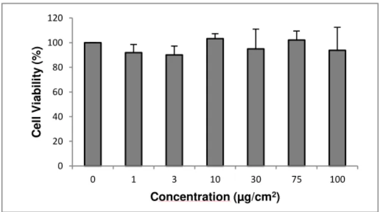
4.3.2 Cytotoxicity Assessment... 44
4.3.3 Genotoxicity Assessment ... 45
4.4. Overview of the cytotoxicity and genotoxicity assessment of the tested NMs ... 47
5. DISCUSSION ... 48
5.1 The analysis of the NMs dispersion ... 49
5.2 Cytotoxicity and genotoxicity of CeO2 nanomaterials ... 49
5.3 Cytotoxicity and genotoxicity of TiO2 nanomaterials ... 52
5.4 Cytotoxicity and genotoxicity of BaSO4 nanomaterials ... 56
6. CONCLUSION ... 58
7. REFERENCES ... 60
8. ANNEXES ... 70
ANNEX I - DLS Analysis ... 70
ANNEX II- Cytotoxic Assays... 72
ANNEX III- Genotoxicity Assays ... 73
xv
LIST OF FIGURES
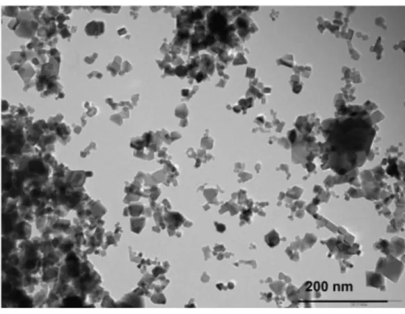
Figure 1.1- TEM image of NM-212, showing irregular and non-homogeneous primary particle size variation (Singh et al. 2014)... 4 Figure 1. 2- TEM-micrograph of NM-100 showing the range in agglomerate and aggregate sizes in the material (Rasmussen et al. 2014). ... 8 Figure 1. 3- Representative TEM micrograph of well-dispersed sample of NM-101 taken for quantitative TEM-analysis; scale bar is 500nm (Rasmussen et al. 2014). ... 8 Figure 1. 4- Micrograph of NM-101, illustrating that the aggregates/agglomerates have a very irregular surface (Rasmussen et al. 2014). ... 8 Figure 1. 5- Schematic representaion of the proposed mechanism for ROS formation and effects in the cell (from Tang et al. 2013)... 9 Figure 1. 6- A549 cells exposed to TiO2 NMs. A- Binucleated cell with one micronucleus; B- Binucleated
cell with one micronucleus and nucleoplasmatic bridge; C- Binucleated cells (one cell with micronucleus) surrounded by TiO2 NMs. Images from the group lab. ... 17
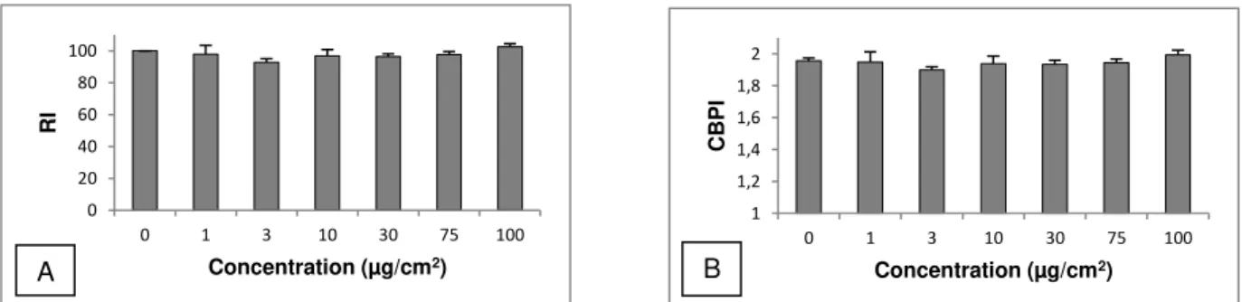
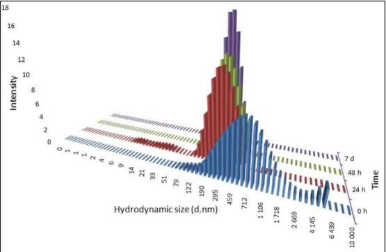
xvi Figure 4. 13- Size distribution of NM- 100 for concentration 240 µg/mL in cell culture medium at 0, 24, 48 hours and 7 days after dispersion and incubation at 37ºC. ... 35 Figure 4. 14- Size distribution of the batch dispersion of NM- 101 (2.56 mg/mL) soon after sonication protocol in BSA water 0.05%. ... 36 Figure 4. 15- Size distribution of NM- 101 for concentration 3.2 µg/mL in cell culture medium at 0, 24, 48 hours and 7 days after dispersion and incubation at 37ºC. ... 36 Figure 4. 16- Size distribution of NM- 101 for concentration 32 µg/mL in cell culture medium at 0, 24, 48 hours and 7 days after dispersion and incubation at 37ºC. ... 37 Figure 4. 17- Size distribution of NM- 101 for concentration 240 µg/mL in cell culture medium at 0, 24, 48 hours and 7 days after dispersion and incubation at 37ºC. ... 37 Figure 4. 18- Results for cell viabiliy after 24 hours of exposure to NM-101 and NM- 100. ... 38 Figure 4. 19- A- Results of CBPI after 48 hours of exposure to NM-100; B- Results of RI after 48 hours of exposure to NM-100. ... 38 Figure 4. 20- Micronucleated binuclated cells after 48 hours of exposure to NM-100. The concentration 30 µg/cm2 does not present any bar because the number of micronuclei scored was 0. ... 39
Figure 4. 21- Microscopical photos of A549 cells after 48 hours exposure to NM-100 (10x40). A- 30 µg/cm2 ; B- 75 µg/cm2; C and D- 100 µg/cm2. It is visible the increase of NM when the concentrations
xix
LIST OF TABLES
xxi
ABBREVIATIONS
A549 Human epithelial lung adenocarcinoma cell line
ATCC American Type culture Collection
BaSO4 Barium Sulphate
BAuA Federal Institute for Occupational Safety and Health
BET Brunauer-Emmett-Teller BSA Bovine Serum Albumin
CBPI Cytokinesis-Blocked Proliferation Index
CeO2 Cerium Dioxide
DLS Dynamic Light Scattering
DMEM Dulbecco’s Modified Eagle Medium
DMSO Dimethyl Sulfoxide
DNA Desoxyribonucleic Acid
EDTA Ethylenediamine Tetraacetic Acid
EMS Ethyl Methanesulfonate FBS Fetal Bovine Serum
FDA Food and Drug Administration
FPG Formamidopyrimidine DNA Glycosylase
HEPES 4-(2-hydroxyethyl)-1-piperazineethanesulfonic Acid
LDH Lactate Dehydrogenase
MMC Mitomycin C
MNBNC Micronucleated Binucleated Cell
MTT 3-(4,5-dimethylthiazol-2-yl)-2,5-diphenyltetrazolium bromide
NM(s) Nanomaterial(s)
NIOSH National Institute for Occupational Safety and Health
OECD Organization for Economic Co-operation and Development
xxii
RI Replication Index SD Standard Deviation SDS Sodium Dodecyl Sulfate
1
1.
INTRODUCTION
Nanotechnology involves the manipulation and application of engineered particles or systems that have at least one dimension under 100 nanometers (nm) in length (Stone et al. 2009; Arora et al. 2012; Ferreira et al. 2013).
Nano-object is defined as a material with one, two, or three external dimensions in the size range from approximately 1–100 nm. There are subcategories of nano-object such as nanoparticle (NP), defined as a nano-object with all three external dimensions at the nanoscale. Nano-objects are commonly incorporated in a larger matrix or substrate referred to as a nanomaterial (NM). The term manufactured nanomaterial describes nanoparticles (NPs) intentionally produced and designed with very specific properties or compositions (e.g., shape, size, surface properties, and chemistry) (NIOSH 2009). In 2011, the European Commission adopted the definition of nanomaterial (NM) as “a natural, incidental or manufactured material containing particles, in an unbound state or as an aggregate or as an agglomerate and where, for 50 % or more of the particles in the number size distribution, one or more
external dimensions is in the size range 1 - 100 nm”.
(http://ec.europa.eu/environment/chemicals/nanotech/faq/definition_en.htm; Consulted on 22/7/2015) Some authors do not distinguish the term nanoparticle (NP) and nanomaterial (NM) in their works, using it indistinguishably and assuming the same definition. Hence, in this work these two terms will be assumed as being the same to facilitate the comprehension.
According to several authors, unique and unusual physical, chemical, and biological properties can be seen at the nanosize level. While the properties of bulk materials at the molecular level are largely understood there are new properties of materials being discovered in the zone between molecule and bulk.When bulk materials are made into smaller and smaller pieces of matter their surface chemistry changes and chemical reactivity increases or, in other words, there are a higher number of molecules available to react with the environment. Also, at the nanoscale, quantum physics can direct the behaviour of a particle; the influence of quantum effects can change important material properties, such as optical, magnetic, and electrical properties (Ferreira et al. 2013; Arora et al. 2012; Elsaesser & Howard 2012; Azqueta & Dusinska 2015).
2 Cemlyn-Jones, and Robalo Cordeiro 2013). Thus, concerns have been raised about their safety profiles. One particular area of concern is that of airborne nanomaterials and the potential harms that may result in the respiratory tract (Ferreira et al. 2013; Frieke Kuper et al. 2015).
1.1
Nanotoxicology
Nanotoxicology was proposed as a new branch of toxicology to address the gaps in knowledge about nanomaterials’ toxic effects to human health and the environment towards the development of a sustainable and safe nanotechnology (Arora et al. 2012; Ferreira et al. 2013). In this sense, nanotoxicology encompasses the physicochemical properties, routes of exposure, biodistribution, molecular determinants, genotoxicity, and addresses also regulatory aspects. In addition, nanotoxicology is involved in proposing reliable, robust, and data-assured test protocols for nanomaterials in human and environmental risk assessment (Ferreira, Cemlyn-Jones, and Robalo Cordeiro 2013).
Toxicological assessment of manmade NMs requires information about the route of exposure (inhalation, oral or dermal). The most common scenarios for human exposure to NMs are occupational, environmental and the consumer ones and one of the most important routes of human exposure to airborne NPs is inhalation, both at the workplace and from the environment (Ferreira, Cemlyn-Jones, and Robalo Cordeiro 2013). When inhaled, particles reach the alveolar epithelial surface where they can interact with alveolar macrophages and epithelial cells (Herzog et al. 2007). The deposition mainly takes place in the alveolar region (Arora, Rajwade, and Paknikar 2012). After translocation across the lung epithelium, NMs can enter the blood and lymph circulation to reach cells in the bone marrow, lymph nodes, spleen and heart, among other organs (Arora, Rajwade, and Paknikar 2012).
Warheit (2008) has put a question “how meaningful are the results of nanotoxicity studies in the absence of adequate material characterization?” This suggests that it is extremely important to characterize the nanomaterials during the study in the culture medium where the cells are seeded. Recent research has demonstrated that NMs polydispersity and agglomeration stability can have profound impacts on NMs’ toxicity in in vitro tests. Harmonized methods for in vitro nanotoxicity assessments must therefore include NMs dispersion preparation and characterization protocols that ensure fairly monodispersed and stabilized NM suspensions suitable for in vitro cellular studies (Cohen et al. 2013). In chapter 1.4.1- Nanomaterials’ dynamic behavior and dispersion in aqueous medium- this subject will be discussed in more detail.
1.1.1. Cytotoxic effects of nanomaterials
3 aggregation and the capacity to generate reactive oxygen species (ROS) also explain the potential for lung damage (Ferreira, Cemlyn-Jones, and Robalo Cordeiro 2013).
NMs can interact with cellular and mitochondrial membranes or alter mitochondrial function, provoking the production of reactive oxygen species and inducing DNA oxidation. ROS include free radicals such as the superoxide anion (O2•−), hydroxyl radicals (.OH) and the non-radical hydrogen peroxide (H2O2), which are also constantly generated in cells under normal conditions, as a
consequence of aerobic metabolism (Arora, Rajwade, and Paknikar 2012). ROS, due to their high chemical reactivity, can react with DNA, proteins, carbohydrates and lipids causing cell death either by apoptosis or necrosis or even inflammatory responses.
1.1.2. Genotoxic effects of nanomaterials
The genotoxicity associated to nanomaterials may be classified as primary or secondary genotoxicity. The former may result from a direct pathway when NPs reach the nucleus (via nuclear pores or during mitosis) and directly interact with DNA organized in chromatin or chromosomes (depending on the phase of cell cycle). During interphase NPs can interact or bind to the DNA molecule and can influence its replication or transcription into RNA. During mitosis NPs can interact with chromosomes, causing clastogenic or aneugenic effects. NPs might induce breaks in chromosomes or disturb the process of mitosis, either mechanically or by chemical binding to DNA bases. Indirect genotoxicity may happen through interaction with nuclear proteins (involved in replication, transcription, repair), or with the mitotic spindle or its components, leading to aneuploidy (aneugenic effect); other indirect effects include the disturbance of cell cycle checkpoint functions, induction of ROS arising from NP surface or inhibition of the cellular antioxidant defense (Stone et al. 2009; Magdolenova et al. 2014). On the other hand, secondary genotoxicity implies a pathway of genetic damage resulting from oxidative DNA attack by ROS that are generated from phagocytes (neutrophils, macrophages) activation during particle-elicited inflammation. NMs can also induce genotoxic effects by depleting the antioxidant defenses or altering the DNA repair systems. All these events may result in pre-mutagenic lesions that can lead to mutations and, possibly, to cancer and other diseases on the long-term (Azqueta & Dusinska 2015; Magdolenova et al. 2014).
Many methods have been developed to assess the genotoxicity caused by nanomaterials that will be discussed in the next chapter 1.4.4.
1.2
The NANoREG Project
The NANoREG project is the first FP7 (7th Framework Programme for Research and
Technological Development) project to deliver the answers needed by regulators and legislators on Environmental Health Science (EHS) by linking them to a scientific evaluation of data and test methods.
4 characterization, toxicity testing and exposure measurements of manufactured nanomaterials, (iii) develop, for the long term, new testing strategies adapted to innovation requirements, (iv) establish a close collaboration among authorities, industry and science leading to efficient and practically applicable risk management approaches for manufactured NMs and products containing manufactured NMs. (http://www.nanoreg.eu/; consulted on 6th August 2016)
The NANoREG project has about 70 partner institutes (Fadeel et al. 2015) and in Portugal, the NANoREG project is represented by Institute of Welding and Quality (ISQ), which is coordinating a platform called PToNANO, formed by four entities whose competencies are complementary in the field of nanotechnology. The other 3 entities are the National Institute of Health Doutor Ricardo Jorge, I.P. (INSA), the Portuguese Institute for Quality, I.P. (IPQ) and the Directorate-General of Health (DGS)(http://www.nanoreg.eu/; consulted on 6th August 2016)
The NANoREG project uses benchmark nanomaterials from the Joint Research Center (JRC) repository (Fadeel et al. 2015).
1.3
Metallic Nanomaterials
1.3.1 Cerium Dioxide (CeO
2)
Cerium (Ce) is the most abundant rare-earth metals found in the earth’s crust which has been recently introduced for specialty applications (Kumari et al. 2014; Dahle et al. 2015) and is found in nature along with other lanthanide elements in the minerals alanite, bastanite, monazite, cerite and samarskite. However, CeO2 has fluorite as the most stable crystalline phase (Prospect Global
Nanomaterials Safety 2010).
5 Larger particles of this material (bulk material) may induce optical lens grinding, itching, sensitivity to heat, skin lesions, pulmonary fibrosis (Rim, Koo, and Park 2013) and pneumoconiosis (Prospect Global Nanomaterials Safety 2010).
Owing to their large surface area to volume ratio, CeO2 NPs (nanoceria, Figure 1.1) have unique
electronic structure and the reduction in the particle size results in the formation of surface oxygen vacancies, which endows it with the ability to exist in either Ce3+ or Ce4+ state on the particle surface
(Kumari et al. 2014).
Nanoceria play several catalytic roles such as catalysts in the petroleum refining industry, as additives to promote combustion of diesel fuels, in catalytic converters in cars enabling them to run at high temperatures, as sub-catalysts for automotive exhaust cleaning and as electrolytes in solid oxide fuel cells (Kumari et al. 2014; Dahle et al. 2015; Rim et al. 2013; Leung et al. 2015; Landsiedel et al. 2014; Keller et al. 2014). The CeO2 NPs can also be employed as polishing agents, UV-scattering
agents in non-irritating lipsticks, outdoor paint, wood care products, gas sensors, and in metallurgic and glass and ceramic applications (Kumari et al. 2014; Manier et al. 2013; Frieke Kuper et al. 2015; Van Koetsem et al. 2015). Recently, CeO2 has been the subject of many studies regarding its use as potential
material for ultraviolet (UV) filtration. In the UV radiation range reaching the Earth’s atmosphere, the ultraviolet type B sub-range (UVB, 290–320 nm) is already well filtered by nanostructured TiO2 in
sunscreen cosmetic products (Truffault et al. 2010; Boutard et al. 2013).
Nanoceria, considered one of the most interesting nanomaterials for their catalytic properties, also are promising for therapeutic applications. Due to the presence of oxygen vacancy on its surface and the autoregenerative cycle of its two oxidation states, Ce3+ and Ce4+, nanoceria can be used as an
antioxidant agent. Because many human disorders are associated with oxidative stress, CeO2 NPs
might be used in future for the treatment of those pathologies (Rim et al. 2013; Leung et al. 2015). It was even proposed that nanoceria protect against hepatic oxidative damage and the progress of cardiomyopathy, which was attributed to their antioxidant properties (Leung et al. 2015). Consequently, they are considered to be of interest for biomedical applications for their antioxidant activity.
Some studies have been conducted with the aim to investigate these assumptions and try to explain why and when are these effects observed. The best explained and currently accepted mechanism to justify these results is the intrinsic material properties, including the mixed valence state of CeO2 (Ce3+ and Ce4+), allowing this material to act as a free radical scavenger (Xia et al. 2009;
Schubert et al. 2006). Moreover, the electron defects in nano-ceria are relatively resistant to the radical damage, thereby allowing an autoregenerative reaction cycle (Ce3+ → Ce4+→ Ce3+) that perpetuates
the scavenging activity (Xia et al. 2009; Das et al. 2007). Hence, it was proposed that Ce3+ produced by
the reduction of Ce4+ interacts with oxygen molecules (O2), and generates superoxide anions. The two
superoxide anion molecules interact and are converted to hydrogen peroxide, and then the hydrogen peroxide converts to hydroxyl radicals. The presence of the mixed valence states of Ce3+ and Ce4+ on
the surface of the nanoceria acts as an anti-oxidant that allows the nanoparticles to scavenge free radicals from the culture system. Ce3+ reacts with hydroxyl radicals (Park et al. 2008).A review reported
6 There are other publications that report this radical scavenging of CeO2 nanoparticles as the
mechanism of neuro-protection in the spinal cord neurons of adult rats (Das et al. 2007) and the cerium NPs are neuro-protective in cultured HT22 cells, which are derived from the rodent nervous system (Schubert et al. 2006). Park et al. (2008) tried to justify their results in Beas-2B cells reporting that the particle which they prepared do not have the same arrangement and do not have the same Ce3+ ionic
state enough to scavenge the oxygen radicals, when they addressed the antioxidant mechanism of cerium oxide nanoparticles. This may be the difference between the neuroprotection in adult rat spinal cord (referred study) and the cytotoxicity in Beas-2B cells obtained by the authors (Park et al. 2008). In fact there are already available some studies that report the effect of the size in the relative amount of cerium ions Ce3+ and Ce4+ saying that, in general, the fraction of Ce3+ ions in the particles increases
with decreasing particle size (Xu & Qu 2014; Schubert et al. 2006). Besides the in vitro studies presented above, it was also related that nanoceria remains deposited in tissues and may decrease ROS in mice, thereby suggesting again that cerium oxide nanoparticles may be a useful anti-oxidant treatment for oxidative stress (Hirst et al. 2009).
However, biomedical and toxicity studies of nanoceria, which focused predominantly on their redox properties, have resulted in contradictory conclusions about their effects. While some authors documented that nanoceria may act as antioxidants and protect cells against oxidative damage and ionizing radiation, and improve cardiac function, others reports found them to be cytotoxic and to induce oxidative stress (Lee et al. 2012; Rim et al. 2013).
Because of the rapidly growing demand of cerium in nanotechnology, the release of this NM is expected to increase in the environment. The majority of Ce NPs residues are likely to be discharged in wastewater treatment plants and/or be partially accumulated in the sludge that is later used for soil amendments. Thus, industrial wastewaters serve as a significant environmental source of CeO2 NPs
(Dahle, Livi, and Arai 2015). For such reason, the Organization for Economic Co-operation and Development (OECD) in the Environment Directorate added CeO2 NPs in the list of 14 NMs for testing
and identified it as commercially relevant to the global economic impact of nanotechnology. It has been suggested that the most common route of CeO2 exposure is likely to be through inhalation and/or
ingestion (Kumari et al. 2014; Manier et al. 2013; Verstraelen et al. 2014; Franchi et al. 2015; Bour et al. 2015; Keller et al. 2014).
There is ongoing exposure of a large population set to new diesel emissions generated from fuel additives containing CeO2 nanoparticles that may be inhaled by humans. Besides alveolar
macrophages, bronchial and alveolar epithelial cells are among the principal cells that get into contact with airborne NPs that penetrate into the lung. As a consequence of this interaction, these cells are capable of producing pro-inflammatory mediators that have the ability to elicit both a local and systemic inflammatory response (Rim et al. 2013; Verstraelen et al. 2014; Kumari et al. 2014).
Concerning toxicity assessment, it was proposed that CeO2 nanoparticles may cause oxidative
7 such as TiO2, ZnO, and CeO2. It has been proposed that the nano metal oxide induced ROS can cause
lipid peroxidation, cell membrane damage (Leung et al. 2015) and, possibly genotoxicity.
As to cell uptake of this NM, a study demonstrated the internalization of 8-20 nm CeO2 NPs in
A549 cell line (Mittal and Pandey 2014). A study in endothelial cells have reported that nanoceria can be uptaken into cells and widely distributed in multiple compartments of the cells. The results indicated that nanoceria can be also uptaken into cells through mediated endocytosis. Nanoceria were distributed throughout the cytoplasma but not into nucleus (S. Chen et al. 2013). Also in monocytes TEM revealed that CeO2 NPs were internalized and found either in vesicles or free in the cytoplasm (Hussain et al.
2012). Moreover, an in vivo study in CD1 mice also showed the internalization, accumulation and translocation of CeO2 NMs behind the pulmonary organs (Aalapati et al. 2014). The presence and
retention of cerium oxide nanoparticles in lung tissue, and alveolar macrophages was also revealed (Ma et al. 2012).
1.3.2 Titanium Dioxide (TiO
2)
Titanium dioxide (TiO2) is considered as an inert and safe material and has been used in many
applications for decades. Furthermore, TiO2 is accepted as a food and pharmaceutical additive. In the
United States it is included in the Food and Drug Administration (FDA) Inactive Ingredients Guide for dental paste, oral capsules, suspensions, tablets, dermal preparations and in non-parenteral medicines. However, with the nanotechnologies’ development, TiO2 NPs (Figures 1.2, 1.3 and 1.4), displaying novel
and useful properties are being increasingly manufactured and used. Therefore, increased human and environmental exposure can be expected, which has put TiO2 NPs under toxicological scrutiny (Skocaj
8
Figure 1. 2- TEM-micrograph of NM-100 showing the range in agglomerate and aggregate sizes in the material (Rasmussen et al. 2014).
Figure 1. 4- Micrograph of NM-101, illustrating that the aggregates/agglomerates have a very irregular surface (Rasmussen et al. 2014).
Among NMs, TiO2 is one of the most manufactured NM worldwide. TiO2 NPs are widely used in
industry for plastics, papers, inks, medicines, food products, cosmetics, toothpastes and skin care products, among others. It also has photocatalytic properties that have resulted in the use of TiO2 NPs
as an environment and wastewater disinfectant. Furthermore, TiO2 NPs have been used as a
photosensitizer for the photodynamic therapy of human colon carcinoma cells (Singh et al. 2009; Medina-Reyes et al. 2015; Jugan et al. 2012; Huerta-García et al. 2014; Chen et al. 2014). In fact, at present, titanium dioxide are the most common ingredient in mineral sunscreens. Because of their high photostability and low photoallergic potential, they are often preferred over organic filters (Boutard et al. 2013).
These particles are in the breathable size range and several toxic effects have been described after their inhalation. Tissue deposition of NPs and their toxicity are closely related to the route of exposure and, in this sense, keratinocytes have been studied as the primary target of dermal exposure, lung tissue as the target for inhalation, and intestines, kidney, and liver for oral exposure. The lung is
9 the best characterized organ regarding the toxic effects induced by TiO2 NPs (Huerta-García et al.
2014).
Although these NPs are considered safe for use in sunscreens by the US FDA, there is a considerable concern with this ruling as sunlight because illuminated TiO2 may induce DNA damage
both in vitro and in vivo. When exposed to UV light, TiO2 catalyses the generation of reactive oxygen
species, such as superoxide anions, hydrogen peroxide, free hydroxyl radicals, and singlet oxygen in aqueous media (N. Singh et al. 2009).
In vivo studies have provided evidence that TiO2 NPs can cause inflammation, fibrosis, pulmonary damage and even DNA damage. Toxicity assessment has been performed in rats tissues and revealed that this NM caused lungs injury and inflammation due the increasing number of red blood cells.The results of lung tissue lipid oxidation accessed by malondialdehyde (MDA) content confirmed that the damage of lung tissue was indeed related with the generation and accumulation of ROS (Tang et al. 2013). These authors explained that the toxicity and ROS formation begun in mitochondria that carried out aerobic respiration. Most of oxygen was combined with electrons which came from electronic transmission chain at inner mitochondrial plasmalemma and formed H2O after a series of oxidation–
reduction reactions. However, a small amount of oxygen formed superoxide which was the major source of ROS in cells. The cumulative ROS attacked the mitochondria and restrained the functional activity of mitochondria and, as a result, the cell was approaching to apoptosis (Tang et al. 2013; Huerta-García et al. 2014). A simple schematic representation of this mechanism is represented in figure 1.5.
Figure 1. 5- Schematic representaion of the proposed mechanism for ROS formation and effects in the cell (from Tang et al. 2013)
However, these animal studies are limited and in vitro studies are required for more mechanistic insights.
Given that oxidative stress and inflammation are associated with indirect and secondary genotoxicity via the damaging activity of ROS, it seems likely that exposure to TiO2 NPs may indirectly
result in DNA lesions and several studies have demonstrated this effect (Singh et al. 2009; Jugan et al. 2012).
Based on the data available, the International Agency for Research of Cancer (IARC) has classified the TiO2 NPs as a possible carcinogen to humans, and the National Institute for Occupational
Safety and Health (NIOSH) has determined that TiO2 is a potential occupational carcinogen by inhalation
(Medina-Reyes et al. 2015).
In an exposure scenario, after absorption, TiO2 NPs can be translocated to all regions of the
10 significantly decreased at 24 and 48 hours, when exposed to 5 to 100 µg/mL of TiO2 NPs (Chen et al.
2014).
Their small size facilitates uptake into cells and transcytosis across epithelial and endothelial cells into the blood and lymph circulation to reach potentially sensitive target sites, such as bone marrow, lymph nodes, spleen, and heart. There is increasing evidence that NPs can cross the blood–brain barrier independent of the route of exposure (Huerta-García et al. 2014). Because TiO2 NPs can translocate to
different organs and into the CNS (Huerta-García et al. 2014), diverse studies have been made in cell lines other than those representative of the respiratoty tract. A study evaluated their possible toxic effect on glial cells (U373 cells) and reported that TiO2 NPs induce a strong oxidative stress in U373 cells,
mediating changes in the cellular redox state, which was correlated with increase in antioxidant enzyme expression and lipoperoxidation (Huerta-García et al. 2014). In human fetal lung fibroblasts (HFL1) the results from MTT indicated that TiO2 NMs (21 nm 80% A, 20% R) induced a decrease in cell viability at
low doses (0.25 to 1.50 mg/mL) (Z. Qiang et al. 2013). A study in normal untransformed human fibroblasts (GM07492) with TiO2 NPs in a range concentrations from 0 to 100 µg/mL also showed
significant decrease in cell viability at 100 µg/mL (Franchi et al. 2015). The MTT assay in human amnion epithelial (WISH cells) cells revealed a concentration-dependent decline in the cell survival after exposure to TiO2 NPs (30,6 nm) for 24 hours at 0.625 to 10 µg/mL (Saquib et al. 2012). Significant
cytotoxicity, intracellular ROS generation, and to some extent G2/M cell cycle arrest were induced at the above specified treatment doses, and attributed to TiO2 NPs mediated oxidative stress in the WISH cells (Saquib et al. 2012).
As to the internalization studies of TiO2 NPs, different techniques have been employed. The
flow cytometry analyses revealed that these NPs were not only internalized but also adhered to the surface of A549 cells (Moschini et al. 2013). Another study also verified the accumulation of the smallest NPs in the cytoplasm and in the nucleus of A549 cells, once again proving the capacity of A549 cells to internalize NMs (Jugan et al. 2012). Other techniques as Raman imaging (Ahlinder et al. 2013) TEM (Aueviriyavit & Phummiratch 2012; Franchi et al. 2015) Static light scattering, in-vitro Raman microspectroscopy (Andersson et al. 2011) have shown the uptake to the nucleus (Ahlinder et al. 2013) and in the cytoplasm relating that the agglomerates can be taken up by the cells and largely accumulated in the endosomes, probably via endocytosis (Aueviriyavit & Phummiratch 2012; Franchi et al. 2015; Tang et al. 2013). All these studies were conducted in A549 cells. The internalization of TiO2 NPs
(30-70 nm) was also verified in the human liver cells (HepG2) through electron microscopy and flow cytometry (Vallabani et al. 2014).
Because NPs can be internalized by cells and some of these NPs also can cross the nuclear membrane, the direct interactions between NPs and the nucleus may be the main cause of genotoxicity, leading to DNA or chromosome breaks. When NPs do not reach the nucleus, the genetic material is exposed to the NMs during cellular division (Elsaesser and Howard 2012). In contrast to these data suggesting a significant genotoxic potential of TiO2 NPs, other authors show that acute exposure to TiO2 NPs do not cause DNA damage (Xie et al. 2010; Bhattacharya et al. 2009; Landsiedel et al. 2010).
This discrepancy may be explained by differences in physicochemical properties of TiO2 NPs
11 different agglomeration status. It may also be explained by the use of different cell lines that may differ in terms of susceptibility to TiO2 toxic potential (Jugan et al. 2012; Chen et al. 2014).
1.3.3 Barium Sulphate (BaSO
4)
Chronic exposure to high levels of micron-scale BaSO4 may induce pneumoconiosis (baritosis)
in miners (Konduru et al. 2014). Baritosis is one of the benign pneumoconioses in which inhaled particulate matter lies in the lungs for years without producing symptoms, abnormal physical signs, incapacity for work, interference with lung function, or liability to develop pulmonary or bronchial infections or other thoracic disease (Doig 1976).
Barium sulfate nanomaterials already have some applications in different industries and are considered a member of the poorly soluble particles (PSP) or poorly soluble low toxicity (PSLT) particle groups, as are cerium dioxide (CeO2) and titanium dioxide (TiO2); this nanomaterial is used as fillers in
coatings (e.g. in motor vehicles) due to their mechanical, optical and chemical properties (Konduru et al. 2014) and it is also introduced into bone cements to increase radio-opacity so that they may be visualized through X-ray imaging, (Gillani et al. 2010). It is being used as a filling material in polymer compositions to increase scratch resistance while conserving transparency (Keller 2015),is a common agent added to catheters or endotracheal tubes which make such medical tubing radiopaque. In addition, BaSO4 polymeric formulations have been shown to exhibit some antimicrobial activity
(Aninwene et al. 2013).
Relatively to toxicity studies, there are only a few publications related to in vivo experiments. A study revealed that pulmonary exposure to instilled BaSO4 NPs caused dose-dependent lung injury and
inflammation. Four-week and 13-week inhalation exposures to a high concentration (50 mg/m3) of
BaSO4 NPs elicited minimal pulmonary response and no systemic effects. Instilled and inhaled BaSO4
NPs were cleared quickly yet resulted in higher tissue retention, exhibited lower toxicity and biopersistence in the lungs compared to other poorly soluble NPs such as CeO2 and TiO2 (Konduru et
al. 2014).
To our knowledge, there are no available studies for in vitro assessment of BaSO4 nanomaterials
through inhalation exposure. Thus, one of the objectives of this work is to assess the cytotoxicity and genotoxicity of this nanomaterial.
1.4
Strategies and challenges in the
in vitro
characterization of
nanomaterials’ toxicity
12 NPs have the tendency to both aggregate and agglomerate. An agglomerate is a “collection of loosely bound particles or aggregates or mixtures of the two where the resulting external surface area is similar to the sum of the surface areas of the individual components,” while an aggregate is defined as a “particle comprising strongly bonded or fused particles where the resulting external surface area may be significantly smaller than the sum of the calculated surface areas of the individual components” (Sauer et al. 2015).
Many attempts have been made to generate stable suspensions or dispersions in case of insoluble NMs. However, it is often the case that the particles settle due to agglomeration and gravity over time in culture. It is probably therefore more appropriate to also express dose in terms of mass per unit surface area of the culture dish (µg/cm2) (Stone, Johnston, and Schins 2009). The effective dosage
is influenced by the sedimentation and diffusion properties that different NMs exhibit under the given cell culture conditions, which largely depend upon the effective densities and diameters of the suspended NM agglomerates (Sauer et al. 2015).
In culture medium or other biological fluids, NMs interact with proteins or phospholipids thereby forming a characteristic ‘corona’ on their surface. NMs’ tendency to agglomerate is governed by its surface properties that can change spontaneously due to protein adsorption. Consequently, corona formation directly affects the type and extent of NM agglomeration, and the type of NM dispersing agent used influences in vitro particle kinetics (Sauer et al. 2015; Cohen et al. 2013). In vitro test systems should be relevant for the in vivo situation they mimic. The biological proteins and lipids surrounding a NP determine its biological fate, since it is this corona that cells encounter and interact with. When nanoparticle-protein complexes pass from one biological fluid to another, the corona is assumed to change due to competitive adsorption of different biomolecules. Only the small fraction of NPs that can pass the air-blood-barrier would experience a corona shift towards the higher affinity of serum components, followed by a comparatively less pronounced, but still considerable agglomeration (Sauer et al. 2015).
Different NM dispersants have been investigated, including natural lung surfactants, phospholipids, organic solvents, and serum or albumin additives (Sauer et al. 2015). A number of studies have now demonstrated that small concentrations of protein (usually albumin below 1% final concentration) improve particle dispersion and the stability of that dispersion over time, especially if incorporated in the medium prior to particle addition, and if combined with a short sonication (e.g. 10 min) (Stone, Johnston, and Schins 2009). Of course, adding protein or other dispersants to the nanoparticles could influence their surface properties and therefore their interaction with cells and other biological molecules. This suggests that either the improved dispersion aids uptake of the smaller agglomerates and/or individual NPs or, alternatively, that the proteins adsorbed to the particle surface interact with cell surface receptors that facilitate uptake into the cells (Sauer et al. 2015).
13 The Brownian motion varies as a function of particle size and causes variation in the intensity of scattered light as function of time (Keld Alstrup Jensen 2014).
1.4.2 Experimental cell models
There are a number of obvious advantages to in vitro toxicity testing of any chemical or particle, including the ethical desire to reduce animal testing, the speed of results, and the relatively lower cost compared to in vivo studies (Stone, Johnston, and Schins 2009). In vitro model systems provide a rapid and effective means to assess NPs for a number of toxicological endpoints. They also allow development of mechanistic evaluations and provide refined information on how NPs interact with human cells in many ways. Such studies can be used to establish concentration–effect relationships and the effect-specific thresholds in cells. The revelation of primary effects on target cells in the absence of secondary effects caused by inflammation and the identification of primary mechanisms of toxicity in the absence of the physiological factors are some of the interactions allowed by in vitro tests (Arora, Rajwade, and Paknikar 2012). These assays are suited for high-throughput screening of an ever increasing number of new NMs obviating the need for in vivo testing of individual materials (Stone, Johnston, and Schins 2009). Other advantages are reduction in variability between experiments, reduced requirement of test materials thereby leading to generation of limited amounts of toxic wastes. (Arora, Rajwade, and Paknikar 2012). A limitation is that an in vitro system is not able to fully replicate the complex interactions that occur between multiple cell types in vivo, both within an organ and also between organs (Stone, Johnston, and Schins 2009),
There are a large number of different tumor and transformed cell-derived cell lines available. It is also possible to increase the complexity of these in vitro systems to include multiple cell types, e.g., the co-culture of epithelial cells and macrophages, with the aim to more closely mimic the in vivo situation (Stone, Johnston, and Schins 2009). The selection of a cell line for in vitro toxicity assessment is frequently driven by the toxicokinetics and toxicodynamics of the NM, namely, the main via of absorption and the primary site of contact or the target organ.
14 the interaction of the nanomaterial with the cellular components. In the present work, internalization was not accessed but the knowledge relies on the results from other laboratories that participated also in the NANoREG project.
1.4.3 Cytotoxicity Assessment
The development and validation of methods to evaluate the toxicity of NMs is regarded as one of the main future challenges relevant to the safety of nanotechnology (Herzog et al. 2007). In past years, a number of methods have been developed to study cell viability and the proliferation ability of cells in culture. The most convenient, modern assays have been optimized for the use of microtiterplates (96-well format). This miniaturization allows many samples to be analyzed rapidly and simultaneously. Colorimetric and luminescence based assays allow samples to be measured directly in the plate by using a microtiterplate reader or ELISA plate reader. Cytototoxicity assays have been developed which use different parameters associated with cell death and proliferation (Weyermann, Lochmann, and Zimmer 2005).
1.4.3.1. The MTT Assay
There are a wide variety of assays to assess cytotoxicity that are frequently based on cell viability measurement. One of the most common is the MTT (3-(4,5-dimethylthiazol-2-yl)-2,5-diphenyltetrazolium bromide) assay, or variations of this assay (MTS, XTT or WST-1). These assays principally determine cell viability through determination of mitochondrial function by measuring the activity of mitochondrial enzymes. The assay generates a colored product (a purple formazan), which can be quantified by spectrophotometric readings at a specific wavelength. The absorbance value generated is representative of both the cell number and the functional viability of those cells (Stepanenko & Dmitrenko 2015; Stone et al. 2009; Weyermann et al. 2005). Such assays can therefore detect proliferation as well as cytotoxicity.
When testing NPs’ cytotoxicity, these assays end up with a suspension containing cell debris, the dissolved formazan, and particles themselves. It could be advantageous to centrifuge the sample at this stage, to transfer the supernatant to a fresh 96-well plate, and therefore to read the absorbance of the supernatant devoid of particles and cell debris. This reduces possible background interference due to the presence of particles. In fact, there are a number of additional control experiments that should be conducted before embarking upon a full MTT assay. First, a number of particles may generate an absorbance at the same wavelength as that used to quantify the colored product, leading to an overestimation of the cell viability (Stone et al. 2009; Stepanenko & Dmitrenko 2015).
15 could include a larger version of the test particle under investigation, or perhaps a polystyrene nanoparticle (negatively charged) (Stepanenko and Dmitrenko 2015).
In the final stages of the MTT assay, solubilization of the formazan product is required using a solvent such as dimethylsulfoxide (DMSO) or isopropanol (Stone et al. 2009; Stepanenko & Dmitrenko 2015).
In order to increase the reliability of cytotoxicity assessment the combination of at least two different cytotoxicity assays should be employed, taking into consideration that they should measure different endpoints and therefore generate complementary results (Stone, Johnston, and Schins 2009).
1.4.3.2. The clonogenic assay
The clonogenic assay is used as an alternative method which avoids the use of any colorimetric or fluorescent indicator dye, thus eliminating the risk of interactions and allowing the assessment of true cytotoxicity. The clonogenic assay, also called colony formation efficiency (CFE) assay, is an in vitro cell survival based assay measuring the ability of a single cell to form a colony (Herzog et al. 2007). This is usually done by simple dilution after generating a single cell suspension and counting the colonies that arise from single cells. For effective and correct counting, a lower threshold, such as five or six doublings (32 or 64 cells/colony), is quantitated, taking into account the doubling time. For instance, the effect of a concentration of a drug on cell survival may be measured with this assay (Longo-Sorbello et al. 2006). Therefore, plating density must not be too high or colonies will coalesce and counting of single colonies will become impossible (Buch et al. 2012).
1.4.4 Genotoxicity Assessment
1.4.4.1. The cytokinesis-blocked micronucleus (CBMN)
assay
Based on the micronucleus (MN) test data on nanomaterials, it was proposed that the in vitro MN test is quite appropriate to screen NPs for potential genotoxicity (Arora, Rajwade, and Paknikar 2012).
16 When using established or primary cell lines from dividing cell populations it is usual to add Cyt-B shortly after exposure to genotoxin to capture all cells undergoing their first nuclear division as binucleated cells — this usually requires an incubation period of about 24 to 48h (1.5 to 2 cycles), depending on the cell cycle duration, before harvesting the cells (Michael Fenech 2000). Micronuclei should only be scored in binucleated cells, i.e., cells that divided in the presence or immediately after exposure to the agent under study (Fenech 2000; OECD 2009).
MN are expressed in dividing cells and contain either chromosome breaks lacking centromeres (acentric fragments) or whole chromosomes that are unable to travel to the spindle poles during mitosis (Figure 1.6). At telophase, a nuclear envelope forms around the lagging chromosomes and fragments, which then uncoil and gradually assume the morphology of an interphase nucleus with the exception that they are smaller than the main nuclei in the cell, hence the term “micronucleus” (Fenech 2000; Fenech et al. 2011; Kotova et al. 2015; Bonassi et al. 2011; Terradas et al. 2010).
As micronuclei may arise from lagging chromosomes, there is the potential to detect aneuploidy-inducing agents that are difficult to study in conventional chromosomal aberration tests. However, the CBMN assay does not allow for the differentiation of chemicals inducing polyploidy from those inducing clastogenicity without special techniques such as fluorescence in situ hybridization (FISH) (OECD 2010). With probes targeted to the centromere region, it is possible to determine if a specific micronucleus contains an acentric chromosome fragment (i.e. resulting from a clastogenic event), or if it holds an entire chromosome (i.e. aneugenic effect) (Stone et al. 2009; Fenech et al. 2011; Terradas et al. 2010).
Micronuclei (MN), nucleoplasmic bridges (NPB) and nuclear buds (NBUD) are nuclear anomalies commonly seen in cancer and they represent a common phenotype of chromosomally unstable cells. Chromosomal instability leads to altered gene dosage and the potential for a cell to rapidly evolve and mutate, due to its genetic plasticity, into diverse abnormal genotypes that can escape the homeostatic control mechanisms and thus become immortalized and evade the immune system (Fenech et al. 2011; Bonassi et al. 2011).
17
Figure 1. 6- A549 cells exposed to TiO2 NMs. A- Binucleated cell with one micronucleus; B- Binucleated cell with one micronucleus and nucleoplasmatic bridge; C- Binucleated cells (one cell with micronucleus) surrounded by TiO2 NMs. Images from the group lab.
When anaphase bridges break unevenly, which they almost always do, one of the daughter cells receives a chromosome with additional copies of genes and the other daughter cell loses some genes (M. Fenech et al. 2011). NPB are usually broken during cytokinesis but they can be accumulated in cytokinesis-blocked cells using the cytokinesis inhibitor cytochalasin-B (M. Fenech et al. 2011).
The NBUD are characterized by having the same morphology as a MN with the exception that they are connected to the nucleus by a narrow or wide stalk of nucleoplasmic material depending on the stage of the budding (M. Fenech et al. 2011).
1.4.4.2 Comet Assay
The comet assay or Single Cell Gel Electrophoresis (SCGE) (Tice et al. 2000; Azqueta & Collins 2013), has become the most popular method for measuring DNA damage of various sorts (Figure 1.7). It is used in genotoxicity testing, to screen novel chemicals and pharmaceuticals, cosmetics, or other chemicals for potential carcinogenic properties; tests can be carried out in vivo (with analysis of various tissues from the experimental animal) or in vitro using suitable cultured cell lines (Azqueta & Collins 2013; Collins 2014; Arora et al. 2012).
Comet assay has demonstrate high sensitivity for detecting low levels of DNA damage, requires small numbers of cells per sample, is flexible, is low costs, is ease to employ, and the study could be completed in a short time (Tice et al. 2000).
Tice et al. 2000reported that the optimal version of the Comet assay for identifying agents with genotoxic activity was the alkaline- pH 13- version of the assay developed by Singh et al. 1988. The alkaline version is capable of detecting DNA single-strand breaks (SSB), alkali-labile sites (ALS), DNA-DNA/ DNA-protein cross-linking, and SSB associated with incomplete excision repair sites (Tice et al. 2000). At this pH, increased DNA migration is associated with increased levels of SSB, SSB associated
with incomplete excision repair sites, and ALS. The induction of increased levels of SSB and ALS results
in an increased ability of the DNA to migrate but, in contrast, the presence of DNA cross-linking reduces the ability of the DNA to migrate (Tice et al. 2000) (Azqueta and Collins 2013). Generally, DNA is denatured and unwound at pH values above 12 because of the disruption of hydrogen bonds between
18 double-stranded DNA. At pH conditions of 12,6 or higher, ALS (e.g., apurinic sites) are quickly transformed to strand breaks. A pH 13 would be expected to maximize the expression of ALS as SSB (Tice et al. 2000).
Because almost all genotoxic agents induce orders of magnitude more SSB and/or ALS than
DSB, this version of the assay offered greatly increased sensitivity for identifying genotoxic agents (Tice et al. 2000).
The enzyme-modified comet assay has been widely used, particularly in human biomonitoring, to determine background levels of oxidised bases, which is a more specific indicator of oxidative attack is the presence of oxidised purines or pyrimidines. The basic comet assay was modified to detect these, by introducing an incubation of the nucleoids (just after lysis- described below) with bacterial repair enzymes. The enzymes combine a specific glycosylase activity, removing the damaged base and creating an apurinic/apyrimidinic (AP) site, and an AP lyase which converts the AP site to a break. Formamidopyrimidine DNA glycosylase (FPG) acts on 8-oxo-7,8 dihydroguanine (8-oxoGua) (Collins 2014; Azqueta & Collins 2013; Collins et al. 2014). An increase in % tail DNA after incubation with the enzyme, compared with an incubation with buffer alone, indicates the presence of oxidised bases (Collins 2014; Azqueta & Collins 2013). These methods can be used to provide mechanistic information on the types of DNA damage induced by a test substance or, in some situations, to eliminate the possibility that the observed increase in DNA migration is due to cytotoxicity (Tice et al. 2000). The relationship between break frequency and % tail DNA is virtually linear, but at around 80% tail DNA, saturation is approached and the relationship departs from linearity. When estimating oxidised bases with the comet assay, it is usual to subtract the value of % tail DNA with buffer incubation, from the % tail DNA with enzyme incubation to obtain ‘net enzyme-sensitive sites’ (Collins 2014).
Using independently coded slides and a blind slides scoring, at least 50 cells should be scored (Tice et al. 2000)in each mini gel.Each individual comet is scored to give a measure of DNA damage; it is often necessary to pool the results of 100 comets to obtain the overall damage level of a population of cells (Azqueta and Collins 2013). According to Tice et al. (2000) and (Azqueta and Collins 2013) the comets near the edges of the gel should not be scored. Also the particles or aggregates can localize at or near comet appearances, and affect their quantification due to their fluorescence or ability to quench DNA-staining agents such as ethidium bromide (Stone, Johnston, and Schins 2009).
19
Figure 1. 7- Different levels of DNA damage. A- Nucleoid without DNA breaks; B- Nucleoid with medium level of DNA breaks; C- Nucleoid with high level of DNA breaks (Araldi et al. 2015).
20
2.
OBJECTIVES
The main objective of this work was to contribute for the hazard assessment of different metallic nanomaterials, through the use of benchmark nanomaterials, standard procedures for NM preparation and toxicity assays, in order to reduce experimental variability.
Specifically, this projects aimed to:
i) characterize the dynamic behavior of the different nanomaterials in the culture medium and detect interferences with the bioassays output;
ii) characterize the potential of CeO2, TiO2 and BaSO4 to induce cytotoxic or genotoxic effects
22
3.
MATERIALS AND METHODS
3.1
Cell Culture/ Cell line
A549 cell line from American Type Culture Collection (ATCC® CCL-185™) delivered from Bundesanstalt für Arbeitsschutz und Arbeitsmedizin (BAuA, in English, Federal Institute for Occupational Safety and Health; Berlin) was the selected cellular model since this cells are from human lung carcinoma epithelia and this way it is possible to mimic environment in the lung when nanoparticles are inhaled. This line derives from an explant culture of lung carcinomatous tissue from a 58-year-old Caucasian male and is classified as level 1 in biosafety. The doubling time of this cell line is approximately 22 hours (Source: www.lgcstandards-atcc.org/Products/All/CCL-185.aspx?slp=1; consulted in 17th May 2016).
The growth medium used for this cells was DMEM (Gibco by Life Technologies, Carlsbad California, USA), supplemented with 10% heat-inactivated Hyclone Fetal Bovine Serum (Thermo Scientific Waltham, Massachusetts, USA), 1% Pen/Strep, 1% Fungizone (Invitrogen, Carlsbad, California, USA) and 2.5% HEPES (Invitrogen, Carlsbad, California, USA). When cells reached about 80% confluence, a subculture was performed: the culture medium was removed, the cells were washed and then incubated with 2 mL trypsin-EDTA (0.05%) for 4 minutes in at 37ºC (Invitrogen, Carlsbad, California, USA) (0.05%). When the cells were detached from the flask, the cells were subcultured in appropriate densities to perform the assays.
3.2
Nanomaterial preparation and characterization of
nanomaterial dispersion in the culture medium
23 concentration of 0,640 mg/mL was prepared for the lowest concentrations (3.2, 9.6 and 32 µg/mL); the concentrations of 96, 240 and 320 µg/mL were prepared directly from batch dispersion.
Table 3. 1- Characteristics of NMs studied (Rasmussen et al. 2014; Singh et al. 2014; and
according to manufacturer information (Fraunhofer IME, Germany)).
Core
material Morphology Crystalline phase (TEM) Size
Surface area (BET) Content of core material [wt%] Coating (organic/ inorganic) Aggregation (TEM/SEM)
NM-212 CeO2 Polyhedral cerionite Cubic 100 nm <10 to 27.2 m2/g 81.62 --- Yes
NM-101 TiO2
Rounded or slightly elongated
Anatase 5 nm m316 2/g 98.1
Silanes, hexa- and oxydecanoic
acids
Yes
NM-100 TiO2
Rounded or slightly elongated
Anatase 20 to >100 nm
9 m2/g 97.7 no Yes
NM-220 BaSO4 --- --- pending 38 m2/g --- no ---
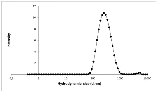
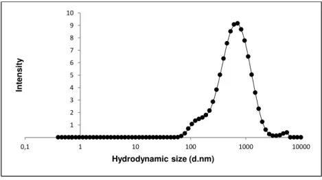
To verify the quality of the dispersion of the nanomaterial in the culture medium the hydrodynamic size-distribution was determined using Dynamic Light Scattering (DLS; Malvern Nano ZS, Malvern Inc., UK) according to SOP for measurement of hydrodynamic Size- Distribution and Dispersion Stability by Dynamic Light Scattering (DLS) (Keld Alstrup Jensen 2014). Samples were placed into 700 µL -polystyrene cuvettes and the most relevant parameters achieved in DLS: the Polydispersity Index (PdI) that indicates if a sample has a broad range of sizes or not; and the Z-Average size (Zav), which is an indicator of the average size of the particle, in nanometers of each suspension were assessed. The analysis was performed in the batch dispersion, soon after the sonication procedure, and in selected concentrations of the NMs diluted in cell culture medium: 3.2, 32 and 240 µg/mL, corresponding to 1, 3 and 10 µg/cm2. Each sample was analyzed at 0, 3, 24, 48 hours following preparation and incubation at 37ºC as well as and after 7 days-incubation. During this time, NM samples were incubated at 37ºC, 5% CO2 to mimic best possible the in vitro test exposure conditions. Each
analysis consisted in ten repeated measurements of hydrodynamic size were performed without pause. Results were compared to values that were obtained within the NANoREG Project and used as benchmark values.
24
3.3
Cytotoxicity Assays
3.3.1 MTT assay
For this assay, A549 cell line was seeded at a density of 0,5x10⁴ cells per well in 96-well plates
and incubated for 24 hours at 37oC with 5% CO2. After this incubation time, the cells were exposed to
the nanomaterials NM-212, NM-101, NM-100 and NM- 220 in a range of concentrations from 0 to 100 µg/cm2 and incubated again in the same conditions for another 24 hours. The positive control used was
a detergent, namely SDS at 0.01%. After this exposure period the treatment was removed and the cells were washed twice with PBS. A volume of 100 µL of MTT (Life Technologies, Carlsbad, California) solution was added in each well at final concentration of 0,5 mg/mL, previously prepared in PBS (5 mg/mL) and finally in culture medium to reach the desired concentration. The cell culture was incubated for a period from 2 to 4 hours in order to enable the viable cells to convert the MTT in the colored compound. After this time, the MTT solution was and DMSO solution was added in order to solubilize the purple precipitate produced by the cells and incubate for approximately for 20 minutes while agitating. The final procedure consisted in measuring the absorbances of each well in a Multiskan Ascent Spectrophotometer (Thermo LabSystems, Waltham, MA)at 570 nm (reference filter: 690 nm). To allow the separation the nanomaterial from the colored supernatant the plate was centrifuged 96 well plate centrifuge in a Sigma 4-16 S and the supernatant was transferred to a new plate; the absorbances were measured again at same conditions.
3.3.2 Clonogenic assay
A549 cell line was plated in a density of 100 cells per well, in a 6-well plate and allowed to attach for 18 hours before exposure. This incubation time was shorter than the doubling time of this cells (22 hours), in order to guarantee that the cells were attached but not divided ate the time of the treatment with nanomaterials. The cell culture were then exposed to the different concentrations (ranging from 0- 100 µg/cm2) of the nanomaterials and incubated for 7 days, at 37oC, with 5% CO2. MMC was used as