ABSTRACT | Purpose: To compare the efficacy of 0.03%
to pical tacrolimus in combination with oral omega (ω) 3 with different ratios of eicosapentaenoic acid (EPA), docosahexaenoic acid (DHA), and antioxidants as adjuvant in the treatment of keratoconjunctivitis sicca (KCS) in dogs. Methods: Forty-five dogs with KCS were evaluated monthly for 6 months. Evalua-tions included performance of the Schirmer tear, fluorescein, and lissamine green tests. Tear film break-up time (TBUT) was assessed. Conjunctival cytology was evaluated at the begin-ning, middle, and end of the study. Conjunctiva was biopsied at the beginning and end of the study. Dogs were randomly assigned to one of the three groups (n=15): Group T (topical tacrolimus 0.03%), Group TO (topical tacrolimus + ω-1.5 EPA: 1 DHA), or Group TOA (topical tacrolimus + ω-1 EPA:4.5 DHA + antioxidants). Results: There was a significant improvement in clinical signs in all groups. TBUT increased throughout treatment in all groups; this effect was most pronounced in Group TO. Cytological analysis performed at the end of the study period, showed decreased levels of lymphocytes, neutrophils, and metaplastic and squamous cells in Groups T, TO, and TOA. Histological analysis performed at the end of the study period showed decreased levels of lymphocytes and neutrophils and increased levels of goblet cells. These effects were most pro-nounced in Group TO. Conclusion: Oral treatment with ω-3 containing a higher proportion of EPA than DHA increased the
effectiveness of topical tacrolimus 0.03% in the treatment of keratoconjunctivitis sicca in dogs.
Keywords: Keratoconjunctivitis sicca; Tacrolimus;
Eicosapen-taenoic acid; Docosahexaenoic acids; Antioxidants; Animal; Dogs
RESUMO | Objetivo: Comparar a eficácia do tacrolimus 0,03% tópico associado ao ômega 3 oral, com diferentes proporções de ácido eicosapentaenoico (EPA), ácidos docosa-hexaenoicos (DHA) e antioxidantes, como adjuvante no tratamento de cães acometidos por ceratoconjuntivite seca. Métodos: Quarenta
e cinco cães atendidos no Hospital Veterinário da UNOESTE portadores de ceratoconjuntivite seca foram avaliados mensal-mente por 6 meses pelo Teste Lacrimal de Schirmer, Teste de Fluoresceína, Tempo de Ruptura do Filme Lacrimal, Teste de Rosa Bengala, citologia da conjuntiva no início, meio e fim do projeto e biopsia da conjuntiva no início e final do projeto. Os cães foram distribuídos aleatoriamente em 3 grupos (n=15): grupo T (tacrolimus 0,03% tópico), grupo TO (tacrolimus + ômegas 1.5 EPA/1 DHA oral) e grupo TOA (tacrolimus + ômegas 1 EPA/4,5 DHA + antioxidantes oral). Resultados: Houve uma melhora significativa nos sinais clínicos em ambos os grupos. No tempo de ruptura do filme lacrimal todos os grupos apresentaram aumento no decorrer do tratamento, sendo que o grupo TO foi o que apresentou melhor resultado em todos momentos quando comparado aos demais grupos. Ao final do experimento, os grupos T, TO e TOA apresentaram na análise citológica, diminuição de linfócitos, neutrófilos, células metaplásicas e escamosas, e na análise histopatológica, diminuição de linfócitos e neutrófilos e aumento das células caliciformes, com o grupo TO com melhor desempenho. Conclusão: O tratamento oral com ω-3 contendo uma maior proporção de EPA do que o DHA aumentou a eficácia do tacrolimus tópico 0,03% no tratamento de ceratoconjuntivite sicca em cães.
Descritores: Ceratoconjuntivite seca; Tacrolimo; Ácido
eico-sa pentaenoico; Ácidos docoeico-sa-hexaenoicos; Antioxidantes; Ani mais; Cães
Oral omega 3 in different proportions of EPA, DHA,
and antioxidants as adjuvant in treatment
of keratoconjunctivitis sicca in dogs
Ômega 3 oral em diferentes proporções de EPA, DHA e antioxidantes
como adjuvante no tratamento de ceratoconjuntivite seca em cães
Danielle Alves Silva1, Gisele Alborgetti Nai2, Rogério Giuffrida1, Marcos Rógério Sgrignoli1, Daniela Rodrigues dos Santos3, Isabela Vasconcelos Donadão3, Felipe Franco Nascimento3, Heloise Rangel Dinallo3, Silvia Franco Andrade1
1. Program in Animal Science, Universidade do Oeste Paulista, Presidente Prudente, SP, Brazil.
2. Department of Anatomy Pathology, Faculdade de Medicina, Universidade do Oeste Paulista, Presidente Prudente, SP, Brazil. 3. Faculdade de Medicina Veterinária, Universidade do Oeste Paulista, Presidente Prudente, SP, Brazil.
Submitted for publication: November 22, 2017 Accepted for publication: February 9, 2018
Funding: No specific financial support was available for this study.
Disclosure of potential conflicts of interest: None of the authors have any potential conflict of interest to disclose.
Corresponding author: Silvia Franco Andrade.
Rodovia Raposo Tavares, km 572, Presidente Prudente, SP - 19067-175 - Brazil E-mail: silviafranco@unoeste.br
INTRODUCTION
Keratoconjunctivitis sicca (KCS), also known as dry eye syndrome, is an ocular disease resulting from in-flammation of the lacrimal gland and decreased lacrimal film. KCS can occur because of reduced production of the aqueous portion of the tear film (quantitative deficiency) and/or excessive evaporation of the tear film (qualitati-ve deficiency). Both phenomena render the pro tecti(qualitati-ve function of the tear film deficient. KCS mainly affects structures of the cornea and conjunctiva(1,2). Among
the causes of KCS in dogs are racial predisposition, hypothyroidism, medications (atropine, sulfonamides), surgical excision of the third eyelid gland, distemper, and, mainly, autoimmune factors(3-5).
The main clinical signs observed are mucoid or mu-copurulent secretion, conjunctival hyperemia, pigmenta-tion, vascularization and opacity of the cornea, blepha-ritis, blepharospasm, and, in cases of greater severity, pre sence of corneal ulcer(6).
Tacrolimus (FK 506) is a macrolide antibiotic isolated from Streptomyces tsukubaensis spp that has an immu-nomodulatory action(4). The effects observed with its use
are the combination of local immunosuppression, goblet cell proliferation, and anti-inflammatory effects(7,8).
Recent studies in humans(9-11) and animals(12,13) showed
good results in dry eye control with the use of essential fatty acids, ω-3 and ω-6, due to their ability to produce anti-inflammatory mediators.
Fish oil (FO) from cold-water fish (e.g., salmon, tuna, herring)(9) is an important source of ω-3. Ingestion of
ma-rine plants that synthesize ω-3 results in the formation of eicosapentaenoic acid (EPA) and docosahexaenoic acid (DHA). Other essential oils, such as flaxseed oil, require conversion of β-linolenic acid (ALA) to EPA and DHA(10,11).
EPA plays an important anti-inflammatory role, whereas DHA contributes to function and development of the retina(14) and brain(15). In addition to EPA and DHA, ω-3
is a precursor of lipid mediators, such as resolvins and protectins, which have anti-inflammatory and immuno-modulatory actions(16,17).
Several studies have shown that nutritional supple-mentation with antioxidants, vitamins (A, C, and E), and minerals improves visual function(18). Antioxidants assist
in the anti-inflammatory response, corneal healing, and tear film stability(19).
The objective of the study was to compare the effi-cacy of 0.03% topical tacrolimus in combination with oral ω-3 with different ratios of EPA, DHA, and antio-xidants as an adjuvant in the treatment of KCS in dogs.
METHODS
Animals
The study was conducted according to the standards of animal experimentation of the UNOESTE Ethical Committee on Animal Use (protocol #2939) and the sta-tement from the Association for Research in Vision and Ophthalmology for the use of animals in ophthalmic and visual research.
We evaluated 45 dogs seen at the Veterinary Hospital of UNOESTE over a 6-month period. Inclusion in the study was not based on any preference in terms of breed or sex. Dogs were diagnosed with KCS and registered under an authorization term (Informed Consent Term). The animals were included in the experiment by ob-serving clinical ophthalmic signs compatible with KCS (ocular secretion, conjunctivitis, corneal opacity, and pigmentation) with a slit lamp (Kowa SL-15, Japan) as well as SST ≤10 mm/min and/or TBUT ≤10 s.
Groups
After the diagnosis of KCS, the dogs were randomly divided in a double-blind study into three treatment groups: T group (n=15): tacrolimus 0.03% eye drops (Eye Pharma Laboratory, São Paulo, Brazil), one drop, 2×/day, topical, in both eyes for 6 months; TO group (n=15) tacrolimus eye drops 0.03% (Eye Pharma Labo-ratory, São Paulo, Brazil), one drop 2×/day, topical in both eyes + oral ω-3 (1.5 EPA: 1 DHA-Ograx®-3, Avert
Laboratory, São Paulo, Brazil, 1 capsule of 500 mg/7 kg/day) for 6 months; Group TOA (n=15) tacrolimus eye drops 0.03% (Eye Pharma Laboratory, São Paulo, Brazil), one drop 2×/day, topical in both eyes + oral ω-3 +
antio-xidants (1 EPA: 4,5 DHA + vitamin E + vitamin C + selenium- Seniox®, Avert Laboratory, São Paulo, Brazil,
1 capsule of 500 mg/10 kg/day), for 6 months.
All groups also received propylene glycol-based lubri-cant (Systane®, Alcon, São Paulo, Brazil), one drop, both
eyes, twice daily for 6 months. If necessary, antibiotic eye drops were used (one drop, four times per day, for 15 days), based on culture and antimicrobial sensitivity from ocular samples secretions of all of the dogs and associated too on the presence of corneal ulcer or cli-nical signs such conjunctivitis and mucopurulent ocular secretion. Anti-inflammatory eye drops containing di-clofenac sodium (Still®, Allergan, São Paulo, Brazil) (one
Ophthalmic exams
Monthly ophthalmic and cytological exams were per-formed, considering zero (T0) the first day of treatment with bilateral KCS diagnosis and the other time points (T1-T6). The cytological examination was performed at moments T0, T3, and T6, and histopathological exami-nation was performed at the time of diagnosis (T0) and at the end of the study (T6).
Clinical ophthalmic signs were identified using the portable slit lamp (Kowa, Japan) with or without conjuncti-vitis, ocular secretion, opacity, and corneal pigmentation according to the scoring described in table 1.
The Schirmer tear test (STT) was performed without anesthetic eye drops to evaluate the quantitative portion of the tear film. A 0.5-cm strip was introduced into the conjunctival sac for 1 min. Results ≤10 mm/min were
considered positive(20). Tear film break-up time (TBUT)
was used to evaluate the qualitative portion of the tear film. The mean of two consecutive measurements was recorded. After instilling one drop of 1% fluorescein (Al-lergan, São Paulo, Brazil), the eye was examined under the slit-lamp (Kowa, Japan). Time from the last blink to the appearance of dark spots on the lacrimal film was measured. TBUT values ≤10 s were considered as
posi-tive results(21).
After TBUT examination, eyes were washed with phy-siological saline. The presence or absence of corneal ulcers was determined using the fluorescein test (FT)(20).
Ulcers were graded according to severity and extent (0: negative; 1: small superficial ulcer; 2: medium su-perficial ulcer; 3: extensive susu-perficial ulcer; 4: small stromal ulcer; 5: medium stromal ulcer; 6: extensive
stromal ulcer; 7: descemetocele; and 8: keratomalacia or melt ulcer).
Green lissamine test (GLT) strips (Ophthalmos, São Paulo, Brazil) were used to evaluate the presence of devitalized cells(22). The lissamine strip was placed in
contact with the tear meniscus at the bottom of the corneal sulcus; measurements were obtained 2-min later. The classification of van Bijsterveld for graduation was used. The palpebral rhyme was divided into three areas: lateral bulbar conjunctiva, cornea, and medial bulbar conjunctiva. In each of these areas, the following graduation was used: 0-absence of staining, 1-fine dots, 2-dots coarse, and 3-plate. The sum of each of these areas determined the final score, which ranged from 0 to 9.
Cytological and histopathological examination
Cytology was evaluated after the eye had been clea-ned with physiological saline solution. Cells were obtai-ned from the lower conjunctiva with a sterile swab that had been moistened with physiological solution. Staining was performed with MGG technique (May-Grunwald-Giemsa). Lymphocytes, neutrophils, metaplastic cells, and squa-mous cells were counted under an optical microscope in 10 fields with a 40× objective lens.
The histopathological examination was performed after instillation of Anestésico® (1% tetracaine
hydro-chloride + 0.1% phenylephrine hydrohydro-chloride, Allergan, São Paulo, Brazil) with a withdrawal of 1-3 mm in the for-nix of the medial inferior conjunctiva using tweezers and conjunctive scissors. The histological section was placed in a standardized 1 × 1 cm paper size fixed in formal-dehyde and embedded in paraffin (Dynamics Analytical Reagents, São Paulo, Brazil). With the help of a rotating microtome, 5-µm-thick sections of the conjunctiva were obtained, stained with hematoxylin and eosin (HE) (Dolles, São Paulo, Brazil), PAS (Merck, USA), and subsequently evaluated for the following parameters. In HE staining: lymphocyte count, neutrophils, presence of squamous metaplasia, and presence of edema. In PAS counting, the goblet cell density (cells/mm2) under the
optical microscope at 40×.
Statistical analysis
Two analyses of variance for Tukey’s sample were used for statistical analysis of results obtained with the STT and TBUT, as well as measurements of goblet cell density and numbers of squamous cells, metaplastic cells, neutrophils, and lymphocytes. For the clinical sign
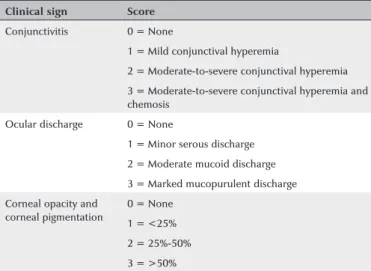
Table 1. Evaluation score of clinical ophthalmic signs
Clinical sign Score
Conjunctivitis 0 = None
1 = Mild conjunctival hyperemia
2 = Moderate-to-severe conjunctival hyperemia 3 = Moderate-to-severe conjunctival hyperemia and chemosis
Ocular discharge 0 = None
1 = Minor serous discharge 2 = Moderate mucoid discharge 3 = Marked mucopurulent discharge Corneal opacity and
corneal pigmentation
variables FT and GLT, we used Friedman’s nonparame-tric test to compare moments, while the Kruskall–Wallis test with Dunn contrast was used to compare the groups. A p<0.05 significance level was adopted. The software used for statistical analysis was R, version 3.2.2. (The R Foundation for Statistical Computing, 2015).
RESULTS
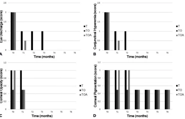
In the clinical signs evaluated (Figure 1), all groups showed significant improvement. In the variables of ocu-lar secretion and conjunctivitis, there was total remis-sion in T1 only in Group TO, whereas in Groups T and TOA, remission was found at T3 and T2, respectively. In the variable of opacity, there was total remission in all groups in T2. Regarding corneal pigmentation, the median showed reduction throughout the treatment in all groups.
In all three groups, STT values significantly increa-sed from baseline at T1 and all subsequent time-points (p<0.05; Figure 2). At the time T6, the T group presented
a significant difference in relation to the other groups. In the TBUT (Figure 2), all groups presented significant di-fferences in relation to the moment T0. However, Group TO presented the best results at all times compared with the other groups.
In FT, all groups presented animals with ulcers of va-rying severity and extension at T0, and all groups showed excellent healing in T1. In the GLT, the performance of all groups was similar, demonstrating improvement in the marking already found in T2.
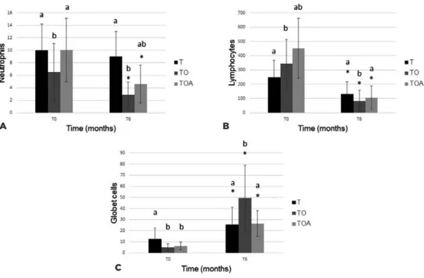
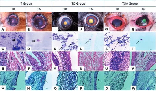
Cytological analysis showed decreased levels of all cell populations examined, but differences at a given time-point were only significant in comparison with baseline levels (Figure 3). In the histopathological study (Figure 4), all groups showed a decrease of inflammatory cells. The goblet cells showed a significance increased (p<0.05) in TO group (49.7 ± 29.4), in relation to other groups, T (25.5 ± 15.6) and TOA (26.5 ± 11.7). Representative images of some eyes and cytological and histopathological results for Groups T, TO, and TOA are presented in figure 5.
Figure 1. Median score of observed clinical signs from T0 to T6 of the T group (Tacrolimus), Group TO (Tacrolimus + Omega 1.5 EPA: 1 DHA), and the TOA group (Tacrolimus + Omega 1 EPA: 4.5 DHA + Antioxidants): (A) ocular discharge, (B) conjunc-tival hyperemia (C), corneal opacity, and (D) corneal pigmentation.
A B
fits, had a portion of EPA. According to another study(23),
efficacy in treatment for dry eye with use of ω-3 is linked to the dose of ingested ω; however, the results of our
study show that effectiveness is related to relative pro-portions of EPA and DHA. Other studies(19) referred to
antioxidants as an adjunct to the inflammatory respon-se, corneal healing, and lacrimal film stability. However, in the present study, this adjunct did not bring additional benefits in the treatment of dry eye in dogs.
According to studies, EPA has a pro-inflammatory effect exerting cellular actions that stimulate the produc-tion of collagenase and increase expression of adhesion molecules necessary for leukocyte extravasation. EPA is the precursor of the E series of resolvins, which includes resolvin E1 (RvE1) and resolvin E2 (RvE2)(24-26). Resolvins
were first described in connection with the formation of mediator molecules with anti-inflammatory capa-city and immunomodulatory properties, including the reduction of proinflammatory leukocytes and cytokine migration, thus leading to a decreased inflammatory response in vivo(27).
DHA is more related to its antioxidant properties and is involved in several cognitive processes while also linked to correct signaling between neurons, in fetal de-velopment and retinal function. DHA can also give rise to resolvins D1 (RvD1) and D2 (RvD2), a protectin (or neuroprotetin, when produced by neural tissues) and a maresine, thus having an important anti-inflammatory function in neuronal systems and the formation, deve-lopment, and functioning of the brain and retina(24,25,27).
In the present study, conjunctival biopsy was perfor-med to assess inflammatory cytology. Neutrophil counts were lower in Group TO compared with other groups at time T6. This effect may be attributable to anti-inflamma-tory mediator RvE1 (produced by EPA), which increases macrophage phagocytic activity(28).
There was a significant increase in the number of goblet cells from baseline to the end of the study period. This effect was most apparent in Group TO. Similar re-sults were reported previously; one study found an in-crease in the number of goblet cells after administration of ω-fatty acids for treatment of KCS(9,10).
The results reported above indicate decreased clini-cal signs and inflammation, as well as increased numbers of goblet cells after treatment with an oral formulation of ω-3 containing a higher proportion of EPA than DHA. This formulation of ω-3 fatty acids enhances the effecti-veness of topical tacrolimus in treating canine KCS. We attribute this effect to higher levels of EPA, which is a DISCUSSION
The use of ω-3 (EPA and DHA) improved the clinical signs of KCS, increasing the STT and TBUT values and promoting resolution of corneal ulcers, because they presented anti-inflammatory characteristics and corro-borated the results of other studies(9,10).
In the present study, resolution of ocular secretion, conjunctivitis, and increased TBUT were better in Group TO compared with the T and TOA groups. Both TO and TOA groups received ω supplementation, at varying
pro-portions: Group TO (1.5EPA:1DHA) and the TOA group (1EPA:4.5DHA + antioxidants), in which addition of antioxidants did not seem to have any treatment
bene-*= p<0.05 (Tukey test to compare moments).
a,b= p<0.05 (Kruskal–Wallis test to compare groups).
Figure 2. (A) Mean and SD of the Schirmer tear test (STT) in mm/min from T0 to T6 of patients in the following groups: T (Tacrolimus), TO (Tacrolimus + Omega 1,5 EPA: 1 DHA), and TOA (Tacrolimus + Omega 1 EPA: 4.5 DHA + Antioxidants). (B) Mean and SD of TBUT (s) T0–T6, of patients in the T, TO, and TOA groups.
A
*= p<0.05 (Tukey test to compare moments).
a,b= p<0.05 (Kruskal–Wallis test to compare groups).
Figure 3. (A) Mean and SD of the neutrophils count in the conjunctival cytology test from T0 to T6 of the T, TO, and TOA groups. (B) Lymphocytes. (C) Squamous cells. (D) Metaplastic cells.
A B
D C
*= p<0.05 (Tukey test to compare moments).
a,b= p<0.05 (Kruskal–Wallis test to compare groups).
Figure 4. Mean and SD of the neutrophils count in T0 and T6 conjunctival histopathology of the T, TO and TOA groups. (B) Mean and SD of the T0 and T6 lymphocyte counts. (C) Mean and deviation pattern of goblet cell counts at T0 and T6.
A B
Figure 5. (A) T group, moment T0 left eye, mucoid secretion, opacity, and corneal pigmentation. (B) Evolution of the patient and persistence of pigmentation. (C) Photomicroscopy of T0 conjunctival cytology, presence of neutrophils, squamous cell (thick arrow), metaplastic cell (fine arrow), MGG coloration, 400× magnification. (D) T6 moment, presence of squamous cells (thick arrow), MGG coloration, 400× magnification. (E) Histopathology at T0, inflammatory infiltrate (*), MGG coloration, 400× magnification. (F) T0, reduction of inflammatory process. (G) T0, absence of goblet cells, PAS staining, increase of 400×. (H) PAS at T6 moment, presence of goblet cells (fine arrow). (I) TO group, T0 moment left eye, mucoid secretion, descemetocele. (J) Ulcer healing. (K) Photomicroscopy of T0 conjunctival cytology, presence of neutrophils, globet cell (fine arrow), MGG, 400× magnification. (L) T6 moment, presence of squamous cell (thick arrow), MGG coloration, 400× magnification. (M) His-topathology at T0, inflammatory infiltrate (*), coloration HE, increase of 400×. (N) T0, reduction of the inflammatory process. (O) T0, absence of goblet cells, PAS staining, increase of 400×. (P) PAS, T6, large amount of goblet cells. (Q) TOA group, moment T0 right eye, mucopurulent secretion. (R) clinical improvement of patient. (S) Photomicroscopy of T0 conjunctival cytology, presence of neutrophils, metaplastic cell (fine arrow), MGG coloration, 400× magnification. (T) T6, presence of globet cell (fine arrow), MGG coloration, 400× magnification. (U) Histopathology at T0, inflammatory infiltrate (*), HE staining, 400× magnification. (V) T0, reduction of inflammatory process (X) T0, absence of goblet cells, PAS staining, increase of 400×. (W) PAS at T6 moment, presence of goblet cells.
precursor of anti-inflammatory and immunomodulatory factors such as RvE1.
REFERENCES
1. Ribeiro AP, Brito FL, Martins BC, Mamede F, Laus JL. Qualitative and quantitative tear film abnormalities in dogs. Cienc Rural. 2008; 38(2):568-75.
2. Williams DL. Immunopathogenesis of keratoconjunctivitis sicca in the dog. Vet Clin North Am Small Anim Pract. 2008;38(2):251-68.
3. Gelatt KN. Veterinary Ophthalmology. 3th ed. Pennsylvania: Lippincott
Williams & Wilkins; 2005. 594p.
4. Miller PE. Lacrimal system. In: Maggs DJ, Miller PE, Ofri R. Slater’s
Fundaments of Veterinary Ophthalmology. 4th ed. St Louis: Elsevier;
2008. p. 157-74.
5. Berdoulay A, English RV, Nadelstein B. Effect of topical 0.02% ta-crolimus aqueous suspension on tear production in dogs with keratoconjunctivitis sicca. Vet Ophthalmol. 2005;8(4):225-32.
6. Koch SA, Sykes J. Keratoconjunctivitis sicca. In: Riis RC. Small ani-mal ophthalmology secrets. Philadelphia: Hanley & Belfuscap; 2002. p. 52-60
7. Hendrix VD, Adkins EA, Ward, DA, Stuffle J, Skorobohach B. An investigation comparing the efficacy of topical ocular applica-tion of tacrolimus and cyclosporine in dogs. Vet Med Int. 2011; 2011:487592.
8. Moskovici BK, Holzchuh R, Sakassegawa-Naves FE, Hoshino-Ruiz DR, Albers MB, Santo RM, et al. Treatment of Sjögren’s syndrome dry eye using 0.03% tacrolimus eyedrop: Prospective double-blind randomized study. Cont Lens Anterior Eye. 2015;38(5):373-8. 9. Rand AL, Asbell PA. Nutritional supplements for dry eye syndrome.
Curr Opin Ophthalmol. 2011;22(4):279-82.
10. Jenkins DJ, Josse AR. Fish oil and omega-3 fatty acids. CMAJ. 2008; 178(2):150.
11. Rashid S, Jin Y, Ecoiffer T, Barabino S, Schaumberg DA, Dana MR. Topical omega-3 and omega-6 fatty acids for treatment of dry eye. Arch Ophthalmol. 2008;126(2):210-25.
12. Neves ML, Yamasaki L, Sanches OC, do Amaral MS, Stevanin H, Giuffrida R, et al. Use of linseed oil to treat experimentally induced A
C
E
G H
I
K
M
O
J Q
S
U
X
T
V
W R
L
N
P F
keratoconjunctivitis sicca in rabbits. J Ophthalmic Inflamm Infect. 2013;3(1):4.
13. Silva DA, Nai GA, Giuffrida R, Barbero RC, Kuhn JM, da Silva AD, et al. Comparison between fish and linseed oils administered orally for the treatment of experimentally induced keratoconjunctivitis sicca in rabbits. Open Vet J. 2017;7(3):277-85.
14. Cheatham CL, Colombo J, Carlson SE. N-3 fatty acids and cognitive and visual acuity development: methodologic and conceptual con-siderations. Am J Clin Nutr. 2008;83(6 Suppl):1458S-66S. 15. Wurtman RJ. Synapse formation and cognitive brain development:
effect of docosahexaenoic acid and other dietary constituents. Metabolism. 2008;57 Suppl 2:S6-10.
16. Calder PC. Omega-3 fatty acids and inflammatory processes. Nu-trients. 2010;2(3):355-74
17. Serhan, CN, Hong S, Gronert K, Colgan SP, Devchand PR, Mirik G, et al. Resolvins: a family of bioactive products of omega-3 fatty acid transformation circuits initiated by aspirin treatment that counter proinflammatory signals. J Exp Med. 2002;196(8):1025-37. 18. Cangemi FE. TOZAL Study: an open case control study of an oral
antioxidant and omega-3 supplement for dry AMD. BMC Ophthal-mol. 2007;7:3.
19. Gus PI, Belló-klein A, Llesuy S, Quinto GG, Matos GH, Bechara SJ. Tear antioxidante potential in Young adults. Arq Bras Oftalmol. 2006;69(4):565-70.
20. Maggs DJ. Basic diagnostic techniques. In: Maggs DJ, Miller PE, Ofri R, editors. Slatter’s Fundamentals of Veterinary Ophthalmology. St. Louis: Saunders Elsevier; 2008. p. 81-106.
21. Saito A, Kotani T. Estimation of lacrimal level and testing methods on normal beagles. Vet Ophthalmol. 2001;4(1):7-11.
22. Machado LM, Castro RS, Fontes BM. Staining patterns in dry eye syndrome: rose bengal versus lissamine green. Cornea. 2009; 28(7):732-4.
23. Escamilla NE. Omega 3 Y su acción terpéutica em el síndrome de ojo seco. Ciência & Tecnologia Para La Salud Visual y Ocular [In-ternet]. 2003[cited 2016 jun 21];1(1):91-8. Available from: https:// revistas.lasalle.edu.co/index.php/sv/article/view/1946
24. Hodge WG, Schachter HM, Barnes D, Pan Y, Lowcock EC, Zhang L, et al. Efficacy of W-3 fatty acids in preventing age-related macular degeneration-a systematic review. Ophthalmology. 2006;113(7): 1165-72; quiz 1172-3, 1178.
25. Seki H, Sasaki T, Ueda T, Arita M. Resolvins as regulators of the immune system. Sci World J. 2010;10:818-31.
26. Arita M, Bianchini F, Aliberti J, Sher A, Chiang N, Hong S, et al. Stereochemical assignment, anti-inflammatory properties, and receptor for the omega-3 lipid mediator resolvin E1. J Exp Med. 2005;201(5):713-22.
27. Bannenberg GL. Therapeutic applicability of anti-Inflammatory and proresolving polyunsaturated fatty acid-derived lipid mediators. Sci World J. 2010;10:676-712.