Estudo
in vitro
dos efeitos do fa tor de crescimen to derivado de plaquetas nos
cera tócitos humanos após ablação com e�cimer laser
João Eduardo C. Ribeiro 11• 2• 31 Fernando Oréfice 111
Roger Beuerman 121
''' Departamento de Oftalmologia, Universidade Federal de Minas Gerais, Belo Horizonte, MG.
'" Laboratory for the Molecular Biology of the Ocular Surface, LSU Eye Center, Nova Orleans, Louisiana -EUA.
"' Universidade de Uberaba, Uberaba, MG. Part ofthe PhD thesis presented to the Postgraduation Course in Ophthalmology of the UFMG which conferred the title of Doctor in Medicine to the author. Perforrned at the Department of Ophthalmology of the UFMG and the Lions Eye Research Laboratories, Laboratory for the Molecular Biology of the Ocular Surface, LSU Eye Center, Louisiana State University Medical Center School of Medicine, New Orleans, Louisiana - USA.
The authors have no proprietary interest in the development or marketing of the instruments or medications referred to in the text.
Address for correspondence: Dr. João Eduardo C. Ribeiro. Av. Santos Dumont, 409. Uberaba (MG) CEP 38060-600.
SUMMARY
Purpose: To evaluate the in vitro effects of platelet derived growth factor (PDGF) on the response ofhuman keratocytes to excimer laser ablation.
Methods: Keratocytes were cultured to confluence in 6-well tissue culture plates and exposed to an excimer laser beam. After the laser they were mantained in one of the following media: Group I -Dulbecco 's Modified Eagle Medium supplemented with heat inactivated fetal bovine serum (FBS); Group II - DMEM + fetal bovine serum (FBS); Group III - DMEM + heat-inactivated FBS pios PDGF at a concentration of 1 ng/ml and Group IV - DMEM + heat inactivated FBS pios PDGF at a concentration of 10 ng/ml. To quantitate the rate of wound closure micrographs were taken at O, 24,
48, 72 and 96 hours.
Results: Ninety-six hours after the excimer laser ablation only keratocytes cultured in FBS (Group II) resurface completely. At evaluations after 48, 72 and 96 hours, the residual area in Groups I, III and IV was bigger than in Group II (p < 0.05). There was no significant difference between groups treated with different concentrations of PDGF.
Conclusion: Cultured human keratocytes proliferate and migrate to the wounded area after excimer laser ablation and PDGF actually does not stimulate these cells in the presence ofheat-inactivated serum.
Keywords : Platelet derived growth factor; Keratocytes ; Excimer laser.
INTRODUCTION
Since the corneal ablation technique utilizing excimer laser has been described, the physical and mechanical aspects of the use of this laser have constantly progressed. Although much knowledge about laser-cornea interaction has been gained in the last years, a better understanding of the corneal healing process after ablation and its modulation is of utmost importance for this surgery. The postlaser corneal remodelling pattern is unpredictable. Formation of the scar, clinically called haze, appears to be related to this process and varies from one individual to the other with or without the use of pharmacological modulators 1
•
Severa! substances have been used in the attempt to regulate corneal healing, either modulating cell population through growth factors and antiproliferative agents or modulating extracellular matrix synthesis 2 Growth factors represent the new therapeutic option.
ln vitro effects of platelet derived growth factor on human keratocyte response to excimer laser ablation
Growth factors represent a system of signals which organize and coordenate cell proliferation. Several growth factors, among which the epidermal growth factor (EGF), fibroblast growth factor (FGF), platelet derived growth factor (PDGF) and transforming growth factor-13 (TGF-13) have shown to play an important role in the stimulation of healing 3• 4
.
The platelet derived growth factor, PDGF, has shown to be important for fibroblast activation. lt is a glycoprotein consisting of two cysteine rich chains with a 60% homology between them, exerting their effects by binding to specific membrane receptors . Cell activation occurs through the tyrosine kinase enzyme system 3•4
•
Studies have shown the action of growth factors as modulators of ocular structure response to trauma 5• 6• The
purpose of this in vitro study is to investigate the response of cultured human keratocytes to excimer laser ablation in the presence of platelet derived growth factor.
MATERIAL AND METHOD
Human keratocytes were cultured from a comea donated by the National Disease Research Interchange (NRDI) - Phila delphia, P A - obtained 24 hours after the donor' s death. The culture method was similar to that described by Gebhardt et al 7
•
The whole procedure was carried out in a laminar flow hood. Summarizing, the corneal epithelium was mechanically removed using Paton' s spatula. Then an approximately 2 x 2 mm fragment was excised from the center of the comea and the endothelium removed using a cotton swab. The remaining stroma was cut into small pieces which were placed in the center of a model T 75 culture flask (Falcom Labware, Becton Dickinson Co., Lincoln Park, NJ, USA). Ten milliliters of Dulbecco' s Modified Eagle Medium - DMEM (Life Technologies, Grand Island, NY, USA) supplemented with 10% fetal bovine serum were then added. Microbial proliferation was avoided by adding 1 o/o antibiotics and antifungais (penicillin G 1 0,000 IU, streptomycin 1 0,000 µg/ml and amphotericin B 25 µ/ml) (Life Technologies).
After 7 to 1 O days the corneal stroma fragments adhered to the surface of the flask and keratocyte proliferation was initiated. After 3 weeks from the beginning of the culture, a keratocyte amount sufficient to cover the whole internai basis of the flask was reached (confluence) and subcultures in new flasks were started.
These cells were subcultured until the fourth passage and then suspended in culture medium and their concentration determined using a hematocytometer (Neubauer, American Optical, Buffalo, NY, USA) . The keratocytes were divided into 1 2 compartments, each containing 8 . 5 x 1 0 5 cells. For this procedure two 6 well culture plates (Costar Inc . , Cambridge, MA, USA) were used for later application of the laser, totalling three cultures per group.
An Excimer Laser VISX, Model Twenty-Twenty (Twenty Twenty Laser System, VISX Incorporation, Sunnyvale, CA, USA), developed for refractive surgery, was used. The laser
was focused on the keratocyte layer covering the internai part of the culture plates and applied to the central part of each plate. Laser parameters for all applications were 1 5 pulses, ablation rate of 0.27 µ/pulse, 5 mm ablation zone, 1 6 8 mJ/cm2 fluence and 5 Hz repetition rate.
Evaluations of cultured keratocytes, before and after the laser, were by microphotographs every 24 hours, using a Zeiss ICM 405 (Zeiss Company, Berlin,Germany) contrast phase microscope. The remaining residual area was quantified on basis of the microphotographs using a computerized imaging analysis system (R.M. Biometrics, Nashville, TN, USA) .
After laser application, the cultures were maintained in one of the following culture media:
- Group I - Plates 1 to 3: DMEM + Heat inactivated fetal bovine serum (Life Technologies) .
- Group II - Plates 4 to 6 : DMEM + fetal bovine serum. - Group III - Plates 7 to 9: DMEM + Heat inactivated fetal bovine serum + 1 ng/ml PDGF (UBI - Upstate Biotechnology Incorporated, Lake Placid, NY, USA) .
- Group IV - Plates 1 0 to 1 2 : DMEM + Heat inactivated fetal bovine serum + 1 0 ng/ml PDGF.
The platelet deri ved growth factor used was its dimeric form BB obtained by genetical engineering (Upstate Biotechnology Incorporated, Lake Placid, NY, USA).
Variance analysis was used to compare the rate of closure of the ablated area between the groups. Tukey-Kramer' s test for multiple comparisons was used for the analysis of the differences between two groups each time, in those cases in which variance analysis detected a difference between the groups. The statistical software used was STA TA (Stata Statistical Software: release 5 .0, Stata Corporation, College Station, TX, USA) . Ali results of this study were considered significant at 5% (p < 0.05).
RESULTS
Evolution of the closure and evaluation of the ablated area were documented at times O, 24, 48, 72 and 96 hours after laser application. The keratocytes formed a single layer on the plate surfaces. They had a uniform size, fusiform format, did not form cell clumps and were confluent. Figure 1 shows these cells before laser application at the time all 1 2 culture plates were evaluated, no difference being observed between them. The time between laser application to the first and the last plate was 1 5 minutes. At the site where the laser beam directly hit the culture plate, the cells and the remaining culture medium were completely eliminated. Alterations unlevelling the plastic surface of the plate can also be observed. The cells adjacent to the area hit by the laser presented normal charac teristics (Figure 1 b).
Evolution of closure of the ablated area
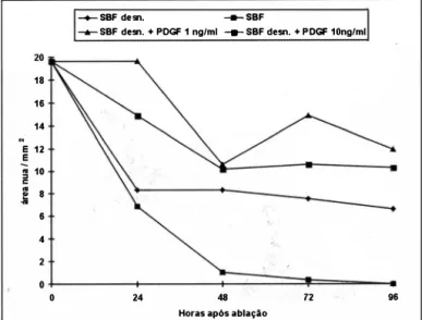
ln order to compare closure rate of the ablated area between the different groups, a graph of the mean remaining area (area without keratocytes in the ablation region) as a function of time and determined for each group was constructed (Graph 1 ) .
Evaluation a/ter 2 4 hours
ln Groups 1, II and IV, a migration of keratocytes invading the wounded area from the periphery towards the center of
ablation could be observed (Figure 2). ln group III no
keratocyte migration towards the ablated area was observed. Regarding evolution of closure of the ablated area, a statistically significant difference was observed between the groups (p = 0.0024) . Table 1 shows the results per group.
Fig. 1 - {a) Mlcrophotograph, uslng phase contrast mlcroscope, of cultured human keratocytes, showlng confluence and fuslform format of these cells {1 00X); {b) Mlcrophotograph lmmedlately after ablatlon wlth exclmer laser. Keratocytes adjacent to ablatlon with normal characterlstics, observing irregularltles on the plastic surface of the
plate at the site of ablatlon {1 00X / group Ili).
Tukey-Kramer' s test for multiple comparisons showed that the remaining area in Groups III and IV, after 24 hours, was greater that the corresponding area in Group II (p < O.O 1 and p < 0.05, respectively) and that the remaining area in Group I was smaller than that in Group III (p < 0.0 1 ) .
Evaluation after 48 hours
There was no progression of closure of the ablated area in Group 1. A statistically significant difference between the groups was found (p = 0.0022). The remaining area in Groups 1, III and
IV was greater than the corresponding area in Group II (p < 0.05 ; p < 0.01 and p < 0.0 1 , respectively). Table 1 shows the values per group after 48 hours.
Evaluation after 72 hours
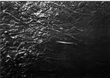
An increase in the central area without keratocytes, caused by the disappearance of some cells that had initiated the covering process of the ablated area, was observed in Groups III and IV. ln Group II the radial arrangement of the migratory keratocyte flux can be observed and these cells begin to reach the center of the ablation area (Figure 3 ) .
Table 1 shows the results o f the groups 72 hours after excimer laser application. A statistically difference between the groups was found (p = 0.0007). The remaining residual area in Groups 1, III and IV was greater than that observed in Group II (p < 0.05 ; p < 0.00 1 and p < 0.0 1 , respectively) and the remaining area in Group III was greater than the corresponding area in Group 1 (p < 0.05 ) .
Evaluation after 9 6 hours
Only in Group II the cells covered the whole ablated area .
18 16 14 E 12 E
�
10.;
8 6 4o
-+- SBF de sn. ... sBF
-+- SBF desn. + POGF 1 ng/ml --e- SBF desn. + PDGF 10nglml
24 48 72 96
Horas após ablação
Graph 1 - Evolutlon of closure of the exclmer laser ablated area accordlng to the compositlon of the culture medlum {heat lnactlvated fetal bovlne
serum, fetal bovlne serum, heat lnactlvated fetal bovlne serum + PDGF
- platelet derlved growth factor - 1 ng/ml, and heat lnactlvated fetal bovlne
serum + PDGF 1 O ng/ml) used after ablation. n = 3 observatlons for each
polnt and treatment condltlon ; values are means.
ln vitro effects of platelet derived growth factor on human keratocyte response to excimer laser ablation
Table 1 . Remai ning residual area (mm2) in the ablation area 24, 48, 72 and 96 hours after excime! laser application according_ to composition of
the cu lture media. Group 1: Heat inactivated fetal bovine serum (hi. FBS); Group li: Fetal bovme serum (FBS); Group 111: h1.FBS + PDGF
-platelet derived growth factor - 1 ng/ml (PDGF 1 ng); Group IV: h i . FBS + PDGF 1 0 ng/ml (PDGF 1 0 ng). Time
(Hours)
24
Group 1 Group li Group Ili Group IV
X :1: SD X :1: SD X :t SD X :t SD
p
48 72 96
8.3 ± 3.7
8.3 ± 3.7 7.5 ± 2.4
6.5 ± 2.3
6.8 ± 2.0 1 9 .6 ± O.O
1 .0 ± 0.4 1 0. 6 ± 1 .7
0.3 ± 0.3 1 4. 7 ± 4.0
o.o ± o.o 1 1 .9 ± 0.6
1 4,9 ± 4,0 = 0.0024 1 0. 1 ± 1 . 1 = 0.0022 1 0 .6 ± 1 .7 = 0.0007
1 0.3 ± 1 .5 < 0.000 1
Note: Value of p is lhe result of the F test of lhe variance analysis; SD: standard deviation of sample; X: mean
Table 1 shows the results in the different groups. Statistically significant differences were found between the groups (p < 0.000 1 ) . Tukey-Kramer' s test showed that the remaining area after 96 hours in Groups I, III and IV was greater than the corresponding area in Group II (p < 0.0 1 ; p < 0.00 1 and p < 0.001, respectively) and that the remaining area in Groups III and IV was greater than that in Group I (p < 0.01 and p < 0.05, respectively).
At all evaluated times there was a difference between Group II and the others . At no time a statistically significant difference w�s found between groups with different PDGF concentrations, that is, Groups III and IV.
DISCUSSION
Despite the recent advances in keratectomy with excimer laser, the main complication of this surgical procedure conti nues to be the formation of corneal opacity and the regression of the refractive effect which might be caused by keratocyte proliferation during the postoperative period 8· 9.
Soluble mediators present in normal plasma, such as platelet derived growth factor, transforming growth factor-�,
Fig. 2 - Mlcrophotograph 24 hours after exclmer laser ablation: the keratocytes already cover the perlphery of the ablated area and mlgrate
towards the center of thls area (200X / Group li).
interleukin 1 , tumor necrosis factor, gamma interferon, somatomedins, epiderma! growth factor, play important roles in the modulation of cell responses to trauma. Synergistic or antagonistic action between different modulators determine final healing 10• The specific action of these factors on the healing process is still unknown.
Proteic growth factors regulate many of the processes essential for normal healing of ocular structures, including migration, mitosis and cell differentiation 5• The effects of growth factors, separately, on human keratocytes remain little studied and documented. Among severa! growth factors, PDGF has cells of mesodermal origin as target, being a chemotactic and mitogenic factor for connective tissue forming mesenchymal cells 4• Since controlled connective tissue proliferation is essential for an optimal repair of wounded tissues, PDGF could have important actions on this process. It is extremely difficult to determine interactions between severa! cell types involved in the repair response through experimental in vivo studies. Using in vitro models for studies on corneal healing it is possible to reduce the complex in vivo tissue repair phenomenon to more simplified aspects 1 1 •
We investigated in vitro the influence of PDGF treatment
Fig. 3 - Microphotograph 72 hours after exclmer laser ablation showlng
on keratocyte response to ablation by excimer laser, at concentrations of 1 and 1 O ng/ml, since previous studies showed a greater effectiveness at these concentrations 12• 13, and also because they are the concentrations recommended by the manufacturer 14• We investigated keratocyte migra tory and growth activities because the activity of this cell is highly correlated with regression and haze formation after photoablation. ln this study model we eliminated the interference of severa! growth factors and inflammatory modulators present in tears and also reduced the complex
- inflammatory response 15 to aspects related only to the keratocyte . We used heat inactivated serum aiming to abolish the action of several mediators present in nomal serum, thus allowing to evaluate the action of PDGF alone.
The size of samples per group was sufficient for a highly significant result and many authors investigating corneal cell response to different drugs used samples of equal size as that of this study 16• 17• Keratocyte responses to excimer laser ablation were evaluated every 24 hours up to 96 hours because this is a necessary and sufficient time for mitoses to occur, since cell responses in vitro are quicker 4• 18.
Closure of the ablated area in the PDGF treated groups in the presence of heat inactivated serum or with heat inactivated serum alone was significantly smaller than that observed in the group with normal serum. These results agree with those authors who, on evaluating cell response to trauma in the presence of heat inactivated serum or in its absence, observed a smaller response when compared with cell responses in the presence of normal serum 6• 19• 20• These responses could bedue
to a series of factors : the studied growth factor could act synergistically with factors present in the serum and/or serum proteins would act as carriers for PDGF and, consequently, would be responsible for its bioavailability. B arber et al. 21 suggest that in the absence of serum, the gene responsible for transcription would be blocked and Funderburgh et al. 22 showed that bovine keratocytes cultured in low serum concentration present slow growth . Absence of other mediators in heat inactivated serum would hinder an adequate PDGF action to increase cell migration.
This study model of biological keratocyte responses to excimer laser allows an in vitro evaluation of both the response of these cells to ablation with this laser and the action of pharmacological modulators on this process. ln spite of the differences existing between in vivo and in vitro healing processes, in vitro investigations are useful in studies which determine migratory, growth, phagocytic and synthetic cell element activities 1 1 •
ln this study, cultured human keratocytes, adj acent to the area to which the excimer laser was applied, proliferated and migrated towards the wounded area, suggesting an active action on regression and haze . formation after excimer ablation. Platelet derived growth factor in the absence of normal serum components actually had no stimulating effect on human keratocytes.
PDGF induced corneal healing modifications are not yet completely defined. Regarding keratocyte activation, it was observed that PDGF acting alone did not present a stimulating
effect. Due to the fact that growth factors act synergistically or antagonistically, it i s possible that PDGF has a more expressive effect on keratocytes when combined with other growth factors. The use of growth factors aiming to regulate corneal healing is a quite promising area of ocular therapy but requires additional studies using growth factor combinations with the purpose to determine precise interactions between different cell response mediators.
RESUMO
Objetivo: Avaliar in vitro os efeitos do fator de crescimento derivado de plaquetas (PDGF) na resposta de ceratócitos
humanos à ablação com excimer laser.
Método : Excimer laser foi aplicado na área central de culturas confluentes de ceratócitos humanos e a seguir estas foram mantidas em um dos seguintes meios de cultura: Grupo I - Dulbecco 's Modified Eagle Medium (DMEM) suplementado com soro bovino fetal desnaturado (SBF d. ); Grupo li - DMEM + soro bovino fetal (SBF); Grupo Ili
-DMEM + SBF d. enriquecido com PDGF a 1 nglml e Grupo
IV - DMEM + SBF d. enriquecido com PDGF a 1 0 nglml. A resposta dos ceratócitos foi documentada por meio de microfotografias O, 24, 48, 72 e 96 horas após ablação.
Resultados : Avaliação 96 horas após ablação, mostrou que apenas os ceratócitos em cultura com SBF (Grupo II) cobriram toda área ablada. Nas avaliações 48, 72 e · 96 horas após aplicação do laser, a área nua residual nos Grupos 1, Ili e IV era maior do que no Grupo li (p < 0, 05). Não se observou diferença na resposta dos ceratócitos com
relação à concentração de PDGF.
Conclusão: Conclui-se que após ablação com excimer laser, ceratócitos humanos em cultura proliferam e migram em
direção à área ablada e PDGF na ausência de componentes
do soro normal efetivamente não tem efeito estimulante sobre ceratócitos humanos.
Palavras-chave: Fator de crescimento derivado de plaquetas;
Ceratócitos; Excimer laser.
REFERENCES
! . Waring GO. The challenge of corneal wound healing after excimer laser refractive corneal surgery. J Refract Surg 1 995; 1 1 : 339-40.
2 . Fuller GC, Cutroneo KR. Pharmaco\ogical intervention. ln: Cohen IK, Diegelmann RF, Lindblad W. eds. Wound healing- Biochemical & Clinicai aspects. Philadelphia: WB Saunders 1 992;20:305 - 1 5 .
3 . Herndon DN, Nguyen TT, Gilpin DA. Growth factors - Local and systemic. Arch Surg 1 993 ; 1 28 : 1 227-33 .
4. Ross R. Platelet-derived growth factor. A n n Rev Med 1 987;38 : 7 1 -9. 5 . Singh G, Poster S . Gross factor in treatment of non healing corneal ulcers and
recurrent erosions. Cornea l 987;8 :45-53.
6. Grotendorst GR, Grotendorst CA, Gilman T. Production of growth factors (PDGF & TGF-B) at the site of tissue repair. ln: Growth factors and other aspects of wound healing: B iological and clinicai implications. San Francisco:
Alan R Liss 1 988;47-54.
7 . Gebhardt BM, Salmeron B , McDonald MB. Effect of excimer laser energy on the growth potential of corneal keratocytes. Cornea 1 990;9:205 - 1 0. 8. Fantes FE, Hanna KD, Waring GO, Pouliquen Y, Thompson PK, Savoldelli M.
ln vitro effects of platelet derived growth factor on human keratocyte response to excimer laser ablation
Wound healing after excimer laser keratomileusis (photorefractive keratectomy) in monkeys. Arch Ophthalmol 1 990 ; 1 08:665-75.
9 . Beuerman RW, Thompson HW. Molecular and cellular response of the corneal epithelium to wound healing. Acta Ophthalmol 1 992;202:7- 1 2.
1 O. Hunt TK. Prospective: A retrospective perspective on the nature of wounds. ln: Growth factors and other aspects of wound healing: Biological and clinicai implications. San Francisco: Alan R Liss, l 988 ;XIIl-XX.
1 1 . Postlethwaite AE, Kang AH. Advantages and limitations of in vitro models of wound healing and tissue repair. ln: Growth factors and other aspects of wound healing: Biological and clinicai implications. San Francisco: Alan R Liss 1988 ;237-42.
12. Assouline M, Chew SJ, Thompson HW, Beuerman R. Effect of growth factor on collagen lattice contraction by human keratocytes. Invest Ophthalmol Vis Sei 1 992;33: 1 742-55.
13. Hoppenreijs VP, Pels E, Vrensen GF, Felten PC, Treffers WF. Platelet-derived growth factor receptor expression in corneas and effects on cornea cells. Invest Ophthalmol Vis Sei 1 993;34:637-49.
14. Upstate Biotechnology Incorporated. UBI catalog and protocols 1 993. Lake Placid, NY 1 993.
15. Wahl LM, Wahl SM. lnflamation. ln: Cohen IK, Diegelmann RF, Lindblad WJE eds. Wound healing- Biochemical & Clinicai aspects. Philadelphia: WB Saunders 1 992;3 :40-62.
,
-1 6 . Soong KZ, McClenic B, Varani J, Hassan T, Huang SC, Brenz R. EGF does not enhance corneal epitelial cell motility. Invest Ophthalmol Vis Sei 1 989;30: 1 808- 1 2 .
1 7 . L u KL, Wee W R , Sakamoto T, McDonell PJ . Comparison o f in vitro antiproliferative effects of steroids and nonsteroids antiinflamatory drugs on human keratocytes. Cornea 1 996;5 : 1 85-90.
1 8 . Tripathi RC, Raja SC, Tripathi BJ. Prospects for epiderma! grouwth factor in the management of corneal disorders. Surv Ophthalmol 1 990;34:457-62. 19. Hoppenreijs VP, Pels E, Vrensen GF, Treffers WF. Basic fibroblast growth
factor stimulates corneal endothelial cell growth and endothelial wound healing of human corneas. lnvest Ophthalmol Vis Sei 1 994;35:93 1 -44. 20. Lambert RW, Anderson JA, Heitzmann J, Sutherland CJ, Moore MM, Binder
PS. Excimer laser effects on human corneal endothelium. Modulation by serum factor(s). Arch Ophthalmol 1 996; 1 1 4: 1 499- 1 505 .
2 1 . Barber JR, Sassone-Corsi P, Verma IM. Proto-oncogene fos expression and postranslational modification. ln: Growth factors and other aspects of wound healing : Biological and clinicai implications. San Francisco: Alan R Liss l 988;23-37.
22. Funderburgh JL, Long CJ, Funderburgh ML, Roth MR, Conrad GW. Modulation of keratocytes differentiation in vitro. Invest Ophthalmol Vis Sei 1 997;38:209.
SIMPOSIO DE ATUALIZACAO E EXAMES COMPLEMENTARES
,A