R E S E A R C H
Open Access
Isolation of biologically active peptides
from the venom of Japanese carpenter
bee,
Xylocopa appendiculata
Hiroko Kawakami
1, Shin G. Goto
2, Kazuya Murata
3, Hideaki Matsuda
3, Yasushi Shigeri
4, Tomohiro Imura
5,
Hidetoshi Inagaki
6and Tetsuro Shinada
1*Abstract
Background:Mass spectrometry-guided venom peptide profiling is a powerful tool to explore novel substances from venomous animals in a highly sensitive manner. In this study, this peptide profiling approach is successfully applied to explore the venom peptides of a Japanese solitary carpenter bee,Xylocopa appendiculata(Hymenoptera: Apoidea: Apidae: Anthophila: Xylocopinae: Xylocopini). Although interesting biological effects of the crude venom of carpenter bees have been reported, the structure and biological function of the venom peptides have not been elucidated yet.
Methods:The venom peptide profiling of the crude venom ofX. appendiculatawas performed by matrix-assisted laser desorption/ionization-time of flight mass spectroscopy. The venom was purified by a reverse-phase HPLC. The purified peptides were subjected to the Edman degradation, MS/MS analysis, and/or molecular cloning methods for peptide sequencing. Biological and functional characterization was performed by circular dichroism analysis, liposome leakage assay, and antimicrobial, histamine releasing and hemolytic activity tests.
Results:Three novel peptides withm/z16508, 1939.3, and 1900.3 were isolated from the venom ofX. appendiculata. The peptide withm/z16508 was characterized as a secretory phospholipase A2(PLA2) homolog in which the characteristic cysteine residues as well as the active site residues found in bee PLA2s are highly conserved. Two novel peptides withm/z1939.3 andm/z1900.3 were named as Xac-1 and Xac-2, respectively. These peptides are found to be amphiphilic and displayed antimicrobial and hemolytic activities. The potency was almost the same as that of mastoparan isolated from the wasp venom.
Conclusion:We found three novel biologically active peptides in the venom ofX. appendiculataand analyzed their molecular functions, and compared their sequential homology to discuss their molecular diversity. Highly sensitive mass analysis plays an important role in this study.
Keywords:Xylocopa appendiculata, Carpenter bee, Venom peptides, Solitary bee, Mass spectrometry analysis
Background
The venom of bees (Hymenoptera: Apoidea: Anthophila) such as honeybees (Hymenoptera: Apoidea: Apidae: Antho-phila: Apinae: Apini) and bumblebees (Apoidea: Apidae: Anthophila: Apinae: Bombini) has attracted significant attention as rich sources of biologically active peptides [1, 2]. Extensive isolation and biological studies on
bee venom have disclosed that it is composed of various biologically active molecules: biogenic amines, peptides and enzymes. Apamine, MCD-peptide, melittin [3], bombolitins [4], phospholipase A2 (PLA2), and hyaluronidase [5] are representative peptide components isolated from the venom of the honeybeeApis mellifera[6] and bumblebees. These peptides reveal a broad range of biological activities such as mast cell degranulating, antimicrobial, histamine releasing, and/or inflammatory activities, and were specu-lated as toxic principles to cause severe pain [1–6]. In con-trast, the venom has been utilized in folk medicine to cure
* Correspondence:[email protected]
1Graduate School of Material Science, Osaka City University, 3-3-138
Sugimoto, Sumiyoshi, Osaka 558-8585, Japan
Full list of author information is available at the end of the article
various diseases for long time. Recently, its potential has been revisited [7, 8].
Mass spectrometry-guided venom peptide profiling has become an indispensable tool for rapid, accurate, and highly sensitive screening of the novel venom substances [9–12]. It contribute to accelerate the elucidation of the venom substances in the molecular structure level. Re-cently, we have successfully applied mass spectrometry-guided venom peptide profiling to explore novel venom substances of social and solitary wasps [13, 14]. In con-junction with our continuous research program on the isolation and biological study of the Hymenoptera venom substances, we were interested in the venom of the Japanese carpenter solitary bee Xylocopa appendiculata (Hymenoptera: Apoidea: Apidae: Anthophila: Xylocopi-nae: Xylocopini). We considered that the target venom is a challenging sample because of the following reasons:
the crude venom of carpenter bees showed significant biological effects such as lethal activities in a small bird and mice [15];
the sting ofXylocopa virginicaandXylocopa
vioraceaseems to be as painful in humans as are honeybee stings [15];
although the significant biological effects of the crude venom has been suggested, the biologically active peptides of the carpenter bee venom including that ofX. appendiculatahas not been isolated yet; it is difficult to collectX. appendiculatabecause of
their solitary lives; and
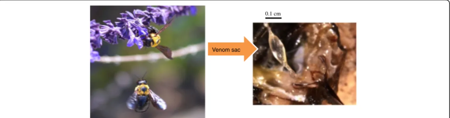
only a small amount of the venom substances is available due to the fact that the venom sac ofX. appendiculatais smaller than those of honeybees and vespid wasps (Fig.1).
Methods
Sample preparation
Fifteen female bees ofX. appendiculatawere collected in Osaka and Sakai, Japan. The venom sacs were dissected
and homogenized with water (50μL). The venom extract was applied to MALDI-TOF MS and HPLC analyses.
MALDI-TOF MS and MS/MS analysis
Matrix-assisted laser desorption/ionization-time of flight mass spectroscopy (MALDI-TOF MS) and tandem mass spectrometry (MS/MS) analysis was performed by Ultra-Flex speed (Bruker Daltonics, Germany). MS and MS/MS analyses (Additional files 1 and 2) were performed in the linear positive ion mode and reflector positive mode, re-spectively.α-Cyano-4-hydroxycinnamic acid (CHCA), tri-fluoroacetic acid (TFA) and all other reagents were purchased from Wako Pure Chemical Industries, Ltd. (Osaka, Japan) or Nacalai Tesque (Kyoto, Japan). The matrix solution was prepared as follows. CHCA was dis-solved in 3:7 acetonitrile/H2O (0.1% TFA) to obtain satu-rated matrix solution. The crude venom or peptide sample were mixed with the matrix solution on a plate, dried for 5 min at ambient temperature, and provided for the mass analysis. Molecular mass and peptide sequencing were analyzed using the FlexAnalysis 3.4 software and BioTools 3.2 (Bruker Daltonics, Germany). The monoiso-topic molecular mass was estimated in a range of m/z 1000 ~ 5000 for short peptides or m/z 5000 ~ 20,000 for PLA2.
High performance liquid chromatography (HPLC) analysis
HPLC analysis and purification of the crude venom was performed by Shimadzu’s Prominence system (Japan). Chromatographic conditions for analysis were as follows:
Column: COSMOSIL 5C18-AR-300, 4.6 mm × 150 mm (Nacalai Tesque).
Eluent: (I) CH3CN containing 0.1% TFA and (II) H2O containing 0.1% TFA.
Elution: linear gradient from (I):(II) = 0.1:99.9 to 60:40.
Flow rate: 1.0 mL/min for 45 min. Detection: UV 210 nm.
0.1 cm
Venom sac
Whereas the conditions for purification were:
Column: COSMOSIL Protein-R, 4.6 mm × 250 mm (Nacalai Tesque).
Eluent: (I) CH3CN containing 0.1% TFA and (II) H2O containing 0.1% TFA.
Elution: linear gradient from (I):(II) = 0.1:99.9 to 60:40.
Flow rate: 1.0 mL/min for 45 min. Detection: UV 210 nm.
Peptide sequence analysis and synthesis
The purified venom peptides were sequenced by automated Edman degradation using ABI model 477A (Applied Biosys-tems, USA). Peptides were synthesized by Fmoc chemistry using a Shimadzu PSSM-8 automated peptide synthesizer (Shimadzu, Japan), and purified by reverse-phase HPLC. The identity and purity of the peptides were confirmed by MALDI-TOF MS. The synthetic Xac-1 and Xac-2 were employed for circular dichroism (CD) analysis, liposome leakage assay, antimicrobial and hemolytic activity tests.
Database search
Peptide database search of the venom peptides was imple-mented by using NCBI database (http://www.ncbi.nlm.-nih.gov/) and Hymenoptera Genome Database (http:// hymenopteragenome.org/).
Circular dichroism (CD) analysis
CD analysis was performed by a spectropolarimeter (J-720 W; JASCO) at room temperature. Spectra were ob-tained at wavelength 190-260 nm. Four scans were accu-mulated for each sample at a scan rate of 20 nm/min. The synthetic peptides were measured at concentration of 0.2 mM in H2O and 50% (v/v) trifluoroethanol (TFE)/H2O.
Liposome leakage experiments
Liposomes were prepared from lecithin from egg yolk (phosphatidylcholine approx. 70%; Nacalai Tesque). The lecithin (28 mg) was dissolved in chloroform (5 mL). The solution was concentrated in vacuo and maintained under the reduced pressure for 10 h to remove the solvent. The dried lecithin was hydrated in 4 mL of 70 mM calcein (Sigma-Aldrich) in aqueous NaOH (pH 7.5). After sonic-ation for 10 min, the vesicles were passed through a col-umn of SephadexTM G-50 (GE Healthcare) in H2O to remove free calcein. The first 5 mL of eluent was collected as calcein-encapsulated vesicles. Water (0.8 mL) was added to the liposome suspension (0.2 mL) in a cuvette. After 10 min, 0.5-20μL of 10 mM solution of mastoparan (Peptide Institute, Inc., Japan) or Xac-1 was added to the cuvette. Fluorescence intensity of calcein was measured by Hitachi P-4500 fluorometer (excitation wavelength of 460 nm and emission wavelength of 530 nm). A 1% (v/v)
solution of Triton X-100 was used as a positive control to obtain maximum fluorescent value at 100% leakage of calcein.
Molecular cloning
RNA was extracted from the venom gland and the venom sac by Trizol reagent (Life Technologies, USA). cDNA was synthesized with oligo(dT)12-18 primer and M-MLV reverse transcriptase (Life Technologies). Degenerate primers were designed on the basis of the nucleotide se-quences of PLA2genes of several Hymenopteran species. PCR was performed with the cDNA by using Xc2 (5′ -AAY GGI -AAY GTN GCN GAR GG-3′) and Xc4 (5′ -AVR TCR AAC CAY TGR TA-3′) primers, and subse-quently the nested PCR was performed with the first PCR product as a template by using Xc2 and Xc3 (5′-GCN GAR GGI CCN GAR GAY-3′) primers.
PCR products were cloned into plasmids using pGEM-T Easy Vector System (Promega, USA). Plasmids were puri-fied with Wizard Plus SV Minipreps DNA Purification Sys-tem (Promega) and sequenced on an ABI PRISM 310 Genetic Analyzer (Life Technologies) or 3130 Genetic Analyzer (Life Technologies) with BigDye Terminator v3.1 Cycle Sequence kit (Life Technologies). To obtain complete sequences of PLA2cDNA, 3′- and 5′-RACEs (rapid ampli-fication of cDNA ends) were performed using a SMART RACE cDNA Amplification kit (Clontech, USA) according to the supplier’s instructions. F3 (5′-CGG CGC CGT AAG
GTT CAC GTA CTT C) and R1 (5′-GCT GAA GGA
GAC CGA CGC CTG TTG T-3′) primers were used for 3′- and 5′RACEs, respectively. The obtained PCR prod-ucts were also cloned into a vector, and sequenced as de-scribed above.
Antimicrobial activity
Hemolytic activity
According to the procedure described by Shigeri et al. [12], hemolytic activities of Xac-1 and Xac-2 were tested. Heparinized rat whole blood from Wistar rats (male, 6 weeks old) was washed twice in NaCl/Pi
(100 mM NaCl, 7.5 mM Na2HPO4 and 2.5 mM
NaH2PO4) by centrifugation at 900 g and suspended in NaCl/Pi to a concentration of 0.5% (v/v). NaCl/Pi and NaCl/Pi containing 0.2% Triton X-100 were used as controls for 0 and 100% hemolysis, respectively. Xac-1 and Xac-2, as well as mastoparan and melittin were employed as comparable standards.
Histamine releasing activity
The histamine-releasing activities of Xac-1 and Xac-2, mastoparan, and melittin were determined with rat peritoneal mast cells, as previously described [17]. The histamine-releasing activity was defined as the ra-tion of the extracellular to the total amount of hista-mine. Spontaneous histamine-releasing activity was 6.9 ± 0.3%.
Results
MALDI-TOF MS and HPLC analysis of the crude venom extract ofX. appendiculata
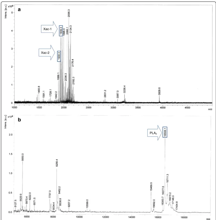
The crude venom extract of X. appendiculata was subjected to MALDI-TOF MS analysis (Fig. 2a and b). The MSPP analysis in a range of m/z 1000 ~ 5000 (Fig. 2a) indicated that peptides in a range of m/z 1850 ~ 2200 are the major in the venom of X. appen-diculata. Characteristic ion signal at m/z 16508 was observed in a range of m/z 5000 ~ 20000 (Fig. 2b). Having these profiles, the crude venom was subjected to HPLC purification with a C18-reversed phase col-umn to provide eight major fractions (A to H) (Fig. 3). Fractions A, D, F, and G included peptides with m/z 2066, 16508, 1939.3 and 1900.3, respectively. These molecular ions were originated from the venom be-cause the same m/z were found in the crude venom analysis. MS analysis of fractions B, C, E, and F showed that these are composed of a mixture of sev-eral peptides.
Peptide sequence of peptides in fractions A, D, F, and G
Edman degradation of fraction F ([M + H]+ m/z 1939.3) provided a partial peptide sequence – GFVALLKKL-PLILKHL–except for the C-terminal amino acid residue. MS/MS analysis (-L/I-L/I-L/I-K-H-L/I-H) indicated that the amino acid residue at the C-terminal was histidine (Additional file 1). Although the sequence was putatively assigned as GFVALLKKLPLILKHLH, the theoretical monoisotopic mass number of GFVALLKKLPLILKHLH-OH (1939.25) differed from the observed mass number (1938.2). These results suggest a possibility of the
C-terminal amidation. To prove this possibility,
GFVALLKKLPLILKHLH-NH2 was prepared and
sub-jected to HPLC analysis to compare retention time. Reten-tion times of the synthetic and naturally occurring peptide were identical. As a result, the peptide of fraction F was
determined to be GFVALLKKLPLILKHLH-NH2. In a
similar manner, the peptide of fraction G was identified as GFVALLKKLPLILKHLP-NH2(Additional file 2).
These peptide sequences were not registered in the NCBI database (http://www.ncbi.nlm.nih.gov/) and Hymenoptera Genome Database (http://hymenoptera-genome.org/). Thus, we named these novel peptides
Xac-1 (GFVALLKKLPLILKHLH-NH2, [M + H]+ m/z
1939.3) and Xac-2 (GFVALLKKLPLILKHLP-NH2, [M
+ H]+ m/z 1900.3). Edman degradation analysis of fraction A ([M + H]+ m/z 2066) was not successfully done, though the reason was unclear. It is speculated that it might be a cyclic peptide with an S-S bond that prevent the Edman analysis. Further sequence analysis is ongoing.
Edman degradation of the peptide of fraction D provided a partial sequence: IIFVG TKWCG NGNVA
EGPED LGSLK E-. Sequence similarity searches
showed that the partial sequence conserved a 70% identity with those of PLA2s isolated from the bumblebee Bombus hypocrite (Apidae: Apinae: Bom-bini) and the social honeybee A. mellifera [18]. We hypothesized that this peptide would be a PLA2 homolog and attempted molecular cloning and RACE to elucidate the full nucleotide sequence encoding this peptide (Fig. 4). The resulting sequence (DDBJ/ GenBank/EMBL accession no. AB731659) was com-pared with those of PLA2s isolated from bee venom, indicating that the PLA2 homolog conserves charac-teristic amino acid residues associated with the cata-lytic activity of PLA2s of honey and bumblebees [18, 19]. It is speculated that the PLA2 of X. appendicu-lata is a product of a post-translational modification due to the fact that the molecular mass number of fraction D ([M + H]+ m/z 16508) was not identical to that of the peptide estimated by the molecular cloning.
Physicochemical properties of Xac-1 and Xac-2: helical wheel projection analysis, CD spectroscopy analysis, and liposome leakage assay
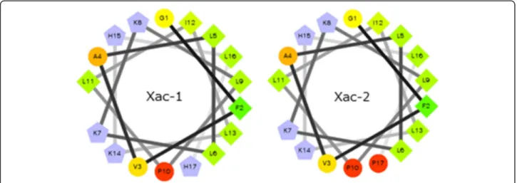
The helical wheel projection of Xac-1 and Xac-2 was made by the database program (http://www.tcdb.org/ progs/?tool=pepwheel) [20]. The results suggest that these peptides possess amphiphilic helical structures in which positively charged amino acid resides, histi-dines and lysines are arranged on one side and hydro-philic residues on the other side (Fig. 5). To obtain
measured. Xac-1 exhibited a mostly disordered con-formation in aqueous solution whereas a higher α -helical content in 50% TFE solution (Fig. 6). The presence of two negative dichroic bands at 208 and 222 nm was consistent with the preferential formation of α-helix. Subsequently, we analyzed liposome leak-age properties of Xac-1 (Fig. 7). Xac-1 revealed lipo-some degradation activity in which its potency was almost the same as that of mastoparan.
Biological activities of Xac-1 and Xac-2
Antimicrobial and hemolytic activities of Xac-1 and Xac-2 were examined. Mastoparan (14 amino acid amphiphilic peptides from wasp venom) was selected as a reference peptide because it is a representative amphiphilic peptide that shows antimicrobial and hemolytic activities due to its potent pore forming ef-fects and mast cell degradation activities [7]. In addition, melittin isolated from the venom of A.
a
Xac-1
Xac-2
b
PLA2
Fig. 2MALDI-TOF MS spectra of the crude venom of X. appendiculata. Positive mode, Matrix:α-CHCA.am/z range from 1000 to
t
Fig. 3HPLC analysis of the crude venom. Column: COSMOSIL 5C18-AR-300, 4.6 mm × 150 mm (Nacalai Tesque). Eluent: (I) CH3CN containing
0.1% TFA and (II) H2O containing 0.1% TFA. Elution: linear gradient from (I):(II) = 0.1:99.9 to 60:40. Flow rate, 1.0 mL/min for 45 min. Detection: UV
210 nm. Purity of each fraction was monitored by MALDI-TOF MS.A:m/z2066;BandC: mixture (m/z2066 was mainly detected);D:m/z16508 (PLA2homolog); E: mixture; F:m/z1939.3 (Xac-1); G:m/z1900.3 (Xac-2); H: mixture
Fig. 4PLA2ofX. appendiculataand other bees. This alignment file was used to construct the phylogenetic tree shown in Figure 9. Hyphen:
alignment gap; yellow: the key amino acid residues for PLA2catalytic activity; green: cysteine. GenBank accession numbers of PLA2are as follows;
mellifera was used as a reference to compare hemolytic activity [3]. These results are summarized in Table 1. Xac-1 exhibited growth inhibitory effects against E. coli, S. aureus, M. luteus, and S. cerevisiae with MIC values in a range from 1.57 to 6.25 μM. The potency is similar to that of mastoparan. Xac-2 showed almost the same or slightly less potency as Xac-1 on the antimicrobial activities using M. luteus and S. cerevisiae. Xac-1 and Xac-2 exerted hemolytic activities (37.5 and 23.5% at 100 μM), respectively. These data were compared with those of mastoparan (40.6% at 100 μM) and melittin (91.8% at 10 μM). These results indicated that the potencies of Xac-1 and Xac-2 were close to that of mastoparan, whereas these potencies were much weaker than that of melit-tin. Bioactive peptides isolated from ant, bee, and wasp have been shown to activate the release of his-tamines from rat peritoneal mast cells [17]. Both Xac-1 and Xac-2 caused a significant and dose-dependent histamine release. At a concentration of 10 μM, Xac-1 and Xac-2 displayed 58.0 and 53.0% of histamine-releasing activities, respectively. These activities
comparable to mastoparan (57.6%), but less effective than melittin (84.8%).
Discussion
Bees are classified into seven families including more than 16,000 described species [21]. Female bees use their venom for defense when they are exposed to dangers and predators. Bee stings are known to be painful. In contrast to the unpleasant effects of bee toxins in humans, its venom has been utilized as a remedy for centuries and recently has attracted much attention as a promising alternative and preventive medicine for the treatment of arthritis, rheumatism, pain, and cancer, etc. [8, 22]. Although many biologically active peptides and enzymes have been isolated from the venom of social honeybees such as A. mellifera and a eusocial bumblebee (Megabombus pennsylvani-cus), the structure elucidation of the venom sub-stances of carpenter bees has not been well examined except for the Nakajima’s study [23] on the analysis of biogenic amines in the venom of X. appendiculate. It revealed that histamine, putrescine and spermidine were detected as major biogenic amines in the venom. Piek [15] predicted that the presence of melittin-like peptides in the venom of X. violacea by comparison with biological activities of the crude venom of X. violacea, A. mellifera, and Bombus ter-restris. To the best of our knowledge, isolation of the peptide substances in the carpenter venom has not been elucidated yet.
In this study, we found two novel amphiphilic pep-tides, Xac-1 and Xac-2, and a new PLA2 homolog in the venom of X. appendiculata for the first time. Our results corroborate that by Nakajima et al. [23] by clearly showing that the venom of X. appendicu-lata is a cocktail of biogenic amines, amphiphilic
peptides, PLA2 and the molecular constitution
Fig. 5Helical wheel projection of Xac-1 and Xac-2.Blue: basic amino acids, others: neutral amino acids
-20 -10 0 10 20 30 40
190 210 230 250
wavelength (nm)
[t
het
a
] x
10
-4 (d
eg
cm
2 dm
o
l) 50%(v/v)TFE
water
resembles those of honeybees and bumblebees. It is supposed that Xac-1 and Xac-2 would be a principle of the melittin-like peptide proposed by Piek [15] since biological activities of Xac-1 and Xac-2 resem-ble those of melittin.
Recently, the research interests for venoms has reached other families of solitary and eusocial bees (Fig. 8). These studies have unveiled the distribution of amphiphilic and biologically active peptides such as melectin from Mellecta albifrons (Apoidea: Melec-tini) [24], codesane from Colletes daviesanus (Colle-tidae) [25], osmin from Osma rufa (Megachilidae) [26], lasioglossins from Lasioglossum laticeps (Halici-tidae) [27], halictines from Halictus sexcinctus
(Hali-citidae) [28], macropin from Macropis fulvipes
(Melittidae) [29] in the bee venom. It is interesting to note that the amino acid sequences of Xac-1 and Xac-2 are similar to those of melectin and osmin isolated from the long-tonged bees, but not to those of bombolitins and melittin isolated from the social bee venom, though carpenter bees, bumblebees and honeybees are closely related. These comparable ana-lyses indicate a possibility that Xac-1, Xac-2, melec-tin and osmin would be derived from a prototype amphiphilic peptide of the ancestor of solitary bees. On the other hand, melittin, bombolitins, masto-paran may have separately developed during the course of
the social evolution. To prove this hypothesis, fur-ther research on isolation and biological studies on the bee venom peptides are required.
PLA2 is known to be the main enzyme component of bee venom. Previously, the presence of PLA2 in
the venom of Anthophora pauperata (Apidae) was
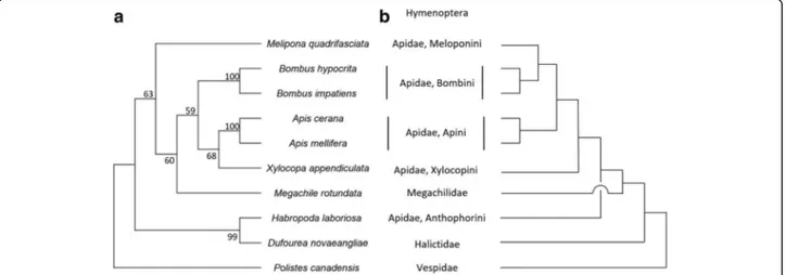
proposed by biological and hematological studies [30]. To the best of our knowledge, structure analysis of the PLA2 of carpenter bees has not been examined yet. We isolated the PLA2 of X. appendi-culata and found that it has a high sequence identity with PLA2 of related species such as bumblebees and honeybees (Fig. 4) [31]. We also analyzed the molecular evolution of bee PLA2s using database sets (Fig. 9) [32, 33]. Interestingly, PLA2 evolution tree did not match with the bee phylogeny that is well-established by large dataset though the charac-teristic amino acid residues of the PLA2 family of honeybees and bumblebees are highly conserved in the PLA2 of X. appendiculata. Our analysis would contribute to discuss the evolution patterns of PLA2s of the bee venoms.
Conclusion
We have analyzed the venom components of the solitary bee X. appendiculata and isolated novel amphiphilic peptides, Xac-1 and Xac-2, and a PLA2
Fig. 7Liposome leakage assay of Xac-1 (blue) and mastoparan (red)
Table 1Biological activities of Xac-1, Xac-2, mastoparan, and melittin
Antimicrobial activity (MIC) Hemolytic
activity at 100μM
Histamine releasing activity at 10μM E. coli(NBRC 14237) S. aureus(NBRC 12732) M. luteus(NBRC 12708) S. cerevisiae(NBRC 10217)
Xac 1 3.12μM 1.57μM 3.12μM 6.25μM 37.5 ± 1.9% 58.0 ± 1.7%
Xac 2 3.12μM 3.12μM 6.25μM 25.0μM 23.5 ± 1.3% 53.0 ± 4.3%
Mastoparan 6.25μM 1.57μM 3.12μM 6.25μM 40.6 ± 2.7% 57.6 ± 1.0%
Melittin – – – – 91.8 ± 1.8%a 84.8 ± 10.1%
aHemolytic activities at 10
homolog. The accurate analysis and structure deter-mination of the venom reveals that it is a cocktail of various biologically active molecules. Our study helps to understand the biological function and
molecular diversity of the solitary bee venom
components. Additionally, it may aid in the design
of biologically active peptides based on the
structures of Xac-1 and Xac-2 to develop more po-tent peptide analogs toward biotechnological and medical applications.
Fig. 8Amphiphilic venom peptides isolated from bees and wasps. The cladogram of bee families is based on Hedtke et al. [21].Blue: basic amino acid residues
Additional files
Additional file 1:MS/MS analysis of Xac-1. (DOCX 175 kb)
Additional file 2:MS/MS analysis of Xac-2. (DOCX 157 kb) Abbreviations
CD:Circular dichroism; CHCA:α-Cyano-4-hydroxycinnamic acid; HPLC: High
performance liquid chromatography; MALDI-TOF MS: Matrix-assisted laser desorption/ionization-time of flight mass spectroscopy; MS/MS: Tandem mass spectrometry; PLA2: Phospholipase A2; TFA: Trifluoroacetic acid
Acknowledgments
YS thanks to the support from JSPS KAKENHI, grant number 15 K01814.
Funding
This work was supported by JSPS KAKENHI, grant number 15 K01814.
Authors’contributions
TS designed this work, prepared this manuscript and sampling ofX. appendiculata. HK contributed to sampling ofX. appendiculata, MALDI-TOF MS analysis, HPLC analysis, and liposome leakage assay. SGG prepared cDNA for PLA2. KM and HM performed sequential analysis of Xac-1, Xac-2, and
PLA2. YS synthesized Xac-1 and Xac-2, and designed biological assays. TI and
HI evaluated biological activities of Xac-1 and Xac-2. All authors read and approved the final manuscript
Competing interests
The authors declare that they have no competing interests.
Consent for publication Not applicable.
Ethics approval and consent to participate
Experiments procedures for hemolytic activity tests were in accordance with the guidelines of the Care and use of Laboratory Animals of AIST.
Publisher’s Note
Springer Nature remains neutral with regard to jurisdictional claims in published maps and institutional affiliations.
Author details
1Graduate School of Material Science, Osaka City University, 3-3-138
Sugimoto, Sumiyoshi, Osaka 558-8585, Japan.2Graduate School of Science,
Department of Biology & Geosciences, Osaka City University, 3-3-138 Sugimoto, Sumiyoshi, Osaka 558-8585, Japan.3Faculty of Pharmacy, Kindai
University, 3-4-1 Kowakae, Higashiosaka, Osaka 577-8502, Japan.4Health
Research Institute, National Institute of Advanced Industrial Science and Technology (AIST), Osaka, Japan.5Research Institute for Chemical Process
Technology, National Institute of Advanced Industrial Science and Technology (AIST), Ibaraki, Japan.6Biomedical Research Institute, National
Institute of Advanced Industrial Science and Technology (AIST), Ibaraki, Japan.
Received: 27 January 2017 Accepted: 9 May 2017
References
1. Palma MS. Hymenoptera Insect Peptides. Kastin AJ, editor. In: Handbook of Biologically Active Peptides. Elsevier; 2013. p. 416-22.http://www. sciencedirect.com/science/book/9780123694423.
2. Piek T. Venoms of the Hymenoptera, biochemical, pharmacological and behavioral aspects. London: Academic; 1986. https://www.elsevier.com/ books/venoms-of-the-hymenoptera/piek/978-0-12-554770-3. 3. Chen J, Guan SM, Sun W, Fu H. Melittin, the major pain-producing
substance of bee venom. Neurosci Bull. 2016;32(3):265–72.
4. Argiolas A, Pisano JJ. Bombolitins, a new class of mast cell degranulating peptides from the venom of the bumblebeeMegabombus pennsylvanicus. J Biol Chem. 1985;260(3):1437–44.
5. Lee G, Bae H. Bee venom phospholipase A2: yesterday’s enemy becomes today’s friend. Toxins (Basel). 2016;8(2):48.
6. de Lima PR, Brochetto-Braga MR. Hymenoptera venom review focusing onApis mellifera. J Venom Anim Toxins Incl Trop Dis. 2003; 9(2):1–24. http://www.scielo.br/scielo.php?script=sci_arttext&pid= S1678-91992003000200002.
7. Moreno M, Giralt E. Three valuable peptides from bee and wasp venoms for therapeutic and biotechnological use: melittin, apamin and mastoparan. Toxins (Basel). 2015;7(4):1126–50.
8. Son DJ, Lee JW, Lee YH, Song HS, Lee CK, Hong JT. Therapeutic application of anti-arthritis, pain-releasing, and anti-cancer effects of bee venom and its constituent compounds. Pharmacol Ther. 2007;115(2):246–70.
9. Escoubas P. Mass spectrometry in toxinology: A 21st-century technology for the study of biopolymers from venoms. Toxicon. 2006;47(6):609–13. 10. Favreau P, Menin L, Michalet S, Perret F, Cheneval O, Stöcklin M, et al. Mass
spectrometry strategies for venom mapping and peptide sequencing from crude venoms: case applications with single arthropod specimen. Toxicon. 2006;47(6):676–87.
11. Nakajima T, Naoki H, Corzo G, Li D, Hisada M, Escoubas P, et al. A trial of mass spectrometric characterization of femto-molar amount from subtropical islands. J Toxicol. 2003;22(4):509–20.
12. Shigeri Y, Yasuda A, Hagihara Y, Nishi K, Watanabe K, Imura T, et al. Identification of novel peptides from amphibian (Xenopus tropicalis) skin by direct tissue MALDI-MS analysis. FEBS J. 2015;282(1):102–13.
13. Murata K, Shinada T, Ohfune Y, Hisada M, Yasuda A, Naoki H, et al. Novel mastoparan and protonectin analogs isolated from a solitary wasp, Orancistrocerus drewseni drewseni. Amino Acids. 2009;37(2):389–94. 14. Murata K, Shinada T, Ohfune Y, Hisada M, Yasuda A, Naoki H, et al. Novel
biologically active peptides from the venom ofPolistes rothneyi iwatai. Biol Pharm Bull. 2006;29(12):2493–7.
15. Piek T. The venom of bumble-bees and carpenter-bees. In: Piek T, editor. The Venoms of the Hymenoptera, biochemical, pharmacological and behavioral aspects. London: Academic; 1986. p. 417–24.
16. Steinberg DA, Lehrer RI. Designer assays for antimicrobial peptides. antibacterial peptide protocols. In: Shafer WM, editor. Methods in molecular biology, vol. 78. Tokowa: Humana Press; 1997. p. 169–86.
17. Inagaki H, Akagi M, Imai HT, Taylor RW, Kubo T. Molecular cloning and biological characterization of novel antimicrobial peptides, pilosulin 3 and pilosulin 4, from a species of the Australian ant genusMyrmecia. Arch Biochem Biophys. 2004;428(2):170–8.
18. Arni RK, Ward RJ. Phospholipase A2–a structural review. Toxicon. 1996;34(8):827–41. 19. Annand RR, Kontoyianni M, Penzotti JE, Dudler T, Lybrand TP, Gelb MH. Active
site of bee venom phospholipase A2: the role of histidine-34,aspartate-64 and tyrosine-87. Biochemistry. 1996;35(14):4591–601.
20. Transporter Classification Database. http://www.tcdb.org/progs/?tool=pepwheel. Accessed 15 May 2017.
21. Hedtke SM, Patiny S, Danforth BN. The bee tree of life: a supermatrix approach to apoid phylogeny and biogeography. BMC Evol Biol. 2013;13:138. 22. Bellik Y. Bee venom: its potential use in alternative medicine. Anti-Infective
Agents. 2015;13(1):3–16.
23. Nakajima T, Yasuhara T, Yoshida N, Takemoto Y, Shinonaga S, Kano R, et al. The pattern analysis of biologieallyaetive amines in some Hymenopteran venoms by high performance liquid chromatography. Jap J Sanit Zool. 1983;34(2):61–71.
24. Cujová S, Bednárová L, Slaninová J, Straka J, Cerovsky V. Interaction of a novel antimicrobial peptide isolated from the venom of solitary beeColletes daviesanus with phospholipid vesicles andEscherichia colicells. J Pept Sci. 2014;20(11):885–95. 25. Cerovský V, Hovorka O, Cvacka J, Voburka Z, Bednárová L, Borovicková L, et
al. Melectin: a novel antimicrobial peptide from the venom of the cleptoparasitic beeMelecta albifrons. Chembiochem. 2008;9(17):2815–21. 26. Stocklin R, Favreau P, Thai R, Pflugfelder J, Bulet P, Mebs D. Structural identification
by mass spectrometry of a novel antimicrobial peptide from the venom of the solitary beeOsmia rufa(hymenoptera: megachilidae). Toxicon. 2010;55:20–7. 27. Cerovský V, Budesínský M, Hovorka O, Cvacka J, Voburka Z, Slaninová J, et al.
Lasioglossins: three novel antimicrobial peptides from the venom of the eusocial beeLasioglossum laticeps(Hymenoptera: Halictidae). Chembiochem. 2009;10(12):2089–99.
29. Monincová L, Veverka V, Slaninová J, Budesínský M, Fucik V, Bednárová L, et al. Structure-activity study of macropin, a novel antimicrobial peptide from the venom of solitary beeMacropis fulvipes(Hymenoptera: Melittidae). J Pept Sci. 2014;20(6):375–84.
30. Zalat S, Nabil Z, Hussein A, Rakha M. Biochemical and haematological studies of some solitary and social bee venoms. Egypt J Biol. 1999;1:57–71. 31. Hou C, Guo L, Wang J, You L, Lin J, Wu W, et al. Cloning and characterization
of phospholipases A2 and hyaluronidase genes from the venom of the honeybeeApis mellifera carnica. Sociobiology. 2012;59(3):945–57.
32. Whelan S, Goldman N. A general empirical model of protein evolution derived from multiple protein families using a maximum-likelihood approach. Mol Biol Evol. 2001;18(5):691–9.
33. Kumar S, Stecher G, Tamura K. MEGA7: molecular evolutionary genetics analysis version 7.0 for bigger datasets. Mol Biol Evol. 2016;33(7):1870–4.
• We accept pre-submission inquiries
• Our selector tool helps you to find the most relevant journal
• We provide round the clock customer support
• Convenient online submission
• Thorough peer review
• Inclusion in PubMed and all major indexing services
• Maximum visibility for your research
Submit your manuscript at www.biomedcentral.com/submit