Design and Evaluation of Antimalarial
Peptides Derived from Prediction of Short
Linear Motifs in Proteins Related to
Erythrocyte Invasion
Alessandra Bianchin1,2,3,4, Angus Bell4, Anthony J. Chubb1,2,3, Nathalie Doolan3, Darren Leneghan4, Ilias Stavropoulos1,2,3, Denis C. Shields1,2,3, Catherine Mooney1,2,3*
1Conway Institute of Biomolecular and Biomedical Science, University College Dublin, Dublin, Ireland,
2Complex and Adaptive Systems Laboratory, University College Dublin, Dublin, Ireland,3School of Medicine and Medical Science, University College Dublin, Dublin, Ireland,4Department of Microbiology, School of Genetics and Microbiology, Moyne Institute of Preventive Medicine, Trinity College, Dublin, Ireland
Abstract
The purpose of this study was to investigate the blood stage of the malaria causing parasite,
Plasmodium falciparum, to predict potential protein interactions between the parasite mero-zoite and the host erythrocyte and design peptides that could interrupt these predicted inter-actions. We screened theP. falciparumand human proteomes for computationally
predicted short linear motifs (SLiMs) in cytoplasmic portions of transmembrane proteins that could play roles in the invasion of the erythrocyte by the merozoite, an essential step in malarial pathogenesis. We tested thirteen peptides predicted to contain SLiMs, twelve of them palmitoylated to enhance membrane targeting, and found three that blocked parasite growth in culture by inhibiting the initiation of new infections in erythrocytes. Scrambled pep-tides for two of the most promising peppep-tides suggested that their activity may be reflective of amino acid properties, in particular, positive charge. However, one peptide showed effects which were stronger than those of scrambled peptides. This was derived from human red blood cell glycophorin-B. We concluded that proteome-wide computational screening of the intracellular regions of both host and pathogen adhesion proteins provides potential lead peptides for the development of anti-malarial compounds.
Introduction
Malaria was the underlying cause of death for 1.24 million people in 2010, the vast majority of these deaths were in Africa and can be attributed to the parasite causing the most virulent form of malaria in humans,P. falciparum[1].P. falciparumhas an extremely complex life cycle that involves different cells and organ systems in both the femaleAnophelesmosquito and human host. When the mosquito bites the human host,P. falciparumsporozoites present in the
a11111
OPEN ACCESS
Citation:Bianchin A, Bell A, Chubb AJ, Doolan N, Leneghan D, Stavropoulos I, et al. (2015) Design and Evaluation of Antimalarial Peptides Derived from Prediction of Short Linear Motifs in Proteins Related to Erythrocyte Invasion. PLoS ONE 10(6): e0127383. doi:10.1371/journal.pone.0127383
Academic Editor:Ulrich Melcher, Oklahoma State University, UNITED STATES
Received:October 22, 2014
Accepted:April 15, 2015
Published:June 3, 2015
Copyright:© 2015 Bianchin et al. This is an open access article distributed under the terms of the
Creative Commons Attribution License, which permits unrestricted use, distribution, and reproduction in any medium, provided the original author and source are credited.
Data Availability Statement:All relevant data are within the paper and its Supporting Information files.
Funding:This work was supported by Science Foundation Irelandhttp://www.sfi.ie/(grant numbers 08/IN.1/B1864, 10/RFP/GEN2749) to DCS and CM and Irish Research Councilhttp://www.research.ie/
mosquito salivary glands are passed into the human subcutaneous tissue and are quickly passed into the bloodstream and on to the liver where they invade the hepatocytes. During this stage the sporozoites develop into pre-erythrocytic schizonts that produce new merozoites. The he-patocyte containing the schizont ruptures and the merozoites re-enter the bloodstream where they bind to and invade erythrocytes and also undergo asexual replication, producing more merozoites. At this stage the merozoites can also form gametocytes which, when ingested by another mosquito feeding on the host, will form gametes which will be fertilised into zygotes. The zygotes penetrate the mosquito midgut wall as ookinetes and subsequently oocysts are formed in which replication of new sporozoites occurs. When the oocysts burst the sporozoites are released into the body cavity of the mosquito and invade the mosquito salivary glands where they are ready to restart the cycle. The characteristic symptoms of malaria are caused by the blood stage of the life cycle and it is the point at which the merozoites invades the erythro-cytes that is under investigation here.
The merozoite is the first cell of the asexual blood stage of malarial infection and is responsi-ble for establishing new infections in erythrocytes, an essential step in the disease pathogenesis of malaria [2]. The cell contains several characteristic organelles, many of which are in the characteristic apical complex, including rhoptries, micronemes and dense granules. Many of the merozoite proteins that are implicated in the invasion of erythrocytes are located in these apical organelles. Merozoite invasion is a multi-step process that begins with a weak attach-ment of the merozoite to the erythrocyte surface, reorientation, tight junction formation and subsequent invasion [3]. This whole process is extremely efficient and lasts just a matter of sec-onds. The surface of the merozoite is covered in membrane proteins such as the glycosylpho-sphatidylinositol (GPI) anchored merozoite surface proteins (MSP) and erythrocyte binding antigens (EBA). Of these MSP-1 is the most abundant and is thought to play a leading role in the initial contact with the erythrocyte [3]. Many other peripheral proteins such as MSP-7 are thought to associate with MSP-1 [4]. However, there is much difficulty in establishing which merozoite proteins are playing a recognition role and thus contributing to the invasion of erythrocytes and this is largely due to the low affinity nature of the interactions. Although this process is important in targeting erythrocytes that are invasion competent, the next step of in-vasion could provide a more promising target for intervention. After reorientation of the mero-zoite, so that the apical prominence is in contact with the erythrocyte membrane, tight
junction formation is triggered. The merozoite then secretes its rhoptry contents into the eryth-rocyte. Many proteins have been predicted to have a role in invasion [5], and some have been shown to be critical for invasion at this stage [6–10].
Peptides have been extensively evaluated as potential antimalarial agents [11] and the idea of blocking erythrocyte invasion using peptides based on key regions of merozoite surface mol-ecules has been explored in dozens of papers. The concentrations of peptide required for inhib-iting invasion (or, to be more precise, development of new ring-infected erythrocytes from schizont-infected erythrocytes) in culture have been typically>1μM and often>100μM, and
in some cases only partial inhibition was achieved. Some published studies lack controls using scrambled peptides of identical amino acid composition to the test peptide, which makes them more difficult to evaluate. A variety of different molecular interactions have been targeted. One of the better characterised examples of merozoite peptides is the 20-mer named R1, based on an erythrocyte binding region of apical membrane antigen 1 (AMA1), that inhibited invasion with an IC50of 4μM [12]. In far fewer cases have peptides based on the known erythrocyte
sur-face receptors for merozoite proteins been investigated: an example is Band 3 [13]. The thera-peutic potential of invasion-blocking peptides is still a long way from being realised.
The full extent of the role that these peptides could play in theP. falciparuminvasion of erythrocytes has yet to be fully examined. There has been significant difficulty in researching
the merozoite-erythrocyte interaction in cultured parasites due to the rapid sequence of events from transient initial recognition to tight junction formation, and the difficulty in producing highly purified proteins that will bind with sufficient affinity for analysis. In a proteome of po-tentially more than 5,000 proteins, this research can be performed more efficiently using bioin-formatics tools (i.e. in silico discovery), followed by in vitro testing of a small set of
predicted peptides.
Here, predicting regions with SLiM-like properties was used to identify peptides that could be involved potentially in the interaction between the host erythrocyte and the parasite mero-zoite [14]. SLiMs are short stretches of a protein sequence consisting of about three to ten amino acids [15]. SLiMs have high levels of conservation in comparison to their surrounding residues, they evolve convergently, usually occur in disordered regions [16] and often form a secondary structure when they bind to their interaction partner. There are four classes of SLiMs and they are categorised in relation to their function: ligand sites, which mediate binding of a ligand protein to its corresponding interaction partner, post-translational modification sites, proteolytic cleavage sites and subcellular targeting motifs [17]. Many strategies for target-ing adhesion molecules or other receptors involved in pathogenicity focus on compounds that bind to these molecules extracellularly. Here, we instead focused on SLiMs that are involved in the intracellular parts of those transmembrane proteins. For well characterised proteins that have a primary role in signaling, this makes sense, since their intracellular regions are typically engaged in transmitting the signal. However, many adhesion molecules not only adhere, but also transmit a signal or alter the binding of other molecules that contribute to the larger pro-tein complexes that form and facilitate shape change or other cellular change upon binding, as seen in integrin or cadherin proteins. Targeting of receptor molecules by palmitoylated pep-tides modeled on sections of their intracellular regions has been shown to facilitate alteration of receptor signaling [18], as well as altering responses mediated by proteins which play a role in adhesion [19,20]. Such peptides can internalize [21], consistent with a model of their loca-tion on the intracellular leaflet of the plasma membrane where they perturb in some way the normal signaling [22–24]. Accordingly, we decided to adopt this strategy to determine if pep-tide targeting of computationally predicted intracellular SLiMs from transmembrane proteins may prove a useful strategy in modulating the merozoite invasion process. The location of the intracellular SLiMs including their distance from a transmembrane (TM) domain and the par-ent protein membrane topology were taken into account in predicting their suitability as candi-dates for peptide design. We then palmitoylated the peptides so they could be targeted to the membrane [22,25] potentially interfering with the binding of the merozoite to the erythrocyte and thus preventing invasion. A small set of the most likely candidates was then tested against culturedP. falciparum.
Materials and Methods
Peptide Design
We took a three-pronged approach in our search for potential peptides. The first was a whole
P. falciparumproteome search. The second and third approaches were targeted towardsP. fal-ciparumproteins and human erythrocytic proteins, which are known to be, or predicted to be, involved in invasion.
SLiMPred [14] to predict SLiM like regions in these sequences. We filtered the predictions keeping only those where at least five residues in a row had a predicted probability of being in a SLiM of 0.5 or greater, and these residues fell within a region that was less than 15 residues from a predicted TM domain. In all, 33 predicted SLiMs in 30 sequences remained, which we examined in detail. We chose regions where the motif was predicted to be on the cytoplasmic side of a TM protein, close to the membrane. These peptides were then checked for solubility and synthesisability [29]. Finally five peptides were selected for testing from four protein se-quences (seeTable 1, Set 1).
The first peptide, Peptide 1, was selected from a putative 609-residue longP. falciparum
transporter protein (Q8II64 (PF11_0310)). The SLiM-like region (QKTPF) is found five resi-dues from the TM region predicted by SCAMPI (Figure A inS1 File). We extended the peptide up to the TM region, added a C-terminal lysine and palmitoylation, and a N-terminal acetyl group. Peptides 2 and 3 were selected from the 1,518 residue long putative protein kinase, Q8ILC4 (PF14_0320). We extended the predicted SLiM (YLYFE) to the TM region (adding
Table 1. Palmitoylated peptides designed around cytoplasmic SLiMs from transmembrane proteins.
Number Sequence Parent Protein
Set 1:P. falciparumproteins
1 Ac-SEQKTPFNINRSK-pal Putative transporter protein (Figure A inS1 File)
2 pal-KKKLYLYFELFF-NH2 Putative protein kinase (Figure B inS1 File)
3 pal-KKKLYLYFE-NH2 Putative protein kinase (Figure B inS1 File)
4 pal-KRKLKEEQRTKKIKID Putative calcium-transporting ATPase (Figure C inS1 File)
5 pal-SSSRKNRFRYLPF-NH2 Putative aminophospholipid-transporting P-ATPase (Figure D inS1 File)
5_scr1 pal-FLPRYRFKNSRSS-NH2
-5_scr2 pal-RFNFYSRSRLPKS-NH2
-Set 2:P. falciparuminvasion proteins
6 pal-KNSNEPHHIFNIFQK-NH2 Reticulocyte binding protein homolog 4 (Figure E inS1 File)
7 pal-KEEIIEIVFDENEEKYF Reticulocyte binding protein 2 (Figure F inS1 File)
8 Ac-LSESIKNLLKNIYKK-NH2 Serine repeat antigen 4 (Figure G inS1 File)
Set 3: human erythrocyte proteins
9 pal-YEKRRKPEDVL-NH2 Basigin (Figure H inS1 File)
10 pal-RRLIKKSP-NH2 Glycophorin-A (Figure I inS1 File)
10_scr1 pal-RIPRSKLK-NH2
-10_scr2 pal-LKPKIRSR-NH2
-11 pal-SYSIRRLIKA Glycophorin-B (Figure J inS1 File)
12 pal-SYTIRRLIKA Glycophorin-B (Figure J inS1 File)
12-Cam pal-SYTIRRLIKA-NH2 Glycophorin-B (Figure J inS1 File)
12_scr1 pal-LKSRIAITYR-NH2
-12_scr2 pal-LITISYKARR-NH2
-12_scr3 pal-IYSLKAIRTR-NH2
-12_scr4 pal-KTSIIALYRR-NH2
-13 pal-KHRKGNNA-NH2 Complement receptor type 1 (Figure K inS1 File)
Peptides selected for testing, the peptide sequence showing palmitoylation (pal) acetylation (Ac) and amidation (NH2) and the protein from which the
peptide originated.
KKKL), palmitoylated the N-terminus and added a C-terminal amidation— pal-KKKLYLY-FELFF-NH2(Figure B inS1 File). As we were concerned that there might be solubility issues
with the original peptide we also included a variation of this peptide, which we predicted to be more soluble, since it had three fewer hydrophobic residues (pal-KKKLYLYFE-NH2).
Peptide 4 was selected from the putative calcium-transporting ATPase, Q76NN8. The pre-dicted SLiM (KKIKI) was extended by ten residues to the TM region, and it was palmitoylated at the N-terminus. We did not add a C-terminal amidation because the peptide included the C-terminal residue of the native protein sequence—pal-KRKLKEEQRTKKIKID (Figure C in
S1 File). Peptide 5 is taken from the aminophospholipid-transporting P-ATPase, Q8I5L4. The predicted SLiM (RFRYLP) was extended by six residues to the TM region, it was palmitoylated at the N-terminus and amidated at the C-terminus—pal-SSSRKNRFRYLPF-NH2(Figure D in
S1 File).
The second set of proteins which we examined wereP. falciparumproteins that are poten-tially involved in the invasion of the erythrocyte byP. falciparum[3]. Predictions were per-formed as previously described, without the requirement of being within 15 residues of a predicted TM domain. Three peptides were selected for testing (seeTable 1, Set 2). Peptide 6 was selected from reticulocyte binding protein homolog 4 (C0H496/PfRh4). The predicted SLiM (FNIFQ) was extended to the TM region, palmitoylated at the N-terminus and amidated at the C-terminus—pal-KNSNEPHHIFNIFQK-NH2(Figure E inS1 File). Peptide 7 was
select-ed from reticulocyte binding protein 2 homolog (C0H5F4/Rh2b). The prselect-edictselect-ed SLiM was ex-tended to the C-terminus of the sequence, therefore the peptide was not amidated. However, although we did not extend the peptide to the TM region in this case because the distance was too great and the resulting peptide would have been too long, we did decide to palmitoylate the N-terminus to allow for targeting to the membrane—pal-KEEIIEIVFDENEEKYF (Figure F in
S1 File). Peptide 8 was selected from the serine repeat antigen 4 (O96164/SERA-4). The pre-dicted SLiM (LLKNIYKK) was extended seven residues and the peptide was acetylated at the N-terminus and amidated at the C-terminus—Ac-LSESIKNLLKNIYKK-NH2(Figure G in
S1 File).
The final set of proteins which we examined were human erythrocytic proteins potentially involved in invasion [3,30,31]. The five proteins (basigin, glycophorin-A, -B and -C, and com-plement receptor type 1) are all TM proteins. We selected peptides from the cytoplasmic side of these TM proteins because SLiMs tend to be enriched in these regions [28]. Preliminary test-ing of the glycophorin-C peptide (pal-AALQEPALQDAGESSRKEYFI) showed no activity and further tests were not conducted on this peptide. The remaining peptides were as follows (see
Table 1, Set 3). Peptide 9 is an 11 residue peptide taken from the C-terminal side of the basigin (P35613) TM protein. We palmitoylated the N-terminus and amidated the C-terminus— pal-YEKRRKPEDVL-NH2(Figure H inS1 File). Peptide 10 is an eight residue peptide taken from
the C-terminal side of the glycophorin-A (P02724) TM protein. We palmitoylated the N-ter-minus and amidated the C-terN-ter-minus—pal-RRLIKKSP-NH2(Figure I inS1 File). Peptide 11 is
taken from the C-terminal side of the glycophorin-B (P06028) TM protein—pal-SYSIRRLIKA. Peptide 12 is a common human polymorphism [32] of peptide 11 where the serine in position three in the peptide has been replaced by threonine—pal-SYTIRRLIKA. Both peptides were palmitoylated but neither was amidated because the peptide extended to the C-terminus of the protein sequence (Figure J inS1 File). The final peptide that we selected for testing, Peptide 13, was from complement receptor type 1 (P17927). The peptide, pal-KHRKGNNA-NH2, extends
Parasite culture
P. falciparumline 3D7, adapted to grow in Albumax1(BioSciences, Dun Laoghaire, Ireland), was cultured in human erythrocytes (Irish Blood Transfusion Board, Ireland) in standard growth medium, in candle jars [33]. Fresh erythrocytes were obtained every three weeks and washed according to lab protocols. Staining of blood smears was with Giemsa stain. The num-ber of infected erythrocytes was counted by microscopy and the parasitaemia was calculated.
Peptide inhibition of parasite growth
The antimalarial activity of test peptides (ChinaPeptides Co. Ltd, Shanghai, China) was deter-mined in culture on the 3D7 line ofP. falciparumusing the parasite lactate dehydrogenase (pLDH) method in 96-well microtitre plates [34,35] with modifications as previously de-scribed [36]. Each test peptide was initially evaluated at three different concentrations, 100μM,
10μM and 1μM, to determine activity range. The effect of the compounds was evaluated at 48
and 72 hours on growth of asynchronous cultures (cultured as described in the previous sec-tion) ofP. falciparum, determined by the assay of pLDH activity. We used unsynchronised cul-tures in order (i) to make no prior assumptions about the stage of growth that might be inhibited and (ii) to avoid possible inhibitory effects of the synchronising protocol, e.g. sorbitol treatment, separation techniques, etc. The appropriate dilutions of the compounds were pre-pared in DMSO and added to the cultures ofP. falciparum(2% haematocrit, 0.8% initial para-sitaemia and a maximum of 0.5% DMSO). The same methodology was then used to assess the dose-response curves of peptides 5, 7, 10, 11, 12, 13 using ten concentrations between 10μM
and 100μM.
Synchronisation of cultures
Routine erythrocyticP. falciparumcultures are an asynchronous culture consisting of rings, trophozoites, schizonts and merozoites. To enrich for the late stages of erythrocytic develop-ment, parasites at high parasitaemia (>15%) were subjected to magnetic selection [37].
Parasit-ised erythrocytes were passed over a CS column (Miltenyi Biotec, Surrey, UK) which was mounted on a VarioMacs magnet (Miltenyi Biotec, Surrey, UK). The column was then washed with incomplete medium (RPMI 1640 supplemented with 25 mM HEPES, hypoxanthine [50
μg/ml], 0.16% w/v glucose) to remove unparasitised erythrocytes and was then removed from
the magnet, a further wash with incomplete medium eluting late-stage parasitised erythrocytes.
Schizont to ring development assay
Parasites synchronised to late stages (trophozoite/schizont) by magnetic selection were incu-bated with either the peptide being tested (100μM concentration) or vehicle only control
(DMSO) in complete medium for two hours. Treated schizonts were washed with complete medium and centrifugation at 350 xg for 10 minutes. Washing was repeated twice. Treated schizonts were then introduced to fresh erythrocytes at 2.5% hematocrit and incubated over-night (14–16 hours) with shaking. Parasitaemia was assessed by counting the number of ring stages in parasitised erythrocytes in Giemsa stained blood smears by light microscopy [37]. This crude assay measures not just invasion but merozoite release and early
ring-stage development.
Hemolysis assay
(ACD) (38 mM citric acid, 75 mM sodium citrate, 124 mM glucose) and was centrifuged at 150 xg for 10 minutes at room temperature. Plasma was removed and erythrocytes were washed twice with buffer A (pH 7.4) (130 mM sodium chloride, 10 mM sodium citrate, 9 mM sodium biocarbonate, 6 mM glucose, 0.9 mM magnesium chloride, 0.81 mM potassium hydro-gen phosphate, 10 mM Tris), by cenrifugation at 1,000 xg for 5 minutes and resuspended in the same buffer. For the hemolysis assay approximately 1.5 × 10⁸erythrocytes were incubated with solutions of peptides 5, 10 or 12-Cam at 100μM and 10μM concentrations for 15 minutes at
37°C. After incubation, samples were centrifuged at 1,000 xg for 5 minutes and the supernatant was analysed for the presence of hemoglobin at an absorbance of 405 nm. Complete hemolysis was achieved using 0.1% (v/v) Triton X-100 yielding the 100% control value. The assay was run in duplicate with three different donors.
Results
Peptide inhibition of parasite growth rate
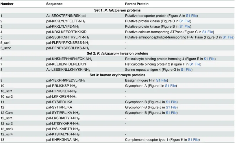
We tested the antimalarial activity of the 13 peptides at three different concentrations (Fig 1). As we used asynchronous culture for the growth inhibition assays the experiments were carried out at two time points to ensure that the peptides were tested over the whole parasite life cycle. Growth inhibition was clearly observed at the 100μM concentration for peptides 5, 10 and 12
at both the 48 hour and 72 hour time points (Fig 1). Peptides 3, 7, 11 and particularly 13 also showed some growth inhibition. These peptides were selected for further investigation (except peptide 3 due to insolubility). At the lowest concentration of 1μM the activity was absent.
Pal-mitate and DMSO were used as controls and showed no inhibitory effect.
Dose-response curve confirms inhibitory effects
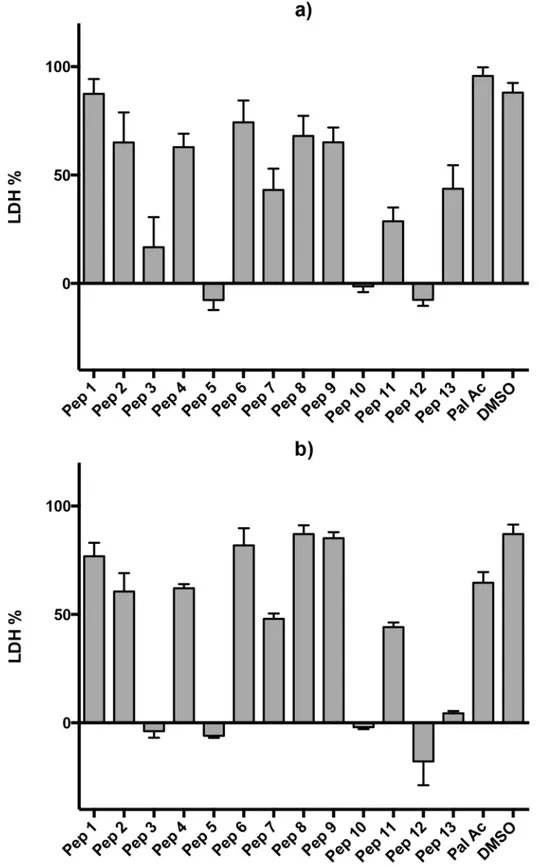
Because the peptides’inhibitory activity dropped dramatically between 10μM and 1μM, we
performed another set of experiments to determine the dose-response curve with 10 data points (Fig 2). Six peptides were tested, the three most active ones (peptides 5, 10 and 12) and peptides 7, 11 and 13. Peptides 7 and 13 were tested because they showed moderate activity at 100μM, however, the curves confirmed no inhibition at 10μM. Peptide 11 was tested as a
con-trol for peptide 12. The concentration-response curves for peptides 5, 10 and 12 showed clear increases in growth inhibition with increasing concentration, while peptide 11 showed no such clear effect, with a non-monotonic dose response relationship (Fig 2).
An important observation was the discrepancy in activity at 10μM between the first and
second sets of experiments (S1AandS1BFig, respectively). Peptides 11 and 12 were selected from glycophorin-B (P06028) which is a natural variant in humans for the amino acid in posi-tion 84, from serine to threonine. The natural variaposi-tion is due to a single nucleotide variaposi-tion C/G. The Population genetics of 1000 Genomes allele frequencies (http://www.1000genomes. org/) show that G is the minor allele with 16% and C is the major allele with 84% frequency; based on that we should expect more people expressing the natural variant T84 corresponding to the sequence of the active peptide 12. It is possible that the donor blood used for the different sets of experiments may be expressing different versions of glycophorin-B, therefore affecting the behaviour of peptide 12, but this requires further investigation.
Peptide inhibition of parasite
“
invasion
”
Fig 1. Growth inhibition assay.Peptides were tested at 100μM. Data were collected at 48 hours (a) and at 72 hours (b). Data are means of three experiments in duplicate and vertical bars indicate standard errors.
Fig 2. Dose-response curves for selected peptides.Peptides 5, 7, 10, 11, 12, 13 and DMSO were evaluated at 48 hours (a) and at 72 hours (b). Data are means of three experiments in duplicate and vertical bars indicate standard errors.
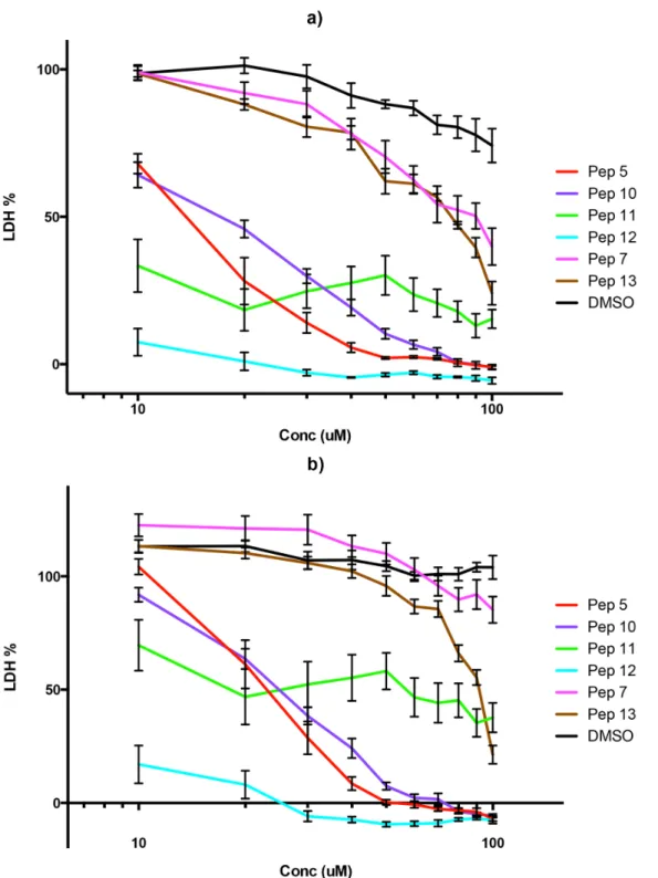
parasites with the peptides. Both peptides 5 and 12 almost totally reduced the ability of mero-zoites to establish new infections in fresh erythrocytes (Fig 3). Peptide 10 also had an effect on the number of new infections but it was not as significant as that of the other two peptides. In the two cultures most affected by the peptides the new ring stage parasites that were counted appeared very unhealthy morphologically (data not shown) and for all intents and purposes the parasitaemia in these cultures was zero. This leads us to the conclusion that peptides 5 and 12 are potent inhibitors of the ability of the parasite to establish new infections in fresh erythro-cytes. It should be borne in mind that this assay does not use isolated merozoites and therefore measures late merozoite development and release as well as invasion. Although it is possible that the effects of the peptides occur just before invasion, during invasion and/or after invasion as the parasites develop into visibly normal ring forms this is the method of choice in many publications to assess invasion [38]. A detailed state-of-the-art invasion assay (for example using flow cytometry) would be appropriate for any highly active peptide leads but was beyond the scope of the present study.
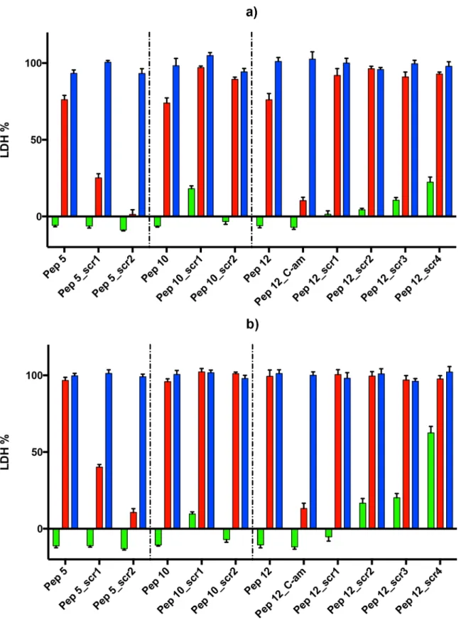
Testing specificity of peptide sequence versus composition
Scrambled peptides were designed for the three most active peptides (5, 10 and 12) (Table 1), to check if the peptide activity was a function of amino acid composition or was sequence spe-cific. For peptide 5 and 10 we selected two scrambled peptides each, with both palmitoylation and amidation; for peptide 12 we selected four scrambled peptides all palmitoylated and ami-dated. We tested peptide 12 with both palmitoylation and amidation to investigate better the effect of peptide charge.
Fig 3. Effect of peptides on merozoite invasion.Vehicle only control (DMSO) and peptides 5, 10 and 12 (100μM concentration) were incubated for 2 hours with schizonts purified by magnetic selection which were subsequently introduced to fresh erythrocytes. The parasitaemia 14–16 hours later was assessed by counting rings in Giemsa stained blood smears microscopically and expressed as a percentage of the control culture (shown as % of control, y axis). Data are means of two experiments in triplicate and vertical bars indicate standard errors.
At 100μM, all of the scrambled peptides tested had some effect on growth inhibition. This
indicates that there is some non-specific contribution to growth inhibition from peptide com-position. This appears to relate to the positive charge of all three candidate peptides (5, 10 and 12) which were each very positively charged (net charge of 4, 4 and 3 respectively) and included no negatively charged residues. Previous work has suggested that peptides of low or high net charge are enriched for bioactivity [39]. Some of the inactive peptides tested above also had a number of positively charged residues, which is a common feature of membrane proximal in-tracellular regions of transmembrane proteins, but these inactive peptides each had a mixture of positive and negative charge. While the inactive peptide 4 had a net positive charge of 5, it had a total of 8 positive and 3 negative charges.
Overall, the results for peptide 5 and 10 were not promising, as the scrambled peptides showed similar or better growth inhibition effect, therefore the effect may be non-specific. Thus, while it is of course possible that these two peptides are indeed acting by mimicking in some way the sequence region of the protein from which they are derived, the fact that scram-bled peptides have a similar effect greatly increases the likelihood that the peptides may be act-ing via some non-specific mechanism, since the number of protein interaction surfaces that involve peptides with a similar amino acid composition greatly exceeds the number of surfaces that share a strongly similar sequence. Interestingly peptide 12 showed more inhibition when amidated and none of the scrambled versions of peptide 12 showed the same degree of inhibi-tion at 10μM (Fig 4). This is suggestive that at the lower dose non-specific effects are lessened
and the sequence-specific effect of peptide 12 is seen.
Testing for erythrocyte lysis in the presence of peptides
Although the antimalarial effects of the peptides were not considered to be due to erythrocyte lysis, as judged by visual inspection of the assay plates, we performed a hemolysis assay to con-firm this. At a concentration of 100μM peptide 10 caused approximately 50% release of
hae-moglobin from erythrocytes (compared with 0.1% (v/v) Triton-X100), whereas the release caused by peptides 5 and 12-Cam was between 10% and 20%. All peptides were non-haemoly-tic at 10μM concentration (S2 Fig).
Discussion
We selected 13 computationally predicted peptides from three sources: (i) the wholeP. falcipa-rumproteome, (ii)P. falciparumproteins potentially involved in erythrocyte invasion and (iii) human erythrocytic proteins potentially involved in invasion. Of the 13 peptides tested, three peptides (5, 10 and 12) inhibited parasite growth at the 100μM concentration at both the 48
hour and 72 hour time points and showed a clear increase in growth inhibition with increasing dose between 10μM and 100μM. Peptide 5, derived from the aminophospholipid-transporting
P-ATPase protein, was predicted using the wholeP. falciparumproteome search strategy. This protein is expressed in the intraerythrocytic asexual cycle, and stage-specifically in mature tro-phozoites and schizonts, with no apparent expression in the ring stage [40]. Peptides 10 and 12 were selected from the human erythrocytic proteins glycophorin A and B respectively.
Fig 4. Growth inhibition by scrambled peptides.Peptides 5, 10 and 12 and their scrambled peptides were tested in three concentrations, 100μM (left bar), 10μM (middle bar) and 1μM (right bar) at 48 hours (a) and at 72 hours (b). Data are means of three experiments in duplicate and vertical bars indicate standard errors.
due to some other unknown mechanism operating around the same stage. We checked wheth-er the peptide action was simply to lyse the wheth-erythrocytes and concluded that the antimalarial ef-fects of the peptides are not primarily due to erythrocyte lysis for peptides 5 and 12-Cam, although it is likely that at 100μM some of the inhibition of parasite growth rate by peptide 10
is due to hemolysis.
Finally, we tested the antimalarial activity of scrambled peptide versions of peptides 5, 10 and 12. The scrambled peptides 5 and 10 showed similar or better growth inhibition effect as the original peptides, therefore the effect seems to be non-specific and may be related to the positive charge of the peptides. The most promising of the peptides tested was the amidated version of peptide 12, which showed additional activity based on its sequence (i.e. it was more active than four scrambled controls of identical amino acid composition). This suggests that it may indeed be acting by mimicking the sequence of glycophorin B’s short cytoplasmic tail. For these reasons, the major conclusion of our analyses is to strongly support further investigation of peptide 12.
It is intriguing that we see some differences in activity for the two polymorphic versions of the glycophorin-derived peptides 11 and 12. While the amino acid properties are highly similar for the variation (between serine and threonine) we would not anticipate that such a difference might impose a functional effect. Nevertheless, the difference in molecular mass and presenta-tion of these two groups may be having some effect. Targeting pathogen proteins is attractive because it is possible to damage the pathogen without harming host molecular components. However, the ability of pathogens to evolve to avoid strategies designed to limit them can sometimes exceed the ability of the human hosts to develop useful solutions either over evolu-tion or in the clinic. Thus, many genetic variants that alter malarial resistance are in fundamen-tal host cell factors (glucose 6-phosphate dehydrogenase and sickle cell) rather than in
components of the immune response mechanisms. It is possible that this particular position may be under some selective adaptive constraint, since the residue is a glycine in chimpanzee, but either serine or threonine in man. However, previous workers have speculated that this var-iant is not important for malarial resistance, focusing instead on studying extracellular amino-acid altering polymorphisms [41]. It will be of interest to test whether the intracellular Ser/Thr polymorphism encoded by SNP rs1132783 does indeed associate with malarial resistance. If it does, it may well be of strong interest to develop more potent compounds targeting glyco-phorin B, for which the peptides developed here may act as initial lead compounds.
We conclude that screening for intracellular short linear protein motifs, and then testing their function when delivered into cells as peptides using palmitylation or other cell penetrating strategies, represents an alternative pathogen targeting strategy. This contrasts with the more usual emphasis on targeting the extracellular regions of both host and pathogen receptors. Such screens need to pay careful attention to both sequence-specific and composition-specific effects of peptides that modulate pathogen invasion.
Supporting Information
S1 File. Images from the SLiMPred server showing the predicted SLiMs and peptides. Pep-tide 1, Ac-SEQKTPFNINRSK-pal Putative transporter protein Q8II64 (PF11_0310) 609 resi-dues in length. Predicted SLiM: QKTPF (resiresi-dues 78–82)(Figure A). Peptide 2,
pal-KKKLYLYFELFF-NH2, and Peptide 3, pal-KKKLYLYFE-NH2Putative protein kinase Q8ILC4
Q8I5L4 (PFL0950c) 1555 residues in length. Predicted SLiM: RFRYLP (residues 463–468)
(Figure D). Peptide 6, pal-KNSNEPHHIFNIFQK-NH2Reticulocyte binding protein homolog
4 C0H496. 1716 residues in length. Predicted SLiM: FNIFQ (residues 1659–1663)(Figure E). Peptide 7, pal-KEEIIEIVFDENEEKYF Reticulocyte binding protein 2 C0H5F4
(RBP2B_PLAF7) 3179 residues in length. Predicted SLiM: IEIVFDE (residues 3167–3173)
(Figure F). Peptide 8, Ac-LSESIKNLLKNIYKK-NH2Serine repeat antigen 4 O96164
(O96164_PLAF7) 962 residues in length. Predicted SLiM: LLKNIYKK (residues 328–335)
(Figure G). Peptide 9, pal-YEKRRKPEDVL-NH2Basigin P35613 (BASI_HUMAN) 385
resi-dues in length(Figure H). Peptide 10, pal-RRLIKKSP-NH2Glycophorin-A P02724
(GLPA_-HUMAN) 150 residues in length(Figure I). Peptide 11, SYSIRRLIKA and peptide 12, pal-SYTIRRLIKA Glycophorin-B P06028 (GLPB_HUMAN) 91 residues in length(Figure J). Pep-tide 13, pal-KHRKGNNA-NH2Complement receptor type 1 P17927 (CR1_HUMAN) 2039
residues in length(Figure K). (PDF)
S1 Fig. Concentration-dependent growth inhibition of peptide 12 compared to peptide 11.
The graphs present peptides 11 (green) and 12 (blue). a) Growth inhibition assay. Light green and light blue refer to peptide 11 and 12 respectively at 48 hours; dark colours refer to 72 hours of inhibition. b) Dose-response curve with fit line.
(EPS)
S2 Fig. Percentage of haemoglobin released from erythrocytes.Peptides 5, 10 and 12-Cam were tested for hemolytic activity. Light grey and red bars represent haemoglobin release when peptides were used at 100μM and 10μM, respectively. Complete hemolysis was achieved using
0.1% (v/v) Triton X-100 yielding the 100% control value. The assay was run in duplicate with three different donors.
(EPS)
Author Contributions
Conceived and designed the experiments: DCS CM A. Bianchin A. Bell DL IS. Performed the experiments: A. Bianchin DL IS. Analyzed the data: DCS CM AJC A. Bianchin. Wrote the paper: CM DCS A. Bell A. Bianchin. Performed computational analysis: CM ND.
References
1. Murray CJ, Rosenfeld LC, Lim SS, Andrews KG, Foreman KJ, Haring D, et al. Global malaria mortality between 1980 and 2010: a systematic analysis. The Lancet. 2012; 379(9814):413–431.
2. Cowman AF, Berry D, Baum J. The cellular and molecular basis for malaria parasite invasion of the human red blood cell. The Journal of Cell Biology. 2012; 198(6):961–971. doi:10.1083/jcb.201206112 PMID:22986493
3. Cowman AF, Crabb BS. Invasion of red blood cells by malaria parasites. Cell. 2006; 124(4):755–766. doi:10.1016/j.cell.2006.02.006PMID:16497586
4. Harvey KL, Gilson PR, Crabb BS. A model for the progression of receptor–ligand interactions during erythrocyte invasion byPlasmodium falciparum. International Journal for Parasitology. 2012; 42 (6):567–573. doi:10.1016/j.ijpara.2012.02.011PMID:22710063
5. Hester J, Chan ER, Menard D, Mercereau-Puijalon O, Barnwell J, Zimmerman PA, et al. De novo as-sembly of a field isolate genome reveals novelPlasmodium vivaxerythrocyte invasion genes. PLoS Ne-glected Tropical Diseases. 2013 Dec; 7(12):e2569. doi:10.1371/journal.pntd.0002569PMID:
24340114
7. Baum J, Richard D, Healer J, Rug M, Krnajski Z, Gilberger TW, et al. A conserved molecular motor drives cell invasion and gliding motility across malaria life cycle stages and other apicomplexan para-sites. Journal of Biological Chemistry. 2006; 281(8):5197–5208. doi:10.1074/jbc.M509807200PMID: 16321976
8. Lamarque M, Besteiro S, Papoin J, Roques M, Vulliez-Le Normand B, Morlon-Guyot J, et al. The RON2-AMA1 interaction is a critical step in moving junction-dependent invasion by apicomplexan para-sites. PLoS Pathogens. 2011; 7(2):e1001276. doi:10.1371/journal.ppat.1001276PMID:21347343
9. Besteiro S, Dubremetz JF, Lebrun M. The moving junction of apicomplexan parasites: a key structure for invasion. Cellular Microbiology. 2011; 13(6):797–805. doi:10.1111/j.1462-5822.2011.01597.x PMID:21535344
10. Srinivasan P, Beatty WL, Diouf A, Herrera R, Ambroggio X, Moch JK, et al. Binding ofPlasmodium mer-ozoite proteins RON2 and AMA1 triggers commitment to invasion. Proceedings of the National Acade-my of Sciences. 2011; 108(32):13275–13280. doi:10.1073/pnas.1110303108
11. Bell A. Antimalarial peptides: the long and the short of it. Current Pharmaceutical Design. 2011; 17 (25):2719–2731. doi:10.2174/138161211797416057PMID:21728986
12. Harris KS, Casey JL, Coley AM, Masciantonio R, Sabo JK, Keizer DW, et al. Binding hot spot for inva-sion inhibitory molecules onPlasmodium falciparumapical membrane antigen 1. Infection and Immuni-ty. 2005 Oct; 73(10):6981–6989. doi:10.1128/IAI.73.10.6981-6989.2005PMID:16177378
13. Goel VK, Li X, Chen H, Liu SC, Chishti AH, Oh SS. Band 3 is a host receptor binding merozoite surface protein 1 during thePlasmodium falciparuminvasion of erythrocytes. Proceedings of the National Academy of Sciences. 2003 Apr; 100(9):5164–5169. doi:10.1073/pnas.0834959100
14. Mooney C, Pollastri G, Shields DC, Haslam NJ. Prediction of short linear protein binding regions. Jour-nal of Molecular Biology. 2012; 415(1):193–204. doi:10.1016/j.jmb.2011.10.025PMID:22079048
15. Davey NE, Van Roey K, Weatheritt RJ, Toedt G, Uyar B, Altenberg B, et al. Attributes of short linear mo-tifs. Molecular BioSystems. 2012; 8(1):268–281. doi:10.1039/C1MB05231DPMID:21909575
16. Fuxreiter M, Tompa P, Simon I. Local structural disorder imparts plasticity on linear motifs. Bioinformat-ics. 2007; 23(8):950–956. doi:10.1093/bioinformatics/btm035PMID:17387114
17. Dinkel H, Van Roey K, Michael S, Davey NE, Weatheritt RJ, Born D, et al. The eukaryotic linear motif resource ELM: 10 years and counting. Nucleic Acids Research. 2014; 42(D1):D259–D266. doi:10. 1093/nar/gkt1047PMID:24214962
18. O'Callaghan K, Kuliopulos A, Covic L. Turning receptors on and off with intracellular pepducins: new in-sights into G-protein-coupled receptor drug development. Journal of Biological Chemistry. 2012; 287 (16):12787–12796. doi:10.1074/jbc.R112.355461PMID:22374997
19. Stephens G, O'Luanaigh N, Reilly D, Harriott P, Walker B, Fitzgerald D, et al. A sequence within the cy-toplasmic tail of GpIIb independently activates platelet aggregation and thromboxane synthesis. Jour-nal of Biological Chemistry. 1998; 273(32):20317–20322. doi:10.1074/jbc.273.32.20317PMID: 9685382
20. Stavropoulos I, Golla K, Moran N, Martin F, Shields DC. Cadherin juxtamembrane region derived pep-tides inhibit TGFβ1 induced gene expression. BioArchitecture. 2014; 4(3):103–110. doi:10.4161/bioa. 32143PMID:25108297
21. Johannessen L, Remsberg J, Gaponenko V, Adams KM, Barchi JJ, Tarasov SG, et al. Peptide Struc-ture Stabilization by Membrane Anchoring and its General Applicability to the Development of Potent Cell-Permeable Inhibitors. ChemBioChem. 2011; 12(6):914–921. doi:10.1002/cbic.201000563PMID: 21365731
22. Covic L, Misra M, Badar J, Singh C, Kuliopulos A. Pepducin-based intervention of thrombin-receptor signaling and systemic platelet activation. Nature Medicine. 2002; 8(10):1161–1165. doi:10.1038/ nm760PMID:12357249
23. Janz JM, Ren Y, Looby R, Kazmi MA, Sachdev P, Grunbeck A, et al. Direct interaction between an allo-steric agonist pepducin and the chemokine receptor CXCR4. Journal of the American Chemical Socie-ty. 2011; 133(40):15878–15881. doi:10.1021/ja206661wPMID:21905700
24. Shields DC, O'Brien KT. Pepducins. In: AccessScience. McGraw-Hill Education; 2014. p. 914–921.
25. Edwards RJ, Moran N, Devocelle M, Kiernan A, Meade G, Signac W, et al. Bioinformatic discovery of novel bioactive peptides. Nature Chemical Biology. 2007; 3(2):108–112. doi:10.1038/nchembio854 PMID:17220901
27. Bernsel A, Viklund H, Falk J, Lindahl E, von Heijne G, Elofsson A. Prediction of membrane-protein to-pology from first principles. Proceedings of the National Academy of Sciences. 2008; 105(20):7177–
7181. doi:10.1073/pnas.0711151105
28. Stavropoulos I, Khaldi N, Davey NE, O'Brien K, Martin F, Shields DC. Protein disorder and short con-served motifs in disordered regions are enriched near the cytoplasmic side of single-pass transmem-brane proteins. PLoS One. 2012; 7(9):e44389. doi:10.1371/journal.pone.0044389PMID:22962613
29. Duffy FJ, Verniere M, Devocelle M, Bernard E, Shields DC, Chubb AJ. CycloPs: generating virtual li-braries of cyclized and constrained peptides including nonnatural amino acids. Journal of Chemical In-formation and Modeling. 2011; 51(4):829–836. doi:10.1021/ci100431rPMID:21434641
30. Tham WH, Wilson DW, Lopaticki S, Schmidt CQ, Tetteh-Quarcoo PB, Barlow PN, et al. Complement receptor 1 is the host erythrocyte receptor forPlasmodium falciparumPfRh4 invasion ligand. Proceed-ings of the National Academy of Sciences. 2010; 107(40):17327–17332. doi:10.1073/pnas.
1008151107
31. Crosnier C, Bustamante LY, Bartholdson SJ, Bei AK, Theron M, Uchikawa M, et al. Basigin is a recep-tor essential for erythrocyte invasion byPlasmodium falciparum. Nature. 2011; 480(7378):534–537. doi:10.1038/nature10606PMID:22080952
32. Kudo S, Fukuda M. Structural organization of glycophorin A and B genes: glycophorin B gene evolved by homologous recombination at Alu repeat sequences. Proceedings of the National Academy of Sci-ences. 1989; 86(12):4619–4623. doi:10.1073/pnas.86.12.4619
33. Fennell BJ, Naughton JA, Dempsey E, Bell A. Cellular and molecular actions of dinitroaniline and phos-phorothioamidate herbicides onPlasmodium falciparum: Tubulin as a specific antimalarial target. Mo-lecular and Biochemical Parasitology. 2006; 145(2):226–238. Available from:http://www.sciencedirect. com/science/article/pii/S0166685105003579. doi:10.1016/j.molbiopara.2005.08.020PMID:16406111
34. Makler MT, Hinrichs DJ. Measurement of the lactate dehydrogenase activity ofPlasmodium falciparum
as an assessment of parasitemia. The American Journal of Tropical Medicine and Hygiene. 1993; 48 (2):205–210. PMID:8447524
35. Makler MT, Ries J, Williams J, Bancroft J, Piper R, Gibbins B, et al. Parasite lactate dehydrogenase as an assay forPlasmodium falciparumdrug sensitivity. The American Journal of Tropical Medicine and Hygiene. 1993; 48(6):739–741. PMID:8333566
36. Cunningham E, Drag M, Kafarski P, Bell A. Chemical target validation studies of aminopeptidase in ma-laria parasites usingα-aminoalkylphosphonate and phosphonopeptide inhibitors. Antimicrobial Agents and Chemotherapy. 2008; 52(9):3221–3228. doi:10.1128/AAC.01327-07PMID:18458130
37. Staalsoe T, Giha HA, Dodoo D, Theander TG, Hviid L. Detection of antibodies to variant antigens on
Plasmodium falciparum-infected erythrocytes by flow cytometry. Cytometry. 1999; 35(4):329–336. doi: 10.1002/(SICI)1097-0320(19990401)35:4%3C329::AID-CYTO5%3E3.3.CO;2-PPMID:10213198
38. Baum J, Maier AG, Good RT, Simpson KM, Cowman AF. Invasion by P. falciparum merozoites sug-gests a hierarchy of molecular interactions. PLoS pathogens. 2005; 1(4):e37. doi:10.1371/journal. ppat.0010037PMID:16362075
39. Parthasarathi L, Devocelle M, Søndergaard C, Baran I, O'Dushlaine CT, Davey NE, et al. Absolute net charge and the biological activity of oligopeptides. Journal of Chemical Information and Modeling. 2006; 46(5):2183–2190. doi:10.1021/ci0600760PMID:16995748
40. Trottein F, Cowman AF. Molecular cloning and sequence of two novel P-type adenosinetriphospha-tases fromPlasmodium falciparum. European Journal of Biochemistry. 1995 Jan; 227(1–2):214–225. doi:10.1111/j.1432-1033.1995.tb20379.xPMID:7851389