(Rho-Kinase 2/ROCK2)
Jeong-Hun Kang1*, Daisuke Asai2, Akira Tsuchiya3, Takeshi Mori3,4, Takuro Niidome3,4, Yoshiki Katayama3,4*
1Department of Biomedical Engineering, National Cerebral and Cardiovascular Center Research Institute, Suita, Osaka, Japan,2Department of Microbiology St. Marianna University School of Medicine, Kawasaki, Japan,3Department of Applied Chemistry, Faculty of Engineering, Kyushu University, Fukuoka, Japan,4Center for Future Chemistry, Kyushu University, Fukuoka, Japan
Abstract
Peptide substrates sensitive for a certain protein kinase could be important for new-drug development and to understand the mechanism of diseases. Rho-associated kinase (Rho-kinase/ROCK) is a serine/threonine kinase, and plays an important part in cardiovascular disease, migration and invasion of tumor cells, and in neurological disorders. The purpose of this study was to find substrates with high affinity and sensitivity for ROCK2. We synthesized 136 peptide substrates from protein substrates for ROCK2 with different lengths and charged peptides. Incorporation of32P [counts per minute (CPM)]
for each peptide substrate was determined by the radiolabel assay using [c-32P]ATP. When the top five peptide substrates
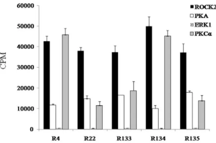
showing high CPMs (R4, R22, R133, R134, and R135) were phosphorylated by other enzymes (PKA, PKCa, and ERK1), R22, R133, and R135 displayed the highest CPM level for ROCK2 compared with other enzymes, whereas R4 and R134 showed similar CPM levels for ROCK2 and PKCa. We hypothesize that R22, R133, and R135 can be useful peptide substrates for ROCK2.
Citation:Kang J-H, Asai D, Tsuchiya A, Mori T, Niidome T, et al. (2011) Peptide Substrates for Rho-Associated Kinase 2 (Rho-Kinase 2/ROCK2). PLoS ONE 6(7): e22699. doi:10.1371/journal.pone.0022699
Editor:Fernando Rodrigues-Lima, University Paris Diderot-Paris 7, France
ReceivedFebruary 8, 2011;AcceptedJune 30, 2011;PublishedJuly 27, 2011
Copyright:ß2011 Kang et al. This is an open-access article distributed under the terms of the Creative Commons Attribution License, which permits unrestricted use, distribution, and reproduction in any medium, provided the original author and source are credited.
Funding:This work was financially supported by a grant-in-aid for Scientific Research from the Ministry of Education, Culture, Sports, Science and Technology (MEXT) of Japan. The funders had no role in study design, data collection and analysis, decision to publish, or preparation of the manuscript.
Competing Interests:The authors have declared that no competing interests exist.
* E-mail: jrjhkang@ri.ncvc.go.jp (J-HK); ykatatcm@mail.cstm.kyushu-u.ac.jp (YK)
Introduction
Phosphorylation by protein kinases plays an essential part in the signal transduction pathways that regulate cellular functions in response to extracellular signals. It is also a general mechanism for the control of intracellular processes [1]. Designing specific peptide substrates for a certain protein kinase is indispensable for the characterization of (or search for) substrate proteins for enzymes, and is also important in explorations for new drugs. In general, peptide substrates specific to a certain protein kinase are identified using genetic variants of protein substrates and synthetic peptides [2–4].
Rho-associated kinase (Rho-kinase/ROCK; hereafter referred to as ROCK) is a serine/threonine kinase and plays an important part in various cellular functions. These include the contraction of smooth muscle, cell adhesion, and cytokinesis [5,6]. Over-expression of this enzyme has been associated with cardiovascular diseases such as hypertension and cerebral and coronary vasospasm [7–9]. Moreover, ROCK is closely associated with the migration and invasion of tumor cells [10,11]. Recent studies suggest that inhibition of ROCK not only increases cerebral blood flow and leads to protection against stroke, but enhances functional recovery in injured spinal cords [12–14]. Thus, ROCK could be a potential therapeutic target for tumors, neurological disorders and cardiovascular diseases.
ROCK is divided into two isozymes: ROCK1 (ROCKb) and ROCK2 (ROCKa) [5,6]. The latter is mainly expressed in the brain, heart, and skeletal muscle, and ROCK1 has been identified
in the spleen, lung, liver, testis, and kidney [15]. Many protein substrates for ROCK and their substrate sequences have been reported. ROCK can phosphorylate calponin [16]; LIM-kinase 1 (LIMK1) [17]; adducin [18]; intermediate filaments (vimentin [19], neurofilament-L [20], and glial fibrillary acidic protein (GFAP) [21]); collapsin response mediator protein 2 (CRMP2) [22]; tau [23]; microtubule-associated protein 2 (MAP2) [23]; ERM family (ezrin/radixin/moesin) [24]; myosin binding subunit (MBS) [25]; myosin light-chain (MLC) [26]; myristoylated alanine-rich C kinase substrate (MARCKS) [27,28]; Rho E [29]; zipper-interacting protein kinase (ZIPK) [30]; LIMK2 [31] and endophilin A1 [32].
The purpose of the present study was to find substrates with high affinity and sensitivity for ROCK2. We synthesized 136 peptide substrates from protein substrates for ROCK2 with different lengths and charged peptides. After determining the incorporation of radioactivity (counts per minute (CPM)) for each peptide substrate by the radiolabel assay using [c-32P]ATP, kinetic properties for the
final five top-ranked substrates were analyzed. We also examined the affinities of five substrates for other kinases (extracellular signal-regulated kinase 1 (ERK1), protein kinase A (PKA), and protein kinase C (PKC)a) showing similar consensus sequences and interactive functions in intracellular signal transduction to ROCKs.
Results
the amino-acid sequences in ROCK protein substrates (Table S1 and Table S2). These peptide substrates showed a different number of charged peptide residues or amino-acid residues. Peptide substrates had a basic amino acid (Arg or Lys) at amino-terminal position22 or23 of the phosphorylated sites (Ser and Thr). Moreover, most amino acids at the +2 carboxyl-terminal position were basic or hydrophobic (Arg, Lys, Phe, Leu, Trp, or Val) (Table S2).
We determined the radioactivity (in counts per minute (CPM)) for each peptide substrate by the radiolabel assay using [c-32P]ATP (Fig. 1). Peptide substrates were divided into four groups from the results of radioactivity. Conversely, five peptide substrates showing CPM values of.35,000 for ROCK2 were in the order R134 (KSARKKRYTVVGNPYWM).R4 (RAKYK-TLRQIR).R22 (KPARKKRYTVVGNPYWM).R133 (KSDR-KKRYTVVGNPYWM).R135 (KADRKKRYTVVGNPYWM) (Fig. 2).
The Ser:Thr ratio for the total number of phosphorylation sites used in this study was 9:12. The substitution of Ser for Thr at the phosphorylation site led to a tendency to a decrease in the radioactivity (see R1–6 and R7–12, R64–67 and R68–71, R72–75 and R76–79, R80–82 and R83–85, and R86–88 and R89–91) (Fig. 1). Although the observation was not universal, the change of negatively charged amino acids (Asp or Glu) into Ala or positively charged amino acids tended to result in an increase in CPM.
However, the length of peptides had no or very little effect on CPMs (Fig. 1 and Table S2).
Furthermore, the kinetic properties of five peptide substrates showing high CPM for ROCK2 were analyzed. Values ofKmand Vmaxfor five peptide substrates ranged from 1.7mM to 3.3mM, and from 8.6 pmol/min/mg to 14.9 pmol/min/mg, respectively (Table 1).
The consensus sequence of the ROCK2 phosphorylation site was considered to be R/KXXS/T or R/KXS/T (R, arginine; K, lysine; X, any amino acid; S, serine; and T, threonine) [5,33] (Table S2). These consensus sequences were similar to other protein kinases (e.g., PKA and PKC). PKA recognizes the phosphorylation motifs R/KXS/T and R/KXXS/T [34,35]. Consensus phosphorylation site motifs for PKC were identified to be R/KXXS/T, R/KXS/T, R/KXXS/TXR/K, or R/KXS/ TXR/K [35,36]. Mitogen-activated protein kinase (MAPK) can recognize the phosphorylation motif XS/TPX (P, proline) [37]. Five peptide substrates that showed CPM of.35,000 for ROCK2 were phosphorylated with PKA, PKCa (isozyme of PKC), and ERK1 (isozyme of MAPK). R4 and R134 showed high CPM for ROCK2 and PKCa. R22, R133, and R135 showed much higher CPM for ROCK2 than that of PKCa(Fig. 3).
Several studies have reported that replacement of phosphor-ylatable amino acids with alanine can lead to an inhibitor peptide for each enzyme [38,39]. Thus, we examined if peptides that
Figure 1. Counts per minute (CPM) levels for 136 peptide substrates.Each peptide substrate was analyzed by the radiolabel assay using [c-32P]ATP.
replace phosphorylatable amino acids with alanine can inhibit the phosphorylation of substrate peptide by ROCK2. The phosphor-ylatable threonine of five peptide substrates (R4, R22, R133, R134, and R135) was replaced with alanine and used as inhibitor peptides. The lowestKi (11.6mM) in a competitive manner was
obtained for R22 with alanine instead of threonine (Table 2).
Discussion
There have been many efforts to search for peptide substrates with high affinity and sensitivity to ROCK2. However, different experimental methods make selecting substrates for ROCK2 quite difficult. Moreover, the affinity and sensitivity of substrates for ROCK2 can be influenced by several factors. These include the peptide length and the number of charged peptides [38,40–42], but these factors were not investigated in previous studies.
We synthesized 136 peptide substrates for ROCK2 with different lengths and charged peptides, and analyzed their ability for phosphorylation. When negatively charged amino acids (Asp and Glu) were replaced by Ala or a positively charged amino acid (Arg), resulting in an increase in the positive charge density in peptide substrate, a tendency to an increase in the radioactivity was identified. For example, a higher CPM value was identified for R4 than for R1, R38 than R33, and R134 than R133. The length of peptide substrate had no or little effect on the level of radioactivity. In general, shortening of peptide substrates de-creased the affinity for the enzyme (i.e., increased theKm value).
However, the peptide length showed minor changes in the Vmax
value [38,40,41]. These data showed that radioactivity was not affected by the length of peptide substrates, but that the change of negatively charged amino acids into Ala or positively charged amino acids could increase the radioactivity values.
PKA and PKCa broadly recognize ROCK phosphorylation sites because of their similar consensus sequences to the ROCK phosphorylation motif. Our previous study showed that ROCK substrates can be good substrates for PKA and/or PKCa. Among 18 ROCK phosphorylation sites, 14 sites (Thr-184 of calponin; Thr-508 of LIMK1; Ser-71 of vimentin; Ser-57 of neurofilament-L; Thr-7, 13, and 34 of GFAP; Thr-555 of CRMP2; Ser-409 of tau; Ser-1796 of MAP2; Ser-854 of MBS; Thr567/564/558 of the ERM family; Ser-19 of MLC; and Ser-159 of MARCKS) were phosphorylated by PKA and/or PKCa [33]. Moreover, there was a close function in intracellular signal transduction between ROCK and PKCa, ERK1, or PKA. The interactive role of ROCK and PKCa has been reported in the actin–myosin interaction [43] and in the contraction of vascular smooth muscle [44,45]. MLC phosphorylation was stimulated by ROCK and
Figure 2. Classification of peptide substrates.Four groups were divided from the results of radioactivity (i.e., CPM).
doi:10.1371/journal.pone.0022699.g002
Table 1.Kinetic properties for five peptide substrates showing CPM.35,000.
Number of peptide
substrate Km(mM) Vmax(pmol/min/mg) Vmax/ Km
R4 1.7 14.2 8.4
R22 1.9 10.3 5.4
R133 3.6 10.0 2.8
R134 3.3 14.9 4.5
R135 3.2 8.6 2.7
doi:10.1371/journal.pone.0022699.t001
Figure 3. Phosphorylation of the top five peptide substrates (R4, R22, R133, R134, and R135) for ROCK2 by other enzymes (PKA, PKCa, or ERK1).Each peptide substrate was analyzed by the
PKCa, but decreased by suppressing ROCK and PKCa using inhibitors [44,45]. RhoA phosphorylation at Ser 188 by PKA blocks the ROCK pathways, resulting in activation of MLC phosphatase and the relaxation of smooth muscle [46]. Activated PKA can also facilitate axon formation by inhibiting RhoA, but ROCK has opposite effects on axon formation [47]. ROCK has an effect on translocation of ERK1 to the cellular nucleus, leading to proliferation of smooth muscle cells in the pulmonary artery [48]. ERK1 activation by ROCK stimulates: 1) force-induced osteopontin expression in human periodontal ligament fibroblasts through focal adhesion kinase signaling [49]; and 2) migration and proliferation of glioblastoma cells [50]. The observations noted above mean that PKA, PKCa, and ERK1 directly and/or indirectly participate in the signal transduction pathways of ROCK. Thus, we examined if five peptide substrates (R4, R22, R133, R134, and R135) showing high CPM values (.35,000) for ROCK2 could be phosphorylated by other enzymes (PKA, PKCa, or ERK1). R22, R133, and R135 displayed higher CPM levels for ROCK2 than the other enzymes, whereas R4 and R134 showed similar CPM levels for ROCK2 and PKCa(Fig. 3). These results suggested that R22 and R133 could be useful peptide substrates for ROCK2, and that R4 and R134 could be useful peptide substrates for ROCK2 and PKCa.
Replacement of phosphorylation sites with alanine is used to develop inhibitor peptides. In general, peptides showing higher affinity (i.e., lowKm) than lower affinity (i.e., highKm) become much
more effective inhibitor peptides when phosphorylation sites are substituted by alanine. In the present study, we examined if five peptides in which a phosphorylatable amino acid (threonine) was replaced with alanine could be used as potent competitive inhibitors of ROCK2. The R22 peptide with alanine instead of threonine showed the lowestKi(11.6mM) (Table 2).
In conclusion, there has been an increasing interest in ROCKs as therapeutic targets of cardiovascular disease, migration and invasion of tumor cells, and neurological disorders. Peptide substrates with high affinity and sensitivity to ROCK2 are important in new-drug developments and in understanding the cellular signals involved. In the present study, 136 peptide substrates from protein substrates for ROCK2 were synthesized and the radioactivity of each peptide substrate analyzed. From CPM results and phosphorylation reactions with other enzymes (PKA, PKCa, and ERK1), we found three peptides (R22, R133, and R135) to be useful peptide substrates for ROCK2.
Materials and Methods
Syntheses of peptide substrates
Peptide substrates were synthesized using an automatic peptide synthesizer according to standard Fmoc-chemistry procedures. After treatment with trifluoroacetic acid (TFA), peptides were
purified on an Inertsil ODS-3 column (250620 mm, 3.5mm; GL Sciences, Tokyo, Japan) using a BioCAD Perfusion Chromatog-raphy System (Ikemoto Scientific Technology, Tokyo, Japan) and a linear A–B gradient at a flow rate of 8 mL/min where eluent A was 0.1% TFA in water and eluent B was 0.1% TFA in acetonitrile. The purity of synthetic peptide was identified by high-performance liquid chromatography and matrix-assisted laser desorption/ionization-time-of-flight mass spectrometry, and the peptide with .95% purity was used for the phosphorylation reaction.
Phosphorylation of peptide substrates by enzymes The synthetic peptide S6K (KRRRLASLR) was used as a control for ROCK2. Alphatomega (FKKQGSFKKK) [4] was used for PKCa assays and ZIPK-T265 (KRRMTIAQSLEHS-WIK) [30] for PKA assays. ERK assays employed 0.33 mg/mL human myelin basic protein (MBP; Chemicon International, Temecula, CA, USA).
The kinase activity of recombinant ROCK2 (Carna Biosci-ences, Kobe, Japan), ERK1 (Upstate, Millipore, Temecula, CA, USA), PKA (Promega, Madison, WI, USA), and PKCa(Sigma– Aldrich, St. Louis, MO, USA) for the peptide substrates was determined by measuring 32P transfer from [c-32P]ATP into substrate peptides according to manufacturer recommendations. Phosphorylation reactions by ROCK2 were carried out in 25mL of buffer (5 mM 3-(N-morpholino)propanesulfonic acid (MOPS) at pH 7.2, 2.5 mM beta-glycerophosphate, 1 mM ethylene glycol tetra-acetic acid (EGTA), 4 mM MgCl2, 0.05 mM dithiothreitol
(DTT), and 100mM adenosine triphosphate (ATP)) containing peptides and 1.25mg/ml ROCK2. For PKCa, phosphorylation reactions were carried out in 25mL of buffer (20 mM HEPES at pH 7.4, 10 mM MgCl2, 1 mM CaCl2, 100mM ATP, 40mg/mL phosphatidylserine, and 20mg/mL diacylglycerol) containing peptides and 0.5mg/mL PKCa. Phosphorylation reactions by PKA were carried out in 25mL of buffer (50 mM HEPES at pH 7.4, 1 mM CaCl2, 10 mM MgCl2, 100mM ATP) containing
peptides and 1.6mg/mL PKA. For ERK1, phosphorylation reactions were carried out in 25mL of buffer (25 mM Tris-HCl at pH 7.5, 20mM EGTA, 15 mM magnesium acetate, and 100mM ATP) containing peptides and 0.5mg/mL ERK1. For each experimental condition, values for control reactions lacking substrate peptides were subtracted as blanks. The assay mixture was incubated for 10 min at 25uC, and the reaction terminated by the addition of 5mL of 30% trichloroacetic acid (TCA). The reaction mixture (24mL) was spotted onto P-81 phosphocellulose membranes. The membranes were washed thrice with 5% TCA, dried with acetone, and the radioactivity of each membrane determined by liquid scintillation counting. Kinetic analyses of enzyme activity were determined using the Lineweaver–Burk plot.
Inhibition of phosphorylation by inhibitor peptides The phosphorylatable amino acid (threonine) of five peptide substrates (R4, R22, R133, R134, and R135) was replaced with alanine. Inhibitory activity of these inhibitor peptides against ROCK2 was examined using S6K peptide (KRRRLASLR) as a substrate; value of Km and Vmax for S6K was 3.1mM and 23.1 pmol/min/mg, respectively. Ki values were obtained from the Dixon plot and Lineweaver–Burk plot. Inhibitor peptides were added into 25mL of buffer (5 mM MOPS at pH 7.2, 2.5 mM beta-glycerophosphate, 1 mM EGTA, 4 mM MgCl2, 0.05 mM
DTT, and 100mM ATP) containing S6K substrate peptide and 1.25mg/mL ROCK2.
Table 2.Kivalues for five peptides with alanine instead of a phosphorylatable threonine.
Number of peptide Ki(mM)
R4 86.6
R22 11.6
R133 .100
R134 29.2
R135 .100
Supporting Information
Table S1 Origins of each peptide substrate for ROCK2. (DOCX)
Table S2 Sequences of peptide substrate for ROCK2. (DOCX)
Author Contributions
Performed the experiments: J-HK YK. Analyzed the data: J-HK DA AT. Contributed reagents/materials/analysis tools: DA TM TN. Wrote the paper: J-HK YK.
References
1. Ghoreschi K, Laurence A, O’Shea J (2009) Selectivity and therapeutic inhibition of kinases: to be or not to be? Nat Immunol 10: 356–360.
2. Kemp BE, Bylund DB, Huang TS, Krebs EG (1975) Substrate specificity of the cyclic AMP-dependent protein kinase Proc. Natl Acad Sci USA 72: 3448–3452. 3. Cheng HC, Matsuura I, Wang JH (1993) In vitro substrate specificity of protein
tyrosine kinases. Mol Cell Biochem 127–128: 103–12.
4. Kang JH, Asai D, Yamada S, Toita R, Oishi J, et al. (2008) A short peptide is a protein kinase C (PKC)a-specific substrate. Proteomics 8: 2006–2011. 5. Kaibuchi K, Kuroda S, Amano M (1999) Regulation of the cytoskeleton and cell
adhesion by the Rho family GTPases in mammalian cells. Annu Rev Biochem 68: 459–486.
6. Amano M, Fukata Y, Kaibuchi K (2000) Regulation and functions of Rho-associated kinase. Exp Cell Res 261: 44–51.
7. Loirand G, Gue´rin P, Pacaud P (2006) Rho kinases in cardiovascular physiology and pathophysiology. Circ Res 98: 322–334.
8. Oka M, Fagan KA, Jones PL, McMurtry IF (2008) Terapeutic poteintial of RhoA/Rho kinase inhibitors in pulmonary hypertension. Br J Pharmacol 155: 444–454.
9. Zhou Q, Liao JK (2009) Rho kinase: an important medicator of atherosclerosis and vascular disease. Curr Pharm Des 15: 3108–3115.
10. Narumiya S, Tanji M, Ishizaki T (2009) Rho signaling, ROCK and mDai1, in transformation, metastasis and invasion. Cancer Metastasis Rev 28: 65–76. 11. Joshi B, Strugnell SS, Goetz JG, Kojic LD, Cox ME, et al. (2008)
Phosphorylation caveolin-1 regulates Rho/ROCK-dependent focal adhesion dynamics and tumor cell migration and invasion. Cancer Res 68: 8210–8220. 12. Shin HK, Salomone S, Ayata C (2008) Targeting cerebrovascular Rho-kinase in
stroke. Expert Opin Ther Targets 12: 1547–1564.
13. Mueller BK, Mack H, Teusch N (2005) Rho kinase, a promising drug target for neurological disorders. Nat Rev Drug Discov 4: 387–398.
14. Kubo T, Yamashita T (2007) Rho-ROCK inhibitors for the treatment of CNS injury. Recent Pat CNS Drug Discov 2: 173–179.
15. Nakagawa O, Fujisawa K, Ishizaki T, Saito Y, Nakao K, et al. (1996) ROCK-1 and ROCK-2, two isoforms of Rho-associated coiled-coil forming protein serine/theronine kinase in mice. FEBS Lett 392: 189–193.
16. Kaneko T, Amano M, Maeda A, Goto H, Takahashi K, et al. (2000) Identification of calponin as a novel substrate of Rho-kinase. Biochem Biophy Res Commun 273: 110–116.
17. Ohashi K, Nagata K, Maekawa M, Ishizaki T, Narumiya S, et al. (2000) Rho-associated kinase ROCK activates LIM-kinase 1 by phosphorylation at threonine 508 within the activation loop. J Biol Chem 275: 3577–3582. 18. Fukata Y, Oshiro N, Kinoshita N, Kawano Y, Matsuoka Y, et al. (1999)
Phosphorylation of adducin by Rho-kinase plays a crucial role in cell motility. J Cell Biol 145: 347–361.
19. Goto H, Kosako H, Tanabe K, Yanagida K, Maki M, et al. (1998) Phosphorylation of vimentin by Rho-associated kinase at a unique amino-terminal site that is specifically phosphorylated during cytokinesis. J Biol Chem 273: 11728–11736.
20. Hashimoto R, Nakamura Y, Komai S, Kashiwagi Y, Tamura K, et al. (2000) Site-specific phosphorylation of neurofilament-L is mediated by calcium/ calmodulin-dependent protein kinase II in the apical dendrites during long-term potentiation. J Neurochem 75: 373–382.
21. Kosako H, Amano M, Yanagida M, Tanabe K, Nishi Y, et al. (1997) Phosphorylation of glial fibrillary acidic protein at the same sites by cleavage furrow kinase and Rho-associated kinase. J Biol Chem 272: 10333–10336. 22. Arimura N, Inagaki N, Chihara K, Menager C, Nakamura N, et al. (2000)
Phosphorylation of collapsin response mediator protein-2 by Rho-kinase. Evidence for two separate signaling pathways for growth cone collapse. J Biol Chem 275: 23973–23980.
23. Amano M, Kaneko T, Maeda A, Nakayama M, Ito M, et al. (2003) Identification of Tau and MAP2 as novel substrates of Rho-kinase and myosin phosphatase. J Neurochem 87: 780–790.
24. Matsui T, Maeda M, Doi Y, Yonemura S, Amano M, et al. (1998) Rho-kinase phosphorylates COOH-terminal threonines of ezrin/radixin/moesin (ERM) proteins and regulates their head-to-tail association. J Cell Biol 140: 647–657. 25. Kawano Y, Fukata Y, Oshiro N, Amano M, Nakamura T, et al. (1999)
Phosphorylation of myosin-binding subunit (MBS) of myosin phosphatase by Rho-kinase in vivo. J Cell Biol 147: 1023–1037.
26. Amano M, Ito M, Kimura K, Fukata Y, Chihara K, et al. (1996) Phosphorylation and activation of myosin by Rho-associated kinase (Rho-kinase). J Biol Chem 271: 20246–20249.
27. Sasaki Y (2003) New aspects of neurotransmitter release and exocytosis: Rho-kinase-dependent myristoylated alanine-rich C-kinase substrate phosphorylation and regulation of neurofilament structure in neuronal cells. J Pharmacol Sci 93: 35–40.
28. Nagumo H, Ikenoya M, Sakurada K, Furuya K, Ikuhara T, et al. (2001) Rho-associated kinase phosphorylates MARCKS in human neuronal cells. Biochem Biophys Res Commun 280: 605–609.
29. Riento K, Totty N, Villalonga P, Garg R, Guasch R, et al. (2005) RhoE function is regulated by ROCK 1-mediated phosphorylation. EMBO J 24: 1170–1180. 30. Hagerty L, Weitzel DH, Chambers J, Fortner CN, Brush MH, et al. (2007)
ROCK1 phosphorylates and activates Zipper-interacting protein kinase. J Biol Chem 282: 4884–4893.
31. Sumi T, Matsumoto K, Nakamura T (2001) Specific activation of LIM kinase 2 via phosphorylation of Threonine 505 by ROKC, a Rho-dependent protein kinase. J Biol Chem 276: 670–676.
32. Kaneko T, Maeda A, Takefuji M, Aoyama H, Nakayama M, et al. (2005) Rho medicates endocytosis of epidermal growth factor receptor through phosphor-ylatin of endophilin A1 by Rho-kinase. Genes Cells 10: 973–987.
33. Kang JH, Jiang Y, Toita R, Oishi J, Kawamura K, et al. (2007) Phosphorylation of Rho-associated kinase (Rho-kinase/ROCK/ROK) substrates by protein kinases A and C. Biochimie 89: 39–47.
34. Kemp BE, Graves DJ, Benjamini E, Krebs EG (1977) Role of multiple basic residues in determining the substrate specificity of cyclic AMP-dependent protein kinase. J Biol Chem 14: 4888–4894.
35. Rearson RB, Kemp RE (1988) Protein kinase phosphorylation site sequences and consensus specificity motifs: tabulations. In: Sefton BM, Hunter T, eds. Protein phosphorylation. San Diego, CA: Academic Press. pp 65–83. 36. Hug H, Sarre TF (1993) Protein kinase C isoenzymes: divergence in signal
transduction? Biochem J 291: 329–343.
37. Davis RJ (1993) The mitogen-activated protein kinase signal transduction pathway. J Biol Chem 268: 14553–14556.
38. Hofmann J (1997) The potential for isoenzyme-selective modulation of protein kinase C. FASEB J 11: 649–669.
39. Nishikawa K, Sawasdikosol S, Fruman DA, Lai J, Songyang Z, et al. (2000) A peptide library approach identifies a specific inhibitor for the ZAP-70 protein tyrosine kinase. Mol Cell 6: 969–974.
40. Jain MK, Rogers J (1989) Substrate specificity for interfacial catalysis by phospholipase A2 in the scooting mode. Biochem Biophys Acta 1003: 91–97. 41. Koivunen P, Hirsila¨ M, Kivirikko KI, Myllyharju J (2006) The length of peptide
substrates has a marked effect on hydroxylation by the hypoxia-inducible factor proly 4-hydroxylases. J Biol Chem 281: 28712–28720.
42. House C, Kemp BE (1990) Protein kinase C psudosubstrate prototope: sturcture-function relationships. Cell Signa 2: 187–190.
43. Madigan JP, Bodemann BO, Brady DC, Dewar BJ, Keller PJ, et al. (2009) Regultaion of Rnd3 localization and function by protein kinase Ca-mediated phosphorylation. Biochem J 424: 153–161.
44. Somlyo AP, Somlyo AV (2003) Ca2+ sensitivity of smooth muscle and nonmuscle myosin II: modulated by G proteins, kinases, and myosin phosphatase. Physiol Rev 83: 1325–1358.
45. Patil SB, Bitar KN (2006) RhoA- and PKC-a-mediated phosphorylation of MYPT and its association with HSP27 in colonic smooth muscle cells. Am J Physiol Gastrointest Liver Physiol 290: G83–G95.
46. Murthy KS (2006) Signaling for contraction and relaxation in smooth muscle of the gut. Annu Rev Physiol 68: 345–374.
47. Leemhuis J, Boutillier S, Schmidt G, Meyer DK (2002) The protein kinase A inhibitor H89 acts on cell morphology by inhibiting Rho kinase. J Pharmacol Exp Therap 300: 1000–1007.
48. Liu Y, Suzuki YJ, Day RM, Fanburg BL (2004) Rho kinase-induced nuclear translocation of ERK1/ERK2 in smooth muscle cell mitogenesis caused by serotonin. Circ Res 95: 579–586.
49. Hong SY, Jeon YM, Lee HJ, Kim JG, Baek JA, et al. (2010) Activation of RhoA and FAK induces ERK-medicated osteopontin expression in mechanical force-subjected periodontal ligament fibroblasts. Mol Cell Biochem 335: 263–272. 50. Zohrabian VM, Forzani B, Chau Z, Murali R, Jhanwar-Uniyal M (2009) Rho/
![Figure 1. Counts per minute (CPM) levels for 136 peptide substrates. Each peptide substrate was analyzed by the radiolabel assay using [c- 32 P]ATP.](https://thumb-eu.123doks.com/thumbv2/123dok_br/17290926.248021/2.918.81.836.510.1051/figure-counts-peptide-substrates-peptide-substrate-analyzed-radiolabel.webp)