R E S E A R C H
Open Access
Characteristics of hemolytic activity induced
by skin secretions of the frog
Kaloula pulchra
hainana
Shuangshuang Wei
1,2†, Tingting Chi
1†, Aiyun Meng
1†, Congwei Chen
1, Tianchen An
1, Manchuriga Wang
2and Yingxia Zhang
1*Abstract
Background:The hemolytic activity of skin secretions obtained by stimulating the frogKaloula pulchra hainanawith diethyl ether was tested using human, cattle, rabbit, and chicken erythrocytes. The skin secretions had a significant concentration-dependent hemolytic effect on erythrocytes. The hemolytic activity of the skin secretions was studied in the presence of osmotic protectants (polyethylene glycols and carbohydrates), cations (Mg2+, Ca2+, Ba2+, Cu2+, and K+), or antioxidants (ascorbic acid, reduced glutathione, and cysteine).
Results:Depending on their molecular mass, osmotic protectants effectively inhibited hemolysis. The inhibition of skin hemolysis was observed after treatment with polyethylene glycols (1000, 3400, and 6000 Da). Among divalent cations, only 1 mM Cu2+markedly inhibited hemolytic activity. Antioxidant compounds slightly reduced the hemolytic activity.
Conclusions:The results suggested that skin secretions ofK. pulchra hainanainduce a pore-forming mechanism to form pores with a diameter of 1.36-2.0 nm rather than causing oxidative damage to the erythrocyte membrane.
Keywords:Amphibian, Skin secretions, Hemolysis, Pore-forming
Background
The skin of amphibians has numerous biochemical and physiological functions to assure amphibian survival. Several bioactive components with specialized functions and molecular structures have been isolated and purified from amphibian skin [1,2]. Some of these components and their analogs have been used for treating diseases such as microbial infections and burns [1,3]. Thus, increasing numbers of studies have focused on amphibian skin secretions to identify biologically active proteins and peptides [1,4].
Cnidarian venom is an abundant source of numerous bioactive molecules such as pore-forming proteins, small cytotoxic peptides, 5-hydroxytryptamine, and histamine. Of the cytolytic, hemolytic, and neurotoxic effects of Cnidarian venom, hemolytic activity is the most commonly
investigated [5-7]. Hemolysis induced by Cnidarian venom toxins has been particularly investigated to identify targets and the attachment of proteins to cell membranes [8,9].
Kaloula pulchra hainana (Anura) is an endemic
amphibian found in the low elevation regions of Hainan Island of China. These frogs always live near pools, which is a harsh environment with numerous pathogens. When these frogs are stimulated, their belly bulges and white secretion is released from their skin. In a previous study, we purified and characterized a 23-kDa trypsin inhibitor from the skin secretions ofK. pulchra hainana, designated
K. pulchra hainanatrypsin inhibitor (KPHTI) [10]. Skin secretions of the frog K. pulchra hainana also exhibit hemolytic activity. This study aimed to establish basic information on erythrocyte hemolysis and cell membrane peroxidation induced by these skin secretions. To determine the toxicological properties of the skin secretions, we investigated the effects of different factors on erythrocyte hemolysis, including osmotic protectants, cations, antioxidants, and chelating agents.
* Correspondence:yingxiazhang@hotmail.com
†
Equal contributors
1Key Laboratory of Tropic Biological Resources, Minister of Education, College
of Marine Science, Hainan University, Haikou, China
Full list of author information is available at the end of the article
Methods
Materials
N-acetyl-D-galactosamine (NAGA), N-methylmannopy-ranose, D-glucose, D-trehalose, D-lactose, reduced glutathione (GSH), and bovine serum albumin (BSA) were purchased from Sigma-Aldrich (USA). Polyethylene glycols (PEGs) of different molecular masses (300, 400, 1000, 3400, and 6000 Da) were obtained from Fluka (USA). The protein concentrations of skin secretions were determined using a protein assay kit (Bio-Rad, USA) with BSA as a standard [11]. All other reagents used were of the highest purity available.
Preparation of skin secretions
Adult specimens ofK. pulchra hainanaof both genders (n = 10; weight range: 80–120 g) were collected in Danzhou City, Hainan Province in southern China. The skin of the frogs was briefly stimulated with diethyl ether and then washed with 50 mM Tris–HCl buffer (pH 7.8) containing 0.1 M NaCl and 5 mM ethylene diamine tetraacetic acid (EDTA). The secretions were centrifuged at 10,000 g for 20 minutes at 4°C to remove insoluble materials. The supernatant was collected, lyophilized, and stored at−80°C until use. Before an experiment, the lyophilized skin secretions were dissolved in phosphate-buffered saline (PBS) (137 mM NaCl, 1.5 mM KH2PO4,
2.7 mM KCl, 8.1 mM Na2HPO4) and then dialyzed
against PBS.
Hemolysis determinations
The hemolytic activity of skin secretions was determined using human, cattle, rabbit, and chicken erythrocytes, as reported by Liuet al. [12]. Erythrocytes from these species were washed with PBS until the supernatant was clear and then resuspended in PBS. Erythrocyte suspensions (5 × 10⁶cells/mL) were incubated with different concen-trations of skin secretions (0.28, 0.56, 1.4, 2.8, 4.2, and 5.6μg/mL) at 37°C for 30 minutes and then centrifuged at 1000 g for 5 minutes at 4°C to precipitate intact erythrocytes and debris. The supernatants were assayed for absorbance at 540 nm to determine the percentage of hemoglobin released from the lysed erythrocytes. We defined 100% lysis as the absorbance of a supernatant obtained using 1% Triton X-100 instead of test samples. The hemolytic activity of the skin secretions was expressed as the percentage of absorbance compared with that observed after 100% lysis induced by Triton X-100. The supernatant of an untreated erythrocyte suspension in PBS was used as a spectrophotometric blank.
Determination of membrane pore diameters
The diameters of membrane pores induced by skin secretions were determined as described previously [13]. In brief, 40 mOsm PEGs of various molecular masses
(300, 400, 1000, 3400, and 6000 Da) were added to PBS to counteract the osmotic pressure of hemoglobin [14]. Following this, by changing the concentration of NaCl, the total osmotic pressure of the extracellular fluid was adjusted to 295 mOsm. After human erythrocytes were suspended in a PEG solution (5 × 10⁶ cells/mL), the hemolytic activity of skin secretions was determined as described above. The hydrodynamic diameters of PEG 300, 400, 1000, 3400, and 6000 were 1.16, 1.36, 2.0, 3.8, and 5.8 nm, respectively [13,15].
Osmotic protectants
The following osmotic protectants were used: 5 mM NAGA, 10 mM N-methylmannopyranose, 25 mM D-glucose, 25 mM D-trehalose, and 25 mM D-lactose. Each of these was added to erythrocytes suspended in PBS. After skin secretions (5.6 μg/mL) were added and incubated at 37°C for 30 minutes, the hemolytic activity was determined as described above. For osmotic protectants with large molecular masses, erythrocytes were preincubated with PEG 6000 for 10 minutes, following which erythrocytes were removed and resuspended in PBS. Following this, skin secretions (0.7, 1.4, or 5.6 μg/mL) were added and hemolytic activity was determined.
Cations and EDTA
Different cations (KCl, BaCl2, MgCl2, CuSO4, or CaCl2)
were individually mixed with erythrocyte suspensions, following which skin secretions (1.4 or 5.6 μg/mL) were added and incubated at 37°C for 30 minutes and hemolytic activity was determined. The final concentration of CaCl2, BaCl2, MgCl2, and KCl was 10 mM, whereas that
of CuSO4 was 1 mM. The effect of EDTA (0.1 mM,
0.2 mM, or 1 mM) was also determined.
Antioxidants
Ascorbic acid, GSH, and cysteine were used to assess possible erythrocyte cell membrane oxidative damage induced by skin secretions. An antioxidant (2 mM) was added to an erythrocyte suspension, following which skin secretions were added (5.6 μg/mL) and hemolytic activity was determined.
Statistical analysis
Results are given as means ± standard error (SE) for ten experiments. Results for different experimental conditions were compared by Student’s t-tests. A p-value < 0.05 was considered significant [16].
Results
secretions were different; the most sensitive erythrocytes were human erythrocytes. For human erythrocytes, the percent hemolysis was 19.8 ± 3% with 0.28μg/mL of skin secretions, while total hemolysis was 90.3 ± 4% with 5.6 μg/mL of skin secretions. Among the four species examined, chicken erythrocytes were the least susceptible to these skin secretions; the hemolytic activity was approximately 60.5 ± 2% with the highest concentration of skin secretions (5.6μg/mL).
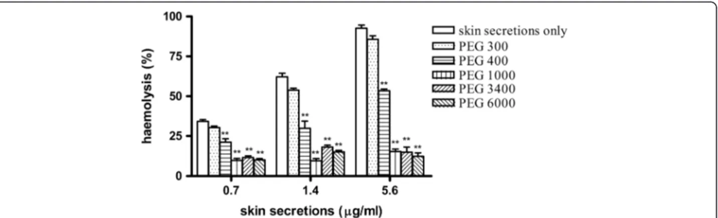
The hemolytic activity of the skin secretions was tested in the presence of PEGs with different hydrodynamic diameters: 300, 400, 1000, 3400, and 6000 Da. These were used to estimate pore diameters in erythrocyte cell membranes. Hemolysis induced by the skin secretions was not affected by treatment with PEG 300, it was partially inhibited by PEG 400, and markedly inhibited by treatment with PEGs 1000, 3400, and 6000 (p < 0.05, compared with no PEGs; Figure 2).
Effects of osmotic protectants on hemolytic activity Erythrocyte suspensions were first pretreated with 25 mM PEG 6000 for 5 minutes, following which erythrocytes were removed and resuspended in PBS. Following this, skin secretions (0.7, 1.4, or 5.6 μg/mL) were added to test their hemolytic activities. The erythrocytes were hemolyzed equally compared with controls, which supported that the larger PEG molecules did not bind to the membrane to reduce the interaction between the cell membrane and skin secretions. However, because PEG 6000 is an osmotic protectant, it maintained the medium as more hypertonic and blocked membrane pores induced by the skin secretions.
Compared with the controls, the osmotic protectants with small molecular masses, including NAGA, D-glucose, methylmannopyranose, trehalose, and lactose, did not significantly inhibit the hemolytic activity of the skin secretions (Table 1).
Effects of cations and EDTA on hemolytic activity
The effects of different cations on the hemolytic activity of the skin secretions were assessed using the divalent cations Ca2+, Mg2+, Ba2+, and Cu2+and the monovalent cation K+. Except for Cu2+, these cations at concentrations of < 10 mM did not affect the hemolytic activity compared with the control (data not shown). Cu2+at a concentration of 1 mM significantly inhibited the hemolytic activity at skin secretion concentrations of 1.4 and 5.6 μg/mL, whereas 10 mM Mg2+, Ca2+, and K+had slight inhibitory effects (Figure 3). EDTA at any of the concentrations used did not inhibit hemolysis (Table 1).
Effects of antioxidants on hemolytic activity
The antioxidants cysteine, GSH, and ascorbic acid (2 mM) reduced the hemolysis induced by skin secretions, with results ranging from 14 to 20% (Table 1). However,
Figure 1Hemolytic activity of skin secretions fromKaloula pulchra hainanatested with erythrocytes from different species: human (□), cattle (Δ), rabbit (○), and chicken (▼).
Figure 2Osmotic protection against hemolytic activity using a series of polyethylenglycols (PEGs; 300, 400, 1000, 3400, and 6000 Da).
Aliquots of skin secretions fromKaloula pulchra hainana(0.7, 1.4, or 5.6μg/mL) were added to erythrocyte suspensions containing PEGs at a final
these results were not significantly different from those of controls.
Discussion
Amphibian skin is a convenient source of substantial amounts of granular gland secretions from which numerous components with specialized functions have been isolated and characterized [1,2,17,18]. In a previous study, we found that the skin secretions from K. pulchra hainana
exhibited diverse biological activities, including protease inhibitory, cytotoxic, and hemolytic activities [10].
Hemolysis can be induced by several protein toxins from animals, plants, and microbes, particularly marine animals [19]. Some of these venoms affect biological
membranes by inducing the formation of pores or channels in natural and model bilayer lipid membranes [20-22]. Thus, hemolytic activity induced by protein toxins has been used as a sensitive toxicological tool to investigate the targeting and attachment of proteins to cell membranes [15].
We used a series of PEGs (300, 400, 1000, 3400, and 6000 Da) with different hydrodynamic diameters to determine their effects on erythrocyte hemolysis induced by the skin secretions of K. pulchra hainana [15,23]. The small polymers PEG 300 did not affect hemolysis, while PEG 400 only partially inhibited hemolysis. However, hemolysis was significantly inhibited by treatment with larger PEGs of 1000, 3400, and 6000 Da (Figure 2). We deduced that these skin secretions had induced the formation of transmembrane pores in erythrocyte membranes. In contrast, small osmolytes, including glucose, trehalose, lactose, methylmanopyranose, and galactosamine, did not affect hemolysis (Table 1). Thus, we hypothesize that the hydrophilic pores in erythrocyte cell membranes induced by these skin secretions caused a colloid osmotic burst that resulted in erythrocyte lysis. The diameters of these pores were approximately 1.36-2.0 nm based on the hydrodynamic diameters of PEG 400 and 1000 of 1.36 nm and 2.0 nm, respectively [24].
In addition to pore-forming mechanisms, lipid peroxi-dation of erythrocyte membranes plays an important role in the hemolysis induced by hemolytic protein toxins, resulting in cell membrane disorder [19,20]. The antioxi-dant compounds GSH, cysteine, and ascorbic acid only minimally reduced the hemolytic activity of these skin secretions. From these results, we deduced that these skin secretions induced erythrocyte lysis by inducing pore formation in bilayer lipid membranes rather than causing oxidative damage.
βγ-CAT, a protein purified from the skin secretions of the frogBombina maxima, has potent hemolytic activity
Table 1 Effect of different agents on the hemolytic activity of skin secretions ofKaloula pulchra hainana
Agent Inhibition (%)
Carbohydrates
D-glucose (25 mM) 12.1
D-trehalose (25 mM) 15.6
D-lactose (25 mM) 11.9
N-methylmannopyranose (10 mM) 6.9
N-acetyl-D-galactosamine (10 mM) 11.7
Antioxidants
GSH (2 mM) 20.9
Cysteine (2 mM) 14.3
Ascorbic acid (2 mM) 14.7
Chelator
EDTA (0.1 mM) 5
EDTA (0.2 mM) 7.3
EDTA (2 mM) 10
Hemolytic activity was assessed using skin secretions with a final concentration of 5.6μg/mL. Percent inhibition with respect to the control is the mean of 10 experiments.
Figure 3Effects of cations (10 mM Ca2+, 10 mM Ba2+, 10 mM Mg2+, 1 mM Cu2+, and 10 mM K+) on the hemolytic activity of skin
secretions ofKaloula pulchra hainana.Each cation was incubated with erythrocyte suspensions, following which 1.4 or 5.6μg/mL of skin
and induces the formation of membrane pores with diameters of approximately 2.0 nm [12]. Antimicrobial peptides from amphibian skin also exhibit hemolytic activity. In that report, the antimicrobial activity of the skin secretions was not detected for some gram-positive bacteria and gram-negative bacteria, suggesting that hemolysis was not caused by antimicrobial peptides. Preincubating these skin secretions with trypsin or colla-genase resulted in a significant loss of hemolytic activity (unpublished observations). Proteins inK. pulchra hainana
skin secretions were analyzed by sodium dodecyl sulfate polyacrylamide gel electrophoresis (SDS-PAGE). Two major bands were identified with molecular weights of approximately 28–60 kDa and 17–25 kDa (unpublished observations). However, we have no data regarding which proteins contributed to the hemolytic activity of these skin secretions.
Cations, particularly Ca2+, affect the hemolytic activities of sea anemone toxins, although different results are obtained depending on the specimens and toxin structures [25-27]. In our study, the hemolytic activity of skin secretions was slightly affected by 10 mM Ca2+, whereas it was significantly inhibited by 1 mM Cu2+. The chelating agent EDTA at any tested concentration did not produce any effects, suggesting that cations were not necessary for the hemolytic activity of these skin secretions.
Conclusion
In this study, we analyzed the effects of osmotic protectants, cations, and antioxidants on erythrocyte hemolysis induced by the skin secretions from the frog K. pulchra hainana. We observed that osmotic protectants of high molecular mass inhibited this hemolytic activity. Cu2+ also signifi-cantly inhibited the hemolytic activity. We deduced that these skin secretions induced erythrocytes lysis by a pore-forming mechanism in the bilayer lipid membrane. This hypothesis needs to be explored in detail in future investigations.
Abbreviations
PEGs:Polyethylene glycols; NAGA: N-acetyl-D-galactosamine; GSH: Reduced glutathione; BSA: Bovine serum albumin; EDTA: Ethylene diamine tetraacetic acid; KPHTI:Kaloula pulchra hainanatrypsin inhibitor; PBS: Phosphate-buffered saline; SE: Standard error; SDS-PAGE: Sodium dodecyl sulfate polyacrylamide gel electrophoresis.
Competing interests
The authors declare that they have no competing interests.
Authors’contributions
ZYX, WSS, CTT, MAY conceived and designed the experiments, and wrote the paper. WSS, CTT, MAY, CCW, ATC, Wang M have made substantial contributions to acquisition of data, and analysis and interpretation of data. All authors have given final approval of the version to be published.
Acknowledgements
We are grateful to Miss Kelly Zheng, Li XC and Dr. Liao CH for reviewing the manuscript and giving some valuable suggestions.
Financial source
This work was supported by the grants from the National Natural Science Foundation (30700128, 30960053), Natural Science Foundation of Hainan Province (312067), and Foundation of Hainan University (hd09xm67qnjj1148).
Author details
1Key Laboratory of Tropic Biological Resources, Minister of Education, College
of Marine Science, Hainan University, Haikou, China.2College of Agriculture, Hainan University, 58, Renmin Road, Haikou, Hainan 570228, China.
Received: 8 January 2013 Accepted: 11 April 2013 Published: 18 April 2013
References
1. Clarke BT:The natural history of amphibian skin secretions, their normal functioning and potential medical applications.Biol Rev Camb Philos Soc
1997,72(3):365–379.
2. Duellman WE, Trueb L (Eds):Biology of Amphibians.New York: McGraw-Hill; 1986:197–228.
3. Lu CX, Nan KJ, Lei Y:Agents from amphibians with anticancer properties.
Anticancer Drugs2008,19(10):931–939.
4. Conlon JM, Iwamuro S, King JD:Dermal cytolytic peptides and the system of innate immunity in anurans.Ann N Y Acad Sci2009,1163:75–82. 5. Alvarez C, Mancheño JM, Martinez D, Tejuca M, Pazos F, Lanio ME:
Sticholysins, two pore-forming toxins produced by the Caribbean Sea anemoneStichodactyla helianthus: their interaction with membranes.
Toxicon2009,54(8):1135–1147.
6. Hu B, Guo W, Wang LH, Wang JG, Lui XY, Jiao BH:Purification and characterization of gigantoxin-4, a new actinoporin from the sea anemoneStichodactyla gigantea.Int J Biol Sci2011,7(6):729–739. 7. Lassen S, Wiebring A, Helmholz H, Ruhnau C, Prange A:Isolation of a Nav
channel blocking polypeptide fromCyanea capillatamedusae-a neurotoxin contained in fishing tentacle isorhizas.Toxicon2012,
59(6):610–616.
8. Edwards LP, Whitter E, Hessinger DA:Apparent membrane pore-formation by Portuguese Man-of-war (Physalia physalis) venom in intact cultured cells.Toxicon2002,40(9):1299–1305.
9. Frazão B, Vasconcelos V, Antunes A:Sea anemone (cnidaria, anthozoa, actiniaria) toxins: an overview.Mar Drugs2012,10(8):1812–1851. 10. Zhang Y, Wang M, Wei S:Isolation and characterization of a trypsin
inhibitor from the skin secretions ofKaloula pulchra hainana.Toxicon
2010,56(4):502–507.
11. Lowry OH, Rosebrough NJ, Farr AL, Randall RJ:Protein measurement with the Folin phenol reagent.J Biol Chem1951,193:265–275.
12. Liu SB, He YY, Zhang Y, Lee WH, Qian JQ, Lai R, Jin Y:A novel non-lens betagamma-crystallin and trefoil factor complex from amphibian skin and its functional implications.PLoS One2008,3(3):e1770.
13. Scherrer R, Gerhardt P:Molecular sieving by theBacillus megateriumcell wall and protoplast.J Bacteriol1971,107(3):718–735.
14. Freedman JC, Hoffman JF:Ionic and osmotic equilibria of human red blood cells treated with nystatin.J Gen Physiol1979,74(2):157–185. 15. Sabirov RZ, Krasilnikov OV, Ternovsky VI, Merzliak PG:Relation between
ionic channel conductance and conductivity of media containing different nonelectrolytes. A novel method of pore size determination.
Gen Physiol Biophys1993,12(2):95–111.
16. Zar JH:Biostatistical analysis.New Jersey: Prentice-Hall; 1984:717–732.
17. You D, Hong J, Rong M, Yu H, Liang S, Ma Y, Yang H, Wu J, Lin D, Lai R:The first gene-encoded amphibian neurotoxin.J Biol Chem2009,
284(33):22079–22086.
18. Zhang YX, Chen CW, Wang M, Wei SS, Guan H, Chi TT, Qi XZ, Hu WT:
Purification and characterization of albumin from frog skin of
Duttaphrynus melanostictus.Protein J2011,30(7):464–470. 19. Parker MW, Feil SC:Pore-forming protein toxins: from structure to
function.Prog Biophys Mol Biol2005,88(1):91–142.
20. García-Sáez AJ, Buschhorn SB, Keller H, Anderluh G, Simons K, Schwille P:
Oligomerization and pore formation by equinatoxin II inhibit endocytosis and lead to plasma membrane reorganization.J Biol Chem
2011,286(43):37768–37777.
22. Savva CG, Fernandes da Costa SP, Bokori-Brown M, Naylor CE, Cole AR, Moss DS, Titball RW, Basak AK:Molecular architecture and functional analysis of NetB, a pore-forming toxin fromClostridium perfringens.J Biol Chem2013,
288(5):3512–3522.
23. Tejuca M, Dalla SM, Potrich C, Alvarez C, Menestrina G:Sizing the radius of the pore formed in erythrocytes and lipid vesicles by the toxin sticholysin I from the sea anemoneStichodactyla helianthus.J Membr Biol
2001,183(2):125–135.
24. Rinaldi AC, Mangoni ML, Rufo A, Luzi C, Barra D, Zhaos H, Kinnunen PK, Bozzi A, Di Giulio A, Simmaco M:Temporin-L: antimicrobial, haemolytic and cytotoxic activities, and effects on membrane permeabilization in lipid vesicles.Biochem J2002,368(Pt 1):91–100.
25. Yu H, Li C, Li R, Xing R, Liu S, Li P:Factors influencing haemolytic activity of venom from the jellyfishRhopilema esculentum Kishinouye.Food Chem Toxicol2007,45(7):1173–1178.
26. Chung JJ, Ratnapala LA, Cooke IM, Yanagihara AA:Partial purification and characterization of a hemolysin (CAH1) from Hawaiian box jellyfish (Carybdea alata) venom.Toxicon2001,39(7):981–990.
27. Marino A, Morabito R, Pizzata T, La Spada G:Effect of various factors on
Pelagia noctiluca(Cnidaria, Scyphozoa) crude venom-induced haemolysis.
Comp Biochem Physiol A Mol Integr Physiol2008,151(1):144–149.
doi:10.1186/1678-9199-19-9
Cite this article as:Weiet al.:Characteristics of hemolytic activity induced by skin secretions of the frogKaloula pulchra hainana.Journal of Venomous Animals and Toxins including Tropical Diseases201319:9.
Submit your next manuscript to BioMed Central and take full advantage of:
• Convenient online submission
• Thorough peer review
• No space constraints or color figure charges
• Immediate publication on acceptance
• Inclusion in PubMed, CAS, Scopus and Google Scholar
• Research which is freely available for redistribution