Arq. Bras. Med. Vet. Zootec., v.68, n.5, p.1275-1282, 2016
Haematological reference for red-browed parrot (Amazona rhodocorytha, Salvadori, 1890) captive in the Atlantic Forest in Eastern Brazil
[Parâmetros hematológicos de papagaios-chauá (Amazona rhodocorytha, Salvatori 1890) em cativeiro]
R.H. Mello¹, R.F. Martins², D.A. Bonissi¹, J.L.B. Júnior¹, T. Melotti3,
H. Bianchi4, M.A.F. Bianchi1*
¹Aluno de graduação Centro Universitário do Espírito Santo UNESC Colatina, ES
2Museu de Biologia Professor Mello Leitão Santa Teresa, ES 3 Médico veterinário autônomo
4Instituto Federal do Espírito Santo IFES Vitória, ES
ABSTRACT
To conduct the survey were used 35 (thirty-five) red-browned parrots (A. rhodocorytha), adults, captive, of both genders and clinically healthy, belonging to the live collection of the Museum of Biology Teacher Mello Leitao, located in Santa Teresa, Espírito Santo, Brazil. Harvests were performed in the morning, by puncture of the brachial vein getting 0.5mL of blood stored in EDTA for a period no longer than 6 hours. Blood smears of fresh material were made at collection, stained using the method of May-Grunwald-Giemsa. Analysis of blood elements was done by cell counting in a mirrored Neubauer chamber using Natt and Herrick solution at a ratio of 2:200 as diluent. For the analysis of the methodology, homoglobinometry cyanide hemoglobin using commercial kits by colorimetry on a semi-automatic biochemical analyzer was used. After completion of the statistical data the following parameters were obtained (mean±standard deviation): Erythrocytes (x106/μl): 2.68±0.56; Hemoglobin (g/dl): 14.27±0.69; Hematocrit (%): 53±3.38; Mean corpuscular volume (fl): 206.7±45.82; Mean corpuscular hemoglobin (pg): 56.4±14.46; Mean corpuscular hemoglobin concentration (%): 27.5±1.19; Thrombocytes (x3/μl): 25.8 ± 10.5; Total plasma protein (g/dl) 5.4±0.5; Leukocytes (x103/dl): 3.1±2; Heterophile (/uL): 1937±1676; Lymphocytes (/uL): 1144±599; Monocytes (/uL): 24.4 ± 28.2; Basophils (/uL): 42.2±46.2; Eosinophils (/uL): 11.7±19.9. In the relation between males and females, no significant differences were found in any hematological parameter evaluated.
Keywords: wild birds, hematology, blood count, parrots, clinical pathology
RESUMO
Para a realização da presente pesquisa, foram utilizados 35 (trinta e cinco) papagaios-chauás (A. rhodocorytha), adultos, cativos, de ambos os sexos e clinicamente saudáveis, pertencentes ao acervo vivo do Museu de Biologia Professor Mello Leitão, localizado em Santa Teresa, Espírito Santo, Brasil. As coletas foram realizadas pela manhã, por meio da punção da veia braquial, obtendo-se 0,5mL de sangue, armazenado em EDTA por um período máximo de seis horas. Foram confeccionados esfregaços sanguíneos de material fresco no momento da coleta, posteriormente corados pelo método May-Grunwald-Giensa. A análise dos elementos sanguíneos foi feita por meio de contagem celular em câmara de Neubauer espelhada, utilizando-se como diluente a solução de Natt e Herrick na proporção de 2:200. Para a análise de hemoglobinometria, foi utilizada a metodologia do cianeto de hemoglobina, utilizando-se kits comerciais por colorimetria em um analisador bioquímico utilizando-semiautomático. Depois de realizada a estatística dos dados, obtiveram-se os seguintes parâmetros (média±desvio-padrão): hemácias (x106/μL): 2,68±0,56; hemoglobina (g/dL): 14,27±0,69; hematócrito (%): 53±3,38; volume corpuscular
Recebido em 18 de julho de 2015 Aceito em 22 de dezembro de 2015
*Autor para correspondência (corresponding author)
médio (fL): 206,7±45,82; hemoglobina corpuscular média (pg): 56,4±14,46; concentração hemoglobina corpuscular média (%): 27,5±1,19; trombócitos (x3/μL): 25,8±10,5; proteína plasmática total (g/dL) 5,4±0,5; leucócitos (x103/dL): 3,1±2; heterófilos (/µL): 1937±1676; linfócitos (/µL): 1144±599; monócitos (/µL): 24,4±28,2; basófilos (/µL): 42,2±46,2; eosinófilos (/µL): 11,7±19,9. Na relação entre machos e fêmeas, não foi encontrada diferença estatística relevante em nenhum parâmetro hematológico avaliado.
Palavras-chave: aves silvestres, hematologia, hemograma, psitacídeos, patologia clínica
INTRODUCTION
The breeding of parrots has increased in recent times, with the growing interest in marketing this kind of bird. This fact makes the knowledge of its physiology essential. Those birds are well known in our country, and they are a choice animal as a companion pet, which is increasing every day, for their ability to mimic human sounds and for their bright colors (Tully et al., 2010).
Since pet birds are fairly common, there is a great demand for veterinary consultations for these types of pet (Jepson, 2010), this means that there is the need to know their physiological parameters, making it possible to obtain correct interpretation of laboratory test results (Bahiense, 2010).
One of the most valuable clinical tests in animals is the complete blood count, representing a test that analyzes the quality and quantity of the blood cells. It can be accomplished easily by veterinary laboratories and promotes fast access and low cost information on the health of the animal (Tarcitano, 2010). Without references, the interpretation of such examination becomes an arduous task, which can generate an incorrect diagnosis, complicating the resolution of problems (Goulart, 2006).
The red-browed parrot (Amazona rhodocorytha) is endemic to the forests in Eastern Brazil, it is an inhabitant of the tall forest, both in the Serra do Mar and high inland regions (Eastern Minas Gerais) and in the coastal lowlands (Fonseca et al., 2008). Today, its distribution is rare by habitat fragmentation and isolation of the Atlantic Forest in Brazil. The most records are in areas in Espirito Santo State, some in southeastern Bahia, Minas Gerais, and Rio de Janeiro. In the past its population was abundant, however, although there are more studies and
records, there is a significant decline in the number of individuals. It is still common to find copies of the free species in nature in Ilha Grande, Linhares and Sooretama, and among 2004-2006 an approximate number of 2295 animals in the state of Espirito Santo was catalogued (Birdlife, 2012).
According to Seama (2003), the red-browed parrot is on the endangered animals list for extinction, and classified as endangered, within threat categories published by the Ministry of Environment (MMA) since 2003.
This research was conducted by collecting and analyzing blood samples from captive red-browed parrots, establishing a reliable source of hematological values for this species, based on bibliographical references, recent theories published in specialized books and scientific articles, giving theoretical support to our work.
The aim was to establish a reliable source of reference values for the red-browed parrot (Amazona rhodocorytha), managing to set a reliable result, providing subsidies to new research and clinicians so that they can compare the normal parameters, to deal with possible changes, contributing to problem solving and maintenance of the health of these birds.
MATERIAL AND METHODS
Santo (CEUA-UNESC), under the number 196180/2013-3 on April, 09, 13.
The animals studied were monitored in their respective enclosures during two days to observe their behavior and identify possible behavioral changes to the group and individuals. The feeding management is standardized for all birds that belong to the enclosure.
The material was collected in the period of May in 2013, within the autumn season, with an average daily temperature of 21 °C.
Physical restraint for capture was made by fine-mesh netting and a device elaborated for the survey, it was used for the physical containment of birds, designed to provide the total capacity of the handled birds’ chest expansion, and reduced stress level generated in the containment, in order to reduce the risk of morbidity at the time of manipulation.
Clinical evaluation of all animals was performed to check their health status, such as visual inspection of skin, feathers, behavior, inspection of the oral cavity, cloaca and uropigian gland, cardiac and lung auscultation. Three wing feathers were taken to carry out examination of sexing by DNA, and sent to the biotechnology department at Sao Camilo Group. Each animal was then identified with a stainless steel ring, receiving a specific number that can be easily identified in the future.
To perform the collection, 1mL syringes were used with a hypodermic needle 12 x 0.38mm in the jugular vein or brachial and disinfected with gauze soaked in 70% isopropyl alcohol. The total blood collected was 0.5mL per animal, amounting to a volume of less than 1% of live animal weight, maintaining the safety of the bird, according to literature data. Microtube test of 0.5mL were used with EDTA anticoagulant, being the best choice for preservation of blood characteristics, and avoid hemodilution. Immediately after collection, they were kept in a refrigerated cool box with recyclable ice sheets until they came to the lab for analysis. The exact venipuncture site and the time of collection were recorded in catalog cards and do not appear longer than 6 hours.
The pre-dried smears were placed on a special crate for staining slides, with the face up, and then the slide was covered with May-Grunwald dye for a 2 minute period, after which 1mL of buffered distilled water (pH 7.0 to 7.2) was added, homogenizing the solution by blowing with a pipette. After 1 minute, the slide was drained without rinsing, and added to 1.5mL of Giemsa, previously prepared, leaving it to act for 15 minutes. Given time, the slides were washed in distilled water and allowed to dry in a vertical position, when dried, they were examined under objective oil immersion.
The VG determination was performed by microhematocrit technique using capillary tubes without heparin and microhematocrit centrifuge.
The capillary tubes were filled with total blood and anticoagulant to over almost 2/3 of its capacity, then the opposite end to the blood inlet was closed by mass modeling. In the next step, the closed capillary tubes were placed on microhematocrit centrifuge for 5 minutes at 15,000 rpm. After centrifugation, the capillaries were read against a ruler microhematocrit, aligning the upper meniscus (plasma) with the top line of the ruler, sliding to match the lower limit of the ruler, and the value expressed in percent.
The determination of total plasma proteins was performed by refractometry using a manual refractometer.
The capillary tubes, used in determining the hematocrit of each bird, were cut at the boundary between the plasma and blood formed elements; the plasma was deposited on a calibrated refractometer, showing the determination of plasma protein in g/dL.
The determination of Hematimetria, Global Leukometria and Trombocitometria was done by single dilution that shows erythrocytes, leukocytes and thrombocytes, using the Natt-Herrick solution as diluent.
An aliquot was removed to fill one of the receptacles of the Neubauer chamber.
After this step, we wait five to ten minutes for sedimentation of cells in the chamber and then carry to the counts in 400x increase.
The erythrocytes were counted using five of the twenty-five square central square, accounting, thereby 1/5mm³. The counting of thrombocytes was carried out in the entire central square, accounting for 1mm³. The overall white blood cell count was performed on four square sides, counting the leukocytes in 4mm³.
The values of the global count in microliters were done taking into consideration the height of the Neubauer chamber, the dilution used and the count area in the chamber. Thus, the calculations performed to obtain the overall score of each factor were: For hematimetria 5x 100x 10 = 5,000 (factor). For Global leukocyte: 1/4x 100x 10 = 250 (factor). For Trombocitometria: 1x 100x 10 = 1000 (factor)
For the accomplishment of specific white blood cell count the hematoscopia technique was performed in increased 1000x (immersion oil) of smears stained using May-Grunwald-Giemsa (MGG). The leukocyte cells were visualized according to the blood smear reading technique, which uses the middle third of the smear, counting leukocytes 100 throughout the length of the blade in a zig zag, thus obtaining the specific leukocyte given in relative percentage to be done after the calculation of the absolute specific leukocyte, expressing the amount of leukocytes per microliter of blood (X 103/microl).
The hemoglobin determination was performed by the cyanide hemoglobin method, which is the most accurate method for bird blood. By virtue of being nucleated erythrocytes, solutions containing lysed cells must be centrifuged for the removal can occur nuclei, preventing reading error. We used commercial kits (Hemoglobin and Hemoglobin Standard) by colorimetry on a semi-automatic biochemical analyzer.
The technique was performed in duplicate, with the initial preparation of the reagent (color
reagent), adding the contents of a vial kit (10mL) to 990mL distilled or deionized water; with the final reagent already prepared in test tubes that were pipetted with 1250uL plus 5uL total homogenate blood. Waiting for the period of 5 minutes (to lyse the erythrocytes and release hemoglobin in the middle), the tubes were centrifuged at 3000 rpm at the same time, then separated the supernatant for further reading on the device. The correction factor was established based on the amount of standard hemoglobin supplied by the manufacturer, making up the photocolorimetric calculations and obtaining the hemoglobin concentration in g/dl.
To determine the VCM divided hematocrit value (SG) found by the value found for the hematimetria (Hem), the division result is multiplied by 10, expressing the result as fentoleters (R).
For the determination of MCHC we divided the value of Hemoglobinometry (Hbm) found for the found VG, the division result is multiplied by 100 expressing the result in percentage (%).
To determine the divided-HCM if the value of Hemoglobinometry (Hb) found for the hematimetria (Hem), the division result is multiplied by 10, expressing the result as picograms (pg).
For statistical analysis of the results obtained in the research the individual results of each animal were considered, and all were placed on table, built into Excel®, commonly used for statistical purposes. Of the surveyed samples statistical description of calculations as mean, standard deviation and significance were performed.
RESULTS
Table 1. Limits of Ht, Hm, Hbm, MCV, MCHC, MCH, T, and PPT, Means and Standard Deviations (SD) of red-browed parrot (Amazona rhodocorytha)
Ht
(%) Hm (x106/μl) Hbm (g/dl) VCM (fl) CHCM (%) HCM (pg) T (x³/µl) PPT (g/dl)
Minimum 47 1.64 13.6 117.9 25.9 37.7 2 4
Maximum 60 3.91 16.2 343.7 30.8 79 42 6.4
Average 53 2.68 14.7 206.7 27.5 56.4 25.8 5.4
SD 3.38 0.56 0.69 45.82 1.19 14.46 10.5 0.5
Subtitles and Units: Ht - Hematocrit (%); Hm - hematimetria (x106/microl); Hbm - Hemoglobinometry (g/dl); VCM - Average Volume Corpuscular (fl); MCHC - Mean Corpuscular Hemoglobin Concentration (%); MCH - Mean Corpuscular Hemoglobin (pg); T - Trombocitometria (x103/microl); PPT - Total Plasma Protein (g/dl).
Table 2. Limits of the relative values of LG, BAS, EOS, LIN, HET, MON, Means and Standard Deviations (SD) of red-browed parrot (Amazona rhodocorytha)
LG
(x10³/μl) BAS (%) EOS (%) LIN (%) HET (%) MON (%)
Minimum 0.5 0 0 23 21 0
Maximum 5.5 7 2 75 79 3
Average 3.1 2 0.4 40 57 0.8
SD 2 1.6 0.6 12.7 13.7 0.7
Subtitles and Units: LG - leukometry Global (x103/microl); BAS - Relative Values of Basophils (%); EOS - Relative Values of eosinophils (%); HET - Relative Values of Heterophile (%); LIN - Relative Values of lymphocytes (%); MON - Relative Values Monocyte (%).
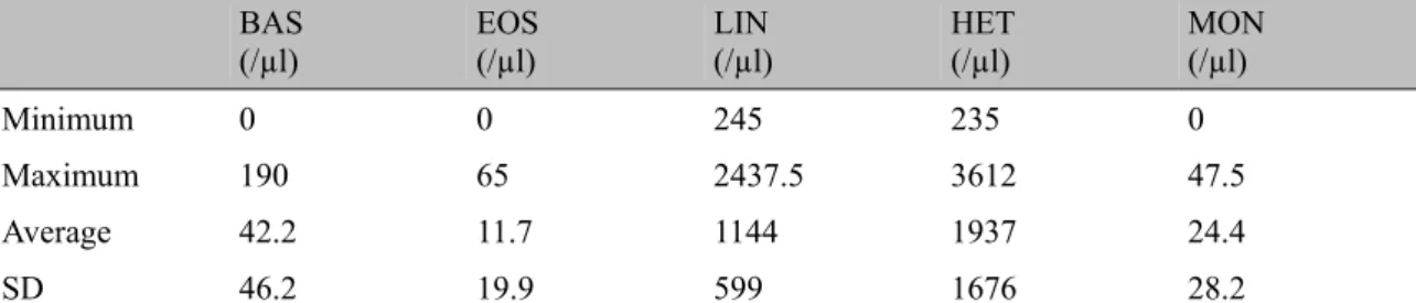
Table 3. BAS of the absolute value limits, EOS, LIN, HET, MON, Means and Standard Deviations (SD) of red-browed parrot (Amazona rhodocorytha)
BAS
(/µl) EOS (/µl) LIN (/µl) HET (/µl) MON (/µl)
Minimum 0 0 245 235 0
Maximum 190 65 2437.5 3612 47.5
Average 42.2 11.7 1144 1937 24.4
SD 46.2 19.9 599 1676 28.2
Subtitles and Units: BAS - Absolute values of Basophils (/ microl); EOS - Absolute values of eosinophils (/microl); Het Absolute values Heterophile (/ microl); LIN - Absolute values of lymphocytes (/microl); MON - Absolute values of monocytes (/microl).
DISCUSSION
In the study, venous blood samples were obtained by venipuncture of the brachial vein, which has proven effective for its ease of access, visualization, and vein tourniquet, the procedure was performed quickly and safely, generating a less stressful effect on birds. The easy viewing and identification of the vessel is confirmed by Gonçalves (2010).
collection is important to decrease the maximum stress generated in the bird, avoiding the increased level of heterophile and a decrease in lymphocytes, caused by arising glucose in these circumstances. The authors also report the importance of keeping the chest and the abdomen of the bird free, in order to avoid circulatory collapse compounded by compression. Weiss and Wardrop (2012), report on the attention that handlers must submit, with claws and beak of birds, since these are the animal's defenses and can cause accidents.
The volume of blood collected, 0.5mL, was enough to practice all procedures necessary for the realization of complete blood count, in addition to maintaining the safety of the bird. Trhall et al. (2006), reports that a safe amount of blood that can be taken from a healthy bird is equal to 1% of their body weight.
Tarcitano (2010), in his work, reports that the low volume of blood that can be reaped from the birds usually do not correspond to the anticoagulant volume found in most tubes, which are marketed, resulting hemodilution. The use of EDTA microtubes with a maximum of 0.5mL capacity, was satisfactory to prevent the occurrence of such misrepresentation.
The mean interval of 6 hours, at which the sample was being cooled in an ice chest with recycled ice until its manipulation, represented insufficient time to bring harm to laboratory tests performed. Trhall et al. (2006) shows that after 24 to 48 hours of EDTA storage, there is an evidence of an increase in cell volume, mean corpuscular volume and erythrocyte lineage cell count.
For each bird, two smears were performed, one with fresh blood at the time of gathering and in the laboratory that was carried out the moment the blood was processed. Both smears were efficient for the realization of leukocyte differentiation and morphological assessment, however, the smears performed in the laboratory indicate the occurrence of toxic heterophils. Tarcitano (2010), recommends that a smear be done with fresh blood without contact with anticoagulant, preventing the occurrence of a cellular morphological change.
The cell staining process used in this study followed the method of May-Grunwald-Giemsa (MGG), which is a combination of two dyes, the May-Grunwald and Giemsa Blood used in poultry, as described by Trhall et al. (2006). The methodology proved to be very efficient in the display and coloration of blood cells. On the work of Tarcitano (2010), he used this method for staining blood cell Cockatiels (cockatiel).
The CBC is an essential tool to assist in the clinical evaluation of red-browed parrot, however, it only becomes usable when there are reliable parameters of their physiology. To be specific, parameters such as the assessment of extrinsic factors is required, which may lead to changes in hematology results of the animals. Examples of factors that can influence the change of these parameters are the health and physical condition of the bird, physical activity performed and nutritional status (Goulart, 2006).
When searching the literature, one can see the lack of information on the complete hematology of Amazona rhodocorytha, raising the need to use values adopted for other species belonging to the same genus or family Psittacidae. The few existing studies adopt a few criteria for data analysis, and are often incomplete, still having those that address only a few variables. An omission in the description of the methods used results in the loss of confidence in the results.
The hematological parameters analyzed in this study were established from a single set of standardized animals, all adult, clinically healthy, and without sexual distinction. In a study by Tarcitano (2010), comparing the blood profile of cockatiels with adult offspring, it was observed that the group of puppies showed average values for erythrocyte, trombocitogram and total plasma proteins slightly smaller than adult animals and, as in the WBC, the results remained more homogeneous. Goulart (2006), in his work with Blue-fronted Parrot (Amazona aestiva) found no significant difference in haematological values between males and females.
use other species of the same genus, for all the results were discussed.
The average values of total red blood cells, hematocrit or VG, VCM, and thrombocytes in the present study were higher than those found by the Goulart (2006), while the hemoglobin, HCM, and MCHC are found at lower levels. The average values of VG and PPT found were also higher than those found by Fonseca et al. (2008).
Fonseca et al. (2008) determined its eosinophil and monocyte value greater than the values found in this work, however, the total WBC values, heterophils, and lymphocytes were lower, with no reference to basophils values.
The differences may be due to the fact it is a different species, which is a constituent of the variable blood count, and the opportunity due to the technique used, since fast Panotic dye was used to stain the slides, in the case of Fonseca’s et al. (2008) work.
In the assessment of the hematological values between males and females a statistical significant difference was observed for all the parameters, as well as the labor Fonseca et al. (2008), for hematocrit, total plasma protein and white blood cell count. In his work, Goulart (2006), found that only basophils had significant difference, indicating that females have approximately the double amount compared to males.
CONCLUSION
Haematological values found through research are adequate to establish the physiological benchmarks for the proposed species, especially considering the animals sampled, allocated within its natural biome, with similar climatic and geographical conditions of their counterparts in nature.
Standardization in the method of obtaining, storage and processing of the samples is a goal to be pursued to reduce extrinsic differences in values and thus, without clinical significance. Likewise, the closer two species are, significant differences demonstrate the fragility of these comparisons.
Yet it can be concluded that the continuous search for knowledge, with regard to Brazilian species, especially those within easy reach, such as captive collections are essential to the process of conservation of the species in question, and therefore a challenge for constant updating and discoveries for veterinary medicine in its social and environmental character.
REFERENCES
BAHIENSE, C.R. Determinação de parâmetros hematológicos e bioquímicos de arara Canindé (Ara ararauna), no estado do Rio de Janeiro. 2010. 42f. Dissertação (Pós-Graduação em Medicina Veterinária – Ciências Clínicas) – Instituto de Veterinária, Universidade Federal Rural do Rio de Janeiro, Seropédica, RJ
BIRDLIFE international. Species factsheet: Amazona rhodocorytha. Diponível em: <Species factsheet>. Acessado em: 02 de jun. 2012. CUBAS, Z.S.; SILVA, J.C.R; CATÂO-DIAS, J.L. Tratado de animais selvagens: medicina veterinária. São Paulo: Roca, 2006. 1354p. FONSECA L.A.; GIRARDI F.M.; MAIA N.L. et al. Determinação do hematócrito, leucograma e proteínas plasmáticas em chauás (Amazona rhodocorytha) criados em cativeiro. In: CONGRESSO DA ASSOCIAÇÃO BRASILEIRA DE VETERINÁRIOS DE ANIMAIS SELVAGENS, 11.; ENCONTRO DA ASSOCIAÇÃO BRASILEIRA DE VETERINÁRIOS DE ANIMAIS SELVAGENS, 17., 2008, Santos. Anais... Santos: ABRAVAS, 2008. p. 260-263.
GONÇALVES, G.A.M. Manual de emergência em aves. São Paulo: MedVet, 2010. 84 p.
GOULART, C.E.S. Valores hematológicos de referência para papagaios-verdadeiros (Amazona aestiva – psittacidae) mantidos em cativeiro. 2006. 80f. Dissertação (Mestrado em Medicina veterinária) – Escola de Veterinária, Universidade Federal de Minas Gerais, Belo Horizonte, MG.
LISTA nacional das espécies da fauna brasileira ameaçadas de extinção. SEAMA, 2003. Diponível em: <http://www.meioambiente. es.gov.br/download/NovaListaFaunaAmeacaMM A2003.pdf>. Acessado em: 02 de jun. 2012. TARCITANO, C.F. Hemograma de Calopsitas (Nymphicus hollandicus) criadas no estado do Rio de Janeiro. 2010. 42f. Dissertação (Pós-Graduação em Medicina Veterinária – Patologia e Ciências Clínicas) – Instituto de Veterinária, Universidade Federal Rural do Rio de Janeiro, Seropédica, RJ.
THRALL, M.A. Hemantologia e bioquímica clínica veterinária. São Paulo: Roca, 2006. 592p. TULLY, T.N.; DORRESTEIN, G.M.; JONES, A.K. Clínica de aves. 2.ed. Rio de Janeiro: Elsevier, 2010. 322 p.