(Annals of the Brazilian Academy of Sciences)
Printed version ISSN 0001-3765 / Online version ISSN 1678-2690 www.scielo.br/aabc
http://dx.doi.org/10.1590/0001-3765201520140069
Plasma nitric oxide, endothelin-1, arginase and superoxide dismutase in the plasma and placentae from preeclamptic patients
FABIANA C. BERNARDI1, FRANCIELI VUOLO1, FABRICIA PETRONILHO2, MONIQUE MICHELS2, CRISTIANE RITTER1 and FELIPE DAL-PIZZOL1
1
Programa de Pós-Graduação em Ciências da Saúde, Laboratório de Fisiopatologia Experimental, Universidade do Extremo Sul Catarinense, Avenida Universitária, 1105, 88006-000 Criciúma, SC, Brasil
2Programa de Pós-Graduação em Ciências da Saúde, Laboratório de Fisiopatologia Clínica e Experimental,
Universidade do Sul de Santa Catarina, Avenida José Acácio Moreira, 88704-201 Tubarão, SC, Brasil
Manuscript received on February 2, 2014; accepted for publication on August 25, 2014
ABSTRACT
The aim of this study was to determine parameters of NO metabolism in plasma and placenta of preeclamptic (PE) patients. It was conducted a case-control study at São José Hospital, Brazil. Thirty-three PE and 33 normotensive pregnant were included in the study. The diagnosis of PE was established in accordance with the definitions of American College of Obstetricians and Gynecologists. Peripheral venous blood and placenta samples were obtained at postpartum period. Plasma NO levels and SOD activity were significantly lower and endothelin-1 levels and arginase activity were significantly higher in PE women when compared to controls. None of the analyzed parameters were different in the placenta between groups. Our findings suggest that parameters associated with NO metabolism are altered only at the systemic level, but not in placenta of PE patients.
Key words: preeclampsia, superoxide dismutase, arginase activity, endothelin-1 levels, nitrite/nitrate.
Correspondence to: Felipe Dal-Pizzol E-mail: piz@unesc.net
INTRODUCTION
Preeclampsia (PE) is a hypertensive and multiple-system disorder that occurs during human pregnancy. It has an unknown etiology, and is
defined by an onset of proteinuria, hypertension
and edema after 20 weeks of gestation (Wagner 2004). PE affects about 2 to 3% of all pregnancies and it is an important cause of maternal death and the leading cause of intrauterine growth restriction (Redman and Sargent 2005). In PE it is described
abnormalities of fetoplacental blood flow, which
are characterized by abnormal umbilical blood
mediators, such as endothelin-1 and thromboxane; and vasodilators mediators such as prostacyclin and nitric oxide (NO). Endothelial injury induces an imbalance between these substances resulting in vasospasm. One of the proposed mechanisms responsible to endothelial damage is oxidative stress (Hubel et al. 1989).
Compelling evidence supports the understan-ding that endothelial activation/dysfunction and increased oxidative stress are a central pathophysiological event in the maternal vascular system in PE (Gu et al. 2006). The steady-state levels of NO depend on several factors, including arginine concentration, nitric oxide synthase (NOS) activity and superoxide anion concentration (Lowe 2000). Lower levels of SOD activity also impede the effects of NO on blood pressure regulation, by allowing it to react with superoxide anion forming peroxynitrite, thereby increasing oxidative stress (Rumiris et al. 2006). Arginase is a key enzyme responsible to the metabolism of nitrogen since it regulates the levels of L-arginine. Thus, an increased activity of arginase could decrease the production of NO (Ash 2004, Xia et al. 1996). Furthermore, there is limited evidence of the occurrence of these alterations in patients with PE, and if they are systemic or limited to the placenta.
In this context, the present study was designed to measure SOD and arginase activities, nitrite/ nitrate (Nox) and endothelin-1 content in plasma and placenta of normal and PE patients.
MATERIAL AND METHODS
PATIENTS
The Ethics Committee of São José Hospital approved study protocol, and informed consent was obtained from all participants. The study population consisted of 33 consecutive PE patients and 33 normotensive pregnant admitted to cesarean delivery at the Obstetrics and Gynecology Department between January 31st and December 31st 2009. The diagnosis of PE was established
in accordance with the definitions of American
College of Obstetricians and Gynecologists (ACOG criteria practice bulletin 2002). Women with ruptured membranes, multiple pregnancy, medical complications (including autoimmune
disorders, diabetes mellitus), inflammatory
conditions (including chorioamnionitis) and cases of chronic hypertension with superimposed PE were excluded. Control patients were matched to PE according to maternal age and gestational age at birth. Peripheral venous blood samples were obtained into heparinized vacutainer tubes, approximately at the same time of placenta collection. The maternal section of the placenta was obtained in the postpartum period and it was immediately washed with saline solution to remove blood cells. All biological specimens were conditioned in liquid nitrogen and then stored at -80°C until assayed.
NITRITE/NITRATE CONCENTRATION
Total concentration of nitrite was determined in
blood samples and placenta by a modified Griess
reaction method (Verdon et al. 1995). Samples were incubated for 3h at 20°C with
glucose-6-phosphate (500 μmol/L), glucose-6-glucose-6-phosphate dehydrogenase (160 U/L), NADPH (1 μmol/L)
and nitrate reductase (20 U/L) in phosphate buffer (80 mmol/L, pH 7.5). The Griess reaction was then initiated by addition of sulfanilamide
to a final concentration of 0.5% (wt/vol),
orthophosphoric acid (1.25%, vol/vol), and N-(1 naphthyl) ethylenediamine hydrochloride (0.05%, wt/vol). After a further incubation at 20°C for 10 min, the absorbance of each sample mixture was measured at 540 nm and corrected for opacity by measuring the absorbance at 750 nm. The corrected absorbance was interpolated in a standard curve of absorbance plotted versus concentration, in order
to find the concentration of nitrite in the sample.
ARGINASE ACTIVITY
Arginase activity was measured as previously
described (Loeb and Stuhlman 1969). Briefly,
biological samples were incubated with 50 mM Tris-HCl, 10 mM MnCl2 and the enzyme was activated by heating for 10 min at 56°C. Arginine hydrolysis was initiated by the addition of 0.5 M L-arginine, pH 9.7, at 37°C for 45 min. The reaction was stopped with H2SO4 (96%)/H3PO4 (85%)/H2O (1/3/7,v/v/v). The urea concentration was measured at 540 nm
after addition of 25 mL α-isonitrosopropiophenone
(dissolved in 100% ethanol) followed by heating at 95°C for 30 min. Arginase activity was expressed as µM arginine/L/h (13).
SUPEROXIDE DISMUTASE (SOD)ACTIVITY
The activity of SOD was assayed by the epinephrine method based on the capacity of SOD to inhibit autoxidation of adrenaline to adrenochrome. One
unit of SOD activity was defined as the amount of
protein causing 50% inhibition of the autoxidation of adrenaline at 26°C, as previously described (Bannister 1987).
ENDOTHELIN-1CONTENT (ET-1)
Concentrations of endothelin-1 were determined by a standard sandwich ELISA, employing comercial available Kits (R&D Systems, Minneapolis, MN) according to manufacturer recommendation.
STATISTICAL ANALYSES
Standard descriptive statistics were calculated to examine baseline characteristics of the study population. Continuous variables with normal distribution were presented as mean ± standard deviation and compared by t-Student test. Continuous variables with a non-normal distribution were reported as median (25%-75% interquartile range) and compared using Mann-Whitney U test. Categorical variables were presented as absolute numbers (frequency percentages) and analyzed by Chi-square test. Correlations were performed by
the Pearson test. It was calculated a sample size of at least 25 patients for each group to detect a difference between groups of 20% (predicting a standard deviation of 25%), a sigma of 0.05 and power of 0.80. A two-tailed p-value < 0.05 was
considered statistically significant.
RESULTS
PATIENTS
Clinical characteristics of included patients were
shown in Table I. Arginase activity was significantly
higher in the plasma of PE group when compared to the control group (p<0.01); but it did not differ
significantly in the placenta (Figure 1). This same
pattern was observed for ET-1 plasma levels (Figure 2). It was demonstrated that the levels of plasma
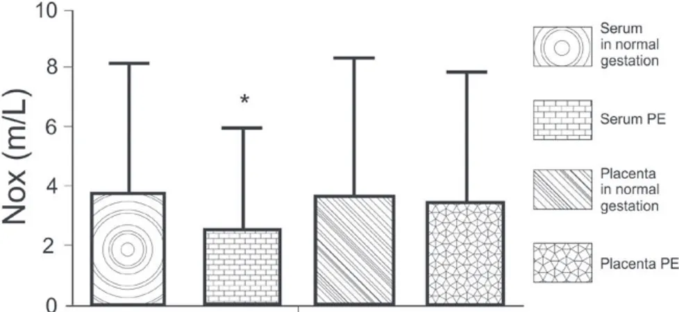
NO were significantly lower in PE when compared
to control patients (p<0.01), but there were no
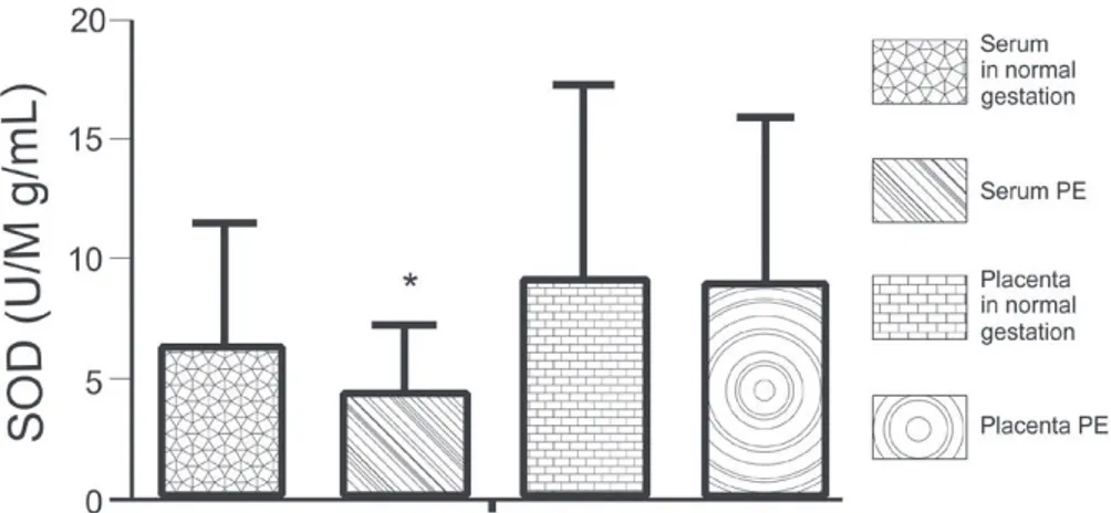
significant differences in the placenta (Figure 3). In addition, SOD activity was significantly lower
in PE group when compared to controls in the plasma (p<0.01), but not in the placenta (Figure 4).
Furthermore, there were no significant correlation
between plasma and placenta levels for any of the measured parameters (data not shown).
Preeclampsia (n=33)
Normal gestation (n=33) p-values
Age (years) 25 ± 6 27 ± 7 0.35
Parity (n) 2.3 ± 0.7 1.9 ± 0.8 0.25 Gestational
age at delivery (weeks ± days)
37.6 ± 8.5 36.1 ± 8.4 0.42 Sistolic blood
pressure (mmHg)
150 ± 12 115 ± 9 0.03
Diastolic blood pressure (mmHg)
98 ± 5 73 ± 4 0.002
Pre-pregnancy
BMI 23 ± 6 22 ± 6 0.40
Birth weight (g) 2495 ± 500 2948 ± 530 0.02
TABLE I
Major clinical characteristics of women with normotensive pregnancy
and preeclampsia.
Figure 1 - Arginase activity in serum and placenta in normal gestation and preeclamptic patients. *p<0.01 compared to normal gestation group.
Figure 2 - Endothelin concentration in serum and placenta in normal gestation and preeclamptic patients. *p<0.01 compared to normal gestation group.
DISCUSSION
We here demonstrated that several different parameters related to NO metabolism were altered in the plasma of PE patients, but differently from what we supposed these same parameters were not altered in the placenta of PE when compared to normal gestation.
PE is a major cause of perinataland maternal morbidity and mortality. PE is characterized by
abnor-malities in fetoplacental blood flow. Interestingly,
during PE plasmanitrate concentrations, which are an index of NO synthesis,are only slightly reduced (Verdon et al. 1995), as we here demonstrated. This is not uniformly demonstrated in the literature. There are reports of normal and even increased NO levels in PE patients, but a recent meta-analyses suggests that in overall there is a decrease in NO levels in PE patients (Dai et al. 2013). These results must be interpreted
with caution since there is significant heterogeneity
between included studies.
Thus, it is possible that placenta could better
reflects the alterations of NO metabolism, but we could not determine any significant differences in
several parameters involved in NO metabolism levels in PE’s placenta. The relative concentration of NO in placenta depends on its siteand rate of synthesis, its half-life, and its site of action.The interaction of
NO with superoxide anion causes its inactivation. Conversely, the activity of NO is prolonged in the presenceof SOD, which removes superoxide anions (Loeb and Stuhlman 1969, Bannister 1987). We here demonstrated that NO levels were reduced in plasma of PE women when compared to normal controls, but there was no difference in the placenta as well as SOD and arginase activities were unaltered reinforcing the lack of alterations in the metabolism of NO.
The expression of arginase was increased in placenta of PE patients (Sankaralingam et al. 2010). Our study examined arginase I and observed an increased activity in the plasma, but not placenta, in patients with PE. Arginase exists in two isoforms, arginase I (cytosolic) and arginase II (mitochondrial). The primary function of arginase I and II in the liver and kidney, respectively, are to regulate urea metabolism. In the vasculature, arginase I is expressed both in vascular smooth muscle cells and the endothelium, while arginase II is highly expressed in the endothelium. Thus, it is possible to speculate that arginase is involved in the regulation of the systemic, but not placental, levels of NO during PE.
Clinical evidence from human studies indicates that endothelin plays an important role in mediating pathophysiological changes that occur during PE. Higher ET-1 plasma concentrations were found
in patients with PE (Myatt et al. 1996). Typically, these levels are highest during the latter stage of the disease, suggesting that ET-1 may not be involved in the initiation of PE, but in the progression of disease into a malignant phase (LaMarca et al. 2005). Previous studies have been reported that
ET-1 plasma levels can have significant long-term
effects on systemic hemodynamics and arterial pressure regulation. Thus, long-term elevations in plasma levels of ET-1 comparable to those measured in women with PE could play a role in mediating the reductions in renal function and elevations of arterial pressure observed in women with PE (Myatt et al. 1996). We observed increase in plasmatic endothelin of patients with PE, but not
in placenta, and this suggest that at final stages of the
disease systemic endothelial, rather than placental, is relevant to the development of the symptoms.
Some limitations of our study must be pointed-out. As we collected a single time point of biological samples, it is not possible to ascertain a cause-effect relationship between alterations in SOD, arginase and nitrite levels. In addition, due to this limitation is possible that placental NO metabolism is altered at earlier times during PE. Thus, at these later time points we are just able to determine a systemic effect of the presence of PE, but not an earlier pathophysiologic placental alteration.
CONCLUSIONS
Our findings suggest that parameters associated
with NO metabolism are altered only at the systemic level, but not in placenta of PE patients. These data suggest that, despite placenta is thought to have a major role in the pathophysiology of PE, some alterations could be restricted to the “systemic compartment”, or only at earlier times that we could not access due to limitations of our sample.
ACKNOWLEDGMENTS
We would like to thank Coordenação de Aperfei-çoamento de Pessoal de Nível Superior (CAPES) and
Conselho Nacional de Desenvolvimento Científico e Tecnológico (CNPq) for financial support.
RESUMO
O objetivo deste estudo foi determinar parâmetros de metabolismo de NO no plasma e placenta de pacientes com pré-eclâmpsia (PE). Foi realizado um estudo de caso-controle no Hospital São José, Brasil. Trinta e três PE e 33 gestantes normotensas foram incluídas no estudo. O diagnóstico de PE foi estabelecido em conformidade com as definições do Colégio Americano de Obstetras e Ginecologistas. Amostras de sangue venoso periférico e placenta foram obtidos no período de pós-parto. Os níveis plasmáticos de NO e atividade SOD foram significativamente menores e os níveis de endotelina-1 e da atividade da arginase foram significativamente maiores em mulheres PE quando comparados aos controles. Nenhum dos parâmetros analisados foram diferentes na placenta entre os grupos. Nossos resultados sugerem que os parâmetros associados ao metabolismo do NO estão alterados apenas no nível sistêmico, mas não na placenta de pacientes PE.
Palavras-chave: pré-eclâmpsia, superoxido dismutase, atividade da arginase, níveis de endotelina-1, nitrito/nitrato.
REFERENCES
ACOG - AMERICAN COLLEGE OF OBSTETRICIANS AND
GYNECOLOGISTS CRITERIA PRACTICE BULLETIN. 2002.
Diagnosis and management of preeclampsia and eclampsia. Obstet Gynecol 99: 159-167.
ASH DE. 2004. Structure and function of arginases. J Nutr 134(10 Suppl): 2760S-2764S.
BANNISTER JV. 1987. Calabrese L. Assays for superoxide
dismutase. Methods Biochem Anal 32: 279-312.
DAI B, LIU T, ZHANG B, ZHANG X AND WANG Z. 2013. The
polymorphism for endothelial nitric oxide synthase gene, the level of nitric oxide and the risk for pre-eclampsia: a meta-analysis. Gene 519: 187-193.
GU Y, LEWIS DF, ZHANG Y, GROOME LJ AND WANG Y. 2006.
Increased Superoxide Generation and Decreased stress Protein Hsp90 Expression in Human Umbilical Cord Vein Endothelial Cells (HUVECs) from Pregnancies Complicated by PE. Hypertens Pregnancy 25: 169-182.
HUBEL CA, ROBERTS JM, TAYLOR RN, MUSCI TJ, ROGERS
GM AND MCLAUGHLIN MK. 1989. Lipid peroxidation in
KIM YJ. 2006. Reduced L-arginine Level and Decrease Placental eNOS Activity in PE. Placenta 27: 438-444.
LAMARCA BB, COCKRELL K, SULLIVAN E, BENNETT W AND
GRANGER JP. 2005. Role of endothelin in mediating tumor
necrosis factor-induced hypertension in pregnant rats. Hypertension 46: 82-86.
LOEB WF AND STUHLMAN RA. 1969. A colorimetric method
for the determination of serum arginase activity. Clin Chem 15: 162-169.
LOWE DT. 2000. Nitric Oxide Disfuncion in the pathophysiology of PE. Nitric Oxide 4: 441-458.
MYATT L, ROSENFIELD RB, EIS ALW, BROCKMAN DE, GREER
I AND LYALL F. 1996. Nitrotyrosine Residues in Placenta.
Hypertension 28: 488-493.
OKUMURA KK, SAGAWA N, KOBAYASHI F, NANNO H,
MATSUMOTO T, ITOH H, KORITA D, TANADA S AND
MORI T. 1999. Activity of platelet-activating-factor-acetylhydrolase and the nitric oxide metabolite level in the plasma of pregnant women who develop transient hypertension during later pregnancy. Reprod Fertil Dev 11(2): 75-79.
REDMAN CW AND SARGENT IL. 2005. Latest advances in
understanding PE. Science 10: 1592-1594.
RUMIRIS D, PURWOSUNU Y, WIBOWO N, FARINA A AND
SEKIZAWA A. 2006. Lower Rate of PEAfter Antioxidant
Supplementation in Pregnant Women with Low Antioxidant Status. Hypertens Pregnancy 25: 241-253.
SANKARALINGAM S, XU H AND DAVIDGE ST. 2010. Arginase
contributes to endothelial cell oxidative stress in response to plasma from women with PE. Cardiovasc Res 85: 194-203.
VERDON CP, BURTON BA AND PRIOR RL. 1995. Sample
pre-treatment with nitrate reductase and glucose-6-phosphate dehydrogenase quantitatively reduces nitrate while avoi-ding interference by NADP+ when the Griess reaction is used to assay for nitrite. Anal Biochem 224: 502-508.
WAGNER LK. 2004. Diagnosis and management of PE. Am
Fam Physican 70: 2317-2324.
WANG Y, LEWIS DF, ALEXANDER JS AND GRANGER DN.
2007. Endothelial barrier function in PE. Front Biosci 12: 2412-2424.
XIA Y, DAWSON VL, DAWSON TM, SNYDER SH AND ZWEIER