UNIVERSIDADE FEDERAL DE MINAS GERAIS FACULDADE DE FARMÁCIA
PROGRAMA DE PÓS-GRADUAÇÃO EM CIÊNCIAS FARMACÊUTICAS
MELINA DE BARROS PINHEIRO
PRÉ-ECLÂMPSIA: INTER-RELAÇÃO DOS SISTEMAS
HEMOSTÁTICO E INFLAMATÓRIO
PRÉ-ECLÂMPSIA: INTER-RELAÇÃO DOS SISTEMAS
HEMOSTÁTICO E INFLAMATÓRIO
Tese submetida ao Programa de
Pós-Graduação em Ciências Farmacêuticas da Faculdade de Farmácia da Universidade Federal de Minas Gerais, como requisito parcial, para
obter o grau de doutor em Ciências
Farmacêuticas.
Orientadora: Profª. Drª. Luci Maria S. Dusse
Co-orientadora: Profª. Drª Karina Braga G. Borges Co-orientador: Profo. Dro Olindo Assis M. Filho
Pinheiro, Melina de Barros.
B277p Pré-eclâmpsia: inter-relação dos sistemas hemostático e
inflamatório / Melina de Barros Pinheiro. – 2012. 129 f. : il.
Orientadora: Profª. Drª. Luci Maria Sant’Ana Dusse.
Coorientadores: Profa . Karina Braga G. Borges, Prof. Dr. Olindo
Assis Martins Filho.
Tese (doutorado) - Universidade Federal de Minas Gerais, Faculdade de Farmácia, Programa de Pós-Graduação em Ciências Farmacêuticas.
1. Pré-eclâmpsia – Teses. 2. Hemostasia – Teses. 3. Fibrinólise – Teses. 4. Inflamação – Teses. 5. Citocinas – Teses. I. Dusse, Luci Maria Sant’Ana. II. Gomes, Karina Braga. III. Martins Filho, Olindo Assis. IV. Universidade Federal de Minas Gerais. Faculdade de Farmácia. V. Título.
Dedico este trabalho
A Deus, por me abençoar e iluminar todos os dias de minha vida.
Aos meus queridos orientadores Profa Luci, Profa Karina e Dr. Olindo,
A Deus, pela incrível tarefa da criação e pela alegria de viver que me foi concedida. Por sua luz, que me ilumina, em todos os momentos de minha vida.
A querida professora Luci Dusse, muito obrigada pela dedicação, carinho e atenção. Obrigada por sempre acreditar e confiar em mim. Você é a principal responsável por essa vitória. Agradeço pela oportunidade de convívio ao lado de uma pessoa tão iluminada como você. Obrigado por tornar todos os momentos deste trabalho um constante aprendizado. Muito obrigada por tudo!
A querida profa. Karina Braga, pela colaboração, carinho, ensinamentos e principalmente amizade. Admiro sua sabedoria e seu profissionalismo . A sua competência e humildade fazem com que você se transforme em um exemplo para todos que convivem ao seu redor. Muito obrigada!
Ao Dr. Olindo A. Martins Filho pela colaboração, dedicação e ensinamentos essenciais no desenvolvimento deste trabalho. Muito obrigada.
As minhas colegas e parceiras de coleta Lara, Patrícia, Fabiana e Letícia pela fundamental ajuda, apoio e incentivo. Sem a colaboração de vocês a realização desse trabalho não seria possível.
A equipe do laboratório Simile Imunologia Aplicada, em especial a Dra. Carla e a Gabrielle Guimarães pela calorosa acolhida e fundamental colaboração em meus experimentos.
Ao pessoal do laboratório de Biomarcadores de Diagnóstico e Monitorização –
Centro de Pesquisa René Rachou – FIOCRUZ, em especial Dra. Andrea Teixeira Carvalho e Amanda Silveira, pela colaboração e grande ajuda no desenvolvimento dessa tese.
Aos amigos do Laboratório de Hematologia Clínica da Faculdade de Farmácia da UFMG pelo convivência, apoio e amizade.
Aos professores, colegas e funcionários da pós-graduação da Faculdade de Farmácia da UFMG que acompanharam e apoiaram o desenvolvimento deste projeto.
A bibliotecária da Universidade de São Paulo (USP) Riberião Preto, Maria Cristina M. Ferreira, pela ajuda fundamental em nossos trabalhos de revisão sistemática e metanálise.
Aos meus pais e meus irmãos Welbinho e Marina, que me apoiaram em todos os momentos e sempre confiaram em mim. Muito obrigada pelo amor, carinho e confiança.
dedicação.
Ao Luis, por ser tão especial e estar sempre ao meu lado me acalmando, torcendo e incentivando. Muito obrigada pelo amor e compreensão.
As minhas queridas amigas Soraya, Larissa e Pollyana, pelo incentivo, amizade e motivação nos momentos mais difíceis. Muito obrigada por estarem presentes em minha vida.
As mulheres que contribuíram voluntariamente com este estudo, sem vocês não seria possível a realização deste trabalho.
A equipe médica e de enfermagem da Maternidade Odete Valadares, Hospital Público Regional de Betim, Unidade Básica de Saúde da Família (UBSF) Guanabara / Betim, Hospital Municipal Odilon Behrens e Santa Casa de Belo Horizonte pela parceria tão fundamental para a realização desse trabalho.
A todos os professores e mestres que passaram pela minha vida, pelos ensinamentos e dedicação.
A pré-eclâmpsia (PE) é uma doença multifatorial, caracterizada por
hipertensão e proteinúria após a 20ª semana de gestação. A etiologia da PE ainda
não é conhecida e a doença ocorre somente na presença da placenta. Clinicamente
é importante diagnosticar a forma grave da doença, na qual a pressão arterial e a
proteinúria estão ainda mais elevadas. A PE está associada à disfunção vascular,
bem como à exacerbação da coagulação, ainda mais acentuada que aquela
observada nas gestantes normotensas. O envolvimento do sistema imune na
patogênese da PE é bem aceito e essa doença está associada a um estado
inflamatório exagerado. Diversos polimorfismos nos genes de citocinas
pró-inflamatórias parecem estar associados ao desenvolvimento da PE. Sabe-se que
componentes de sistema hemostático são capazes de ativar o sistema inflamatório e
vice-versa. Dessa forma, o objetivo desse estudo foi investigar a inter-relação dos
sistemas hemostático e inflamatório na PE grave, por meio da determinação dos
níveis plasmáticos de marcadores hemostáticos e citocinas, bem como avaliar a
relação de polimorfismos nos genes das citocinas e a ocorrência de PE. Foram
avaliadas 331 mulheres, sendo 108 mulheres não gestantes, 107 gestantes
normotensas e 116 gestantes com PE forma grave. A PE grave foi definida por
pressão arterial ≥160/110mmHg e proteinúria > 2 gL-1. Os níveis plasmáticos de
PAI-1 e D-Di foram determinados por ELISA (Kit IMUBIND® PLASMA PAI-1 e Kit
IMUCLONE® D-Dimer American Diagnostica® Inc., Stamford, USA ,
respectivamente). As citocinas IL-8, IL-6, IL-1β, TNF-α, IL-12, IFN-γ, 4, 5 e
IL-10 foram determinadas por citometria de fluxo (Cytometric Beads Array – CBA; BD
Biosciences Pharmingen, USA). A determinação dos polimorfismos nos genes das
citocinas IL-6, IL-10, IFN-γ e TNF-α foi feita por PCR-SSP (Cytokine Genotyping
Tray; One Lambda, Inc. Canoga Park, CA). Os dados obtidos neste estudo permitem
concluir que os marcadores plasmáticos da coagulação/fibrinólise e as citocinas
inflamatórias IL-6, IL-8 e IFN-γ estão elevados na PE grave e não há correlação forte
entre os mesmos; a PE grave está associada a maior frequência do genótipo T/T no
gene IFN-γ (+874) e esse genótipo determina o aumento desta citocina, enquanto os
revisão sistemática e metanálise investigando os níveis de D-Di na PE, revelaram que esse marcador é um candidato promissor para a monitoração da PE.
Preeclampsia (PE) is a multifactorial disease characterized by hypertension and
proteinuria after 20 weeks of gestation. The PE etiology is not known yet, and the
disease occurs only in the presence of the placenta. Clinically it is important to
diagnose the severe form of the disease, in which blood pressure and proteinuria are
even higher. PE is associated with vascular dysfunction, as well as to exacerbation of
coagulation, which is higher than those observed in normotensive pregnant women.
The involvement of the immune system in the PE pathogenesis is well accepted and
this disease is associated with a high inflammatory condition. Several polymorphisms
in the genes of pro-inflammatory cytokines appear to be associated with PE
occurrence. It is known that components of the hemostatic system are able to
activate the inflammatory system and vice versa. Thus, the aim of this study was to
investigate the relationship between hemostatic and inflammatory systems in severe
PE, by determining plasma levels of hemostatic markers and cytokines, as well as
evaluating the relationship of polymorphisms in cytokine genes and the PE
occurrence. A total of 331 women were evaluated (108 non-pregnant women, 107
normotensive pregnant women, and 116 pregnant women with severe PE). Severe
PE was defined as blood pressure ≥ 160/110mmHg and proteinuria > 2 g L-1. PAI-1
and D-Di Plasma levels were measured by ELISA (Kit IMUBIND® PLASMA PAI-1
and IMUCLONE® Kit D-Dimer American Diagnostica® Inc., Stamford, USA,
respectively). The cytokines IL-8, IL-6, IL-1β, TNF-α, IL-12, IFN-γ, 4, 5 and
IL-10 were determined by flow cytometry (Cytometric Beads Array - CBA; BD
Biosciences Pharmingen, USA). The determination of polymorphisms in the 6,
IL-10, IFN-γ and TNF-α genes was performed by PCR-SSP (Cytokine Genotyping Tray;
One Lambda, Inc. Canoga Park, CA). The data obtained in this study indicate that
plasma markers of coagulation/fibrinolysis and inflammatory cytokines IL-6, IL-8 and
IFN-γ are elevated in severe PE and there is not a strong correlation between them.
Furthermore, severe PE is associated with high frequency of T/T genotype in IFN-γ
gene (+874) and this genotype determines the increase of this cytokine, while the
other polymorphisms do not exert any role in this disease. The systematic review and
meta-analysis investigating the D-Di levels in PE revealed that this marker is a
α2-AP α2-antiplasmin
α2-M α2-macroglobulin
ACOG American College of Obstetricians and Gynecologists
ALT Alanine aminotransferase
APC Activated protein C
AST Aspartate amino transferase
CBA Cytometric Bead Array
CID Coagulação intravascular disseminada
COX-2 Cyclooxygenase-2
D-Di Dímero-D / D-Dimer
ELISA Enzyme-linked immunosorbent assay
FVII Factor VII
HELLP Haemolysis, elevated liver enzyme activity, low platelets
IFN-γ Interferon do tipo gama
IL Interleucina
MCP-1 Monocyte chemoattractant protein-1
MPs Microparticles
NF-κB Transcription factor κB
NO Nitric oxide
O2- Superoxide anion
PAI-1 Inibidor do ativador de plasminogênio do tipo 1 / Plasminogen activator
inhibitor type 1
inhibitor type 2
PARs Protease activator receptors
PBMC Peripheral blood mononuclear cells
PCR Reação em cadeia da polimerase
PE Pré-eclâmpsia / preeclampsia
RFLP Polimorfismo de tamanho de fragmentos de restrição
ROC Receiver operator characteristics
ROS Reactive oxygen species
sPE Severe preeclampsia
STBM Syncytiotrophoblast
TAFI Throbin activatable fibrinolytic inhibitor
TAT Complexo trombina-antitrombina
TCLE Termo de Consentimento Livre e Esclarecido
TF Tissue factor
TGF-β Fator transformador de crescimento beta
TNF-α Fator de necrose tumoral alfa
t-PA Tissue plasminogen activator
1 INTRODUÇÃO E RELEVÂNCIA…... 13
2 OBJETIVOS... 17
2.1Objetivo geral... 17
2.2 Objetivos específicos... 17
3 DELINEAMENTO EXPERIMENTAL... 18
4 RESULTADOS... 19
4.1 Artigos publicados... 19
4.1.1 Pre-eclampsia: Relationship between coagulation, fibrinolysis and inflammation – Clinica Chimica Acta... ... 19 4.1.2 D-dimer plasma levels in preeclampsia: a systematic review and metanalysis - Clinica Chimica Acta... 24
4.1.3 Fibrinolytic system in preeclampsia - Clinica Chimica Acta ... 29
4.2 Artigos submetidos... 36
4.2.1 Severe preeclampsia: association of genes polymorphisms and maternal cytokines production - Cytokine... 36 4.2.2 Severe Preeclampsia: Does Cytokine Network Drive To An Excessive Systemic Inflammatory State? – Clinical Immunology... geral…... 54 4.2.3 Severe Preeclampsia: How Is The Relationship Between Hemostatic And Inflammatory Parameters? - Arteriosclerosis, Thrombosis, and Vascular Biology………….……….……..…. 86
4.3 Outras publicações junto ao grupo de pesquisa... 100
4.3.1 Artigo aceito - Molecular Biology Reports... 100
4.3.2 Artigos em fase final de redação... 101
4.3.3 Resumos publicados………. 104
5.1 Limitações do estudo... 112 6 CONCLUSÕES...
…...
113 7 PERSPECTIVAS DE ESTUDOS... 114 REFERÊNCIAS BIBLIOGRÁFICAS...
…...
A pré-eclâmpsia (PE), na sua forma pura, caracteriza-se pelo aparecimento
em grávida normotensa, após a vigésima semana de gestação de hipertensão e
proteinúria. De acordo com o Working Group on High Blood Pressure in Pregnancy
(2000) (1) e o The American College of Obstetricians and Gynecologists - ACOG
Practice Bulletin (2002) (2), os parâmetros para diagnóstico da PE são hipertensão
(pressão sanguínea sistólica ≥140 mmHg ou pressão sanguínea diastólica ≥90
mmHg, em no mínimo duas ocasiões e o intervalo entre as medições não deve ser
inferior a duas horas ou superior a uma semana) e proteinúria (excreção de proteína
≥0,3 g em urina de 24 horas ou ≥30 mg/dL , ou seja, ≥+1 pelo método qualitativo de
fita, em amostras isoladas).
A etiologia da PE ainda não é conhecida e a doença ocorre somente na
presença da placenta. A PE constitui a principal causa de morte materna em
diversos países do mundo e contribui significativamente para a prematuridade, baixo
peso fetal e o aumento da mortalidade neonatal. Esta doença está associada a um
elevado custo social, uma vez que frequentemente resulta na internação da gestante
e do recém- nascido por vários dias. (3)
Um dos aspectos mais intrigantes da PE é o seu desfecho. Ainda não está
elucidado por que algumas gestantes com PE vão até o puerpério sem maiores
complicações, enquanto outras evoluem para a eclâmpsia (com surgimento de
alterações neurológicas e convulsões, que podem evoluir para o coma e morte),
síndrome HELLP (Haemolysis, elevated liver enzyme activity, low platelets) ou
coagulação intravascular disseminada (CID). (3)
Segundo os critérios estabelecidos pela American College of Obstetricians
and Gynecologists (ACOG) a PE pode ser clinicamente caracterizada nas formas
leve e grave. Na forma grave da PE, os sintomas clínicos são ainda mais
acentuados e os parâmetros para diagnóstico são hipertensão (pressão sanguínea
sistólica ≥160mmHg ou pressão sanguínea diastólica ≥110mmHg, em no mínimo
duas ocasiões e o intervalo entre as medições não deve ser inferior a seis horas ou
superior a uma semana) e proteinúria (excreção de proteína ≥5g em urina de 24
horas ou ≥+3 pelo método qualitativo de fita, em amostras isoladas, coletadas em
intervalo de no mínimo 4 horas). (2) Esta classificação tem sido amplamente
orientando a condução da gestação. Porém, de modo geral, os obstetras não
esperam a obtenção de níveis tão elevados de proteinúria, pelo risco de
complicações e morte da gestante, e é feita a interrupção da gestação.
Embora os sintomas da PE se manifestem após a vigésima semana de
gestação, atualmente tem sido aceito que a patogênese é estabelecida muito antes
e a doença ocorre em duas fases. A primeira fase se dá nas primeiras doze
semanas de gestação, quando ocorre de forma defeituosa a diferenciação dos
trofoblastos, invasão da decídua e remodelamento das artérias espiraladas. Isto
resulta na entrada abrupta do sangue materno no espaço interviloso causando dano
mecânico aos sinciciotrofoblastos, além de um suprimento irregular de sangue na
placenta, com eventos de hipoperfusão e reperfusão. (4, 5) A segunda fase ocorre
no segundo ou terceiro trimestres e resulta da hipoperfusão e isquemia placentária.
A placenta isquêmica libera citocinas e radicais livres do oxigênio que induzem a
disfunção endotelial materna sistêmica e a resposta inflamatória excessiva. (4)
O entendimento da PE como síndrome e sua diversidade de repercussões na
gestante e concepto vêm sendo investigados à luz de uma nova classificação,
baseada no momento do surgimento de manifestações clínicas. Dessa forma, a PE
é classificada como precoce ou tardia, de acordo com a idade gestacional na qual
aparecem os sintomas da doença (6). Tem sido sugerido que a PE precoce e tardia
constituem entidades distintas, que refletem o mecanismo etiopatogênico que se
manifestam em momentos diferentes da gestação. (7) A PE precoce, tem início
antes da 34ª semana de gestação, é menos frequente, mas associa-se à forma
clinicamente mais grave, refletindo lesões isquêmicas placentárias. Seu componente
genético é mais acentuado (6), há maior taxa de recorrência e seu prognóstico é
mais sombrio para a gestante e seu concepto. (7, 8) Nestes casos, a restrição do
crescimento intrauterino é mais frequente. (9) A PE tardia, tem início a partir da 34ª
semana gestacional, é a mais frequente e, em geral, é associada a uma placentação
adequada ou levemente comprometida. (6) Caracteriza-se por ausência ou leve
resistência ao fluxo nas artérias uterinas, menor comprometimento do crescimento
fetal e resultados perinatais mais favoráveis. (10)
A gestação normal está associada a elevação dos níveis de fatores da
coagulação e diminuição dos anticoagulantes naturais, o que resulta em um estado
de hipercoagulabilidade. (11-13) Esse estado constitui uma adaptação fisiológica,
fibrina na microcirculação placentária (18) e que a ativação e/ou dano das células
endoteliais parece desempenhar um papel chave na fisiopatologia da PE e
certamente contribuem para as alterações hemostáticas observadas nessa
síndrome. (19, 20)
Evidências recentes sugerem que a disfunção na angiogênese (21), bem
como alterações na tensão local de oxigênio (22, 23) e na resposta imunológica
(24-27), constituem fatores fisiopatológicos importantes na PE. O envolvimento do
sistema imune na patogênese dessa doença tem sido sugerido, principalmente pelo
contexto inflamatório observado. (24, 26-28)
O modelo de regulação imunológica durante a gravidez tem por base a
mudança da resposta imune materna para um estado pró-inflamatório modulado.
(29-31) Este modelo baseia-se na observação de que em uma mulher saudável não
gestante, a resposta imune a um antígeno dependerá, em parte, do microambiente
de citocinas. Assim, um microambiente rico em interleucina (IL) 12, IL-18 e interferon
do tipo gama (IFN-γ) irá favorecer o desenvolvimento de células pró-inflamatórias
que secretam citocinas inflamatórias, como o fator de necrose tumoral alfa (TNF-α),
IL-2 e IFN-γ. Além disso, promoverá a ativação de macrófagos e linfócitos T
citotóxicos. Por outro lado, um microambiente rico em IL-10 e IL-4 irá promover a
expansão de linfócitos regulatórios. (29-31)
Nas mulheres não gestantes, há um equilíbrio entre as respostas
pró-inflamatória e regulatória. No entanto, durante a gestação, o equilíbrio é
significativamente alterado pela presença da placenta, uma vez que progesterona e
citocinas são capazes de modular as células do sistema imunológico favorecendo o
estado regulatório. (32) Na PE, o desvio da resposta imune para o estado regulatório
provavelmente não ocorre, ou é revertido em fases muito precoces da doença.
Níveis elevados da citocina pró-inflamatória IFN-γ e reduzidos da regulatória IL-4
têm sido descritos. (33-36) Sabe-se que as citocinas pró-inflamatórias podem
provocar alterações funcionais e estruturais, incluindo danos oxidativos e
comprometimento dos mecanismos de vasoconstrição e relaxamento de vasos, o
que resulta em alterações da integridade vascular e da hemostasia. (37) No entanto,
o fator que desencadeia a resposta inflamatória excessiva na PE não é ainda
Diversos estudos têm sido realizados visando elucidar as alterações
genéticas que explicariam o desenvolvimento da PE. Estes estudos têm como
objetivos a análise de genes relacionados aos mecanismos de alterações
fisiológicas da doença, e visam definir marcadores moleculares capazes tanto de
prever o desenvolvimento da doença, como melhorar a resposta ao tratamento
clínico e farmacológico. A presença de polimorfismos em um determinado gene
pode ou não acarretar alterações funcionais. Polimorfismos funcionais em genes de
citocinas, que podem conferir diferenças interindividuais na síntese e secreção
destas proteínas, têm sido associados a doenças que têm patogênese inflamatória.
(39, 40) A investigação da associação de polimorfismos nos genes de citocinas e a
ocorrência de PE têm resultado em conclusões conflitantes (41-51), o que indica a
necessidade de estudos em outras populações.
A principal motivação para a realização deste estudo foi o maior entendimento
da inter-relação dos processos hemostático e inflamatório na PE, uma vez que
poderá contribuir para a adoção de medidas importantes na sua monitoração.
Sabendo que a PE é uma doença de caráter multifatorial e que os fatores genéticos
podem estar associados à sua ocorrência, foi também investigado neste estudo se
os polimorfismos nos genes das citocinas estariam associados à ocorrência dessa
doença no nosso meio.
Considerando a complexidade da PE, bem como das lacunas existentes na
literatura com relação à sua etiologia, diagnóstico e tratamento, este estudo se
2.1 Objetivo geral
Investigar a inter-relação dos sistemas hemostático e inflamatório na
pré-eclâmpsia grave, por meio da determinação dos níveis plasmáticos de marcadores
hemostáticos e citocinas, bem como os polimorfismos nos genes das citocinas e a
ocorrência de pré-eclâmpsia grave.
2.2 Objetivos específicos
Nos três grupos avaliados, mulheres não gestantes, gestantes
normotensas e gestantes com PE grave:
• Determinar os níveis plasmáticos dos marcadores da coagulação e
fibrinólise, D-Di e PAI-1.
• Determinar os níveis plasmáticos das citocinas IL-8, IL-6, IL-1β, TNF-α,
IL-12, IFN-γ, IL-4, IL-5 e IL-10, por citometria de fluxo.
• Correlacionar os níveis plasmáticos de D-Di e PAI-1 e de citocinas.
• Determinar a frequência dos polimorfismos nos genes das citocinas IL-6,
IL-10, IFN-γ e TNF-α.
• Estabelecer a relação entre os polimorfismos dos genes das citocinas IL-6,
IL-10, IFN-γ e TNF-α e os níveis plasmáticos dessas citocinas.
Além desses, também foi objetivo:
• Realizar uma revisão sistemática e metanálise sobre a associação dos
4.1 Artigos publicados
4.1.1 Pre-eclampsia: Relationship between coagulation, fibrinolysis and inflammation – Clinica Chimica Acta
Invited critical review
Pre-eclampsia: Relationship between coagulation,ଏbrinolysis and inଏammation
Luci M. Dussea,b,, Danyelle R.A. Riosb, Melina B. Pinheirob, Alan J. Cooperc,d, Bashir A. Lwaleeda,d aFaculty of Health Sciences, University of Southampton, Southampton, UK
bClinical and Toxicological Department, Faculty of Pharmacy, Federal University of Minas Gerais, Brazil cSchool of Pharmacy and Biomedical Sciences, Portsmouth University, UK
dDepartment of Urology, Southampton University Hospitals NHS Trust, UK
a b s t r a c t
a r t i c l e i n f o
Article history:
Received 24 September 2010 Accepted 25 September 2010 Available online 1 October 2010 Keywords:
Pre-eclampsia Coagulation Fibrinolysis Inଏammation Microparticles
Pre-eclampsia (PE) is a multi-system disorder of human pregnancy, characterised by hypertension and proteinuria. Although the pathogenesis of PE is not fully understood, predisposition to endothelial dysfunction is thought to play a crucial part. Despite intensive research there is no reliable test for screening purposes or to inform decision making towards effective treatment for PE. Understanding the link between PE, abnormal haemostatic activation and inଏammation may help to elucidate some of the patho-physiology of the disease; primary preventative measures and targeted therapies at an early stage of the disease could then be considered. In the present paper we discuss potential causal links between PE, haemostasis and inଏammation. The potential implications of such interaction on the pathogenesis of PE are also addressed.
© 2010 Elsevier B.V. All rights reserved.
Contents
1. Introduction . . . 17 2. Haemostasis and pre-eclampsia . . . 18 3. Inଏammatory response and pre-eclampsia . . . 18 4. Pre-eclampsia, haemostasis and inଏammation . . . 18 5. Pre-eclampsia and microparticles . . . 19 6. Future perspectives on pre-eclampsia . . . 20 Acknowledgement . . . 20 References . . . 20
1. Introduction
Pre-eclampsia (PE) is a multi-system disorder of human pregnancy, whose etiology remains poorly understood[1]. It is characterised by hypertension (diastolic blood pressureN110 mmHg on one occasion, or greater than 90 mmHg on two or more consecutive occasions at least 4 h apart) and proteinuria (either≥300 mg protein per day or a urinary protein/creatinine ratio≥30 mg/mmol), occurring after the 20th week of pregnancy in women who have had no previous symptoms[2]. During past decades many theories related to the etiology of PE have been proposed and challenged, while several others remain the subject of ongoing investigation. Although its pathogenesis is not fully
understood, predisposition to endothelial dysfunction is thought to play a crucial part. This may trigger abnormal activation of the haemostatic and/or inଏammatory systems. Indeed, maternal endothe-lial cell disorder can explain many of the clinical aspects associated with PE. For example, hypertension is probably due to endothelial disruption or uncontrolled vascular tone,ଏuid retention is a consequence of increased endothelial permeability, and clotting dysfunction results from increased blood borne pro-coagulant-microparticles[3–6]. Risk factors for PE such as chronic hypertension, renal disease and diabetes, are all conditions known to be associated with endothelial dysfunction. Pre-eclampsia is also associated with increased inଏammatory responses compared to uncomplicated pregnancy[7–9]. A history of PE increases the risk of future hypertension, ischaemic heart disease, stroke, venous thromboembolism, and the risk of PE occurring earlier in subsequent pregnancies[10–12]. Similarly, women with inherited thrombophilias are at increased risk of PE and venous thromboem-bolic disease[13,14].
Clinica Chimica Acta 412 (2011) 17–21
Corresponding author. Faculdade de Farmácia, Universidade Federal de Minas Gerais, Av. Antônio Carlos, 6627 Sala 4104 - B3, Campus Pampulha, Belo Horizonte/ Minas Gerais CEP: 31270-901, Brazil. Tel.: +55 31 3409 6880; fax: +55 31 3409 6985.
E-mail addresses:[email protected],[email protected](L.M. Dusse). 0009-8981/$–see front matter © 2010 Elsevier B.V. All rights reserved. doi:10.1016/j.cca.2010.09.030
Contents lists available atScienceDirect Clinica Chimica Acta
2. Haemostasis and pre-eclampsia
Pre-eclamptic women are known to have an increased hypercoag-ulable state compared to those with a normal pregnancy[4,15,16]. Activation of blood coagulation in women with PE occurs at an early stage of the disease and often antedates clinical symptoms and abnormal changes in other laboratory parameters. For example, there is a reported increase in factor VIII, von Willebrand factor, thrombin-antithrombin complex (TAT), D-dimers, solubleଏbrin and thrombo-modulin levels[17–22]. There is also increased resistance to the
anticoagulant property of activated protein C (APC)[23]. However, antithrombin levels are described as reduced[24]and tissue factor pathway inhibitor levels unchanged[25]; platelets have a reduced half-life[26]and platelet counts are also decreased[27]. Interestingly, antithrombin, thrombomodulin and platelet counts correlate positively with the severity of the disease[22,24,26,28]. Fibrin deposition is usually found in the sub-endothelium of the glomerulus, the decidual segments of spiral arteries and occlusive lesions in placental vasculature (atherosis or atheroma-like lesions)[15,16]. Clinical manifestations of PE are considered secondary to hypoperfusion, which results from micro-thrombus formation and excessଏbrin deposition affecting multiple maternal organs as well as the placenta[4].
Theଏbrinolytic system is also involved in PE. A signiଏcant increase in plasma plasminogen activator inhibitor type-1 (PAI-1) was reported[27,29]. Measurements of end products ofଏbrinolysis in both peripheral and uteroplacental circulation in normotensive and pre-eclamptic pregnancies, including soluble ଏbrin, TAT complex, plasmin-α2-antiplasmin complex and D-dimers plasma, showed an abnormal haemostatic pattern occurring in women with PE compared to normal pregnancy. In the uteroplacental circulation, decreased level of solubleଏbrin is consistent with increasedଏbrin formation as well asଏbrin degradation products[21].
3. Inଏammatory response and pre-eclampsia
Expatiated inଏammatory reactions usually occur in women with PE compared to those with a normal pregnancy[8]. PE is associated with circulatory disturbances caused by systemic maternal endothelial cell dysfunction and/or activation; however, the causes of such dysfunction
are not well understood. Pathological alterations in the endothelium have been observed in the kidney as glomerular endotheliosis[3]. The endothelium is an integral part of the inଏammatory network; thus, its activation stimulates leukocytes and vice versa [30]. In PE both monocytes and granulocytes are activated and pro-inଏammatory cytokines released into the circulation [31,32]. Increased cytokine concentration in PE is a potential stimulus for nicotinamide adenine dinucleotide phosphate oxidase activation, which results in increase superoxide generation[9]. Enhanced superoxide generation by placenta
[33]or neutrophils[34,35]leads to an increase in oxidative stress in pre-eclamptic women. Shedding of syncytiotrophoblasts is a feature of healthy pregnancy and it has been viewed as part of syncytial renewal
[36]. In PE shedding of syncytiotrophoblasts is increased. Placental ischaemia and reperfusion, as a consequence of oxidative stress, have been regarded as a major cause of syncytiotrophoblast apoptosis[36]. Oxidative stress in PE is not localised to the placenta but disseminate into the maternal circulation. Postmortem observations indicate that in some cases the lethal pathologic condition resembles that of the Shwartzman reaction, a particular form of inଏammatory response to endotoxin[8].
4. Pre-eclampsia, haemostasis and inଏammation
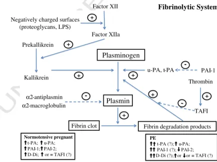
Coagulation,ଏbrinolysis and inଏammation are integral parts of the host immune response. Activation of inଏammatory and coagulation pathways is important in the pathogenesis of vascular disease and both systems interact strongly, so that coagulation and inଏammatory activity mutually modulates each other (Fig. 1). Such processes appear to be intrinsically related to PE since the disease is associated with endothelial cell dysfunction, increased inଏammatory responses and hypercoagulability[37].
Activation of blood coagulation produces proteases that not only interact with coagulation protein but also with speciଏc cell receptors involved in inଏammatory responses. Binding of coagulation proteases (such as thrombin and/or tissue factor) or anticoagulant proteins (e.g., APC) to protease activated receptors (PARs) may affect cytokine production or inଏammatory cell apoptosis[38,39]. These receptors are localised on the vasculature on endothelial cells, mononuclear cells, platelets,ଏbroblasts and smooth muscle cells. Stimulation of PARs by
Placental ischemia
Pre-eclampsia
Inflammatory cytokines
Microparticles Maternal circulation and
endothelial cells TF-expression Coagulation activation PAR´s Fibrinolysis Protein-C Inflammation Fibrin Platelet activation TF-expression factor VIIa Monocytes
Apoptosis (platelets, T cells, monocytes, granulocytes,
syncytiotrophoblasts)
Fig. 1.A potential relationship between haemostasis, inଏammation and pre-eclampsia. Pre-eclampsia is associated with placental oxidative stress and subsequent placental ischaemia and cellular apoptosis. Consequently, there is endothelial dysfunction and release of cytokines as well as microparticles that fall into the maternal circulation. Inଏammatory cytokines trigger inଏammation reaction which modulates coagulation. Microparticles are also able to modulate both inଏammatory and coagulation pathways. Continuous arrows represent activation. Dotted arrows represent inhibition. TF, tissue factor; PARs, protease-activated receptors.
coagulation proteases leads to an induction of a number of pro-inଏammatory mediators including IL-6, IL-8, tumour growth factor-β, monocyte chemoattractant protein-1 (MCP-1), platelet-derived growth factor, intercellular adhesion molecule-1 and P-selectin[40,41].
In addition, the TF/Factor VII (FVII) complex induces pro-inଏammatory effects in macrophages/monocyte leading to the production of reactive oxygen species (ROS) including superoxide anion (O2-)[42]. Superoxide causes vasoconstriction, either directly
through contracting smooth muscle[43]or indirectly by inactivating nitric oxide and reducing the release of prostacyclin[44]. Vasocon-striction is associated with slow bloodଏux and platelet activation. In PE, superoxide generation is increased in neutrophils[34]and the placenta [33]. High concentrations of superoxide stimulate the arachidonic acid pathway in cells to produce thromboxane A2,
which is a potent stimulator of platelet activation[45]. It is well established that activated platelets secrete an array of pro-inଏ amma-tory and pro-coagulant substances stored in their alpha and dense granules. These substances induce TF synthesis in monocyte[46]and contribute to the production of interleukin IL-1, TNF-α, IL-8 and MCP-1[47]. On the other hand, platelets can be directly activated by pro-inଏammatory mediators, such as platelet activating factor [48]. Cytokines increase platelet reactivity, due to the release of large multimers of von Willebrand factor from the endothelium, which are particularly effective in promoting high shear stress rates[49]. Platelet activation and increased cytokine release are commonly seen in pre-eclamptic women[26].
Endogenous anticoagulant pathways also inଏuence inଏammatory responses[50]. Beneଏcial cytoprotective activities of APC include APC-mediated alteration of gene expression proଏles, anti-inଏammatory and anti-apoptotic activities [51,52]. Such activities require endothelial protein C receptors and PAR-1[51,53,54]. Anti-inଏammatory effects of APC on endothelial cells involve inhibition of inଏammatory mediator release and expression of vascular adhesion molecules with the net result of inhibiting leukocyte adhesion and tissue inଏltration. In addition, by helping to maintain endothelial barriers, APC reduces extravascular inଏammatory processes through the inhibition of med-iators released by leukocytes or endothelial cells[5,26,49,55]. Although there is no consensus, some groups have demonstrated a signiଏcant decrease of protein C levels in pregnancy-associated hypertensive disorders[56,57]. Thrombin–antithrombin complex can activate pro-thrombin activatableଏbrinolysis inhibitor (pro-TAFI) to active-TAFI [58]. Activated TAFI plays a role in vascular responses to inଏammation by removing the carboxyl-terminal arginine residues from C3a and C5a. It also has an important role in the regulation of inଏammation by interfering in the cleavage of bradykin, osteopontin or C5a and modulating their pro-inଏammatory functions [59]. Plasmin and thrombin can also activate pro-TAFI[60]. Pro-inଏammatory mediators are known to up-regulate genes that stimulate α2-macroglobulin
production, which upon binding plasmin abrogates its action in degradingଏbrin[61]. Thus, pro-inଏammatory mediators contribute to maintaining theଏbrin clot formation, as seen in PE.
Central regulators of plasminogen activators and inhibitors during inଏammation are TNF-α and IL-1β [62]. The presence of these cytokines in the circulation leads to the release of plasminogen activators, particularly tissue-type plasminogen activator and uroki-nase-type plasminogen activator, from storage sites into vascular endothelial cells. However, this increase in plasminogen activation and subsequent plasmin generation is counteracted by a delayed but sustained increase in PAI-1[63]. The resulting effect onଏbrinolysis is complete inhibition and, as a consequence, inadequateଏbrin removal, contributing to microvascular thrombosis. Inଏammation is also associated with increased concentrations of plasma acute phase reactant proteins (e.g.,ଏbrinogen and C reactive protein - CRP)[64]. High levels ofଏbrinogen increase blood viscosity favouring platelet activation[61,64], while CRP facilitates monocyte-endothelial cell interactions[64]and TF expression[65]. In addition,ଏbrin itself may
act as a pro-inଏammatory agent, speciଏcally during edema accompa-nying acute inଏammatory reactions. Fibrinogen andଏbrin directly inଏuence the production of pro-inଏammatory cytokines (including TNF-α, IL-1β, and MCP-1) by mononuclear cells and endothelial cells [66]. It is known that PE is associated with decreaseଏbrinolysis, as shown by higher PAI-1 levels[29]. Taken together these suggest that haemostatic abnormalities are associated with abnormal inଏ amma-tory responses[67] and that the two systems (haemostasis and inଏammation) are implicated in the ethio-pathogenesis of PE.
5. Pre-eclampsia and microparticles
An additional pathway through which the coagulation and inଏ am-matory systems are generally activated is linked to microparticles (MPs) (Fig. 1). Microparticles wereଏrst described nearly 30 years ago and initially called“platelet dust.”They were described as small vesicles (N0.1 mm) and were shown to promote coagulation activation[68].
However, MPs are now considered to be membrane nano-fragments (0.05–1̀m) with pro-coagulant and pro-inଏammatory properties[69]. Microparticles are generated after cell activation or apoptosis. This usually occurs following the disturbance of membrane phospholipid asymmetry and the pumps responsible for phospholipid transport. Changes in phosphatid MPs composition are not yet elucidated, but appear to differ depending on the cell origin and the stimulatory mechanisms behind their generation[70]. Microparticles are able to act on both endothelial cells[71]and smooth muscle cells [72]; as a result, they regulate vasomotor reactivity as well as angiogenesis [73]. Microparticles can provide as well as interact with TF to generateଏbrin clot. In order for TF to gain its fully activity it requires the presence of PS which is exposed on apoptotic cell and MPs surfaces[74]. Moreover, MPs accelerate the interaction between TF and factor VIIa [75]. Microparticles participate in the regulation of vascular tonus, notably by decreasing the production of nitric oxide (NO). The latter is a powerful vasodilator, anti-platelet agent and a major factor for endothelial cells survival[76]. Microparticles are also able to inଏuence smooth muscle cells directly through the activation of the transcription factorκB (NF-κB), leading to enhanced expression of inducible NOS (iNOS) and cyclooxygenase-2 (COX-2) with subsequent increase in NO and prostacyclin productions respectively, ending in a blunting of vascular contractility to agonists[72]. Microparticles also act as potent pro-inଏammatory mediators, initiating an array of signal transduction pathways and gene expression proଏles in endothelial cells, thereby affecting their function. They can also directly activate and stimulate monocytes to produce cytokines and ROS, resulting in an inଏammatory response[75].
low, and antioxidative pathways are able to inactivate endogenous ROS thereby limiting placental damage. However, in PE these adaptive mechanisms are overwhelmed by enhanced production of ROS leading to an apoptotic/necrotic cascade in STBM MPs[89]. This may promote the release of syncytial products including STBM MPs. The presence of STBM MPs was speciଏcally demonstrated to promote cell death and/or reduce proliferation of endothelial cells and to activate superoxide production in neutrophils isolated from women with PE[5,77,89–91].
In conclusion, several interfaces link coagulation and inଏ amma-tion. Pro-inଏammatory cytokines can affect coagulation pathways, while activated coagulation proteases and endogenous anticoagulants can modulate inଏammation though speciଏc cell receptors. Both systems have been shown to impinge on the ethio-pathogenesis of PE. However, the relationship between these and PE is complex and is far from being understood. Thus, detailed studies are required to elucidate the mechanisms governing these interactions and their relation to PE presence and/or progression.
6. Future perspectives on pre-eclampsia
•Despite intensive research, PE remains one of the leading causes of
maternal death worldwide. The only deଏnitive treatment is to deliver the baby and placenta, often prematurely, in the interest of the baby, the mother, or both. Several randomised trials have reported different means of reducing the rate or the severity of PE. These trials have some limitations (e.g., small sample size) and the results show at best minimal beneଏt. Thus, the classical prophylactic treatment continues i.e., control of blood pressure using antihypertensive drugs and seizure prophylaxis with magnesium sulphate (a cerebral vasodilator)[92].
•Attempts to manage inଏammation and oxidative stress have not improved outcome. PE is associated with endothelial cell injury, haemostatic abnormalities and systemic inଏammatory processes. Whether these events are primary mechanisms or secondary to PE needs clariଏcation.
•PE may be linked to homeostasis involving blood coagulation,
ଏbrinolysis and inଏammation. Detailed understanding of the relationship between these three systems and PE may improve our knowledge on the patho-physiology of PE. A large-scale study correlating key markers of coagulation,ଏbrinolysis and inଏ amma-tion and PE is required. Apart from shedding light on mechanisms, new therapeutic targets might be identiଏed.
•The role of pro-coagulant microparticles in P-EC needs to be clariଏed
further. Recently there have been more studies involving micro-particles but the deଏnitive role of these in PE and indeed other disease processes remains lacking.
•Finally, the causal effect of the proposed association of PE with risk
of delayed cardiovascular disease and with the risk of PE occurring earlier in subsequent pregnancies should be examined further.
Acknowledgement
The authors thank CAPES, FAPEMIG and CNPq/Brazil. LMSD is grateful to CNPq Research Fellowship (PQ) and CAPES(BEX-2694.05.0).
References
[1] Schuiling GA, Koiter TR, Faas MM. Why pre-eclampsia? Hum Reprod 1997;12: 2087–91.
[2] National High Blood-Pressure Education-Program Working Group-Report on high blood-pressure in pregnancy. Am J Obstet Gynecol 1990;163:1691–712. [3] Roberts JM, Taylor RN, Musci TJ, Rodgers GM, Hubel CA, McLaughlin MK.
Preeclampsia - an endothelial-cell disorder. Am J Obstet Gynecol 1989;161: 1200–4.
[4] Brown MA. The physiology of preeclampsia. Clin Exp Pharmacol Physiol 1995;22: 781–91.
[5] Knight M, Redman CWG, Linton EA, Sargent IL. Shedding of syncytiotrophoblast microvilli into the maternal circulation in pre-eclamptic pregnancies. Br J Obstet Gynaecol 1998;105:632–40.
[6] Huppertz B, Kingdom J, Caniggia I, et al. Hypoxia favours necrotic versus apoptotic shedding of placental syncytiotrophoblast into the maternal circulation. Placenta 2003;24:181–90.
[7] Sacks GP, Studena K, Sargent IL, Redman CWG. Normal pregnancy and preeclampsia both produce inଏammatory changes in peripheral blood leukocytes akin to those of sepsis. Am J Obstet Gynecol 1998;179:80–6.
[8] Redman CWG, Sacks GP, Sargent IL. Preeclampsia: an excessive maternal inଏammatory response to pregnancy. Am J Obstet Gynecol 1999;180:499–506. [9] Redman CWG, Sargent IL. Pre-eclampsia, the placenta and the maternal systemic
inଏammatory response - a review. Placenta 2003;24:S21–7.
[10] Magee LA. Pre-eclampsia and increased cardiovascular risk. Br Med J 2007;335: 945–6.
[11] Bellamy L, Casas JP, Hingorani AD, Williams DJ. Pre-eclampsia and risk of cardiovascular disease and cancer in later life: systematic review and meta-analysis. Br Med J 2007;335:974–7.
[12] Magnussen EB, Vatten LJ, Lund-Nilsen TI, Salvesen KA, Smith GD, Romundstad PR. Prepregnancy cardiovascular risk factors as predictors of pre-eclampsia: popula-tion based cohort study. Br Med J 2007;335:978–81.
[13] Paidas MJ, Ku DHW, Arkel YS. Screening and management of inherited thrombophi-lias in the setting of adverse pregnancy outcome. Clin Perinatol 2004;31 783-+. [14] Lin J, August P. Genetic thrombophilias and preeclampsia: a meta-analysis. Obstet
Gynecol 2005;105:182–92.
[15] He S, Bremme K, Blomback M. Acquired deଏciency of antithrombin in association with a hypercoagulable state and impaired function of liver and/or kidney in preeclampsia. Blood Coagul Fibrinolysis 1997;8:232–8.
[16] DG M. Hematologic evidence of disseminated intravascular coagulation in eclampsia. Obstet Gynecol Surv 1972:399–417.
[17] Howie PW, Prentice CR, McNicol GP. Coagulation,ଏbrinolysis and platelet function in pre-eclampsia, essential hypertension and placental insufଏciency. J Obstet Gynaecol Br Commonw 1971;78:992.
[18] Redman CWG, Denson KWE, Beilin LJ, Bolton FG, Stirrat GM. Factor-VIII consumption in pre-eclampsia. Lancet 1977;2:1249–52.
[19] Aznar J, Gilabert J, Estelles A, Espana F. Fibrinolytic-activity and protein-c in preeclampsia. Thromb Haemost 1986;55:314–7.
[20] Schjetlein R, Abdelnoor M, Haugen G, Husby H, Sandset PM, Wisloff F. Hemostatic variables as independent predictors for fetal growth retardation in preeclampsia. Acta Obstet Et Gynecol Scand 1999;78:191–7.
[21] Higgins JR, Walshe JJ, Darling MRN, Norris L, Bonnar J. Hemostasis in the uteroplacental and peripheral circulations in normotensive and pre-eclamptic pregnancies. Am J Obstet Gynecol 1998;179:520–6.
[22] Dusse LM, Carvalho MG, Getliffe K, Voegeli D, Lwaleed BA, Cooper AJ. Increased circulating thrombomodulin levels in pre-eclampsia. Clin Chim Acta 2008;387: 168–71.
[23] VanWijk MJ, Boer K, Berckmans RJ, et al. Enhanced coagulation activation in preeclampsia: the role of APC resistance, microparticles and other plasma constituents. Thromb Haemost 2002;88:415–20.
[24] Weiner CP. Plasma antithrombin III activity: an aid in the diagnosis of preeclampsia-eclampsia. In: J B, editor. Lancet; 1977. p. 1249–52.
[25] Dusse LM, Carvalho MG, Getliffe K, Voegeli D, Lwaleed BA, Cooper AI. Total plasma tissue factor pathway inhibitor levels in pre-eclampsia. Clin Chim Acta 2008;388: 230–2.
[26] Redman CWG, Bonnar J, Beilin L. Early platelet consumption in pre-eclampsia. Br Med J 1978;1:467–9.
[27] Cadroy Y, Grandjean H, Pichon J, et al. Evaluation of 6 markers of hemostatic system in normal-pregnancy and pregnancy complicated by hypertension or preeclampsia. Br J Obstet Gynaecol 1993;100:416–20.
[28] Rakoczi I, Tallian F, Bagdany S, Gati I. Platelet life-span in normal-pregnancy and pre-eclampsia as determined by a non-radioisotope technique. Thromb Res 1979;15: 553–6.
[29] Estelles A, Gilabert J, Grancha S, et al. Abnormal expression of type 1 plasminogen activator inhibitor and tissue factor in severe preeclampsia. Thromb Haemost 1998;79:500–8.
[30] Mantovani A, Dejana E. Cytokines as communication signals between leukocytes and endothelial-cells. Immunol Today 1989;10:370–5.
[31] Vince GS, Starkey PM, Austgulen R, Kwiatkowski D, Redman CWG. Interleukin-6, tumor-necrosis-factor and soluble tumor-necrosis-factor receptors in women with preeclampsia. Br J Obstet Gynaecol 1995;102:20–5.
[32] Lim KH, Rice GE, Degroot CJM, Taylor RN. Plasma type-II phospholipase A(2) levels are elevated in severe preeclampsia. Am J Obstet Gynecol 1995;172:998–1002. [33] Takagi Y, Nikaido T, Toki T, et al. Levels of oxidative stress and redox-related
molecules in the placenta in preeclampsia and fetal growth restriction. Virchows Arch 2004;444:49–55.
[34] Tsukimori K, Fukushima K, Tsushima A, Nakano H. Generation of reactive oxygen species by neutrophils and endothelial cell injury in normal and preeclamptic pregnancies. Hypertension 2005;46:696–700.
[35] Tsukimori K, Maeda H, Ishida K, Nagata H, Koyanagi T, Nakano H. The superoxide generation of neutrophils in normal and preeclamptic pregnancies. Obstet Gynecol 1993;81:536–40.
[36] Huppertz B, Frank HG, Kingdom JCP, Reister F, Kaufmann P. Villous cytotropho-blast regulation of the syncytial apoptotic cascade in the human placenta. Histochem Cell Biol 1998;110:495–508.
[37] Levi M, van der Poll T. Inଏammation and coagulation. Crit Care Med 2010;38:S26–34. [38] Coughlin SR. Thrombin signalling and protease-activated receptors. Nature
2000;407:258–64.
[39] Levi M, van der Poll T, Buller HR. Bidirectional relation between inଏammation and coagulation. Circulation 2004;109:2698–704.
[40] Hou L, Howells GL, Kapas S, Macey MG. The protease-activated receptors and their cellular expression and function in blood-related cells. Br J Haematol 1998;101: 1–9.
[41] Naldini A, Carney DH, Bocci V, et al. Thrombin enhances T-cell proliferative responses and cytokine production. Cell Immunol 1993;147:367–77. [42] Cunningham MA, Romas P, Hutchinson P, Holdsworth SR, Tipping PG. Tissue factor
and factor VIIa receptor/ligand interactions induce proinଏammatory effects in macrophages. Blood 1999;94:3413–20.
[43] Katusic ZS, Vanhoutte PM. Superoxide anion is an endothelium-derived contract-ing factor. Am J Physiol 1989;257:H33–7.
[44] Gryglewski RJ, Palmer RMJ, Moncada S. Superoxide anion is involved in the breakdown of endothelium-derived vascular relaxing factor. Nature 1986;320:454–6. [45] Cosentino F, Sill JC, Katusic ZS. Role of superoxide anions in the mediation of
endothelium-dependent contractions. Hypertension 1994;23:229–35. [46] Ernofsson M, Siegbahn A. Platelet-derived growth factor-BB and monocyte
chemotactic protein-1 induce human peripheral blood monocytes to express tissue factor. Thromb Res 1996;83:307–20.
[47] Neumann FJ, Marx N, Gawaz M, et al. Induction of cytokine expression in leukocytes by binding of thrombin-stimulated platelets. Circulation 1997;95:2387–94. [48] Osterud B. Tissue factor expression by monocytes: regulation and
pathophysio-logical roles. Blood Coagul Fibrinolysis 1998;9:S9–S14.
[49] Bernardo A, Ball C, Nolasco L, Moake JF, Dong JF. Effects of inଏammatory cytokines on the release and cleavage of the endothelial cell-derived ultralarge von Willebrand factor multimers underଏow. Blood 2004;104:100–6.
[50] Levi M, van der Poll T. The role of natural anticoagulants in the pathogenesis and management of systemic activation of coagulation and inଏammation in critically ill patients. Semin Thromb Hemost 2008;34:459–68.
[51] Esmon CT. New mechanisms for vascular control of inଏammation mediated by natural anticoagulant proteins. J Exp Med 2002:561–4.
[52] Okajima K. Regulation of inଏammatory responses by natural anticoagulants. Immunol Rev 2001;184:258–74.
[53] Grifଏn JH, Fernandez JA, Gale AJ, Mosnier LO. Activated protein C. J Thromb Haemost 2007;5:73–80.
[54] Esmon CT. The endothelial cell protein C receptor. Thromb Haemost 2000;83: 639–43.
[55] Mosnier LO, Grifଏn JH. Inhibition of staurosporine-induced apoptosis of endothelial cells by activated protein C requires protease-activated receptor-1 and endothelial cell protein C receptor. Biochem J 2003;373:65–70. [56] Lao TT, Yin JA, Ng WK, Yuen PMP. Relationship between maternal antithrombin-iii
and protein-c protein-s levels before, during and after delivery. Gynecol Obstet Investig 1990;30:87–90.
[57] Krauss T, Augustin HG, Osmers R, Meden H, Unterhalt M, Kuhn W. Activated protein C resistance and Factor V Leiden in patients with hemolysis, elevated liver enzymes, low platelets syndrome. Obstet Gynecol 1998;92:457–60. [58] Nesheim M. Thrombinଏbrinolysis Chest 2003;124:33S–9S.
[59] Myles T, Nishimura T, Yun TH, et al. Thrombin activatableଏbrinolysis inhibitor, a potential regulator of vascular inଏammation. J Biol Chem 2003;278:51059–67. [60] Mao SS, Cooper CM, Wood T, Shafer JA, Gardell SJ. Characterization of
plasmin-mediated activation of plasma procarboxypeptidase B - modulation by glycosa-minoglycans. J Biol Chem 1999;274:35046–52.
[61] Esmon CT. The interactions between inଏammation and coagulation. Br J Haematol 2005;131:417–30.
[62] Biemond BJ, LM, Ten Cate H, Van der Poll T, Büller HR, Hack CE, Ten Cate JW. Plasminogen activator and plasminogen activator inhibitor I release during experimental endotoxaemia in chimpanzees: effect of interventions in the cytokine and coagulation cascades. Clin Sci 1995:587–94.
[63] Vanderpoll T, Levi M, Hack CE, et al. Elimination of interleukin-6 attenuates coagulation activation in experimental endotoxemia in chimpanzees. J Exp Med 1994;179:1253–9.
[64] Han KH, Hong KH, Park JH, et al. C-reactive protein promotes monocyte chemoattractant protein-1-mediated chemotaxis through upregulating CC chemo-kine receptor 2 expression in human monocytes. Circulation 2004;109:2566–71. [65] Devaraj S, Xu DY, Jialal I. C-reactive protein increases plasminogen activator
inhibitor-1 expression and activity in human aortic endothelial cells. Circulation 2003;107:398–404.
[66] Szaba FM, Smiley ST. Roles for thrombin andଏbrin(ogen) in cytokine/chemokine production and macrophage adhesion in vivo. Blood 2002;99:1053–9.
[67] Shrivastava S, McVey JH, Dorling A. The interface between coagulation and immunity. Am J Transplant 2007;7:499–506.
[68] Wolf P. Nature and signiଏcance of platelet products in human plasma. Br J Haematol 1967;13:269.
[69] Meziani F, TA, Andriantsitohaina R. Microparticles are vectors of paradoxical information in vascular cells including the endothelium: role in health and diseases. Pharmacol Rep 2008:75–84.
[70] Distler JHW, Huber LC, Gay S, Distler O, Pisetsky DS. Microparticles as mediators of cellular cross-talk in inଏammatory disease. Autoimmunity 2006;39:683–90. [71] Martin S, Tesse A, Hugel B, et al. Shed membrane particles from T lymphocytes
impair endothelial function and regulate endothelial protein expression. Circu-lation 2004;109:1653–9.
[72] Tesse A, Martinez MC, Hugel B, et al. Upregulation of proinଏammatory proteins through NF-kappa B pathway by shed membrane microparticles results in vascular hyporeactivity. Arterioscler Thromb Vasc Biol 2005;25:2522–7. [73] Kim HK, Song KS, Chung JH, Lee KR, Lee SN. Platelet microparticles induce
angiogenesis in vitro. Br J Haematol 2004;124:376–84.
[74] Gando S, Kameue T, Morimoto Y, Matsuda N, Hayakawa M, Kemmotsu O. Tissue factor production not balanced by tissue factor pathway inhibitor in sepsis promotes poor prognosis. Crit Care Med 2002;30:1729–34.
[75] Barry OP, Pratico D, Savani RC, FitzGerald GA. Modulation of monocyte–endothelial cell interactions by platelet microparticles. J Clin Investig 1998;102:136–44. [76] Martinez MC, Tesse A, Zobairi F, Andriantsitohaina R. Shed membrane
micro-particles from circulating and vascular cells in regulating vascular function. Am J Physiol Heart Circ Physiol 2005;288:H1004–9.
[77] Lok CAR, Van Der Post JAM, Sargent IL, et al. Changes in microparticle numbers and cellular origin during pregnancy and preeclampsia. Hypertens Pregnancy 2008;27:344–60.
[78] Meziani F, Tesse A, David E, et al. Shed membrane particles from preeclamptic women generate vascular wall inଏammation and blunt vascular contractility. Am J Pathol 2006;169:1473–83.
[79] VanWijk MJ, Nieuwland R, Boer K, van der Post JAM, VanBavel E, Sturk A. Microparticle subpopulations are increased in preeclampsia: possible involve-ment in vascular dysfunction? Am J Obstet Gynecol 2002;187:450–6. [80] Lok CAR, Jebbink J, Nieuwland R, et al. Leukocyte activation and circulating
leukocyte-derived microparticles in preeclampsia. Am J Reprod Immunol 2009;61:346–59.
[81] Toth B, Lok CAR, Boing A, et al. Microparticles and exosomes: impact on normal and complicated pregnancy. Am J Reprod Immunol 2007;58:389–402. [82] Bretelle F, Sabatier F, Desprez D, et al. Circulating microparticles: a marker of
procoagulant state in normal pregnancy and pregnancy complicated by preeclampsia or intrauterine growth restriction. Thromb Haemost 2003;89:486–92. [83] Gonzalez-Quintero VH, Jimenez JJ, Jy W, et al. Elevated plasma endothelial
microparticles in preeclampsia. Am J Obstet Gynecol 2003;189:589–93. [84] Gonzalez-Quintero VH, Smarkusky LP, Jimenez JJ, et al. Elevated plasma
endothelial microparticles: preeclampsia versus gestational hypertension. Am J Obstet Gynecol 2004;191:1418–24.
[85] Germain SJ, Sacks GP, Soorana SR, Sargent IL, Redman CW. Systemic inଏammatory priming in normal pregnancy and preeclampsia: the role of circulating syncytiotrophoblast microparticles. J Immunol 2007;178:5949–56.
[86] Redman CWG, Sargent IL. Circulating microparticles in normal pregnancy and pre-eclampsia. Placenta 2008;29:S73–7.
[87] Redman CWG, Sargent IL. Microparticles and immunomodulation in pregnancy and pre-eclampsia. J Reprod Immunol 2007;76:61–7.
[88] Goswami D, Tannetta DS, Magee LA, et al. Excess syncytiotrophoblast micropar-ticle shedding is a feature of early-onset pre-eclampsia, but not normotensive intrauterine growth restriction. Placenta 2006;27:56–61.
[89] Guller S. Role of the syncytium in placenta-mediated complications of preeclamp-sia. Thromb Res 2009:389–92.
[90] Aly AS, Khandelwal M, Zhao J, Mehmet AH, Sammel MD, Parry S. Neutrophils are stimulated by syncytiotrophoblast microvillous membranes to generate superoxide radicals in women with preeclampsia. Am J Obstet Gynecol 2004;190:252–8. [91] Gupta AK, Rusterholz C, Huppertz B, et al. A comparative study of the effect of
three different syncytiotrophoblast micro-particles preparations on endothelial cells. Placenta 2005;26:59–66.
4.1.2 D-dimer plasma levels in preeclampsia: a systematic review and metanalysis - Clinica Chimica Acta
UNCORRECTED PR
OOF
1 Invited critical review
2 D-dimer in preeclampsia: Review and meta-analysis
3 Melina de Barros
Q1 Pinheiroa,b,Daniela Rezende GarciaJunqueiraa,c,Fernanda FonsecaCoelhoa, 4 Letícia G.Freitasa,Maria G.Carvalhoa,Karina BragaGomesa,Luci Maria Sant'AnaDussea, 5 aDepartment of Clinical and Toxicological Analysis, Faculty of Pharmacy,Universidade Federal de Minas Gerais,Brazil
6 bSchool of Medicine,Universidade Federal de São João Del Rei,Brazil
7 cCentre of Drug Studies (Cemed), Department of Social Pharmacy, Faculty of Pharmacy, Universidade Federal de Minas Gerais,Brazil
8
9
a b s t r a c t a r t i c l e i n f o
10 Article history:
11 Received 30 May 2012
12 Received in revised form 10 July 2012
13 Accepted 4 August 2012
14 Available online xxxx
15 16 17 18 Keywords: 19 Preeclampsia 20 D-dimer 21 Diagnosis
22 Systematic review
23 Meta-analysis
24
Preeclampsia is a multifactorial disease characterized by high blood pressure and proteinuria after the 20th week
25
of pregnancy. Preeclampsia is associated with microvasculatureଏbrin deposition and maternal organ
dysfunc-26
tion. D-dimer (D-Di) has been used as a marker of production/degradation ofଏbrin in vivo. D-Di has emerged
27
as a useful diagnostic tool for thrombotic conditions because its plasma concentration has a high negative
predic-28
tive value for venous thromboembolism. The aim of this study was to evaluate publications that assessed plasma
29
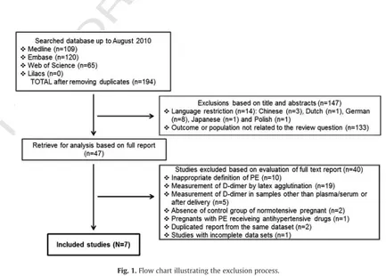
D-Di in preeclampsia and normotensive pregnant subjects to deଏne its diagnostic value. A total of 194
publica-30
tions were identiଏed. Following the exclusion process, seven studies were in accordance with the pre-deଏned
el-31
igibility criteria. This systematic review was performed with methodologic accuracy, including a careful
32
deଏnition of preeclampsia and a high sensitivity literature search strategy. Quality of the included studies was
33
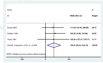
assessed in accordance with widely accepted literature recommendations. Our meta-analysis indicates that
in-34
creased plasma D-Di is associated with preeclampsia in the third trimester of gestation vs normotensive
preg-35
nant subjects. These preliminaryଏndings in this select group of patients clearly highlight the need for
36
additional comprehensive studies throughout pregnancy, including the establishment of an appropriate
37
cut-off, in order to fully elucidate the diagnostic/prognostic role of D-Di in preeclampsia.
38
© 2012 Elsevier B.V. All rights reserved.
39 40 41 42 43 44 Contents
45 1. Introduction . . . 0
46 2. Methods . . . 0
47 2.1. Data sources and searches . . . 0
48 2.2. Study selection . . . 0
49 2.3. Data extraction . . . 0
50 2.4. Data analysis . . . 0
51 3. Results . . . 0
52 3.1. Participants . . . 0
53 4. Discussion . . . 0
54 Conଏict of interest statement . . . 0
55 Acknowledgments . . . 0
56 References . . . 0
57
58
1. Introduction
59
Preeclampsia is a multifactorial disease characterized by systolic
60
blood pressure≥140 mm Hg or diastolic≥90 mm Hg at bed rest on
61
at least two occasions6 hapart, and proteinuria≥0.3 g/24 h, measured
62
after the 20th week of pregnancy[1,2]. Symptoms frequently observed
63
in preeclampsia include headache, blurred vision, and abdominal pain.
64
The etiology of preeclampsia is unknown and the delivery of placenta
Clinica Chimica Acta xxx (2012) xxx–xxx
Corresponding author at: Department of Clinical and Toxicological Analysis, Faculty of
Pharmacy, Federal University of Minas Gerais, Av. Antônio Carlos, 6627, Pampulha CEP:
31270901, Belo Horizonte, Minas Gerais, Brazil. Tel.: +55 31 3409 6880/6900; fax: +55
31 3409 6985.
E-mail addresses:[email protected],[email protected](L.M.S. Dusse).
CCA-12776; No of Pages 5
0009-8981/$–see front matter © 2012 Elsevier B.V. All rights reserved.
http://dx.doi.org/10.1016/j.cca.2012.08.003
Contents lists available atSciVerse ScienceDirect
Clinica Chimica Acta
j o u r n a l h o m e p a g e : w w w . e l s e v i e r . c o m / l o c a t e / c l i n c h i m
UNCORRECTED PR
OOF
65 remains the only known treatment. This disease can progress to66 eclampsia (characterized by seizures as a sign of affection of the cere-67 bral vessels), syndrome HELLP (hemolysis, elevated liver enzyme, low
68 platelets) or disseminated intravascular coagulation [2]. Although
69 some laboratory tests such as platelet count and liver enzymes can be
70 used to monitor the risk of preeclampsia, the diagnosis is more effective
71 when established by blood pressure and proteinuria measurement[2]. 72 Preeclampsia is associated with the deposition ofଏbrin in
micro-73 vasculature, which results in placental perfusion compromise,
in-74 trauterine fetal growth retardation[2]and dysfunction of some
75 maternal organs[3].
76 In the early stages ofଏbrin clot formation, activated thrombin
77 cleavesଏbrinogen, a soluble plasma protein. Molecular polymerization
78 is observed due to the formation of solubleଏbrin, which is subsequently
79 stabilized by covalent cross-linking with factor XIII—producing an insol-80 ubleଏbrin matrix. Degradation is immediately initiated by plasmin,
81 resulting in a variety of relatively stable dimeric fragments orଏbrin
deg-82 radation products. The smallest fragment, D-dimer (D-Di), is resistant
83 to plasmin degradation. Therefore, D-Di speciଏcally reଏects bothଏbrin
84 polymerization and breakdown[4–7].
85 Plasma D-Di is a well established clinical laboratory marker of this
86 process in vivo. Additionally, D-Di is a useful diagnostic tool due to its
87 high negative predictive value for venous thromboembolism[6,8,9].
88 Several studies have shown increased D-Di in preeclampsia vs
nor-89 motensive pregnant subjects[10–14]. The aim of this meta-analysis 90 was to compile and evaluate publications that assessed the D-Di by
91 enzyme-linked immunosorbent assay (ELISA) to deଏne its diagnostic
92 value in preeclampsia.
93 2. Methods
94 2.1. Data sources and searches
95 An electronic database search was conducted for four databases
96 (Medline, Embase, LILACS, and Web of Science) from the earliest re-97 cord to August 2010. A sensitive search strategy using controlled
vo-98 cabulary and free text terms was developed for each database with a
99 combination of relevant key words such as D-Dimer, preeclampsia,
100 eclampsia, pregnancy induced hypertension and gestational
hyper-101 tension (full details of the search strategy are available on-request
102
from the authors). Citation tracking was performed by manually
103
screening reference lists of eligible studies. Studies included in the
re-104
view were restricted to English, Spanish and Portuguese languages.
105
2.2. Study selection
106
Eligible studies included those that evaluated D-Di by ELISA,
consti-107
tuted by preeclamptic women and controls (normotensive pregnant).
108
Preeclampsia was deଏned as systolic blood pressure≥140 mm Hg or
109
diastolic≥90 mm Hg at bed rest on at least two occasions6 hapart
110
and proteinuria≥0.3 g/24 h after the 20th week of pregnancy[2].
Stud-111
ies with inappropriate or unclear deଏnition of preeclampsia and those
112
presenting insufଏcient results were excluded.
113
The retrieved papers were submitted to a rigorous selection
pro-114
cess using a standardized protocol applied to papers by three authors
115
independently. Disagreements were resolved by consensus.
116
2.3. Data extraction
117
For each included study, two reviewers independently extracted
118
data such as study design, preeclampsia deଏnition, number of
pre-119
eclamptic and normotensive pregnant women in each study,
gesta-120
tional age at which blood collection occurred, D-Di concentration
121
and author's conclusions. Data were adjusted to include only
preg-122
nant women in the third trimester of gestation.
123
Quality of the included studies was performed according to the
124
Newcastle-Ottawa Scale recommendations[15]for nonrandomized
125
studies in meta-analyses[16]and STROBE guidelines[17]. Five
do-126
mains were considered: appropriate selection of participants,
appro-127
priate measurement of variables and outcomes, adequate follow-up
128
rate, control for confounding via statistical adjustment and the
exis-129
tence of conଏict of interest. This approach was designed to provide
130
an overall quality assessment of the speciଏc domains associated
131
with potential source of bias in studyଏndings and was not designed
132
to provide a score to each individual study[18].
133
2.4. Data analysis
134
D-Di (median and standard deviation or median and ranges) from
135
the participants (case or control group) were weighted in a
meta-Fig. 1.Flow chart illustrating the exclusion process. 2 M.B. Pinheiro et al. / Clinica Chimica Acta xxx (2012) xxx–xxx