UNIVERSIDADE FEDERAL DE UBERLÂNDIA INSTITUTO DE GENÉTICA E BIOQUÍMICA PÓS-GRADUAÇÃO EM GENÉTICA E BIOQUÍMICA
O USO TERAPÊUTICO DE MEDIADORES ANTI-INFLAMATÓRIOS DA VIA DO ÁCIDO ARAQUIDÔNICO
Aluno: Carlos Antonio Trindade da Silva
Orientador: Prof. Dr. Carlos Ueira Vieira
ii
UNIVERSIDADE FEDERAL DE UBERLÂNDIA INSTITUTO DE GENÉTICA E BIOQUÍMICA PÓS-GRADUAÇÃO EM GENÉTICA E BIOQUÍMICA
O USO TERAPÊUTICO DE MEDIADORES ANTI-INFLAMATÓRIOS DA VIA DO ÁCIDO ARAQUIDÔNICO
Aluno: Carlos Antonio Trindade da Silva
Orientador: Prof. Dr. Carlos Ueira Vieira
Tese apresentada à Universidade Federal de Uberlândia como parte dos requisitos para obtenção do Título de Doutor em Genética e Bioquímica (Área Genética)
Dados Internacionais de Catalogação na Publicação (CIP) Sistema de Bibliotecas da UFU, MG, Brasil.
S586u 2016
Silva, Carlos Antonio Trindade da, 1985
O Uso terapêutico de mediadores anti-inflamatórios da via do ácido araquidônico / Carlos Antonio Trindade da Silva. - 2016.
92 p. : il.
Orientador: Carlos Ueira Vieira.
Tese (doutorado) - Universidade Federal de Uberlândia, Programa de Pós-Graduação em Genética e Bioquímica.
Inclui bibliografia.
1. Genética - Teses. 2. Tireoíde - Câncer - Teses. 3. Prostaglandinas - Teses. I Vieira, Carlos Ueira, 1981. II. Universidade Federal de Uberlândia. Programa de Pós-Graduação em Genética e Bioquímica. III. Título.
iii
UNIVERSIDADE FEDERAL DE UBERLÂNDIA INSTITUTO DE GENÉTICA E BIOQUÍMICA PÓS-GRADUAÇÃO EM GENÉTICA E BIOQUÍMICA
O USO TERAPÊUTICO DE MEDIADORES ANTI-INFLAMATÓRIOS DA VIA DO ÁCIDO ARAQUIDÔNICO
ALUNO: Carlos Antonio Trindade da Silva
COMISSÃO EXAMINADORA
Presidente: Prof. Dr. Carlos Ueira Vieira
Examinadores: Dr. Carlo Jose Freire de Oliveira Dr. Siomar de Castro Soares Dra. Cássia Regina da Silva Dra. Lara Vecchi
Data da Defesa: 29/06/2016
As sugestões da Comissão Examinadora e as Normas PGGB para o formato da Dissertação/Tese foram contempladas
iv
SUMÁRIO
LISTA DE FIGURAS………...…vi
LISTA DE ABREVIATURAS...vii
RESUMO...viii
ABSTRACT...iv
APRESENTAÇÃO GERAL ...x
CAPITULO 1...12
1.Ácido Araquidônico...12
1.2 Via das Ciclooxigenases ...12
1.2.1 15-deoxi-Δ12,14-PGJ 2 ...14
1.2.2 15-deoxi-Δ12,14-PGJ 2 e cancer ...16
1.3 Via da lipoxigenase...17
1.4 Via do Citocomo P450 epoxygenase...18
1.4.1 Epoxido Hidrolase solúvel (sEH) ...19
Bibliografia ...22
CAPÍTULO 2. 15-Deoxy-Δ12,14-prostaglandin J2 induces apoptosis and upregulates SOCS3 in human thyroid cancer cells……….27
ABSTRACT………....28
1. INTRODUCTION……….….29
2. MATERIALS AND METHODS………..…….…30
2.1 Cell line………..………. 30
2.2 Analysis of cell viability………..30
2.3 Evaluation of apoptosis via annexin V staining
...312.4 Cytokine Analysis ……….….31
2.5 mRNA expression analyses………..……...31
2.6 Statistical Analysis………..…32
3. RESULTS………..32
3.1 In vitro effect of 15d-PGJ2 on TPC-1 and FG11 cell proliferation and viability……….32
3.2 Apoptotic effects of 15d-PGJ2 on thyroid cancer cells ……….…34
3.3 Relative IL-6 mRNA expression and IL-6 release by TPC-1………...…35
3.4 Relative expression of SOCS3, SOCS1 and STAT3………36
4. DISCUSSION………37
5. CONCLUSION……….…….40
REFERENCES………..40
CAPITULO 3. Soluble epoxide hydrolase pharmacological inhibition decreases alveolar bone loss by modulating host inflammatory response, RANK-related signaling, ER stress and apoptosis……….…………..47
Abstract ……….50
Introduction.. .. ………..51
Materials and Methods ………...………53
Bacterial viability………..………..54
Animals and Animal Care………...………..54
Periodontitis model and treatments………...……….55
Quantification of alveolar bone loss……….….…..56
v
Statistical analysis……….56
Results………..……..57
TPPU does not affect bacterial viability ……….…………57
Chemical inhibition of sEH reduces bone loss ……….…...….57
Genetic ablation of sEH recapitulates the effects of sEH inhibitor on bone loss …………59
Inhibition of sEH alters key regulators of bone remodeling ………60
Discussion………...67
References………....…….72
vi
LISTA DE FIGURAS
CAPITULO 1.
Figura 1. Síntese da 15d-PGJ2, modificado e adaptado de Surh et al. 2011
Figura 2. Metabolismo do ácido araquidônico e efeitos fisiológicos dos metabolitos do ácido araquidônico.
CAPITULO 2.
Figure 1. 15d-PGJ2 decreased the viability of TPC1 cells.
Figure 2. Fibroblast (FG11) cell proliferation under 15d-PGJ2 treatment.
Figure 3. 15d-PGJ2 induced apoptosis in TPC1 cells.
Figure 4. decreased relative IL-6 mRNA expression and release - TPC1 cells treated with 15d-PGJ2.
Figure 5. SOCS 3 and SOCS1 increased in TPC-1 cells treated with 15d-PGJ2.
CAPITULO 3.
Figure 1. Inhibition of sEH with a potent and orally available small molecule TPPU decreases.
Figure 2. Genetic inhibition of sEH by gene knockout decreases bone loss similar to chemical inhibitor
Figure 3. The dysregulated RANK/RANKL/OPG system in periodontal disease is restored by chemical or genetic ablation of sEH.
Figure 4. Periodontal disease mediated phosphorylation of pro-inflammatory p38 and JNK1/2 is reduced by chemical or genetic ablation of sEH
Figure 5. ER stress sensors are activated in gingival tissues of mice with periodontal disease and reversed by inhibition of sEH.
Supplemental Figure 1. TPPU or its vehicle does not affect bacterial viability.
vii
LISTA DE ABREVIATURAS
15d-PGJ2= 15-deoxi-Δ12,14-Prostaglandina J2
AA = ácido araquidônico
COX = Ciclooxigenase
DHET= ácidos dihidroxieicosatrienoicos
EET= ácidos epoxieicosatrienoicos
IL-1β = Interleucina 1 β
IL-6= Interleucina 6
LOX = Lipoxigenase
LT= Leucotrieno
MCP-1= Proteína quimiotática de monócitos-1
NF-Қb= Fator nuclear-қB
PLA2= Fosfolipase A2
PGE2= Prostaglanina E2
OPG= Osteoprotegerina
PERK= PKR- like endoplasmic reticulum kinase
PPAR = Receptor de Ativação de Proliferação de Peroxissomo
RANK/RANKL= Receptor ativador do ligante fator nuclear-kB
TNF-α= Fator de Necrose Tumoral α
sHE= Epoxido hidrolase solúvel
viii
RESUMO
O ácido araquidônico (AA) é precursor na formação dos eicosanoides, que são mediadores lipídicos com uma série de funções na fisiologia e patologia humana. A maioria dos eicosanoides atuam como mediadores pró-inflamatórios e contribuem para o desenvolvimento e proliferação de tumores. Nesta tese foram avaliados dois mediadores: a 15-deoxi-Δ12,14-PGJ2 (15d- PGJ2) e os ácidos Epoxieicosatrienoicos (EETs), ambos apresentam uma atividade oposta a da maioria dos eicosanoides, ou seja, com uma ação anti-inflamatória e antitumoral. Esses dois mediadores distintos da via do AA foram utilizados nesta tese em dois projetos distintos. Primeiro: A 15d- PGJ2 possui uma atividade antiproliferativa e induziu apoptose para diversos tipos de células tumorais, entretanto, o efeito da15d- PGJ2 em células de cancer da tireoide ainda estava desconhecido. Neste sentido, foram cultivadas in vitro células tumorais da tireoide, da linhagem TPC1, e tratadas
com diferentes concentrações de 15d- PGJ2 (0 ate 20 µM), as células tratadas demonstraram uma diminuição na proliferação, e aumento na apoptose, e uma diminuição na liberação e expressão relativa de IL-6. Estes resultados em conjunto sugerem que a 15d- PGJ2 pode ser utilizada como uma nova terapia para o cancer da tireoide. Segundo: Os EETs são metabolizados em seus diois pela epóxi hidrolase solúvel (sEH), para manter a estabilidade dos EETs e a sua atividade antiiflamatoria, foi utilizado um inibidor (TPPU) para sEH em um modelo de periodontite induzida por Aggregatibacter actinomycetemcomitans. O tratamento
oral com TPPU, assim como o uso de animais sEH Knockout, levou a uma redução na perda óssea acompanhada da diminuição de moléculas osteoclastogenicas como RANK, RANKL e OPG, demonstrando que a inibição farmacológica da sEH pode ter um valor terapêutico na periodontite e doenças inflamatórias que envolvem a reabsorção óssea.
ix
ABSTRACT
Arachidonic acid (AA) a precursor in the formation of eicosanoids which are lipid mediators with a number of functions in human physiology and pathology. The most of the eicosanoids act as proinflammatory mediators and contribute to the development and proliferation of tumors. In this thesis we evaluated two mediators: 15-deoxy-Δ12,14-PGJ2 (15d- PGJ2) and epoxieicosatrienoic acids (EETs) both act with an opposite activity of most eicosanoids, with an anti-inflammatory and and anti-tumoral action these two distinct mediators from AA pathway were used in this thesis in two different projects. First: 15d- PGJ2, was described that to have an antiproliferative activity and to induce apoptosis in several types of tumor cells however, the effect of 15d- PGJ2 in thyroid cancer cells was unknown in this sense, we tested
in vitro cultured thyroid tumor cells, here in TPC1 cells, and treated with different
concentrations of 15d- PGJ2 (0 to 20 uM) the treated cells showed a decrease in proliferation and an increase in apoptosis and a decrease in IL-6 release and relative expression. These key results together demonstrate that 15d- PGJ2 can be used as a new therapy for thyroid cancer. Second: The EETs are converted to their diols by soluble epoxy hydrolase (sEH) to maintain the stability of EETs and their anti-inflammatory activity, an inhibitor (TPPU) against was used to sEH in a periodontitis model induced with Aggregatibacter
actinomycetemcomitans. The oral treatment in mice with TPPU and sEH Knockout animals
showed bone loss reduction accompanied by a decrease in the osteoclastogenic molecules, like RANK, RANKL and OPG, demonstrating that pharmacological inhibition of sEH may have therapeutic value in periodontitis and inflammatory diseases that involve bone resorption.
x
APRESENTAÇÃO GERAL
A via do ácido araquidônico (AA) resulta na síntese de mediadores lipídicos indispensáveis na fisiologia e patologia humana. Dentre estes mediadores, chamados de eicosanoides, encontram-se as prostaglandinas, leucotrienos e os ácidos epoxieicosatrienoicos que desempenham diversas funções bioquímicas nas células, agindo como mensageiros de curta distância, com ação autócrina e parácrina.
Nas últimas décadas, diversos esforços têm sido feitos para o controle e modulação da via do ácido araquidônico, culminando no uso terapêutico das ações dos eicosanoides com o intuito de prevenir ou tratar uma serie de patologias, como a hipertensão arterial, artrite reumatoide, carcinomas e doença periodontal.
A 15-deoxi-Δ12,14-prostaglandina J2 (15d-PGJ2), é um ligante do Receptor Ativado por Proliferadores de Peroxissoma ɣ, esta prostaglandina possui diversas ações biológicas, como a capacidade anti-inflamatória e antitumoral. Neste sentido o seu uso exógeno tem sido utilizado para modular a resposta inflamatória, e também tem demonstrado um potencial terapêutico para inibir o crescimento de tumores como: mama, bexiga, pulmão e próstata, estes efeitos foram eficientes tanto em modelos experimentais in vivo quanto in vitro, no
entanto o seu efeito sob o câncer de tireoide ainda está desconhecido, neste sentido foi avaliado o efeito in vitro da 15d-PGJ2 em células da linhagem TPC1, no câncer da tireoide.
xi
A doença periodontal é uma doença inflamatória crônica que acomete cerca de 70% da população adulta. Mediadores inflamatórios gerados na doença periodontal ativam osteoclastos, que promovem a reabsorção óssea.
O periodonto é constituído por ligamentos, gengiva e osso alveolar. Estas estruturas promovem a fixação dos dentes na boca. A doença periodontal é caracterizada pela destruição destas estruturas. Sendo assim a modulação da resposta inflamatória na doença periodontal tem sido alvo, no intuito de reduzir a perda óssea, que se não for tratada pode levar a perda dos dentes.
12
CAPÍTULO 1.
Fundamentação teórica
1. Ácido Araquidônico
O AA é oriundo do ácido linoleico, um ácido graxo essencial. A partir da formação do AA, esta molécula passa a constituir os fosfolipídios, que são componentes da estrutura da membrana das células. Cada fosfolipídio é constituído de uma molécula de glicerol que interliga a um grupo fosfato e dois ácidos graxo. Desta forma, o AA é um dos ácidos graxos que compõem a membrana celular (LEHNINGER & NELSON 2006).
Para iniciar a síntese da cascata do AA, é necessário que este ácido graxo esteja livre da membrana da célula. O processo de liberação do AA depende da ação de enzimas que são capazes de liberar o AA da membrana para o citoplasma. A fosfolipase A2 (PLA2), é a principal enzima responsável pela liberação do AA da membrana das células. No entanto, a clivagem do AA a partir do diacilglicerol pela diacilglicerol lipase, é uma via alternativa relacionada com a produção de endocanabinóides (LEHNINGER & NELSON 2006).
A PLA2 é ativada pela fosforilação, que ocorre em resposta a eventos de transdução de sinal desencadeados por vários estímulos, como, por exemplo, a ação da trombina nas plaquetas, do C5a nos neutrófilos, da bradicinina nos fibrobrastos, reações antígeno anticorpo nos mastócitos e lesão celular em geral, leva a ativação da fosfolipase A2, requer uma ação catalítica dependente de fosforilação, liberando o AA para o citoplasma da célula. O AA livre no citoplasma pode ser direcionado e metabolizado por três vias diferentes, a via da ciclooxigenase, lipoxigenase e do Citocromo P 450 (ONI-ORISAN, et al. 2016, MRUWAT, et al 2015).
1.2 Via das Ciclooxigenases
13 produto (enzima COX 1) promove a regulação de diversas funções, como a proteção da mucosa gastrointestinal de ulceração. O gene codificador da enzima COX2 induzivél, é localizado no cromossomo 1 e sua expressão é rapidamente aumentada em resposta a uma variedade de citocinas pró-inflamatórias e lesões teciduais (BACCHI, et at 2012). A enzima COX1 participa da síntese das prostaglandinas responsáveis pela citoproteção gastrointestinal e função plaquetária, enquanto a enzima COX2 induz a síntese de prostaglandinas, como por exemplo a prostraglandina E2, que é responsável por induzir dor e inflamação (KRAEMER et al 1992; SIMMONS et al 2004). As duas isoformas, tanto a da COX-1 quanto a COX-2 ligam-se ao retículo endoplasmático promovendo uma ação de endoperóxido sintase, pela catalize da incorporação de duas moléculas de oxigênio ao araquidonato levando à uma ciclização para produzir o endoperóxido cíclico (PGG2). Em seguida as COXs tem uma ação de peroxidase que converte a PGG2 em outro endoperóxido cíclico, a PGH2 (HANG et al. 2008). A PGH2 é convertida através de várias sintases especificas de cada célula nas principais prostaglandinas como: prostaglandina sintetase converte para prostaglandina I2, (PGI2), a prostaglandina E2 sintetase converte para prostaglandina E2 ( PGE2), a tramboxano-A sintetase converte para Tramboxano A2 (TXA2) e a prostaglandina D2 sintetase converte para prostaglandina D2 (PGD2); (figura 1) a PGD2 sofre uma desidratação e espontaneamente é formada a prostaglandina J2, Em seguida esta molécula sofre um rearranjo intramolecular formando a Δ12-PGJ2 que perde mais uma molécula de água formando a 15-deoxi-Δ12,14-PGJ2 (15d-PGJ2) (SCHER, PILLINGER, 2004; LAMANO-CARVALHO, 2007)
A 15d-PGJ2 é produzida por uma variedade de células, incluindo macrófagos, mastócitos, células T e plaquetas (STRAUS et al, 2000). A maioria das
14 Durante o início do processo inflamatório, no qual encontramos um aumento do influxo de polimorfonucleares, há um aumento da atividade da COX2 e síntese de PGE2, a qual possui um papel proinflamatório intenso. No entanto, aparentemente, a COX2 participa da resolução da fase aguda da inflamação alternando o padrão da síntese de prostaglandina para a família J. (SURH et at 2011). Além da fase aguda da inflamação, a PGE2 está envolvida também na inflamação crônica e no desenvolvimento de tumores malignos (OBERMAJER et al 2012).
Figura 1. Síntese da 15d-PGJ2, modificado e adaptado de Surh et al. 2011.
1.2.1 15-deoxi-Δ12,14-PGJ2
proliferator-15
activated receptor gamma) PPAR-γ (FORMAN et al., 1995; RICOTE et al., 1998),
o que sugere que esta molécula possui um importante papel na regulação da reposta inflamatória e no desenvolvimento de diversos carcinomas.
A subfamília de receptores nucleares PPAR consiste de três diferentes isoformas, PPAR-α, PPAR-β/δ e PPAR-γ, as quais são codificadas por diferentes genes além de possuírem diferentes promotores (ZHU et al., 1995). As três isoformas possuem padrões distintos em relação à distribuição celular e tecidual. O PPAR-γ foi descrito inicialmente expresso em adipócitos e hepatócitos, porém atualmente, sabe-se que o PPAR-γ é encontrado em macrófagos/monócitos, miócitos, fibroblastos e células precursoras de medula (BRAISSANT et al., 1996).
Acredita-se que o PPAR-γ em estado inativado esteja conjugado com proteínas co-repressoras, localizado no citoplasma ao invés do núcleo (BISHOP-BAILEY, HLA, 1999). A ligação do PPAR e a 15d-PGJ2, induz a dissociação do PPAR de seus repressores, e permite a interação com co-ativadores (por exemplo; receptores esteróides), o que resulta na translocação deste do citoplasma para o núcleo (Zhu et al., 1997). O resultado desta translocação é a ativação da expressão ou repressão de uma variedade de genes que estão envolvidos no processo inflamatório e com o desenvolvimento e progressão de tumores (NEGISHI, KATOH, 2002, MORIAI et al 2009).
Napimoga et al. 2008 demonstrou que a 15d-PGJ2 (ligante natural dos receptores PPAR-γ) inibe a migração de neutrófilos para o sítio inflamatório inibindo a expressão de ICAM-1 (do inglês Intercelular Adhesion Molecule 1) no endotélio sendo esta ação dependente da via do óxido nítrico. Por outro lado, a 15d-PGJ2 também foi capaz de inibir a dor inflamatória devido à liberação de opióides locais.
Devido a sua capacidade eletrofílica a 15d-PGJ2 pode se ligar a várias proteínas do meio extracelular como, por exemplo, na albumina que é a proteína encontrada em maior concentração no plasma. Experimentos in vitro demonstram
16 A capacidade eletrofílica da 15d-PGJ2 ocorre no anel ciclopentônico, através da reação de Michael, que consiste na adição nucleofílica de um carbânion ou outro nucleófilo a um composto carbonil α,β insaturado, que prontamente reage com substâncias que contenham grupos nucleofílicos, como cistenil tiol de proteínas. A reação seletiva da adição tiol ocorre na posição endocíclica, dentro do anel ciclopentônico (UCHIDA, CHIBATA, 2008).
Como citado anteriormente, devido a sua característica eletrofílica, quando administrada de forma exógena sua maior parte é perdida ao se ligar a proteínas do soro como a albumina, sendo rapidamente degradada. Alves et al 2011, na tentativa de modular esta característica eletrofílica da 15d-PGJ2 desenvolveu um novo sistema de liberação sustentada em nanopartículas poliméricas biodegradáveis. Neste trabalho, a 15d-PGJ2 apresentou um efeito 33 vezes mais potente, quando comparada a molécula livre, na diminuição da migração de neutrófilos e produção de citocinas próinflamatórias em um modelo de peritonite induzida. Ainda utilizando este modelo de liberação sustentada, Napimoga et al 2012, demonstrou que usando baixas doses de 15d-PGJ2 hove uma inibição da reabsorção óssea em um modelo de periodontite induzida. Ainda envolvendo o metabolismo ósseo em processos patológicos, foi investigado a atividade da inibição da perda óssea associada com metástase de câncer de mama e a deficiência de estrógeno causada pelo tratamento de câncer. A 15d-PGJ2 preveniu a destruição trabecular do osso femoral, atuando de forma benéfica para o tratamento de metástases ósseas associadas com o câncer de mama (DIERS et al 2010)
1.2.2 15-deoxi-Δ12,14-PGJ2 e cancer
O câncer e a inflamação estão relacionados pela epidemiologia, histopatologia, perfis inflamatórios e a eficiência de drogas anti-inflamatórias. Estas observações proporcionaram um impulso para a investigação e hipóteses sobre os mecanismos e a relação entre câncer e inflamação (RAKOFF-NAHOUM 2006).
17 desenvolvimento e proliferação na leucemia mieloide e câncer de mama (OBERMAYER et al 2012, BISHOP-BAILEY et al 2002), de outro modo a 15d-PGJ2 uma prostaglandina com efeitos anti-inflamatórios, foi demonstrado que células do câncer de próstata foram tratadas com 15d-PGJ2, diminuiram a expressão e sinalização dos receptores de andrógenos, que são essenciais para o desenvolvimento e progressão no câncer de próstata (KAIKKONEN et al. 2013)
O PPAR-γ está expresso em diversos tipos de câncer, (HERRERA et al 2015). A 15d-PGJ2 e outros ligantes de PPAR-γ não prostanóides, como a classe dos glitazones, tem demonstrado em diversos experimentos in vitro e in vivo,
elevada ativada antitumoral e antiangiogênico, inibindo consideravelmente o crescimento de células tumorais e induzindo apoptose em células de câncer de mama, cólon e pulmão (ALLRED et al. 2005; FROHLICH; WAHL, 2015; HORITA et al 2015).
Foi demonstrado que enquanto a 15d-PGJ2 é capaz de inibir a atividade do NF-B e a produção de óxido nítrico em células de cultura de micróglia, outro agonista de PPAR- o troglitazone não teve nenhum efeito, indicando que a 15d-PGJ2 pode ser mais eficaz que outros agonistas de PPAR- (PETROVA, AKAMA, VAN-ELDIK, 1999) e com mecanismos independentes de PPAR-; (CHAWLA et al., 2001) alguns destes efeitos podem ser mediados através de interações covalentes entre a 15d-PGJ2 e proteínas intracelulares conferindo melhores propriedades imunofarmacológicas se comparado aos glitazonas. Além do fator nuclear-қB (NF -қB) e a via de sinalização ERK também sido modulada pela 15d-PGJ2 independente de PPAR (SCHER, PILLINGER, 2005).
1.3 Via da lipoxigenase
18 (HAEGGSTROM; WETTERHOLM, 2002). Os leucotrienos atuam como mediadores próinflamatórios onde o grupo LTC4, LTD4 e LTE4 são produzidos predominantemente por basófilos, mastócitos e eosinófilos enquanto macrófagos e neutrófilos geram LTB4 (FUNK; CHEN, 2000; FUNK, 2001). Inibidores da via da lipoxigenase tem demonstrado efeitos promissores na redução de mediadores proinflamatorios (WERZ; STEINHILBER, 2006).
1.4 Via do Citocromo P450 epoxIgenase
Os ácidos Epóxieicosatrienoicos (EETs) são produtos do metabolismo do ácido araquidônico, catalisados pelo citocromo P450 epoxigenase, os EETs possuem importantes atividades biológicas como vasodilatadores e propriedades anti-inflamatórias. Através da ação da enzima Epoxido Hidrolase solúvel (sEH) os EETs são convertidos em ácidos Dihidroxieicosatrienoicos (DHET), esta forma de diol, tem uma ação biológica bem reduzida, sendo assim a inibição da sEH tem sido proposto com uma abordagem para o tratamento da hipertensão e modulação das respostas inflamatórias (NORWOOD et al 2010).
A inflamação crônica é um fator importante que contribui para uma variedade de doenças humanas, incluindo artrite reumatoide, doença periodontal, psoríase e aterosclerose. Em condições fisiológicas o ácido araquidônico encontra-se esterificado nos fosfolipídios de membrana sendo mobilizados durante o processo inflamatório pela fosfolipase A2 que é ativada por estímulos químicos, mecânicos e produtos bacterianos, a epoxidação do ácido araquidônico por enzimas do citocromo P450 promovem a conversão para EETs, que tem uma variedade de efeitos biológicos, incluindo a modulação da inflamação (HUANG et al 2016).
Recentemente, foi demonstrado que a administração de EETs em ratas ovariectomizadas, impediu a perda óssea em modelo experimental de osteoporose, e inibiu a osteoclastogênese tanto in vitro como in vivo. Este resultado foi possível,
pois o EETs inibiram múltiplas vias intracelulares após a ativação do RANK (Receptor ativador do fator nuclear k B), incluindo MAPKs (Proteíno-quinases
ativadas por mitógenos), NF-kB, AP-1 (Ativator protein 1), PI3K, e ROS (Espécie
19 RANKL/OPG, TNF-α e IL-1β, que podem também ter contribuído para a atenuação da osteoclastogênese (GUAN et al., 2015).
No entanto, os EETs como descrito acima, tem uma meia vida curta o que dificulta seu uso farmacológico, porém, existem alguns inibidores da enzima sEH a fim de impedir a sua metabolização em diol e que poderiam ser utilizados como ferramenta farmacológica.
1.4.1 Epoxido Hidrolase solúvel (sEH)
20 Figura 2: Metabolismo do ácido araquidônico e efeitos fisiológicos dos metabolitos do ácido araquidônico. COX=Ciclooxigenase, CYP2C=Citocromo P450 2C, CYP2J=Citocromo P450 2J, EETs=ácidos epóxi-eicosatrienóico, DHETs= dihidroxi-eicosatrienóico, sEH=epóxi hidrolase solúvel, LOX=Lipoxigenase. Adaptado e modificado de Nortwood et al 2010.
Kundu et al 2013, investigando a importância da quimiotaxia por MCP-1, descobriram que inibidores da ciclooxigenase (COX) não impediram a migração de monócitos tratados com MCP-1, entretanto no mesmo experimento curiosamente a migração de macrófagos foi diminuída ao inibirem de forma farmacológica o citocromo P450 e a sEH demonstrando que este efeito era dose dependente com uso de inibidores. Por outro lado quando foi adicionado os DHETs este processo de migração foi restaurado, demonstrando que os DHETs são importantes mediadores no processo de quimiotaxia mediado pelo MCP-1.
22 Bibliografia
ALVES, C; DE MELO, N; FRACETO, L et al. Effects of 15d-PGJ₂-loaded poly(D,L-lactide-co-glycolide) nanocapsules on inflammation. Br J Pharmacol.
162(3):623-32.2011.
ALLRED, C.D; KILGORE, M.W.Selective activation of PPARgamma in breast, colon, and lung cancer cell lines. Mol Cell Endocrinol.12;235(1-2):21-9. 2005.
ATSMON, J; FREEMAN M.L; MEREDITH, M.J; SWEETMAN, B.J; ROBERTS, L.J. Conjugation of 9-deoxy-D9,D12(E)-prostaglandin D2 with intracellular glutathione and enhancement of its antiproliferative activity by glutathione depletion.Cancer Res. 50:1879–85.1990.
BETTAIEB, A; CHAHED, S; BACHAALANY, S et al. Soluble Epoxide Hydrolase Pharmacological Inhibition Ameliorates Experimental Acute Pancreatitis in Mice.
Mol Pharmacol. 88(2):281-90. 2015.
BACCHI, S; PALUMBO, P; SPOINTA, A; COPPOLINO, M. F. Clinical pharmacology of non-steroidal anti-inflammatory drugs: a review. Antiinflamm
Antiallergy Agents Med Chem. 2012;11(1):52-64.2012.
BISHOP-BAILEY, D; HLA, T. Endothelial cell apoptosis induced by the
peroxisome proliferator-activated receptor (PPAR) ligand 15-deoxy-Delta12, 14-prostaglandin J2. J Biol Chem. 274(24):17042-8.1999.
BISHOP-BAILEY, D; CALATAYUD, S; WARNER et al. Prostaglandins and the regulation of tumor growth. J Environ Pathol Toxicol Oncol. (2):93-101.2002.
BRAISSANT, O; FOUFELLE, F; SCOTTO, C et al. Differential expression of peroxisome proliferator-activated receptors (PPARs): tissue distribution of PPAR-alpha, -beta, and -gamma in the adult rat. Endocrinology.137(1):354-66.1996.
CLARK, R. B; BISHOP-BAILEY, D; ESTRADA-HERNANDEZ et al. The nuclear receptor PPAR gamma and immunoregulation: PPAR gamma mediates inhibition of helper T cell responses. J Immunol. 164(3):1364-71.200.
CHAWLA, A; BARAK, Y; NAGY, L; LIAO, D; TONTONOZ, P; EVAN, R. M. PPAR-gamma dependent and independent effects on macrophage-gene expression in lipid metabolism and inflammation. Nat Med. 48-52.2001.
CRONIN, A; MOWBRAY, S; DURK, H; et al. The N -terminal domain of
mammalian soluble epoxide hydrolase is a phosphatase. Proc Natl Acad Sci U S A. 100: 1552-1557. 2003.
DIERS, A. R; DRANKA, B. P; RICART et al. Modulation of mammary cancer cell migration by 15-deoxy-delta(12,14)-prostaglandin J(2): implications for anti-metastatic therapy. Biochem J. 15;430(1):69-78. 2010.
23 FROHLICH, E; WAHL, R. Chemotherapy and chemoprevention by thiazolidinediones. Biomed Res Int. 2015:845340 2015.
FUNK, C.D.; CHEN, X. 5-Lipoxygenase and Leukotrienes Transgenic Mouse and Nuclear Targeting Studies. Journal of Respiratory and Critical Care Medicine, v.161, p.S120-S124, 2000.
FUNK, C.D. Prostaglandins and leukotrienes: advances in eucosinoids therapy. Science, v.294, p.1871-1875, 2001.
GILROY, D.W; COLVILLE-NASH P.R; WILLISi D et al. Inducible cycloxygenase may have anti-inflammatory properties. Nature Med. 1999;5:698–701. 1999.
GUAN, H; ZHAO, L; CAO, H; CHEN, A; XIAO, J. Epoxyeicosanoids suppress osteoclastogenesis and prevent ovariectomy-induced bone loss. FASEB
J. (3):1092-101.2015.
HANG, H.P.; DALE, M.M.; RITTER, J.N.; MOORE, P.K. Farmacologia, 6ª ed. Rio de Janeiro. 2008.
HANGENS, W I; MATTOS, A; GREUPINK, R; et al. Targeting 15d-prostaglandin J2 to hepatic stellate cells: two options evaluated. Pharm Res. 24(3):566-74. 2007.
HERRERA, C. L; KIM, D. Y; KUMAR, S. R; BRYAN, J. N. Peroxisome proliferator activated receptor γ protein expression is asymmetrically distributed in primary lung tumor and metastatic to lung osteosarcoma samples and does not correlate with gene methylation. BMC Vet Res. 4;11:230. 2015.
HIKIJI, H. et al. The roles of prostanoids, leukotrienes, and platelet-activating factor in bone metabolism and disease. Progress in Lipid Research, v.47, p.107-126, 2008.
HORITA, S; NAKAMURA, M; SATOH, H et al.Thiazolidinediones and Edema: Recent Advances in the Pathogenesis of Thiazolidinediones-Induced Renal Sodium Retention. PPAR Res. 2015:646423.2015.
HAEGGSTROM, J. Z; WETTERHOLM, A. Enzymes and receptors in the leukotriene cascade. Cell Mol Life Sci. 742-53.2002.
HUANG, H; AL-SHABRAWEY, M; WANG, M. H. Cyclooxygenase- and cytochrome P450-derived eicosanoids in stroke. Prostaglandins Other Lipid
Mediat.122:45-53. 2016.
JIANG, C; TING, A.T; SEED, B. PPAR-gamma agonists inhibit production of monocyte inflammatory cytokines. Nature. 391:82-86.1998.
KAIKKONEN, S; PAAKINAHO, V; SUTINEN, P et al. Prostaglandin 15d-PGJ(2) inhibits androgen receptor signaling in prostate cancer cells. Mol
24 KRAEMER, S. A; MEADE, E. A. DE WITT, D. L. Prostaglandin endoperoxide synthase gene structure: Identification of the transcriptional start site and 5’-flanking regulatory sequences. Arch. Biochem. Byophys., vol. 293, p. 391-400.1992.
KE, J; YANG, Y; CHE, Q et al. Prostaglandin E2 (PGE2) promotes proliferation and invasion by enhancing SUMO-1 activity via EP4 receptor in endometrial cancer.
Tumour Biol. 2006.
KUNDU, S; ROOME, T; BHATTACHARJEE, A. et al. Metabolic products of soluble epoxide hydrolase are essential for monocyte chemotaxis to MCP-1 in vitro and in vivo. J Lipid Res. 54(2):436-47. 2013.
LAMANO-CARVALHO, T. L. Efeito dos anti-inflamatórios não-esteroidais convencionais e seletivos para COX-2 sobre o reparo ósseo. Acta ortop. bra, vol.15,
n.3, pp. 166-168. 2007.
LARSSON, C; WHITE, I; JOHANSSON, C, STARK, A and MEIJER, J. Localization of the human soluble epoxide hydrolase gene (EPHX2) to chromosomal region 8p21-p12. Hum Genet 1995; 95: 356-358.
LEHNINGER, A. L.; NELSON, K. Y. Princípios de Bioquímica. 4. ed. São Paulo, p. 599-630. 2006.
LIU, C; CHEN, S; WANG, X; CHEN, Y; TANG, N.15d-PGJ₂ decreases PGE₂ synthesis in HBx-positive liver cells by interfering EGR1 binding to mPGES-1 promoter.Biochem Pharmacol. 1;91(3):337-47.2014.
MORIAI, M; TSUJI, N; KOBAYASHI, D et a. Down-regulation of hTERT expression plays an important role in 15-deoxy-Delta12,14-prostaglandin J2-induced apoptosis in cancer cells. Int J Oncol. 34(5):1363-72.2009.
MRUWAT, R; KIVITY, S; LANDSBERG, R; et al.Phospholipase A2-dependent release of inflammatory cytokines by superantigen-stimulated nasal polyps of patients with chronic rhinosinusitis. Am J Rhinol Allergy. 29(5):122-8. 2015.
NAPIMOGA, M.H; VIERIA, S. M; DAL-SECCO, D et al. Peroxisome proliferator-activated receptor-gamma ligand, 15-deoxy-Delta12,14-prostaglandin J2, reduces neutrophil migration via a nitric oxide pathway. J Immunol. 180:609-17. 2008.
NAPIMOGA, M. H; DA SILVA, C. A; CARREGARO, V et al. Exogenous administration of 15d-PGJ2-loaded nanocapsules inhibits bone resorption in a mouse periodontitis model. J Immunol.189(2):1043-52. 2012.
NEGISHI, M; KATOH, H. Cyclopentenone prostaglandin receptors. Prostaglandins
Other Lipid Mediat. 68-69:611-7.2002.
25
NORWOOD, S; LIAO, J; HAMMOCK, B. D; YANG, G.Y.
Epoxyeicosatrienoic acids and soluble epoxide hydrolase: potential therapeutic tar gets for inflammation and its induced carcinogenesis. Am J Transl Res.
2(4):447-57.2010.
OH, J.Y; GILES, N; LANDAR, A et al. Accumulation of 15-deoxy-delta(12,14)-prostaglandin J2 adduct formation with Keap1 over time: effects on potency for intracellular antioxidant defence induction. Biochem J. 15;411(2):297-306. 2008.
OBERMAJER, N; WONG, J.L; EDWARDS, R.P. et al. PGE(2)-driven induction and maintenance of cancer-associated myeloid-derived suppressor cells. Immunol
Invest. 41(6-7):635-57. 2012.
ONI-ORISAN, A; EDIN, M. L.; LEE, J. A. et al. Cytochrome P450-derived epoxyeicosatrienoic acids and coronary artery disease in humans: a targeted metabolomics study. J Lipid Res. 57(1):109-19. 2006.
PETROVA, T.V; AKAMA, K. T; VAN-ELDICK, L.J. Cyclopentenone prostaglandins suppress activation of microglia: down-regulation of inducible nitric-oxide synthase by 15-deoxy-Delta12,14-prostaglandin J2. Proc Natl Acad Sci U S A.
1999;96:4668-73.1999.
RAKOFF-NAHOU, S. Why Cancer and Inflammation? Yale J Biol Med. 79(3-4):
123–130.2006.
RICOTE, M; HUANG, J; FAJAS, L, et at. Expression of the peroxisome proliferator-activated receptor gamma (PPARgamma) in human atherosclerosis and regulation in macrophages by colony stimulating factors and oxidized low density lipoprotein.
Proc Natl Acad Sci U S A. 23;95(13):7614-9.1998.
SANDBERG, M and MEIJER, J. Structural characterization of the human soluble epoxide hydrolase gene (EPHX2). Biochem Biophys Res Commun 1996; 221: 333-339.
SCHER, J.U; PILLINGER, M. H. 15d-PGJ2: The anti-inflammatory prostaglandin?
Clin Immunol. 2005; 114:100-109.
SIMMONS, D. L; BOTTING, R. M. HLA, T. Ciclooxigenase Isozymes: The biology of Prostaglandin Synthesis and Inhibition. Pharmacol. Rev. vol. 56(3), p.
387-437.204.
STRAUS, D.S; PASCUAL, G; LI M; et al. 15-Deoxydelta 12,14-prostaglandin J2 inhibits multiple steps in the NFkappa B signaling pathway. Proc Natl Acad Sci USA 97: 4844–4849. 2000.
SURH, Y. J; NA, H. K; PARK, J. M et al. Yoon and D. D. Kim, 15-Deoxy-Δ¹²,14 -prostaglandin J2, an electrophilic lipid mediator of anti-inflammatory and pro-resolving signaling. Biochem Pharmacol. vol. 15;82, no. 10, pp. 1335-51. 2011.
26 VANELLA, L; CANESTRARO, M; LEE, C. R. et al. Soluble epoxide hydrolase null mice exhibit female and male differences in regulation of vascular homeostasis.
Prostaglandins Other Lipid Mediat.120:139-47. 2015.
WERZ, O.; STEINHILBER, D. Therapeutic options for 5-lipoxygenase inhibitors.
Pharmacology and Therapeutics, v.112, p.701-18, 2006.
ZHU, Y; QI, C; KORENBERG, J. R et al.Structural organization of mouse peroxisome proliferator-activated receptor gamma (mPPAR gamma) gene:
alternative promoter use and different splicing yield two mPPAR gamma isoforms.
Proc Natl Acad Sci U S A. 15;92(17):7921-5. 1995.
ZHU, Y; QI, C; JAIN, S et al. Isolation and characterization of PBP, a protein that interacts with peroxisome proliferator-activated receptor. J Biol
27
CAPÍTULO 2.
15-Deoxy-Δ12,14-prostaglandin J2 induces apoptosis and upregulates SOCS3
in human thyroid cancer cells
Carlos Antônio Trindade-da-Silva,1,2,3 Carolina Fernandes Reis,1,5 Lara Vecchi,1 Marcelo Henrique Napimoga,4 Marcelo Sperandio,4 Bruna França Matias Colombo,1 Patrícia Terra Alves,1 Laura Sterian Ward,5 Carlos Ueira Vieira,2 Luiz Ricardo Goulart Filho.1,6
1Laboratory of Nanobiotechnology, Federal University of Uberlândia, 38400902 Uberlândia, MG, Brazil.
2Laboratory of Genetics, Federal University of Uberlândia, 38400902 Uberlândia, MG, Brazil.
3Hammock Laboratory of Pesticide Biotechnology, University of California Davis, 95616 Davis, CA, United States.
4Laboratory of Immunology and Molecular Biology, São Leopoldo Mandic Institute and Research Center, 13045-755 Campinas, SP, Brazil.
5Laboratory of Cancer Molecular Genetics, University of Campinas, 13081-970 Campinas, SP, Brazil.
28
ABSTRACT
29
1. INTRODUCTION
Thyroid cancer combined with some of the commonest endocrine cancers make up the 5th commonest neoplastic disease in humans, which are increasing in incidence more rapidly than any other. The treatment of thyroid cancer consists mainly of surgical excision and ablation of the remaining tissue using radioactive iodine, which is only effective in non-metastatic primary tumors. Metastatic disease and recurrence are mostly incurable and require advanced therapeutic strategies to improve survival [1].
The molecular pathogenesis of thyroid cancer and several signaling pathways involve Signal Transducers and Activators of Transcription (STATs), which are a family of transcription factors that regulate cell proliferation, differentiation, apoptosis, immune and inflammatory responses, as well as angiogenesis [2,3]. Cumulative evidence has established that STAT3 plays a critical role on the development [4] and mediation of oncogenic signaling in many different cancers [5]. Phosphorylation of STAT3 can be induced via stimulation of the heterodimeric receptor complex by the IL-6 cytokine family, including IL-6, leukemia inhibitory factor, ciliary neurotrophic factor, oncostatin M, IL-11 and cardiotrophin-1 [6]. Moreover, STAT3 phosphorylation must be precisely controlled to maintain cellular homeostasis during both embryonic and adult development, requiring the participation of several negative regulators [7].
These negative regulators include cytoplasmic tyrosine phosphatases, e.g., protein tyrosine phosphatase 1B STAT, suppressor of cytokine signaling (SOCS) proteins, which block the cytokine receptor [8]. Loss of SOCS is known to contribute to abnormal activation of STAT3 in leukemia, lymphoma, hepatocellular carcinoma and non-small cell carcinoma of the lung [9].
30 transducers and activators of transcription 3 (STAT3), nuclear factor-erythroid 2p45 (NF-E2)-related factor (Nrf2), activator protein-1 (AP-1), hypoxia inducible factor, p53 and peroxiredoxins [16]. The electrophilic carbonyl group present in the 15d-PGJ2 cyclopentenone ring has been suggested as the main culprit for most such non-prostaglandin-like effects, since it promptly reacts with cysteine thiol groups of proteins that can be critical in the proliferative machinery of the cell [10].
Considering the cumulative evidence pointing towards a potent anti-neoplastic effect of 15d-PGJ2 as well as the scarcity of studies investigating its effects on thyroid malignancies [17], the aim of this study was to evaluate the chemotherapeutic effect of 15d-PGJ2 in thyroid cancer cells in vitro.
2. MATERIALS AND METHODS. 2.1 Cell line.
A papillary thyroid cancer (TPC-1) cell line was selected and cultured in Dulbecco Modified Eagle Medium (DMEM) supplemented with 10% fetal bovine serum (FBS) in humidified 5% CO2 atmosphere at 37ºC. A normal fibroblast cell line (FG11) was cultured under the same conditions and used as control for cytotoxicity.
2.2 Analysis of cell viability
31
2.3 Evaluation of apoptosis via annexin V staining
Drug-induced apoptosis was measured using annexin V-fluorescein isothiocyanate (Annexin V-FITC) and PI co-staining using an Annexin V-FITC apoptosis detection kit (Sigma-Aldrich). After 24 hours of treatment with 15d-PGJ2, cells were rinsed and resuspended in 100 μL of staining solution (Annexin V-FITC and PI in HEPES buffer). Cells were then incubated at room temperature in the dark for 15 min, followed by the addition of 400 μL of binding buffer. The percentage of apoptotic cells was established by flow cytometry using a FACS Accuri C6 Flow (BD eBiosciences).
2.4 Cytokine Analysis
The effect of 15d-PGJ2 on cytokines production by TPC-1 cells was evaluated in IMDM medium from 0 to 24 hours at 37ºC and 5% CO2. This experiment was performed in triplicate using 24-well plates (1×10⁴ cells/well). Cells suspensions were supplemented with 15d-PGJ2 at 9.3 µM per well. Cytokines present in the culture supernatants were analyzed by BD Cytometric Bead Array (CBA) for Human Th1/Th2/Th17. This method uses beads with different fluorescence intensities in conjunction with a cytokine-specific capture antibody. Measurements were performed using the FL2 and FL3 channels of the Flow Cytometer Accuri C6 Flow (BD eBiosciences). A specific detection kit for IL-6 was used according to the manufacturer’s protocols (BD eBioscience). Analysis output was obtained in the form of tables and charts using the FCAP ArrayTM Version 3.0 Software (BD eBioscience).
2.5 mRNA expression analyses
Quantitative real-time PCR (RT-qPCR) assays were performed using the Applied Biosystems 7500 Sequence Detecting system (Applied Biosystems, California, USA) and SYBRPremix Ex Taq II (Takara, Shiga, Japan) under the following reaction conditions: 40 cycles of PCR (95°C for 15 s and 60°C for 1 min) after an initial denaturation (95°C for 1 min). The primers used for amplification were as follows: SOCS3 - Forward: TCACCGAAAACACAGGTTCCA, and Reverse:
GAGTATGTGGCTTTCCTATGCTGG; β-actin - Forward:
CTACAATGAGCTGCGTGTGGC, and Reverse:
CAGGTCCAGACGCAGGATGGC. Amplification of the housekeeping gene β-actin
32 data were presented as cycle threshold levels and were normalized against the corresponding β-actin control cycle threshold values. Relative gene expression was calculated using the 2(−ΔΔC(T)) method, as described previously [18].
2.6 Statistical Analysis
The data were analyzed on GraphPad Prism (v.6.0c) software to compare the effects of different treatments. Two-way ANOVA and Bonferroni's post-hoc tests
were used to analyze the data.
3. RESULTS
3.1 In vitro effect of 15d-PGJ2 on TPC-1 and FG11 cell proliferation and
viability.
33 Figure 1. 15d-PGJ2 decreased the viability of TPC1 cells. TPC1 cells were treated with 15d-PGJ2,
(A) represent the cell culture without treatment (B) cells treated with 10 µM of 15d-PGJ2 (C) viability
of the TPC1 cells treated with 15d-PGJ2 in the concentrations of 0 to 20 µM (D) IC50 from cell viability
following treatment with 15d-PGJ2. The data are presented as means + - standard deviation of three
34 Figure 2.Fibroblast (FG11) cell proliferation under 15d-PGJ2 treatment. FG11 cells were treated with 5 to 15 µM of 15d-PGJ2. The data are presented as means ± standard deviation of three
replicates from at least three independent tests. 15d-PGJ2 did not show significant difference from
the control at the doses of 5 µM, 10µM and 15µM.
3.2 Apoptotic effects of 15d-PGJ2 on thyroid cancer cells
35 Figure 3. 15d-PGJ2 induced apoptosis in TPC1 cells. The Annexin-V assay revealed that 15d-PGJ2 induced 47% apoptosis in TPC-1, compared to 5% in the control group. The data are presented as means + - standard deviation of three replicates from at least three independent tests. An asterisk * indicates statistically significant difference from the control (*** p> 0.001).
3.3 Relative IL-6 mRNA expression and IL-6 release by TPC-1
36 Figure 4: decreased relative IL-6 mRNA expression and release - TPC1 cells treated with
15d-PGJ2. TPC1 cells were treated with 15d-PGJ2 (9,8 uM) for 0 to 24 h (A) shows the relative IL-6
expression (B) quantitative IL-6 released by TPC-1 cells treated with 15d-PGJ2 against the control group. The data are presented as means + - standard deviation of three replicates from at least three independent tests. An asterisk * indicates statistically significant difference from the control group (* P> 0.01; *** p> 0.001).
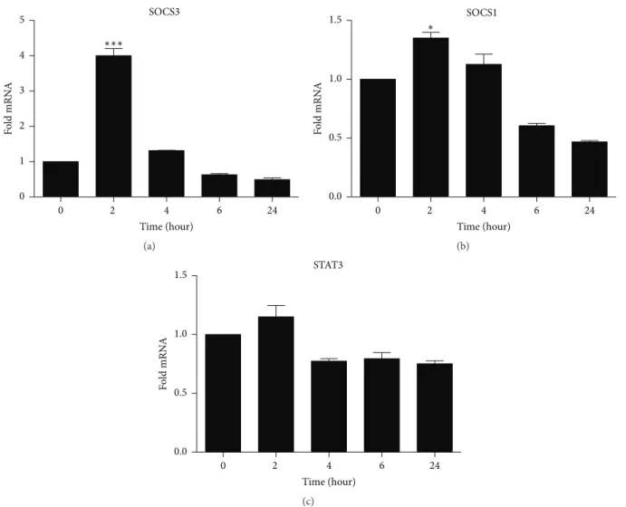
3.4 Relative expression of SOCS3, SOCS1 and STAT3
37 Figure 5: SOCS 3 and SOCS1 increased in TPC-1 cells treated with 15d-PGJ2. TPC1 cells were treated with 15d-PGJ2 (9,8 uM) for 0 to 24 h (A) shows the relative expression of SOCS3 (B) SOCS
1 (C) and STAT3 (C) in the first two hours of treatment and decreased STAT3 four hours after the treatment (C). The date are presented as means + - standard deviation of three replicates from at least three independent tests. An asterisk * indicates statistically significant difference from the control (* P> 0.01; *** p> 0.001).
4. DISCUSSION
38 Several studies have demonstrated that although 15d-PGJ2 is an endogenous ligand of PPAR-γ, most of its anti-neoplastic effects are PPAR-γ-independent [22, 23]. The effects of PPAR-γ ligands may also act by independent mechanisms because they differ widely amongst carcinoma types, and thus must be individually examined.
The present study investigated the role of exogenous 15d-PGJ2 on papillary thyroid carcinoma cells, the TPC-1 cell line. The drug reduced cell viability at the doses of 10 and 20 µM (Figure 1C). Similar results have been found in cell viability in cultures with other cell lines of breast cancer, lung cancer, lymphoma [24, 25], colorectal [26, 27], ovarian [22], gastric [21], pancreatic [28] and prostate cancer [29].
Despite the overall anti-tumoral effect of 15d-PGJ2, most studies have reported both dose and time-dependent responses, with lower doses often promoting opposing effects to the cytotoxic doses [23]. Micromolar doses of 15d-PGJ2 are required to induce lymphoma cell death [30, 31], whereas physiological concentrations of the metabolite are in the range of picomolar to nanomolar [23, 32]. It has also been reported that high doses of 15d-PGJ2 (≥5 μmol/L) caused cytotoxicity in cultured neurons, whereas low concentrations of the agonists (15d-PGJ2, ≤1 μmol/L) suppress rat and human neuronal apoptosis and necrosis induced by H2O2 treatment [32].
39 Differently from normal cells, which phosphorylate STAT under stringent control, STAT3 is continuously phosphorylated in several neoplastic diseases via overproduction of agonists, such as specific cytokines, namely IL-6, and their respective cytokine receptors [40]. This cycle can be further enhanced via antagonism of negative regulators, such as SOCS and tyrosine phosphatases [43]. STAT3 has been reported to play an important role in maintaining cancer stem cells both in vitro and in vivo, implicating an integral involvement of STAT3 in tumor
initiation, progression and maintenance [4]. In fact, this signaling route is so relevant in tumorigenesis that targeting STAT3 in neoplastic bone marrow disease practically interrupted the progression of metastasis [44, 45, 46, 47]. Cumulative evidence points to a clear STAT3-inhibitory effect of 15d-PGJ2 in inflammatory diseases [48, 49, 10]. However, our findings show a small and stable decrease in the relative expression of STAT3 in thyroid cancer cells treated with 15d-PGJ2 (Figure 5C), although not significant. It is possible that STAT3 phosphorylation was prevented by 15d-PGJ2 through upregulation of SOCS3, which results in the inhibition of STAT3 activation, as shown elsewhere [50].
Upregulation of both SOCS3 and SOCS1 was also followed by downregulation of IL-6 expression in TCP-1 cells related to exposure to 15d-PGJ2. SOCS3 is an inducible endogenous negative regulator of STAT3, and it is suggested as a tumor suppressor gene [51]. Negative modulation of SOCS1 and SOCS3 is a survival strategy in most cancer cells [52, 53, 54]. Conversely, overexpression of such cytokine inhibitors may indicate an anti-proliferative response. Indeed, our results have demonstrated that 15d-PGJ2 increased SOCS3 on TPC-1 cells within two hours of contact with the drug, thus supporting the anti-oncogenic nature of this gene (Figure 5B). Interestingly, cells presented diminished levels of SOCS3 and SOCS1 six hours post treatment, which was extended to 24 hours post treatment (Figure 5 A and B), probably because 15d-PGJ2 was already driving cells into apoptosis (Figure 3).
40 Our data demonstrated that apoptosis was detectable in nearly 50% of the TPC-1 cells treated with 15d-PGJ2, compared to 5% in the control group. We have also demonstrated that SOCS3 overexpression was an early event in treated cells, while STAT3 remained stable over 24 hours. It is known that activation of STAT3 in cancers leads to gene expression promoting cell proliferation and resistance to apoptosis [58], but 15d-PGJ2-induced SOCS3 overexpression may have prevented STAT3 phosphorylation [50]. Despite the premature and short-lasting effect of 15d-PGJ2 on SOCS3, its expressive upregulation (Figure 5A) may have been high enough to mediate apoptotic signaling within cells [59].
5. CONCLUSION
The present study shows important anti-proliferative and apoptotic activities in human thyroid cancer cells induced by 15d-PGJ2, Such events are linked with overexpression of SOCS3 that inhibits IL-6 signaling, a key factor in many cancers. This is the first report on 15d-PGJ2-induced SOCS3 expression, which evidences a novel therapeutic option for the treatment of thyroid cancer, and other cancers that are dependent on IL-6 signaling.
Conflict of Interests
The authors declare no conflict of interests.
Authors´ Contribution
Carlos Antonio Trindade da Silva and Carolina Fernandes Reis have equally contributed to this work.
REFERENCES
1. R. Vigneri, P. Malandrino and P. Vigneri, ´´The changing epidemiology of thyroid cancer: Why is incidence increasing? Current Opinion Oncology. vol. 27. no.1, pp
1-7, 2015.
41 3. H.Yu, D. Pardoll, and R. Jove, STATs in cancer inflammation and immunity: a leading role for STAT3. Nature Reviews cancer. vol. 9. Pp 798-809, 2009.
4. A. Xiong, Z. Yang, Y. Shen, J. Zhou and Q. Shen, ´´ Transcription Factor STAT3 as a Novel Molecular Target for Cancer Prevention´´ Cancers, vol. 6, no. 2, pp.
926-957. 2014.
5. H. K. Resemann, C. J. Watson and B. Lloyd-Lewis. ´´The Stat3 paradox: A killer and an oncogene. Molecular and Cellular Endocrinology, vol. 382, pp. 603-611,
2014.
6. C. Schindler, D. E. Levy and T. Decker. ´´ JAK-STAT Signaling: From Interferons and Cytokines´´ The Journal of Biological Chemistry, vol. 282. pp 20059-20063,
2007.
7. D. E. Levy and J. E. Darnell, Jr. ´´STATs: transcriptional control and biological impact´´, Nature Reviews Molecular Cell Biology, vol. 3, pp. 651-662, 2002.
8. D. L. Krebs and D. J. Hilton. ´´SOCS Proteins: Negative Regulators of Cytokine Signaling´´ Stem Cells. vol. 19. no 5. pp. 378-387, 2001.
9. B. Groner, P. Lucks and C. Borghouts. ´´The function of Stat3 in tumor cells and their microenvironment´´, Seminar in Cell & Developmental Biology, vol. 19, no. 4,
pp. 341-350, 2008.
10. Y. J. Surh, H. K. Na, J. M. Park, H. N. Lee, W. Kim, I. S. Yoon and D. D. Kim, 15-Deoxy-Δ¹²,14-prostaglandin J2, an electrophilic lipid mediator of anti-inflammatory and pro-resolving signaling. Biochem Pharmacol. vol. 15;82, no. 10, pp. 1335-51. 2011.
11. T. S. Farnesi-de-Assunção, C. F. Alves, V. Carregaro, et al., ´´ PPAR-gamma agonists, mainly 15d-PGJ2, reduce eosinophil recruitment following allergen challenge´´. Cellular Immunology, vol. 273, pp. 23-29, 2012.
12. M. H. Napimoga, C. A. T. Silva, V. Carregaro et al. ´´ Exogenous administration o 15d-PGJ2-loaded in nanocapsules inhibits bone resorption in a mouse periodontitis model´´, The Journal of Immunology, vol. 189, pp. 1043-1052, 2012.
42 14. V. Paulitschke, S. Gruber, E. Hofstätter et al., ´´Proteome analysis identified the PPARγ ligand 15d-PGJ2 as a novel drug inhibiting melanoma progression and interfering with tumor-stroma interaction´´ PLoS One. vol. 7, no. 9, e46103, 2012.
15. C. D. Allred and M. W. Kilgore, ´´Selective activation of PPARγ in breast, colon and lung cancer cell lines´´Molecular and Cellular Endocrinology, vol. 235, no. 1–2,
pp. 21–29, 2005.
16. E. H. Kim and Y. J. Surh, 15-deoxy-D12,14-prostaglandin J2 as a potential endogenous regulator of redox-sensitive transcription factors. Biochem Pharmacol,
vol. 72, pp. 1516–28, 2006.
17. A. Aiello, G. Pandini, F. Frasca, et al., Peroxisomal proliferator-activated receptor-γ agonists induce partial reversion of epithelial-mesenchymal transition in anaplastic thyroid cancer cells. Endocrinology,vol. 147, no. 9, pp. 4463-4475, 2006.
18. K. J. Livak and T. D. Schmittgen, Analysis of relative gene expression data using real-time quantitative PCR and the 2(-Delta Delta C(T)) Method. Methods, vol. 25,
no. pp. 402–408, 2001.
19. Y. Zhao, Y. Zhang, X.J. Liu, B.Y. Shi. Prognostic factors for differentiated thyroid carcinoma and review of the literature. Tumori, vol. 98, pp. 233–237, 2012.
20. A. Rapoport, O. A. Curioni, A. Amar and R. A. Dedivitis, Review of survival rates 20-years after conservative surgery for papillary thyroid carcinoma. Braz J
Otorhinolaryngol. vol.81, no. 4, pp. 389-93, 2015.
21. N. Takahashi, T. Okumura, W. Motomura, Y. Fujimoto, I. Kawabata and Y. Kohgo, Activation of PPARr inhibits cell growth and induced apoptosis in human gastric cancer cells. FEBS Lett, vol. 455, pp. 135–139, 1999.
22. K. Bräutigam, J. Biernath-Wüpping, D.O. Bauerschlag, C.S. von Kaisenberg, W. Jonat, N. Maass, N. Arnold and I. Meinhold-Heerlein, Combined treatment with TRAIL and PPARγ ligands overcomes chemoresistance of ovarian cancer cell lines.
J Cancer Res Clin Oncol. vol. 137, no. 5, pp. 875-86, 2011.
43 24. J, Eucker, J. Sterz, H. Krebbel et. al., ´´Peroxisome proliferator-activated receptor-gamma ligands inhibit proliferation and induce apoptosis in mantle cell lymphoma´´, Anti-Cancer Drugs. vol. 17, no. 7, pp. 763-769, 2006.
25. J. Yuan, A. Takahashi, N. Masumori et al., ´´ Ligands of peroxisome proliferator-activated receptor gamma have potent antitumor effect against human renal cell carcinoma´´, Urology, vol. 65, no. 3, pp. 594-599, 2005.
26. M. Cekanova, J. S. Yuan, X, Li, K. Kim, S. J. Baek, Gene alterations by peroxisome proliferator-activated receptor gamma agonists in human colorectal cancer cells. Int J Oncol. vol. 32, no. 4, pp. 809-8019, 2008.
27. A. Cerbone, C. Toaldo, S. Laurora, F. Briatore, S. Pizzimenti, M. U. Dianzani, C. Ferretti and G. Barrera, 4-Hydroxynonenal and PPARgamma ligands affect proliferation, differentiation, and apoptosis in colon cancer cells. Free Radic Biol Med. vol. 1;42, no 11, pp. 1661-1670, 2007.
28. S. Kawa, T. Nikaido, H. Unno, N. Usuda, K. Nakayama and K. Kiyosawa, Growth inhibition and differentiation of pancreatic cancer cell lines by PPAR gamma ligand troglitazone. Pancreas , vol. 24, pp. 1–7, 2002.
29. C. L. Chaffer, D. M. Thomas, E.W. Thompson and E. D. Williams, PPARgamma-independent induction of growth arrest and apoptosis in prostate and bladder carcinoma. BMC Cancer. vol. 6, no. 6, pp. 53, 2006.
30. J. Padilla, K. Kaur, H. J. Cao, T. J. Smith and R. P. Phipps. Peroxisome proliferator activator receptor-gamma agonists and 15-deoxy-Δ(12,14)(12,14) -PGJ(2) induce apoptosis in normal and malignant B-lineage cells. J Immunol. vol.
165, pp. 6941-6948, 2000.
31. S. G. Harris and R. P. Phipps, Prostaglandin D(2), its metabolite 15-d-PGJ(2), and peroxisome proliferator activated receptor-gamma agonists induce apoptosis in transformed, but not normal, human T lineage cells. Immunology. vol. 105, pp.
23-34, 2002.
32. T. N. Lin, W. M. Cheung, J. S. Wu, J. J. Chen, H. Lin, J.Y. Liou, S. K. Shyue and K. K. Wu, 15d-prostaglandin J2 protects brain from ischemia-reperfusion injury.
Arterioscler Thromb Vasc Biol. vol. 26, pp. 481-487. 2006.
33. W. E. Naugler, T. Sakurai, S. Kim et. al., ´´Gender disparity in liver cancer due to sex differences in MyD88-dependent IL-6 production´´, Science, vol. 317, no.
44 34. S. P. Gao, K. G. Mark, K. Leslie et. al., ´´Mutations in the EGFR kinase domain mediate STAT3 activation via IL-6 production in lung adenocarcinomas´´, Journal of
Clinical Investigation, vol. 117, no. 12, pp. 3846-3856, 2007.
35. P. Sansone, G. Storci, S. Tavolari, T. Guarnieri, C. Giovannini, M. Taffurelli, C. Ceccarelli, D. Santini, P. Paterini and K. B. Marcu, et al., IL-6 triggers malignant features in mammospheres from human ductal breast carcinoma and normal mammary gland. J Clin Invest vol. 117, pp. 3988–4002, 2007.
36. D. Reynaud, E. Pietras and K. Barry-Holson, ´´IL-6 Controls Leukemic Multipotent Progenitor Cell Fate and Contributes to Chronic Myelogenous Leukemia Development´´, Cancer Cell, vol. 20, no. 5, pp. 661-673, 2011.
37. J. F. Rossi, Z. Y. Lu, M. Jourdan and B. Klein,´´Interleukin-6 as a Therapeutic Targe´´, Clinical Cancer Research. Published online DOI: 10.1158/1078-0432,
2015.
38. L. S. Angelo, M. Talpaz and R. Kurzrock, ´´Autocrine interkin-6 production in renal cell carcinoma: evidence for evolvement of p53´´, Cancer Research, vol. 63,
no. 3, pp. 932-940, 2002.
39. K. Ito, T. Asano, H. Yoshii, A. Satoh, et. al., ´´Impact of thrombocytosis and C-reactive protein elevation on the prognostics for patients with renal cell carcinoma´´,
International Journal of Urology, vol. 13, no. 11, pp. 1365-1370, 2006.
40. Q. Chang, E. Bournazou, P. Sansone, et al. The IL-6/JAK/Stat3 Feed-Forward Loop Drives Tumorigenesis and Metastasis. Neoplasia, vol. 15, no. 7, pp. 848-862,
2013.
41. F. Penas, G. A. Mirkin, E. Hovsepian, A. Cevey, R. Caccuri, M. E. Sales, N. B. Goren, PPARγ ligand treatment inhibits cardiac inflammatory mediators induced by infection with different lethality strains of Trypanosoma cruzi. Biochim Biophys Acta.
vol. 1832, no.1, pp. 239-248, 2013.
42. M. Q. Silva, M. H. Napimoga, C. G. Macedo, F. F. Freitas, H. B. Abdalla, R. Bonfante and J. T. Clemente-Napimoga, 15-deoxy-Δ12,14-prostaglandin J2 reduces albumin-induced arthritis in temporomandibular joint of rats. Eur J
Pharmacol. vol. 740, pp. 58-68, 2014.
43. H. Yu and R. Jove, The STATs of cancer—new molecular targets come of age.
45 44. M. Kortylewski, P. Swiderski, A. Herrmann, L. Wang, C. Kowolik, M. Kujawski, H. Lee, A. Scuto, Y. Liu, C. Yang, et al., In vivo delivery of siRNA to immune cells by conjugation to a TLR9 agonist enhances antitumor immune responses. Nat
Biotechnol , vol. 27, pp. 925–932, 2009.
45. A. Herrmann, M, Kortylewski, M. Kujawski, et al., Targeting Stat3 in the myeloid compartment drastically improves the in vivo antitumor functions of adoptively transferred T cells. Cancer Res, vol.70, pp. 7455–7464, 2010.
46. M. Kujawski, M. Kortylewski, H. Lee, A. Herrmann, H. Kay, and H. Yu, Stat3 mediates myeloid cell–dependent tumor angiogenesis in mice. J Clin Invest,
vol.118, pp. 3367–3377, 2008.
47. M. Kortylewski, M. Kujawski, T. Wang, S. Wei, S. Zhang, S. Pilon-Thomas, G. Niu, H. Kay, J. Mule, W. G. Kerr, et al. Inhibiting Stat3 signaling in the hematopoietic system elicits multicomponent antitumor immunity. Nat Med , vol. 11, pp.1314–
1321, 2005.
48. T. Hosoi, S. Matsuzaki, T. Miyahara, K. Shimizu, Y. Hasegawa, K and Ozawa. Possible involvement of 15-deoxy-Δ(12,14) -prostaglandin J2 in the development of leptin resistance. J Neurochem. vol, 133, no. 3, pp. 343-351, 2015.
49. Y. I. Kim, K. Park, J. Y. Kim, H. S. Seo, K. O. Shin, Y. M. Lee, W. M. Holleran, P. M. Elias and Y. Uchida. An endoplasmic reticulum stress-initiated sphingolipid metabolite, ceramide-1-phosphate, regulates epithelial innate immunity by stimulating β-defensin production. Mol Cell Biol. vol. 34, no. 24, pp. 4368-4378,
2014.
50. B. Carow and M. E. Rottenberg, SOCS3, a Major Regulator of Infection and Inflammation. Front Immunol. vol.19, no. 5, pp. 58. 2014.
51. B. He, L. You, K. Uematsu et. al., ´´SOCS-3 is frequently silenced by hypermethylation and suppresses cell growth in human lung cancer´´, Proceedings
of National Academy of Sciences of the United States of America, vol 100, no. 24,
pp 14133-14138, 2003.
52. G. Li, J. Xu, Z. Wang, et. al., ´´Low expression of SOCS1 and SOCS3 is a poor prognostic indicator for gastric cancer patients´´. Journal of Cancer Research and
Clinical Oncology, [Epub ahead of print]. 2014.
46 Function thought Down-Regulation of Cyclin-Dependent Kinases´´. The American
Journal of Pathology. vol. 174, no. 5, pp. 1921-1930, 2006.
54. I. Bellezza, H. Neuwirt, C. Nemes, et. al ´´ Suppressor of cytokine signaling-3 antagonizes cAMP effects on proliferation and apoptosis and is expressed in human prostate cancer´´. The American Journal of Pathology. vol. 169, no. 6, pp.
2199-2208, 2006.
55. S. Grivennikov, E. SKarin, J. Terzic, et al. IL-6 and Stat3 are required for survival of intestinal epithelial cells and development of colitis-associated cancer. Cancer
Cell. vol.15. no.2.pp.103-13.2009.
56. E. J. Park, J. H. Lee, G. Y. Yu , et al., Dietary and genetic obesity promote liver inflammation and tumorigenesis by enhancing IL-6 and TNF expression. Cell.
vol.140. no.2. pp.197-208. 2010.
57. L. A. Gilbert and M. T. Hemann, DNA damage-mediated induction of a chemoresistant niche. Cell. vol.143.no3. pp.355-66.2010.
58. O. A. Timofeeva, N. I. Tarasova, X. Zhang, .et al., STAT3 suppresses transcription of proapoptotic genes in cancer cells with the involvement of its N-terminal domain. Proc Natl Acad Sci U S A. vol. 110, no.4. pp.1267-72. 2013.
59. Z. Liu, L. Gan, Z. Zhou, W. Jin and C. Sun, SOCS3 promotes inflammation and apoptosis via inhibiting JAK2/STAT3 signaling pathway in 3T3-L1 adipocyte.
47
CAPÍTULO 3.
Soluble epoxide hydrolase pharmacological inhibition decreases alveolar bone loss by modulating host inflammatory response, RANK-related
signaling, ER stress and apoptosis
Carlos Antonio Trindade-da-Silva1,2,3Ϯ, Ahmed Bettaieb4Ϯ, Marcelo Henrique
Napimoga3, Kin Sing Stephen Lee1, Bora Inceoglu1, Carlos Ueira-Vieira2, Donald
Bruun6, Sumanta Kumar Goswami1, Fawaz G. Haj5 and Bruce D. Hammock1.
Ϯ
Contributed equally to this work.
1
Department of Entomology and Nematology and UC Davis Comprehensive Cancer Center, University of California, Davis, CA 95616, United States;
2
Institute of Genetics and Biochemistry, Federal University of Uberlândia, Uberlândia, 38400-902, Brazil;
3
Laboratory of Immunology and Molecular Biology, São Leopoldo Mandic Institute and Research
Center, Campinas, 13045-755, Brazil;
4
Department of Nutrition, University of Tennessee-Knoxville, Knoxville, TN 37996 5
Nutrition Department, University of California, Davis, CA 95616; 6
Department of Molecular Biosciences, School of Veterinary Medicine, University of California, Davis, CA 95616, United States.
48 [R01ES002710, P42ES004699, ES025598-01A1, 1K99ES024806]; Brazilian funding agencies São
Paulo Research Foundation (FAPESP) and National Council for Scientific and Technological
Development (CNPq); MHN was supported by grant [2015/23556-0](FAPESP); [303555/2013-0]
(CNPq); F.G.H laboratory is funded by NIH [R01DK090492, R01DK095359]; A.B is
funded by NIH/NIDDK [R00DK100736]. Authors A.B., F.G.H, B.I., S.K.L, and
B.D.H are co-inventors on patents related to sEH by the University of California,
B.D.H. and B.I. are co-founders of Eicosis
LLC. The authors declare no competing financial interests.
Running Title: Inhibition of sEH blocks alveolar bone loss
Corresponding author:
Bruce D. Hammock
Professor
Department of Entomology and Nematology & UCD Comprehensive Cancer Center, Director,
NIEHS-UCD Superfund Research Program PI, NIH Biotechnology Training Program
University of California – Davis
One Shields Ave, Davis, CA 95616, USA
(530) 752-7519 office
(530) 752-8465
message (530)
752-1537 fax
bdhammock@ucd
49 Text page count: 26
Number of tables: none
Number of figures: 6
Number of supplementary figures: 2
Number of references: 45
Word count, abstract: 177
Word count, introduction: 699
Word count, discussion: 1280
Recommended section assignment: Drug Discovery and Translational Medicine