Universidade de Lisboa
Faculdade de Farmácia
Glycosylation in cancer
Mechanisms and clinical implications
- Pancreatic cancer, an overview -
Ana Clara Almeida Aparício
Mestrado Integrado em Ciências Farmacêuticas
Universidade de Lisboa
Faculdade de Farmácia
Glycosylation in cancer
Mechanisms and clinical implications
- Pancreatic cancer, an overview -
Ana Clara Almeida Aparício
Monografia de Mestrado Integrado em Ciências Farmacêuticas
apresentada à Universidade de Lisboa através da Faculdade de
Farmácia
Orientador: Professora Doutora Ana Cristina Ferreira da Conceição
Ribeiro, Professora auxiliar FFUL
Resumo
Atualmente, a Glicobiologia desempenha um papel fulcral na investigação do cancro, dada a sua participação em diversos mecanismos e o seu acesso a uma ampla gama de alvos de elevado interesse diagnóstico e terapêutico. As aberrações na glicosilação de proteínas e polissacáridos desempenham um papel determinante na génese do tumor pancreático, influenciando a progressão do cancro, metástase, resposta imune e resistências a quimioterapia. A expressão anormal de glicanos pode afetar a atividade de várias glicoproteínas, incluindo mucinas, recetores de superfície, adesinas, proteoglicanos, bem como dos seus alvos e ligandos, culminando assim num aumento da agressividade do cancro e num microambiente favorável para o crescimento tumoral. Recentes avanços na área glicoproteómica, glicómica e noutras técnicas de bioquímica, abriram caminho para uma compreensão mais próxima do mecanismo complexo de eventos de glicosilação que rodeiam a génese tumoral, e a forma como estes coordenam as atividades moleculares a nível genómico, proteómico e metabólico implicadas no adenocarcinoma pancreático. Várias estratégias foram exploradas visando a glicosilação de proteínas e polissacáridos para o desenvolvimento diagnóstico e terapêutico do cancro pancreático.
Abstract
Nowadays, glycobiology plays a major role in cancer research, given its part in many cancer mechanisms and its access to a series of targets with valuable diagnostic and therapeutic purposes. Aberrations in protein glycosylation and polysaccharides play a decisive role in pancreatic tumorigenesis, through influencing cancer progression, metastasis, immunoresponse and chemoresistance. Abnormal expression in sugar moieties can impact the activity of various glycoproteins, including mucins, surface receptors, adhesive proteins, proteoglycans, as well as their effectors and binding ligands, culminating in an increase in pancreatic cancer invasiveness and a cancer privileged microenvironment. Recent progress in glycoproteomics, glycomics and other chemical biology techniques has cleared the path to better understand the complex mechanism of glycosylation events and how they mediate molecular activities in genomics, proteomics and metabolomics implicated in pancreatic adenocarcinoma. A wide range of strategies have been demonstrated targeting protein glycosylation and polysaccharides for diagnostic and therapeutic development.
Agradecimentos
A elaboração desta tese, assim como de todo o mestrado integrado, teria sido impossível sem o apoio incondicional da minha família e amigos.
À professora Doutora Ana Cristina Ribeiro, agradeço toda a sua paciência, apoio e dedicação, por tudo o que me ensinou. Sinto-me muito privilegiada por ter tido a oportunidade de ser tão bem orientada, muito obrigada por tudo.
À minha família, que sempre acreditou em mim e sempre me motivou a seguir, por toda a paciência e apoio, teria sido impossível sem vocês. O meu refúgio, obrigada. Aos meus amigos, obrigada por todas as palavras amigas e de motivação nos momentos certos, por nunca me deixarem desistir, por saber que poderei sempre contar convosco.
Acronyms
AFP – α-fetoprotein Asn – Asparagine
C1GALT1C1 – C1GalT1-specific chaperone 1 C2GnT – β1,6‑Nacetylglucosaminyltransferase CA19-9 – Carbohydrate antigen 19-9
CRC – Colorectal cancer ECM – Extracellular matrix
EGFR – Epidermal growth factor receptor FAK – Focal adhesion kinase
FGFR – Fibroblast growth factor FUC-T – Fucosyltransferase GAG – Glycosaminoglycan Gal – Galactose GlcNAc – N-acetylglucosamine GalNAc – N-acetylgalactosamine GnT-V – N-acetylglucosaminyltransferase V GnT-III – N-acetylglucosaminyltransferase III GPI – Glycosylphosphatidylinositol
HBP – Hexosamine biosynthetic pathway HCC – Hepatocellular carcinoma
HER – Human Epidermal growth factor Receptor 2 HSPGs – Heparan sulfate proteoglycans
IPMN – Intraductal papillary mucinous neoplasm MCN – Mucinous cystic neoplasms
MET – Hepatocyte growth factor-β
MGAT5 – Mannoside acetylglucosaminyltransferase 5 MMP – Matrix metalloproteinases
O-GalNAc – O-linked β-N-acetylgalactosamine OGA – O-GlcNAcase
OGT – O-GlcNAc transferase
PanIN – Pancreatic intraepithelial neoplasias PDAC – Pancreatic ductal adenocarcinoma PDGFR – Platelet derived growth factor receptor ppGalNAcTs – Polypeptide GalNAc transferases PSA – Prostate-specific antigen
RTK – Receptor tyrosine kinase Ser – Serine
SLea – Sialyl Lewis a
SLex – Sialyl Lewis x
ST6GalNAc-I – α-GalNAc α-2,6-sialyltransferase I STn – Sialyl Tn antigen
Thr – Threonine
T antigen – Thomsen-Friedenreich antigen Tn antigen – Monosaccharide GalNAc
VEGFA – Vascular endothelial growth factor A
Table of contents:
1 Introduction ... 1
1.1 Cell glycome and glycosylation ... 1
2 Modified glycosylation in cancer ... 3
2.1 Sialylation ... 5
2.2 Fucosylation... 6
2.3 Branching and bisecting GlcNAc N-glycans ... 7
2.4 Truncated O-glycans ... 8
3 Impact of glycosylation in cancer cells ... 11
3.1 Glycosylation in tumor cell-cell adhesion ... 11
3.1.1 GnT-V expression ... 11
3.1.2 GnT-III expression ... 11
3.1.3 Sialylated glycans expression ... 12
3.2 Glycosylation in cell-matrix interaction and signaling ... 13
3.2.1 Heparan sulfate proteoglycans ... 13
3.2.2 CD44 expression ... 13
3.2.3 Integrin expression ... 14
3.2.4 Modifications in N-linked β1,6-branching ... 15
3.3 Glycosylation in cancer metabolism and signaling ... 16
3.3.1 O-GlcNAcylation ... 17
3.3.2 N-glycan branching ... 17
3.3.3 Gangliosides ... 19
4 Glycosylation in cancer immune response ... 19
5 Glycans in cancer diagnosis and treatment ... 20
5.1 Cancer biomarkers ... 21
6 Pancreatic Cancer – an overview ... 23
6.1 Aberrant glycosylation in pancreatic cancer ... 23
6.2 The Sialyl Lewis antigens (SLa and SLex) ... 23
6.3 Truncated O-glycans ... 24 6.4 N-glycans ... 25 6.5 Mucins ... 26 6.6 The HBP pathway ... 27 6.7 Proteoglycans ... 27 6.8 Galectins ... 28 6.9 Future perspectives ... 31 7 Conclusions ... 32 Bibliography ... 33
1 Introduction
Nowadays, glycobiology plays a major role in cancer research, given its part in many cancer mechanisms and its access to a series of targets with valuable diagnostic and therapeutic purposes.
Glycosylation steps up as a crucial regulatory mechanism, as it controls most physiopathological processes. Human glycome keeps an impressive amount of biological information, linking disease to defects in glycosylation, making it indispensable to be researched.
Most secretory and membrane-bound proteins produced by mammalian cells contain covalently linked sugar chains with diverse structures. The glycosylation form and density of glycans on a protein can be altered significantly in association with changes in cellular pathways and processes resulted from diseases, such as malignancy. In fact, altered glycosylation patterns have long been recognized as hallmarks in epithelial cancer (1–4), including pancreatic ductal adenocarcinoma (PDAC), which accounts for about 90% of pancreatic cancer.
Glycan diversity arises from differences in monosaccharide composition (for example, galactose (Gal) or N-acetylgalactosamine (GalNAc)), in linkage between monosaccharides (for example, between carbons 1 and 3 or carbons 1 and 4), in anomeric state, in branching structures, in other substitutions (such as sulfation state) and in linkage to their aglycone part (protein or lipid). (5)
Understanding the biological functions of each glycan along with the glycan-binding proteins (including galectins and sialic acid-binding immunoglobulin-type lectins (siglecs)), promises to accomplish important contributions to the cancer field. (6) The various types of glycoconjugates interfere with key cancer cell mechanisms, along with tumor microenvironment, resulting in cancer progression. This thesis is focused on the role of glycans in the genesis and progression of cancer, as well as the developments in glycobiology and their applications in the oncology field.
1.1 Cell glycome and glycosylation
The cellular membrane exhibits a glycan component that is considered a cellular fingerprint of cells of different tissues and different organs.
Glycosylation is the enzyme-catalyzed covalent attachment of a carbohydrate to a polypeptide, lipid, polynucleotide, carbohydrate, or other organic compound, generally catalyzed by glycosyltransferases, using specific sugar nucleotide donor substrates. (5) Protein glycosylation occurs in the endoplasmic reticulum and Golgi apparatus in multiple enzymatic steps. The resulting glycoconjugates are categorized according to the nature and linkage to their aglycone (non-glycosyl) part. Glycoproteins, linked to the cell membrane, carry glycans covalently attached, via nitrogen or oxygen linkages, to a polypeptide backbone, resulting in N-glycans or O-glycans, respectively. (7)
N-linked glycans are attached to the amide group of asparagine residues in a defined
Asn-X-Ser/Thr sequence (where X can be any aminoacid except proline). O-linked glycans are bound to the hydroxyl group on serine or threonine residues (8). One unique subclass of glycosylation is the phosphorylation-like, reversible O-GlcNAcylation (9). Less common forms of glycosylation include glycosylphosphatidylinositol anchors attached to protein carboxyl terminus, C-glycosylation that occurs on tryptophan residues (10) and S-linked C-glycosylation through a sulfur atom on cysteine or methionine (11). In addition to protein glycosylation, proteoglycans and hyaluronan are major components of the extracellular matrix (ECM), which are implicated in cell proliferation and migration.
An average protein O-glycosylation begins via GalNAc, which is the first monosaccharide that binds serine or threonine in specific forms of protein O-glycosylation, and it can be elongated into a multitude of diverse structures. (12) The different types of O-glycans are attached by distinct paths, such as via O-mannose or the nucleocytoplasmic glycan O-linked β-N-acetylglucosamine (O-GlcNAc). (13) Furthermore, other considerable classes of glycoconjugates include the proteoglycans and glycosphingolipids. The proteoglycans have one or more glycosaminoglycan (GAG), such as heparan sulfate, keratan sulfate and chondroitin sulfate. (5)
The glycosphingolipids are composed of a glycan linked to a lipid ceramide, which is a sphingosine and a fatty acid linked. (14) Glycosphingolipids are classified according to their glycan part of the molecule, both structurally and functionally. (5) Typically the first sugars linked to ceramide are β-linked galactose (galactosylceramide) or glucose (glucosylceramide). In vertebrate glycosphingolipids, the glucose moiety is typically switched by a β-galactose, conceiving a lactosylceramide (D-galactosyl‑1,4‑β-D-glucosylceramide). Glycosphingolipids also include a series of neutral ‘core’ structures and gangliosides, which usually carry one or more sialic acids and have been shown to regulate receptor tyrosine kinase (RTK) signaling. (15)
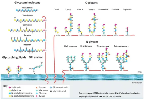
Figure 1. Glycan classes present on the cellular membrane – The main classes of glycans are represented on this figure: glycosaminoglycans (GAGs), N-glycans, O-glycans, glycosphingolipids, and glycosylphosphatidylinositol (GPI) anchor. Heparin sulfate, chondroitin sulfate, hyaluronic acid, dermatan sulfate, and keratin sulfate, are the GAGs portrayed. NS, 2S, 4S, and 6S illustrate the sulfation positions on the GAGs chains. Representative examples of complex-type N (bi–tri–tetra–antennary) and high-mannose N-glycans are illustrated. Also depicted are core 1–4 glycans, high-mannose, fucose, and O-glucose structures. Glycan linkages are identified by the anomeric configuration (a or b) of the donor saccharide and by the ring position (1–6) of the acceptor sugar. The GPI anchor and examples of glycosphingolipids are also represented. (figure adapted from “Glycosylation and Integrin Regulation in Cancer”, Marsico, G; Russo, L; Quondamatteo, F; Pandit, A.; 2018, Elsevier)
2 Modified glycosylation in cancer
Over more than six decades, changes in glycosylation were associated with oncogenic events. (16,17) Those associations were supported with the major innovation that is monoclonal antibody technology, which proved that tumor-specific antibodies were linking straight to carbohydrate epitopes and, in most cases, these were oncofetal antigens existent on tumor glycoproteins and glycosphingolipids. (18)
The glycosylation of proteins broadens the molecular heterogeneity along with the functional diversity within cell populations. This event occurs due to the specificity of the aberrant glycan modifications, site, cell and protein wise.
Asn asparagine; ECM extracellular matrix; Etn–P phosphoethanolamine; PI phosphatidylinositol; Ser, serine; Thr, threonine
Two main mechanisms of tumor-associated modifications of carbohydrate structures were first described by Hakomori and Kannagi, as incomplete synthesis and neo-synthesis process.(19) The incomplete neo-synthesis process, characteristic of early stage cancers, is a result of the impairment of a normal synthesis of complex glycans expressed in normal epithelial cells, leading to the biosynthesis of truncated glycans, such as sialyl Tn (STn) expression in breast cancer. (20) As for neo-synthesis, occurring more often in advanced stages of cancer, is the cancer-associated induction of genes implicated in the expression of carbohydrate determinants, such as the de
novo expression (expression of protein sequences not based on existing natural
sequences) of certain antigens like sialyl Lewis a (SLea) and SLex in various cancers.
(21)
Generally, the modifications from the common glycosylation pathway takes place in cancer cells, leading to altered expression due to various factors. In the first place, altered expression of glycans can be a result of under or overexpression of
glucosyltransferases (due to dysregulation at the transcriptional level (21–24),
alteration of chaperone function (25,26), and/or modified glycosidase activity (27)). Secondly, altered glycan expression can also be attributed to changes in the tertiary
conformation of the peptide backbone and the conformation of the nascent glycan
chain. Moreover, the diversity of various acceptor substrates in conjunction with the
availability and abundance of the sugar nucleotide donors and cofactors can
cause differences in the glycosylation pathway. (28) Lastly, the expression and
localization of the key glucosyltransferases in the Golgi apparatus can also lead to
changes in glycan expression.(29,30)
Modified localization and/or shifts in the activity of the glucosyltransferases stems the synthesis of immature core glycan structures. (31,32) Research shows that early acting enzymes synthesizing core O-glycans, as the GalNAc transferases, core 1 GalNAc β1,3‑galactosyltransferase 1 (C1GalT1) and core 2 β1,6‑N-acetylglucosaminyltransferase (C2GnT), are enriched in cis- and medial-Golgi cisternae. (30–33) The overexpression of α-GalNAc α-2,6‑sialyltransferase I (ST6GalNAc‑I; encoded by ST6GALNAC1), which is the enzyme responsible for STn biosynthesis, leads to expression of enzymes in all Golgi cisternae, inevitably disrupting glycosylation by early adding sialic acid to form the STn antigen. (21,34) The most common glycosylation alterations in cancer are sialylation, fucosylation,
2.1 Sialylation
Sialylation plays a crucial role in cellular glycosylation, since sialylated carbohydrates are involved in cellular recognition, cell adhesion and cell signaling. Moreover, it has been closely associated with cancer an increase in global sialylation, particularly in α2,6- and α2,3-linked sialylation, as a result of altered glycosyltransferases expression. (38)
The lactosamine chains are commonly terminated with a sialic acid. For instance, β‑galactoside α2,6‑sialyltransferase I (ST6Gal‑I) is an enzyme with an altered expression in many cancers, such as colon, ovarian and stomach, and it gives origin to α2,6‑sialylated lactosamine (Sia6LacNAc). Furthermore, this enzyme is disclosed as a predictive marker of very poor prognosis in colon cancer. (39,40)
SLea and SLex are other two major sialylated antigens closely associated with
malignant cancers, and SLex expression levels have been correlated with poor
prognosis in cancer patients. (41,42)
SLex is a ligand for selectins (43), which are a family of three proteins that mediate
adhesive interactions between leukocytes and the endothelium and between
leukocytes and platelets in the blood vascular compartment, known as L(leukocyte)-, P(platelet)-, and E(endothelial)-selectin. (43) Thus, selectins are vascular cell adhesion molecules that belong to a family of C-type lectins, that require calcium for binding. In inflammatory events, these proteins mediate the attachment of leukocytes to the endothelium throughout the process of leukocyte extravasation. (43) The metastatic cascade in cancer is regulated by SLex interactions with selectins, through the
formation of emboli of cancer cells and platelets, causing their arrest on endothelia, thus determining the malignant behaviour and metastasis development.(44) The use of specific GAGs (Glycosaminoglycans), such as heparin, has been shown to attenuate tumor metastasis in animal models, through the inhibition of P-selectin-mediated interactions of platelets with carcinoma cell-surface ligands. (45)
The SLea tetrasaccharide, detectable by the serological assay CA19-9 (it detects the
epitope of SLea on mucins, and other adhesive molecules such as carcinoembryonic
antigen), is closely associated with cancer, and is currently widely used in the clinical practice. The CA19-9 assay is mostly used as a monitor for clinical response to therapy in patients with an established diagnosis of pancreatic, gastric, colorectal or biliary
cancer. (46,47) Also, high preoperative concentrations of CA19-9 have been shown
Another form of increased sialylation is the elevated expression of polysialic acid in
cancer, which is correlated with many types of cancer and is regularly expressed in
high-grade tumours. Polysialic acid is frequently present in neural cell adhesion molecule 1 (NCAM1), resulting in aggressiveness and poor clinical outcomes in cancer, including neuroblastomas, gliomas and lung cancer. (49,50)
Gangliosides, acidic glycosphingolipids containing one or more sialic acid (N-acetylneuraminic acid or N-glycolylneuraminic acid) residue(s) in their carbohydrate moiety, are too overexpressed in tumours such as neuroblastomas, melanomas and
breast cancer, where they mediate cell proliferation, tumor growth and cancer cell
migration. (15,51,52)
2.2 Fucosylation
Fucosylation is another event associated with cancer. Fucosyltransferases are the enzymes responsible for the synthesis of Fucosylated glycans, and they include Fuc-Ts, Fuc-TI–Fuc-TXI (encoded by FUT1–FUT11, in which FUT3 is disclosed as the Lewis gene, Le). As a non-extendable modification, fucosylation is typically subdivided into: terminal fucosylation (creating specific Lewis blood-group antigens, such as Lex
and Ley, and Lea and Leb) and core fucosylation. (53) The last steps of the
biosynthesis of SLe antigens consist of the α1,3- or α1,4-fucosylation of a previously α2,3-silaylated type 1 (SLea) or type 2 (SLex) chains. (54)
It has been demonstrated that the elevated expression of SLex in adult T cell leukemia
cells is apparently dependent on Fuc-TVII activity. This leukemia is provoked by the human T-lymphotropic virus 1 (HTLV-1), and this virus encodes a transcriptional activator protein, TAX, that regulates the FUT7 gene encoding Fuc-TVII, the enzyme responsible for controlling the SLex synthesis in leukocytes. (55)
The expression of SLex appears to be mainly regulated by Fuc-TVI, the
fucosyltransferases that is encoded by FUT6, in breast tumours. (56) Nonetheless, in gastrointestinal cancer, the synthesis of SLe antigens can depend on the integrated expression of various glycosyltransferases. In colon cancer tissues, the glycolipidic expression of SLex and of SLea antigens is associated to the activation of
a certain β1,3GlcNAc transferase. This last enzyme is responsible for the synthesis of a sugar chain that is a precursor for both type 1 ad 2 Lewis structures. (57) In the gastritis caused by the bacteria Helicobacter pylori,(58,59) a similar mechanism occurs. The bacterium expresses adhesins able to recognize glycan receptors expressed by the gastric epithelium, ergo provoking gastric ulcers and, potentially,
gastric carcinogenesis. (60) Fuc-TVI is also closely associated as a dominant enzyme modulating the SLex biosynthesis in colorectal cancer (CRC). (61)
Core fucosylation is disclosed as the addition of a α1,6-fucose to a core GlcNAc residue of a N-Glycan, as a result of Fuc-TVIII’s (encoded by FUT8) action. It can be observed in cancers like lung cancer and breast cancer, the overexpression of FUT8 and core fucosylation.(62,63) This increased core fucosylation can be observed in the serum levels during an event of hepatocarcinogenesis. (64) Curiously, core fucosylation of α-fetoprotein is an approved biomarker for the early diagnosis of hepatocellular carcinoma (HCC), distinguishing it from chronic hepatitis and liver cirrhosis. (65) Furthermore, in breast cancer, increased core fucosylation of epidermal growth factor receptor, the also known as EGFR, is correlated to increased dimerization and phosphorylation, resulting in increased EFGR-mediated signaling giving origin to malign cell growth and tumours. (62,63)
2.3 Branching and bisecting GlcNAc N-glycans
In malignant cancer, it is frequent to observe an increased expression of complex β1,6-branched N-linked glycans, thus making it a very common glycosylation change in cancer cells.(35,66)
The raised expression of GlcNAc-branching N-glycan, is a consequence of an increment in the activity of GnT-V (N-acetylglucosaminyltransferase V), which is encoded by the mannoside acetylglucosaminyltransferase 5 (MGAT5) gene. The RAS-RAF-MAPK signaling pathway is responsible for the regulation of MGAT5 expression, and it is activated during cancer processes. (66) As the branched N-glycans start to be expressed, they are further modified by the β-1,4-GalTs, and elongated with
poly-N-acetyllactosamine (repeats of Galβ1,4GlcNAcβ1,3) through the action of β1,3-GnTs,
being further terminated with sialic acid and fucose.
The poly-N-acetyllactosamine structure connects with galectins. Galectins are a group of conserved carbohydrate-binding proteins, with important roles in cancer, such as contributing to neoplastic transformation, tumor cell survival, angiogenesis and tumor metastasis. The binding between a poly-N-acetyllactosamine and a galectin forms galectin-glycan structures named “lattices”. (67)
It has been reported that the overexpression of MGAT5 in an immortalized lung epithelial cell line resulted in the loss of contact inhibition, increased cell motility and
tumor formation in athymic mice (68), and also it enhanced invasion and metastasis
regulator element in breast carcinoma formation in a Her2-transgenic mouse mammary tumor model. (70) Moreover, downregulation of GnT-V in mouse mammary cancer cell lines showed a significant suppression of tumor growth and metastasis. (69) The progression of breast cancer and its metastasis induced by a viral oncogene in transgenic mice is considerably noticeably suppressed in MGAT5-deficient background. (71) Also, GnT-V-mediated glycosylation has been shown to regulate the cancer stem cell compartment and tumor progression trough WNT signaling. (72) As opposed to the function of GnT-V, GnT-III (which is encoded by MGAT3) catalyses the addition of bisecting GlcNAc N-glycans in a β-1,4 linkage, inhibiting the additional processing and elongation of N-glycans, like the β1,6-branching structures. GnT-III assumes an opposite role to GnT-V in cancer, as it is involved in the suppression of
cancer metastasis. (73) It was tested in mouse melanoma cells the transfection of
MGAT3 into this high metastatic potential tissue, and it resulted in a significant reduction of β1,6GlcNAc branching (as a result of the enzymatic competition between GnT-III and GnT-V), which led to a notable suppression of lung metastasis in mice. The mechanism of tumor metastasis suppression carried out by GnT-III is through the
regulation of key proteins, such as EGFR, integrins and cadherins (63,74), as will
be explained further.
2.4 Truncated O-glycans
The overexpression of truncated O-glycans is another prevalent trait of tumors. The GalNAc-type O-glycans, also known as mucin-type O-glycans, are most commonly found in transmembrane and secreted glycoproteins. Throughout the malignant phase of tumors, abnormal glycosylation also takes place in glycoproteins with aberrant expression of shortened or truncated glycans, such as the disaccharide Thomsen-Friedenreich antigen (T antigen, also known a core 1) and the monosaccharide GalNAc (also known as Tn), and their respective sialylated forms (ST and STn (Neu5Acα2-6GalNAcα-R)), which result from the incomplete synthesis of O-glycans.(75)
The enzymes responsible for initiating the mucin-type O-glycosylation (7,12), polypeptide GalNAc transferases (also known as ppGalNAcTs), have often an altered expression in cancer events. (76,77) The ppGalNAcTs handle the sites and density
of O-glycan occupancy (7,12), and any alteration in their expression leads to
alteration in O-glycosylation. Another way of inducing the expression of truncated glycans exposure is through the enzymatic competition for the same substrate, and exposure of protein epitopes that would otherwise be hidden in the normally
glycosylated protein. The activities of both C2GnT and α2,3-sialyltransferase I (ST3Gal-I) have been reported as to determine the O-glycan structure in cancer cells. (78) These relative activities are in the foundation of the aberrant expression of tumor-associated epitopes in glycoproteins, such as mucins in breast (78) and gastric cancer. (79)
STn is hardly ever expressed in healthy tissues, but it can be detected in most carcinomas, namely those from the pancreas (80,81), stomach (82,83), colorectum (82,84), breast (34), bladder (85) and ovary (86), associating it with increased cell adhesion, increased tumor growth, increased tumor cell migration, invasion and poor prognosis. The overexpression of ST6GalNAc‑I results in the aberrant synthesis of
STn in cancer. When a mutation occurs in the T-synthase C1GalT1-specific
chaperone 1 (C1GALT1C1), it can block further the O-glycan elongation and shift the pathway towards the generation of Tn, and this can also lead to STn expression through the action of ST6GalNAc‑I.(87,88) Hence, STn is being considered as a crucial prognostic marker and target for the design of anticancer vaccines. (89)
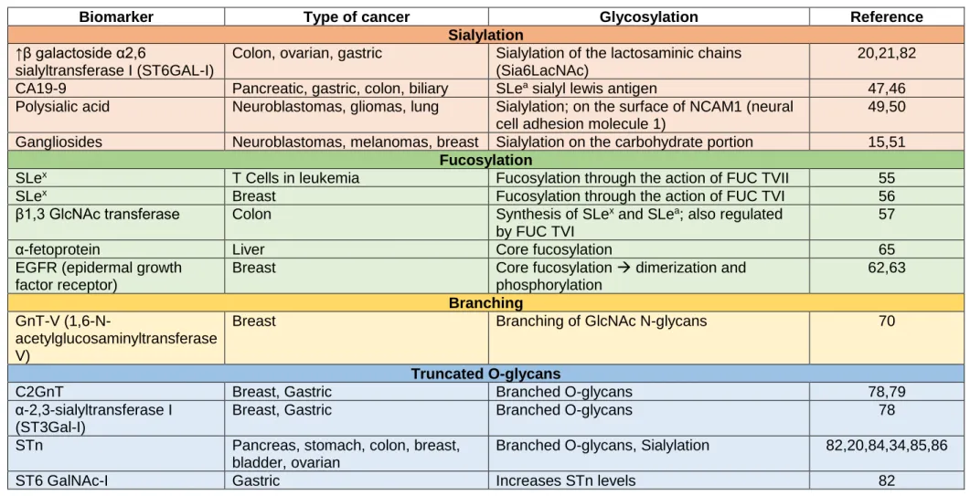
The following table summarizes some of the different types of biomarkers according to their glycosylation mechanism and type of cancer.
Table 1 – Different biomarkers from different types of cancer, according to their type of glycosylation
Biomarker Type of cancer Glycosylation Reference Sialylation
↑β galactoside α2,6
sialyltransferase I (ST6GAL-I)
Colon, ovarian, gastric Sialylation of the lactosaminic chains (Sia6LacNAc)
20,21,82 CA19-9 Pancreatic, gastric, colon, biliary SLea sialyl lewis antigen 47,46
Polysialic acid Neuroblastomas, gliomas, lung Sialylation; on the surface of NCAM1 (neural cell adhesion molecule 1)
49,50 Gangliosides Neuroblastomas, melanomas, breast Sialylation on the carbohydrate portion 15,51
Fucosylation
SLex T Cells in leukemia Fucosylation through the action of FUC TVII 55
SLex Breast Fucosylation through the action of FUC TVI 56
β1,3 GlcNAc transferase Colon Synthesis of SLex and SLea; also regulated
by FUC TVI
57
α-fetoprotein Liver Core fucosylation 65
EGFR (epidermal growth factor receptor)
Breast Core fucosylation → dimerization and phosphorylation 62,63 Branching GnT-V (1,6-N-acetylglucosaminyltransferase V)
Breast Branching of GlcNAc N-glycans 70
Truncated O-glycans
C2GnT Breast, Gastric Branched O-glycans 78,79
α-2,3-sialyltransferase I (ST3Gal-I)
Breast, Gastric Branched O-glycans 78
STn Pancreas, stomach, colon, breast, bladder, ovarian
Branched O-glycans, Sialylation 82,20,84,34,85,86
3 Impact of glycosylation in cancer cells
Inflammation, immune surveillance, cell-cell adhesion (74,90,91), cell-matrix interaction (74), inter- and intracellular signaling (92–95) and cellular metabolism, are all processes involved in cancer events, and they all involve glycans in their mechanisms. (96,97) Glycans modulate the functional activity of proteins through the modification of protein conformation and structure (98), making it crucial to understand the glycan-based interactions in cancer, as it can contribute immensely to understand the cancer processes.
3.1 Glycosylation in tumor cell-cell adhesion
What defines a malignant tumor is its ability to overcome cell-cell adhesion and to invade the surrounding tissue. Epithelial cadherin (E-cadherin) is a transmembrane glycoprotein (99) and a predominant epithelial cell-cell adhesion molecule in cancer. (100) As such, when glycans interfere with E-cadherin functions they have a crucial impact on tumor cell-cell adhesion, as they cause loss of cell-cell adhesion.
3.1.1 GnT-V expression
The GnT-V overexpression in gastric cells (mentioned above) promotes E-cadherin cellular mislocalization from the membrane into the cytoplasm, thus causing its functional impairment. (90,91) The binding between E-cadherin and GnT-V-mediated
β1,6GlcNAc-branched N-glycans generates non-functional adherens junctions,
hence compromising cell-cell adhesion (90,91,101) and downregulating signaling
pathways (102), leading to tumor invasiveness and metastases. (103) There is a way
of avoiding this abnormal glycosylation through a specific Asp site, thus improving E-cadherin functions in cancer. (104) Curiously, a correlation is observed between gastric carcinoma patients with loss of E-cadherin function (not explained either at the genetic nor the structural level) and an increase in β1,6GlcNAc-branched N-glycans on E-cadherin. (60,91) Also, cadherins depend on calcium ions to function, and lectins spend considerable amounts of calcium and magnesium whilst functioning, hence the removal of this calcium abolishes adhesive activity and turns the cadherins vulnerable to proteases.
3.1.2 GnT-III expression
GnT-III emerges again counteracting GnT-V activity, through the interaction between E-cadherin and GnT-III mediated bisecting GlcNAc N-glycans. (73,91) A connection has been reported associating this E-cadherin glycan modification with a delayed
turnover rate at cell membrane (91,105), an inhibition of endocytosis (91), a diminished phosphorylation of β-catenin that remained in complex with E-cadherin(106), and an elevated stability of adherens junctions, thus boosting tumor suppression. (60,90,91) Furthermore, research has associated GnT-III with suppression of epithelial-to-mesenchymal transition. (24,107)
Accordingly, the competitive action of GnT-III towards GnT-V establishes a mechanism between E-cadherin-mediated cell-cell adhesion and its glycosylation, determining either the tumor suppression or the tumor metastasis, respectively. (60,108)
3.1.3 Sialylated glycans expression
High levels of sialylated glycans, a common feature in cancer events, leads to the high expression of tumor associated antigens. (1,35) The sialylated antigens promote cell detachment from the tumor mass through electrostatic repulsion of negative
charges, detachment which inhibits and disrupts the cell-cell adhesion.(109,110) A
research experiment with breast cancer cells transfected with ST6Gal-I resulted in augmented cell migration and diminished cell-cell adhesion in vitro. (111)
Moreover, sialylated glycans, SLex for instance, can aid the adhesion of tumor cells to
vascular endothelial cells, via their interaction with selectins, which are glycoproteins, consisting of an extracellular lectin-like domain, and calcium dependent to interact with fucosylated ligands. E-selectin for instance, also known as CD62 antigen-like family member E (CD62E), is a selectin cell adhesion molecule expressed only on endothelial cells activated by cytokines, thus making selectins moderators of the initial phases of cancer metastases. (35) Also, de novo expression of STn in gastric carcinoma cells regulates the malignant phenotype, promoting aggressive cell behaviour, augmented matrix interaction and decreased cell-cell aggregation, migration and invasion of other tissues. (83) Gene silencing, mediated by RNA interference, of ST6GALNAC1 conceals the metastatic potential of gastric cancer cells, due to a reduction in expression of the insulin growth factor I (IGF-1) and decreased activation of signal transducer and activator of transcription, STAT5B. (112) Furthermore, somatic mutations and hypermethylation of C1GALT1C1 (C1GALT1 Specific Chaperone 1, a protein coding gene) showed that loss of C1GALT1C1 function leads to STn expression, thus inhibiting and cell-cell interaction and contact inhibition of cell growth in cancer cells. (80) At the clinical level, the increase in sialylation is frequently associated with malignant and invasive tumors, with a decidedly poor prognosis of cancer patients. (41,44)
3.2 Glycosylation in cell-matrix interaction and signaling
The ECM, extracellular matrix, is a material composed of a dynamic and complex array of glycoproteins, collagens, GAGs and proteoglycans. Its function is to provide mechanical and structural support, and also spacial context, for signaling events, making it a direct intervenient in tumor development, maintenance of stem cell niches and cancer progression.(113)
3.2.1 Heparan sulfate proteoglycans
Heparan sulfate proteoglycans (HSPGs), components of the ECM at the surface of
the cell, and are responsible for cell growth and differentiation, controlling embryogenesis, angiogenesis and homeostasis. HSPGs are composed of one or more heparan sulfate GAG chains covalently attached. (114) HSPGs can be cast into groups according to their location: membrane HSPGs, as the syndecans and the GPI-anchored proteoglycans, the glypicans; the ECM HSPGs, like agrin, perlecan and type XVIII collagen; and the secretory-vesicle HSPG, serglycin.(114) HSPGs can bind with chemokines, cytokines and growth factors, providing protection against proteolysis. Furthermore, HSPGs act as co-receptors for numerous growth factors for tyrosine kinase receptors, by lowering activation thresholds for these receptors or through the change of the duration of their signaling reactions. (114)
In several cancers it is common to observe a overexpression of proteoglycans, in which the covalently bound heparan sulfate chains to the proteoglycans modulate the activation of various protein receptors, for instance HER2, EGFR, MET (also termed hepatocyte growth factor-β (TGFβ). (115) Heparan sulfate are in charge of regulating the interactions,(116) and increasing the solubility, of several signaling molecules,(117) as such they increase the access to receptors and facilitate signal transduction. Heparan sulfate chains can release HGF, leading to cell growth and inducing motility through interaction with MET (116), receptor which is commonly activated in cancer cells. (95) Heparan sulfate chains are also able to release vascular
endothelial growth factor A (VEGFA), a factor responsible for regulating angiogenesis through growth stimulation, motility and tubulogenesis in vascular
endothelial cells, while interacting with VEGF receptor 1 (VEGFR1) and VGFR2. (116)
3.2.2 CD44 expression
CD44 is another important membrane receptor participating in matrix-dependent cell
motility and migration, and it is the major receptor for hyaluronic acid. As a multifunctional cell surface molecule it is involved in cancer cell proliferation,
differentiation, migration and signaling.(118) CD44 splicing variants are correlated with tumor development and progression (119), however it remains unknown the role of
CD44 glycosylation in matrix-dependent cell adhesion, motility, and migration. Still,
research has reported that changes in the glycosylation of CD44 considerably influence the recognition and binding of hyaluronic acid ligands, therefore changing cancer cell signaling. (120) Consequently, treatment tests with CD44 inhibitors of
glycosylation and de-glycosylation enzymes were performed, and they showed
significative changes to the binding rate of hyaluronic acid, modulating CD44-dependent signaling and function. (121) Furthermore, transfection of α1,2-Fuc-T inducing glycosylation modifications of CD44 resulted in enhanced cell motility and tumorgenicity in rat carcinoma cells. (122) Moreover, GAG structures of CD44 containing chondroitin and heparin sulfate chains mediates the binding of tumor cells to fibronectin. (123)
Biogenesis and recognition of exosomes also involve proteoglycans, as they are secreted vesicles of endosomal origin participant in signaling processes. (124)
Syndecans, membrane heparan sulfate proteoglycans, control the communication
with crucial accessory components of the endosomal-sorting complexes required for the transport machinery.
Moreover, heparanase, an heparan sulfate degrading enzyme, controls the syndecan-mediated pathways, promoting endosomal membrane budding and exosome biogenesis through the trimming of the heparan sulfate chains on syndecans, and also through the control of the selection of specific cargo to exosomes. (124)
Hyaluronidases have several roles in cancer metastasis as well, through the
participation in the degradation process of ECM surrounding the tumor, through enabling the dissemination from the primary tumor and allowing invasion as a consequence of the degradation of the basement membrane, and also clearing the ECM off the secondary site. (125)
3.2.3 Integrin expression
Recent research has shown that a way of facilitating the integrin clustering is through the expression of bulky glycoproteins in the cancer cell glycocalyx, as it funnels active integrins into adhesions and applies tension to the matrix-bound integrins, without the influence of actomyosin contractility. (126) The expression of large-associated glycoproteins in healthy cells facilitates the integrin-dependent factor signaling to aid cell survival, thus confirming that alterations in these glycoproteins expression in the cancer cell glycocalyx promotes invasion and metastasis through mechanically
improving cell-surface receptor function, as it provides more available physical space for these modifications (invasion and metastasis). (126)
Interactions involving cell-ECM perform essential roles during the gaining of migration and invasive behaviour of tumor cells. (127) Integrins, N-glycan carriers, are crucial receptors for signals in the ECM and mediate several biological functions, like protection against apoptosis, cell proliferation and malignant transformation. (126) Although, integrin expression is increased in migratory tumor metastasis
associated cells. (128) In order to accomplish proper integrin-matrix interaction and
αβ-heterodimer formation, N-glycans on α5β1 integrin (which is a receptor for fibronectin (and is encoded by FN1)), are demanded. (74) Several alterations in N-glycans in cancer have consequences in integrin functions. For instance, transformation of NIH3T3 cells containing an oncogenic RAS gene culminated in improvement of cell dispersion on fibronectin owing to elevated modifications on α5β1 integrins with β1,6GlcNAc‑branching N-glycans present, as a result of upregulation of the RAS-RAF-MAPK signaling pathway and consequent activation of MGAT5 transcription. (129) Equivalently, increased expression of human fibrosarcoma cells containing GnT-V rises the cell migration rate towards fibronectin and invasion through the Matrigel (gelatinous protein mixture secreted by Engelbreth-Holm-Swarm (EHS) mouse sarcoma cells) owing to an increase in β1,6GlcNAc‑branching N-glycans on α5β1 integrin.(130) Furthermore, the definition of carbohydrate moieties of α3β1 integrin, the receptor for laminin-5 demonstrated that β1,6GlcNAc‑branched
structures are vastly expressed in metastatic human melanoma cells. (131)
3.2.4 Modifications in N-linked β1,6-branching
Modifications in N-linked β1,6-branching change cell-matrix adhesion and migration, through the inhibition of integrin clustering and consequent signal transduction pathways, in oncogenic processes. (131) As opposed to the high expression of GnT-V, the overexpression of GnT-III inhibits the α5β1 integrin-mediated cell spreading and migration, and also the phosphorylation of focal adhesion kinase (FAK). The strength of the bond between α5β1 integrin and fibronectin is greatly affected by the introduction of a bisecting GlcNAc N-glycans on the α5 subunit. (132) Equivalently, in MKN45 gastric cancer cells, the high expression of GnT-III suppresses α3β1 integrin-mediated cell migration on laminin-5, canceling out the V activity. (133) In summary, GnT-III is disclosed as a suppressor of cancer metastases by two main mechanisms: the enhancement of cell-cell adhesion, and the downregulation of cell-ECM adhesion. (134)
Moreover, terminal α2,6-sialylation of integrins N-glycans is closely associated to cancer cell migratory and metastatic potential, being able to control it through interference with the ligand-binding properties of integrins. (97,135) Research in cancer cells that have high expressions of ST6GAL1 steadily indicates a remarkable modified adhesion of cells to ECM substrates, for instance collagen fibronectin and laminin in colon cancer cases (136) and breast cancer cell lines. (111)
Modified N-glycosylation of integrins is also able to impact their cis-interaction with membrane associated receptors, such as EGFR (137) and the tetraspanin family of proteins, along with gangliosides in the microdomain. The interactions among tetraspanin CD151 and α3β1 integrin have been studied, and they appear to modulate cell spreading and motility. (138) Accordingly, any change in the N-glycosylation
profile of integrins reflects on the tumor cell motility and migration, by interfering
with the supramolecular complex formation (tumor cell focal adhesions) on the surface of the cell. In the genesis of these focal adhesions, integrins reach the HSPG on the surface of tumor cells. (139) Furthermore, syndecan-4 binds to fibronectin and laminin-5 improving the function of β1 integrin amidst cell spreading(140), being upregulated in a wide range of cancers. (141) Another syndecan that is associated with cancer events is syndecan-1, and research shows that it functionally couples with αvβ3 integrin in breast cancer cells, terminating in elevated αvβ3-dependent cell spreading and migration. (142)
3.3 Glycosylation in cancer metabolism and signaling
A main element in cancer cell metabolism is the Warburg effect, (143) which is the switch from oxidative phosphorylation to aerobic glycolysis, characterized by elevated rates of glucose uptake to deal with the raised energetic and biosynthetic needs to generate the tumor. In order to help reach the increased biosynthetic requirements, the glutamine uptake also increases. Logically, the affluence of glucose in the cytoplasm of the cancer cells rises the glycolysis rate and it also increases its flux into the metabolic branch pathways, such as the hexosamine biosynthetic pathway (HBP). Nearly 3-5% of the glucose entering a cell is shunted through this pathway. (144) The
increased uptake of glucose and glutamine by cancer cells is most likely the
responsible for the increased HBP flux. The final product of HBP is a uridine
diphosphate (UDP)-GlcNAc, which is a key metabolite that is used for
3.3.1 O-GlcNAcylation
O-GlcNAcylation acts as a “nutritional sensor”, given its responsiveness to the glucose
flux. (146)
It has been reported that O-GlcNAc transferase (OGT) is overexpressed in breast
cancer, and the knockdown (experimental reduction of gene expression) of OGT in
vitro clearly decreased the cancer hyper-O-GlcNAcylation and blocked tumor growth, invasion and metastasis, thus confirming that elevated levels of O-GlcNAc promote cancer progression. (147–149) Also, O-GlcNAc mediates key protein functions through the regulation of protein phosphorylation, modifying protein degradation, defining protein localization and modulating transcription. (150) As such, O-GlcNAc alterations are involved in key molecular events occurring in cancer processes, such as tumor cell proliferation (through the regulation of the activities of transcription factor forkhead box protein M1 (FoxM1) and cyclin D1, both involved in cell cycle progression (147), cancer cell survival and angiogenesis (by the effect of hyper-O-GlcNAcylation (via activation of the nuclear factor κB-mediated signaling (149)) and upregulation of VEGFA and matrix metalloproteinases (MMPs) (151) and metastasis (through O-GlcNAc regulation of E-cadherin trafficking and function). (152)
O-GlcNAc also modifies various oncogene and tumor-suppressor gene products. (153)
MYC, for instance, goes through O-GlcNAcylation at Thr58, which is also a phosphorylation site. Actually, O-GlcNAcylation has an extensive interference with phosphorylation and acts as a nutrient sensor to control signaling, transcription and cytoskeletal functions. Modified phosphorylation processes influence GlcNAcylation levels mutually.(153) As such, increased MYC O-GlcNAcylation competes with phosphorylation, stabilizing MYC and therefore contributing to oncogenesis.(154) This type of “give-and-take” also happens with the p53 tumor-suppressor protein.(155)
3.3.2 N-glycan branching
In the same way as O-GlcNAcylation, N-glycan branching is also nutrient sensitive, which results in functional consequences for the cancer cell. The level of N-glycan branching controls the activity and/or signaling and surface retention of various proteins belonging to the cell surface, such as growth factor receptors.(93)
Cell surface glycoprotein receptors have various and specific N-glycan sites. The number of N-glycans is dictated by the protein sequence of each glycoprotein, and the type of N-glycan structure is defined by the Golgi N-glycan-processing pathway and metabolite supply to sugar-nucleotide pools. (156) The receptors that have more N-glycan sites (8-16 Asn-X-Ser/Thr sites, in which X is any aminoacid) per 100
aminoacids, are the receptors that stimulate cell proliferation, growth and
oncogenesis such as: EGFR; IGF receptor (IGFR); fibroblast growth factor (FGFR);
and platelet derived growth factor receptor (PDGFR). Consequently, these receptors have longer extracellular domains. On the other hand, growth-arrest receptors implicated in organogenesis and differentiation (like TGFβ receptor 1 (TGFβR1) and TGFβR2) have very few N-glycan sites. (156) A mechanism was proposed for metabolic regulation of cellular progression from cell proliferation and arrest to differentiation, that arises from the cooperation of complex N-glycan number and the level of branching structures. (156) Modifications in the metabolic flux through the HBP (hexosamine biosynthetic pathway) influence the stability and retention of receptors on the cell surface by mediating the interaction of branched N-glycans with galectin-3. (157,158) The galectin-3 lattice limits receptor endocytosis, improving the signaling(67,156). Thus, the more N-glycan sites, the more β1,6 branching structures are added, which connect with galectins, ruling out endocytosis and therefore increasing signaling. (156,157) Mammary carcinoma cells derivative from polyomavirus middle T (PyMT) Mgat5-/--transgenic mice are not fully responsive to IGF,
EGF, PDGF, FGF and TGFβ when confronted with Mgat5+/+-tumor cells, displaying
diminished galectin-3 binding and endocytosis of receptors from the cell surface. (159) Correspondingly, human cancer cells with targeted silencing of the MGTA5 gene also show a reduced EGFR signaling. (160) Hexosamine supplementation with UDP-GlcNAc and GnT-V expression show an increase in sensitivity to EGF and TGFβ cytokines rescue, further confirming that remodeling of N-glycans in tumor cells is metabolism sensitive. (156) Similarly, the decline of galectin lattice interactions promoted by the addition of bisecting GlcNAc N-glycans compensates the amplified branched N-glycosylation of EGFR and PDGFR, limiting it downstream signaling and subsequently delaying mammary tumor progression. (161)
GnT-III elevated expression minimizes the ability of EGF to connect with its receptor, thus blocking EGFR-mediated ERK phosphorylation and raising EGFR endocytosis. (162) Expanding intracellular metabolic flux with UDP-GlcNAc induces a hyperbolic
activation profile for high-n receptors (receptors with an extensive number of
N-glycan sites (growth receptors for instance)) and a sigmoid or switch-like profile for low-n receptors (receptors with a low low-number of N-glycalow-n sites (arrest receptors for instance)), subsequently controlling the transition between cell growth and differentiation. (156) In general, the nutrient flux that coordinates complex N-glycan biosynthesis regulates the cellular response of tumor cells, thereby determining growth, invasion and drug sensitivity. (96) Curiously, the interaction of VEGFR2 with
galectin-1 in the presence of branching N-glycans, determines the abnormal and
compensatory angiogenesis mechanism so closely related with tumor growth in tumors resistant to anti-VEGF treatment. (163)
3.3.3 Gangliosides
Gangliosides play a major role in modulation of signal transduction. A deranged
expression or inhibition of specific glycosyltransferases altering gangliosides modulates RTK signaling. Amidst glycolipid-enriched microdomains, RTKs can be regulated by glycans, culminating in the restriction of ligand-induced dimerization and autophosphorylation or even in the activation of receptor signaling without any ligand binding. The modulation of RTK is dependent on the glycan structure. As such, monosialogangliosides (like GM3 and GM1) are disclosed as negative regulators of RTKs, while disialogangliosides (such as GD2, GD3, GD1a and GD1b) are considered activators of RTKs. (15) Moreover, some physiopathological changes in cell membrane have been correlated with different cellular responses. (164) Gangliosides regulate various growth factor receptors, such as EGFR, FGFR, PDGF, MET and IGFR. (15,50,165)
RTKs are positioned in glycolipid-enriched microdomains, and alterations in gangliosides alter the molecular composition and the structure of glycolipid-enriched microdomains, resulting in modifications in the organization and location of RTKs on the cell membrane and subsequently modified activation. (51,165) It has been observed in gliomas that additional regulation of specific ganglioside GD3 due to formation of 9-O-acetyl GD3 turns GD3 unable to promote apoptosis. (166)
4 Glycosylation in cancer immune response
Glycans also play various roles in the immune response that have consequences in tumor editing. Such roles are modulated by several lectins (galectins, C-type lectins and siglecs for instance), that are responsible for binding glycans and modulate immune processes involved in pathogen recognition, thus determining the course of adaptive immune responses. (167,168) In order to monitor the host’s carcinogenesis and maintain cellular homeostasis it’s is crucial to perform a close cancer immune surveillance. Altered cells can be eradicated by immune effector cells, culminating in immune selection of tumor cell variants with diminished immunogenicity and resistance to immune effector cells. Glycan-specific natural and caused antibodies (like the ones against GM2, globo H and Ley) can modulate tumor cell killing and tissue elimination
through complement-dependent cytotoxicity. (169) Furthermore, abnormal
O-glycosylation on cell surface of cancer cells is an inductive factor of
antibody-dependent cellular cytotoxicity (ADCC) (170) and can also determine dendritic cell-specific intercellular adhesion molecule-3 grabbing non-integrin 1 (DC-SIGN, also known as CD209) (171) and macrophage galactose-type C-type lectin (172) present on dendritic cells. Research shows that galectins can also control the immune and inflammatory responses and may have a crucial role helping tumors to escape immune surveillance, thus having direct diagnostic and prognostic applications. (167,173–175) An interesting approach to immunotherapy in cancer treatment would be targeting altering glycosylation, for instance anticancer vaccines that target tumor-associated carbohydrate antigens. (89,176) Ideas go from vaccines targeting the mucin-related Tn, STn, and T antigens for suppression of breast cancer, to using gangliosides GM2 and GD3 for treatment of melanoma cases, or even glycosphingolipid globo-H for prostate cancer treatment. (177)
The benefit of using these anticancer vaccines is the chance of being custom designed to incorporate only the elements required for a desired immune response. (178–180) Several clinical trials have been performed using antibodies targeting GD2
disialoganglioside in neuroblastoma, and curiously remarkable antitumor effects
were observed, with positive survival outcomes. (181)
Moreover, passive immunotherapy employing antibodies directed to glycoform-specific targets expressed in tumor cells has shown effectiveness at inducing ADCC. (170) Research also shows that ADCC is a crucial mechanism by which some antibodies used currently as therapy mediate their antitumor effects. Alterations in glycosylation on the heavy chain of the therapeutic antibodies can boost the affinity between the antibody and Fcγ receptor, thus increasing ADCC. (182)
5 Glycans
in
cancer
diagnosis
and
treatment
New methods for cancer diagnosis, risk prediction and treatment are an urgent demand, as cancer strikes shocking incidence numbers worldwide nowadays. Glycans emerge as a source for development of new non-invasive biomarkers.
5.1 Cancer biomarkers
The most-common clinically used serological biomarkers for cancer diagnosis and monitoring of malignant progression, along with prognostic biomarkers of disease recurrence, are glycoproteins. (46,47)
Glycoproteins comprise biomarkers that widely used in patients with: prostate cancer (prostate-specific antigen (PSA)) (183); ovarian cancer (carcinoma antigen 125 – CA125; also known as mucin-16 (MUC16)) (184); colon cancer (SLea, CA19-9 (46,47)
and carcinoembryonic antigen (CEA)(185)); breast cancer (aberrantly glycosylated MUC1 (also known as CA15-3)) (186,187); gastric cancer (SLea , CA19-9) (46,47); and pancreatic cancer (SLea, CA19-9) (188).
Logically these serological biomarkers also have limitations, due to their relatively low specificity and low precocity, ruling out their application for screening strategies and diagnostic potential, even though they have an aberrant glycosylation in cancer. (189– 191) Although, the limited specificity and sensitivity of these tests has driven a search for new biomarkers based on the detection and measurement of specific glycostructures of a certain protein that could lead to the establishment of a biomarker with superior specificity for the early detection of cancer or for diagnostics at a precancerous stage.
The case of α-fetoprotein (AFP) in the detection of liver diseases is an example of the application of a glyco-biomarker. AFP is widely accepted as a protein for diagnosis of HCC (hepatocellular carcinoma) (65), though its serum levels are not enough to discriminate between HCC and benign liver diseases. As such, an association was proposed, based on a glycosylated form of AFP (the AFP-L3 fraction), form which presents a highly significant increase in the fucosylation index in HCC when compared to chronic liver diseases. (192) AFP-L3 has a fucosylated fraction that was approved by the FDA as biomarker for early detection of HCC, as this fraction emerges in serum at the stage of liver cirrhosis, the stage immediately before the onset of HCC, thus being disclosed as the best approved marker in patients with HCC. (65,192) Moreover, other liver-secreted proteins, such as HP73, kininogen and haptoglobin, have revealed to be fucosylated, thus emerging as promising biomarkers for the early detection of HCC and monitor factor for disease progression. (193)
As technology evolves and new methods for glycan analysis arise, several examples of abnormal glycans associated to cancer events are discovered. (194) For instance, the late application of precise and stable glycogene editing in mammalian cell lines joined with high-throughput mass spectrometry approaches provides an access to the
characterization of the O-glycoproteome of cancer cells, acknowledging new biological information and achieving new putative disease biomarkers. (195) Further, the lately developed high-throughput platform technologies enable the analysis of large cohorts of samples in a remarkably efficient way. (194,196) Research using these methods shows an increased serum concentration of fucosylated haptoglobin in patients with pancreatic cancer, when compared with other kinds of cancer, gastric cancer or CRC for instance, and with healthy control groups. (197) Another finding is that STn antigen is found in circulating CD44 in serum from patients with gastric cancer. (198) STn has also been found in plasminogen in serum of patients with intestinal metaplasia and gastric carcinoma. (199) Other studies have demonstrated altered glycosylation (both fucosylation and sialylation) in PSA as a specific biomarker for prostate cancer, being enough to distinguish it from benign prostate hyperplasia. Thereby, targeting glycans in combination with the protein backbone is a promising association in the field of diagnostics and prognostics of cancer, providing enough sensitivity and specificity for clinical applications. (183,200)
Furthermore, exosomes enriched in certain glycoconjugates that are in circulation have a critical potential for early detection of cancer. For instance, proteoglycan
glypican 1 (GPC1) has shown accuracy in the identification of circulating pancreatic cancer exosomes, providing the chance to reach an early detection of this cancer.
(201)
Another potential association with applications as biomarker for early cancer detection is antibodies against tumor-associated glycan antigens. (202) Amazingly, the detection of aberrant glycosylated MUC1-specific autoantibodies correlates very closely with
CRC (colorectal cancer), predicting this cancer with 95% specificity. (203) Although,
this assay shows decreased sensitivity, turning it necessary to associate another marker, suggesting that a combination of antibody signatures may eventually make possible a biomarker panel for the early detection of cancer. (203)
Moreover, microarrays of glycopeptides exhibiting cancer-related glycans broadens the horizons for the expansion of glycoconjugates and glycoforms with clinical applications as cancer biomarkers. (202) Therefore, glycans stand as very promising biomarkers with direct application in the clinical setting as appealing targets for personalized medicine.
6 Pancreatic Cancer – an overview
The pancreatic adenocarcinoma is a lethal disease with the lowest 5-year survival rate of all types of cancer, 5%. Currently, the diagnosis of pancreatic cancer relies on imaging and tissue biopsy, and the only curative therapy is surgical resection. This type of cancer has a natural tendency to metastasize since the early stages, and the majority of patients are diagnosed at stages too advanced to be treated with surgical resection. Therefore, an urgent need emerges to identify new biomarkers to enable early diagnosis, and to develop new therapeutic strategies. As mentioned above, the most widely used serological marker in pancreatic cancer is the carbohydrate antigen CA19-9, containing a glycan known as sialyl Lewis A (SLea). Again, sensitivity and
sensibility issues rise against CA19-9’s ability as a diagnostic biomarker. However, a wide range of alterations to other glycans occur simultaneously to SLea: increases in
the sialyl Lewis X antigen (SLex); increase in truncated O-glycans (Tn and STn);
increased branched and fucosylated N-glycans; upregulation of specific proteoglycans and galectins; and increased O-GlcNAcylation.
6.1 Aberrant glycosylation in pancreatic cancer
In the normal pancreas glycosylated proteins have important functions, including protection and lubrication of the pancreatic ducts (204). In pancreatic cancer glycosylation of proteins becomes deregulated, and the aberrant expression of specific glycans is associated with disease progression and poor prognosis. Changes to the glycome in pancreatic cancer include increases in the sialyl Lewis antigens (SLea and
SLex), an increase in truncated O-glycans (Tn and STn), increased branched and
fucosylated N-glycans, upregulation of specific proteoglycans and galectins, and increased O-GlcNAcylation.
6.2 The Sialyl Lewis antigens (SL
aand SLe
x)
CA 19-9 is the most widely used serological assay in the management of pancreatic cancer, as it detects a cancer associated carbohydrate antigen that contains a glycan known as sialyl Lewis A (SLea)(205–209). SLea belongs to the Lewis family of blood
group antigens, named after the discoverer of a series of antigens found on red blood cells. Research show that SLea has low expression in healthy tissue, higher levels in
embryonic tissue (210), and is overexpressed in epithelial cancers (211). In a healthy pancreas, SLea is found on the epithelial surfaces of the ducts, while in pancreatic
cancer SLea is heavily secreted into the lumen of proliferating ducts, and go to the
The CA19-9 assay detects not only the SLea motif but also with additional glycans,
lipids and proteins to which it is attached. SLea is found in several proteins such as
mucins, carcinoembryonic antigen and circulating apolipoproteins. (213) The CA19-9 assay is used to monitor response to treatment in patients already diagnosed with pancreatic cancer (214,215), but again the sensitivity and sensibility of it as a diagnostic biomarker still stands as an issue, and it is not used in screening. (211,216– 218) Mucin glycoproteins have multiple roles in pancreatic cancer are major carriers of glycans including CA19-9. (204) Altered mucin glycoforms are observe not only in early stages of pancreatic cancer, but also observed in late stage metastatic disease (219). It has been suggested that measuring the CA19-9 antigen on specific protein carriers (such as mucins), and detecting additional related glycans could improve the performance of the CA19-9 assay (217,220,221). Targeting mucin glycosylation may also limit pancreatic cancer growth (222).
Along with SLea, other Lewis antigens also play important roles in pancreatic cancer.
For instance, an isomer of SLea (known as sialyl Lewis X (SLex)) is also overexpressed
in some pancreatic cancers, and it can be detected in the blood of many patients. (223– 226) The sialyl antigens are the minimal recognition motif for ligand of selectins, a family of lectins with roles in leukocyte trafficking, tumor extravasation and cancer metastasis. (227) In pancreatic cancer, SLex is found migrating lymphocytes and
connected to invasion. (228) An increased level of SLex on the glycoprotein
ceruloplasmin is observed in pancreatic malignancy (226), and several proteins involved in pancreatic cancer (such as KRAS, SPARC, and Wnt7b) have shown to express SLex glycans. (229) The levels of multiple glycans have been profiled in the
plasma of 200 patients with either benign pancreatic disease or pancreatic cancer in 2016 (221), and pancreatic cancer showed increased levels of CA19-9, SLex and
Dupan-2 (a sialylated type 1 LacNAc). Each of these three glycans are elevated in some pancreatic cancer patients but not in all of them, making the authors suggest a three glycan panel for diagnosis purposes, facilitating the pancreatic cancer sub-classification. (221)
6.3 Truncated O-glycans
Truncated O-glycans are a very common characteristic of almost all epithelial cancer cells. (230) In pancreatic cancer, the expression of the truncated cancer associated O-glycans Tn and sialyl-Tn (STn) are connected to very poor patient outcome (231), and linked to cancer cell growth and metastasis (80,232). An healthy pancreas doesn’t express either Tn nor STn (81), as opposed to a cancerous pancreas, that expresses