Induction of Apoptosis and Reduction of
Endogenous Glutathione Level by the
Ethyl-Acetate Soluble Fraction of the Methanol
Extract of the Roots of
Potentilla fulgens
in
Cancer Cells
Debabrata Tripathy1, Alka Choudhary2, Uttam Chand Banerjee2, Inder Pal Singh3, Anupam Chatterjee1*
1Molecular Genetics Laboratory, Department of Biotechnology & Bioinformatics, North-Eastern Hill University, Shillong, Meghalaya-793022, India,2Department of Pharmaceutical Technology Biotechnology, National Institute of Pharmaceutical Education and Research, Sector-67, S.A.S. Nagar, 160062, Punjab, India,3Department of Natural Products, National Institute of Pharmaceutical Education and Research, Sector-67, S.A.S. Nagar, 160062, Punjab, India
Abstract
Potentilla fulgensroot traditionally used as a folk remedy in Meghalaya, India. However, systematic evaluation of its anticancer efficacy was limited. We investigated the anticancer potentials of the various extracts prepared by partitioning of the methanol extract of the root with the aim to discover major contributing factors from the most effective fractions. Metha-nol extract ofP.fulgensroots (PRE) was prepared by maceration which was subsequently fractionated into hexane, ethyl-acetate (EA) andn-butanol soluble fractions. Various assays (clonogenic assay, Flow cytometry analysis, western blot, semiquantitative RT-PCR and the level of endogenous glutathione) were used to evaluate different parameters, such as Cell survivability, PARP-1 proteolysis, expression pattern of anti-apoptotic andγ -glutamyl-cysteine synthetase heavy subunit (GCSC) genes in both MCF-7 and U87 cancer cell lines. Since the EA-fraction showed most efficient growth inhibitory effect, it was further purified and a total of nine compounds and some monomeric and dimeric flavan-3-ols were identi-fied and characterized. Three compounds viz., epicatechin (EC), gallic acid (GA) and ursolic acid (UA) were taken on the basis of their higher yield and 10μg/ml of each was mixed together. The concentration used in this study for PRE, EA- and Hex-fraction was 100μg/ ml, which was higher than the IC50value. Apoptotic cell death in the PRE, EA-fraction and EC+GA+UA treated cancer cell cultures was significantly greater than in normal cells due to suppression of anti-apoptotic protein Bcl2 following treatment. Depletion of glutathione by downregulating GCSC was also observed. Induction of apoptosis and lowering the level of glutathione are considered to be positive activity for an anticancer agent. Therefore, modu-lation of GSH concentration in tumor cells by PRE and its EA-fraction opened up the possi-bility of a new therapeutic approach because these plant products are not harmful to normal
OPEN ACCESS
Citation:Tripathy D, Choudhary A, Banerjee UC, Singh IP, Chatterjee A (2015) Induction of Apoptosis and Reduction of Endogenous Glutathione Level by the Ethyl-Acetate Soluble Fraction of the Methanol Extract of the Roots ofPotentilla fulgensin Cancer Cells. PLoS ONE 10(8): e0135890. doi:10.1371/ journal.pone.0135890
Editor:Ying-Jan Wang, National Cheng Kung University, TAIWAN
Received:April 22, 2015
Accepted:July 27, 2015
Published:August 18, 2015
Copyright:© 2015 Tripathy et al. This is an open access article distributed under the terms of the Creative Commons Attribution License, which permits unrestricted use, distribution, and reproduction in any medium, provided the original author and source are credited.
Data Availability Statement:All relevant data are within the paper and its Supporting Information files.
Funding:This work was supported by DBT, Govt. of India (BT/59/NE/TBP/2010) to AC, UCB, and IPS; DBT fellowship and DST-Inspire fellowship (IF 120044) to Alka Choudhary; DBT-fellowship to DT.
cells and may regulate the tumor cellular response to different anticancer treatments. Thus, it would be interesting to examine efficacy of these plant products or EA-fraction in human cancer treatment.
Introduction
Traditional medicinal plants as potential cancer preventive and therapeutic agents have received increasing attention during the past years [1,2]. The genusPotentillabelongs to family
Rosaceaewhich includes about 500 species, and is mainly distributed in temperate, arctic and alpine zones of the Northern hemisphere [3].Potentillaextracts are considered to be safe and non-toxic when applied to humans [4,5]. Extracts from the root ofPotentilla anserinahave been used for the treatment of certain viral infections as folk medicinal herbs in Tibet [6]. It was proposed that the presence of higher amount of hydrolysable and condensed tannins, fla-vonoids and triterpenes in most plant parts ofPotentillaspecies could be an important factor for the observed biological effects [7].
Potentilla fulgensWall. Ex Hook, commonly known as Himalayan Cinquefoil in English, is an important medicinal plant of higher reaches of the Himalaya. Different ethnic groups of North-East India use different plant parts as a source of medicine, though their mode of action is yet to be established. Inhabitants of this region traditionally chew betel-nut (Areca catechu), betel leaves (Piper betel) and the tap root ofP.fulgensfor various ailments [8] but despite its extensive use little is known about its phytochemistry and mechanism of action. Earlier studies on methanol extract of the root ofP.fulgenshave shown better survival of the mice bearing Ehrlich ascites cells, and also a dose-dependent inhibitory effect on growth of MCF-7 cells [9]. A recent attempt to isolate and characterize pure compounds from the ethyl-acetate soluble fraction of the methanol extract of the roots ofP.fulgenshave yielded nine compounds includ-ing two new ursane type triterpenoids Fulgic acid A and Fulgic acid B. Both these new com-pounds show good antioxidant activity [10]. More extensive studies using various analytical techniques have established that flavans including oligomeric flavanols followed by triterpene acids are the major constituents of the root ofP.fulgens[11].
The present study aims to investigate the effects of different fractions ofP.fulgensroot extract on cell survival, cell proliferation, apoptosis and endogenous GSH-level in mammalian cancer cells and to determine the major contributing factors for such effect from the most effec-tive fraction of this root extract. We demonstrate thatP.fulgensroot extract and its ethylacetate soluble fraction inhibit the growth of cancer cells by inducing apoptosis. We have also analyzed the status of endogenous glutathione (GSH) in the treated cancer cells and shown its depletion, considered to be a positive effect of any chemotherapeutic drug because lowering the level of endogenous GSH makes the cell more sensitive to drug [12]. Since GSH was found to play an important role in cell death regulation and its depletion requires for the execution of apoptosis [13], therefore effort to develop anticancer drugs targeting the redox systems, for example, glu-tathione and thioredoxin, have attracted attention.
Materials and Methods
Plant Material
voucher specimen was deposited in the herbarium of the Department of Botany, North-Eastern Hill University, Shillong (accession number 11906). The root material (1 kg) was air dried under shade for a week and ground in a mixer grinder to a coarse powder.
Extraction and isolation
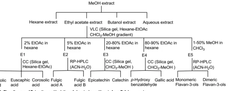
Ursolic acid, euscaphic acid, corosolic acid, fulgic acid A and B, epicatechin, catechin, gallic acid, p-hydroxybenzaldehyde along with several dimeric flavan-3-ols were isolated as described in our previous communications [10]. Briefly, methanol extract (MeOH) (80:20) ofP.fulgens
roots (PRE) was prepared by maceration which was subsequently fractionated into hexane (Hex), ethyl-acetate (EA) andn-butanol soluble fractions. The ethyl-acetate extract was sub-jected to vacuum liquid chromatography using hexane-ethyl acetate and chloroform-methanol gradients to yield five pooled fractions, E1 to E5. Column chromatography of fraction E1 yielded ursolic acid, euscaphic acid and corosolic acid. Two stereoisomeric triterpene acids, ful-gic acid A and fulful-gic acid B were separated from fraction E2 by HPLC. Fractions E3 to E5 after column chromatography and reverse phase HPLC yielded phenolics catechin, epicatechin, gal-lic acid, p-hydroxybenzoic acid, various monomeric and dimeric flavan-3-ols [10,11]. Isolation scheme is shown inFig 1.
Cell line and clonogenic cell survival assay. MCF-7 (human breast cancer cell line) and U87 (human malignant glioma cell line) were purchased from the National Centre for Cell Sci-ence (Pune, India). Cells were cultured in Dulbecco’s MEM medium (DMEM, Invitrogen-Gibco) supplemented with 10% Fetal Calf serum (Invitrogen-Invitrogen-Gibco), 100 u/ml Penicillin and 100 mg/ml Streptomycin (Invitrogen-Gibco) and 2 mM L-Glutamine (Invitrogen-Gibco).
The cell survivality was evaluated using a clonogenic assay in both the cancer cell lines. Briefly, cells were trypsinized and appropriate cell numbers (2000 and 3000 cells) were seeded into three 25 cm2flasks each and for untreated controls, four flasks were plated at one cell den-sity (1000 cells). Five hours after seeding, the cells were exposed for 24 h with 5 to 150μg/ml of
the root extract (PRE) ofP.fulgensor 10 to 120μg/ml of EA- or Hex- ornbutanol-fraction.
After the treatment cells were washed twice with the medium and finally flasks were incubated in a humidified incubator with 5% CO2at 37°C with fresh Dulbecco’s MEM medium with 10%
foetal calf serum for 10 days. The colonies were fixed and stained with 0.2% crystal violet in
Fig 1. Flow diagram of Extraction and Isolation of chemical constituents fromP.fulgensroots.
70% ethanol. Each assay was performed in triplicate and colonies containing at least 50 cells were counted. The number of surviving colonies (treated samples) was normalized against the number of colonies in untreated samples.
Treatment details
In all the experiments that were carried out after colonogenic assay, cancer cells were treated with 100μg/ml of PRE, EA-fraction and Hex-fraction of the methanol extract of the roots ofP.fulgens.
In case of mixture of three compounds which were isolated from the EA-fraction i.e epicatechin (EC), ursolic acid (UA) and gallic acid (GA), each was taken 10μg/ml. These three compounds
were taken from three different EA-subfractions (fraction 1, 3 and 4) and their selection was based on their higher yield. In all these experiments the cells were exposed for 24h.
Cell killing ability by PRE and EA-fraction to normal and cancer cells
This was determined by Trypan blue exclusion assay. Freshly collected human peripheral blood lymphocytes was used as normal cells and MCF-7 and U87 cells were used as cancer cells.
Heparinized peripheral blood from two healthy male donors (HPBL) was obtained after having their written consent for participation and used soon after its collection. Peripheral blood mononuclear cells were isolated by Ficoll-Hypaque (Sigma Diagnostics, St. Louis, MO) density gradient centrifugation (specific gravity 1.077 g/ml) for 30 min at 400gfrom these freshly drawn heparinized whole blood. Lymphocytes cultures were set up in RPMI 1640 medium supplemented with 10% heat inactivated FCS. Penicillin (100 U/ml) and streptomycin (100 mg/ml) and 2 mM L-Glutamine were added to the medium. Lymphocytes were stimu-lated with PHA and after 24 h lymphocytes were treated with PRE and EA-fraction (100 and 150μg/ml) for 24 h. These cultures were incubated at 37° C and were harvested soon after the
treatment. This study was approved by the Institutional and Human Ethics Committee. In case of cancer cells, MCF-7 and U87 cells were used and treated with PRE and EA-fraction (100 and 150μg/ml) for 24 h. Cells were washed with fresh medium and then incubated for 10 min
at room temperature with 0.4% final trypan blue. Dead cells were stained blue, and live ones were unstained. Experiments were repeated three times.
Cell killing ability by PRE and EA-fraction was also evaluated by flow-cytometric analysis (S1 File).
Effect on cell proliferation in MCF-7 and U87 cells
The cellular proliferation assay was performed in MCF-7 and U87 cells after treating the cells with PRE, EA and Hex-fractions. Cells at a density of 5 x 10⁵were grown in 25 cm2flasks in DMEM medium at 37°C and 5% CO2. After 24 h of seeding, the cells were exposed to PRE, EA
or Hex-fraction at a final concentration of 100μg/ml for 24 h and finally fixed in 70% ethanol.
All the above fixed cells were washed in PBS and resuspended in 500μl of propidium iodide
solution (20μg/ml propidium iodide, 0.2 mg/ml RNase) for 1 h incubation at room
tempera-ture in the dark. 10,000 cells were acquired for each sample and analyzed them in a FACS Cali-bur (Becton-Dickinson) using Cellquest software in order to quantify cell cycle compartments to estimate the percentage of cells distributed in the different cell cycle phases.
Flow cytometric analysis of mitochondrial membrane potential
membrane permeability, known as the mitochondrial permeability transition [14]. Mitochon-drial damage was evaluated using the lipophilic fluorescent probe 5,50,6,60-tetrachloro-1,10,3,30 -tetra-ethyl-benzimidazol-carbocyanine iodide according to the manufacturer’s protocol (JC-1; BD Mitoscreen JC-1 kit; Cat 51302). Apoptotic cell death was evaluated in MCF-7 and U87 cells treated with and without PRE, EA-fraction, Hex-fraction and mixture of EC, UA and GA using JC-1 stain. In brief, JC-1 working solution was prepared in 1xAssay buffer and added 0.5 ml JC-1 stain to 1x10⁶MCF-7 or U87 cells for 10 min at 37°C in CO2incubator. The cells were washed
twice with 1x Assay buffer at room temperature and finally JC-1 fluorescence was measured using a Becton Dickinson FACScalibur analytical flow cytometer (BD Biosciences, San Jose, CA). The percentage of cells of green (530 nm) and red (590 nm) fluorescence of JC-1 was analyzed.
Immunoblotting
Immunoblot analyses were performed to detect the expression of PARP andβ-actin proteins in untreated and treated MCF-7 and U87 cells. Cells were plated in 25 cm2flasks at a concentra-tion of 4 x 10⁵and 20 h after they were exposed to PRE, EA, Hex and mixture of EC+GA+UA for 24 h. Cells were collected soon after the treatment.
All the collected cells were washed with PBS and proteins were extracted in cell lysis buffer (50mM Tris (pH 8.0), 150mM Nacl, 10% glycerol, 1% NP–40, 0.5% Sodium deoxycholate, 0.1% SDS and 0.42% NaF) containing protease inhibitor (1mM phenylmethylsulfonyl and 100 U/ml aprotinin). Protein concentrations were determined using the bicinchonic acid pro-tein assay and 40μg protein lysates from each sample was loaded in Novex Tris-Glycine
4–20% gradiaent gels and electrophoresis was performed in NuPAGE electrophoresis system from (Invitrogen, USA). Proteins were transferred to PVDF membranes (Sigma) following the standard protocol. The membranes were probed with a 1:1000 dilution of a rabbit polyclonal antibody against PARP Ab-3 (Neo-Markers, USA) and mouse monoclonal antibody against
β-actin (AC-15; ab6276; abcam, USA). The membranes were blocked in 5% non-fat dried milk, freshly made in TBST (10mM Tris-Cl, 150mM NaCl, 0.05% Tween 20) buffer. Alkaline-phos-phatase conjugated anti-mouse IgG (Abcam, USA) used as secondary antibody, and immuno-detection was obtained by treating the blot with the substrate solution of BCIP/NBT
(Bangalore Genei, India). The whole experiment was repeated twice.
DNA fragmentation assay
Fragmentation of DNA as an indication of apoptosis is a commonly used assay in drug-cell inter-action studies. The MCF7 and U87 cells (5 × 10⁵cells/flask) were cultured for 24 h and then exposed to PRE, EA or Hex-fraction at a final concentration of 100μg/ml for 24 h. Cells were
washed once with ice-cold PBS, and resuspended in 250μl lysis buffer (10 mM Tris–HCl,
pH7.6, 20 mM EDTA, pH 8.0 and 0.5% (w/v) Triton X-100). After centrifugation at 12,000 rpm for 5 min, the supernatant was extracted once with phenol/chloroform (1:1) and once with chloroform / isoamyl alcohol (24:1). DNA was precipitated with sodium acetate (pH 5.2) at−20°C overnight. The DNA was then pelleted and subsequently digested with DNase-free RNAase (Amer-sham Biosciences UK Ltd., Little Chalfont, Buckinghamshire, UK) at 37°C for 20 min. Electropho-resis was carried out in a 2.0% (w/v) agarose gel and was examined under UV transillumination following ethidium bromide staining to determine the extent of apoptotic DNA fragmentation.
Semiquantitative RT-PCR (reverse transcription polymerase chain
reaction)
Hex and mixture of EC+GA+UA. Total RNA was extracted from cells using RNAeasy kit (Qia-gen GmbH, Hilden Germany). cDNAs were reverse transcribed from 1μg of total RNA from
each sample using Quantiscript Reverse Transcriptase, Quantiscript-buffer and RT-primer-mix of QuantiTect Reverse Transcription kit (Qiagen GmbH, Hilden Germany) according to the manufacturer’s protocol.
Amplification of cDNA was carried out in 20μl solution containing 2μl cDNA, 10 pmol
primer pairs for Bcl2, Survivin, XIAP, cIAP and GAPDH (for primer sequences- Table A inS1 File) and 10μl of RT qPCR Master mix (Qiagen GmbH, Hilden, Germany). The PCR consisted
of initial denaturation at 94°C for 5 min, followed by 25 reaction cycles (30 seconds at 94°C, 30 seconds at 60°C, and 30 seconds at 72°C) and a final cycle at 72°C for 10 min. GAPDH was used as internal control. The amplified PCR products were separated by agarose gel electropho-resis and visualized with ethydium bromide. The abundance of each target mRNA was detected and normalized to that of GAPDH mRNA.
Determination of GSH level
Total cellular GSH content was estimated by the method of Akerboom and Sies [15] in MCF-7 and U87 cells after treatment with PRE, EA, Hex and EC+GA+UA. Cells were seeded in 25 cm2flasks at a concentration of 5 x 10⁴and when the cells reached confluency between 75–80%, the PRE or EA-fraction or Hex-fraction or EC+GA+UA was added for 24 h. Both treated and untreated cells were flashed into ice-cold 0.1 M phosphate buffered saline solution (pH 7.4), and the volume was made up to 1ml. Cells were counted in a haemocytometer and processed for determination of total GSH level as described earlier [16]. Briefly, after deprotei-nization by 10% ice-cold 5-sulfosalicylic acid 190μl sample suspension was taken and added to
700μl NADPH (0.3 mM), 100μl DTNB (6 mM) and 10μl GSH reductase (6 units/ml) and the
optical density of the samples was measured at 412 nm using the UV-visible spectrophotome-ter (Cecil series 1000, Camlab, UK). The GSH concentrations were despectrophotome-termined by comparison with standards.
Statistics
The results are expressed as mean plus or minus Standard deviation (SD). Statistical analysis of the data was performed by paired t-test and p values<0.05 were considered significant.
The data obtained from clonogenic survival assay are represented as a sigmoidal fit curve with a linear X scale, the function used was Boltzmann. The Y value at X0is considered to be
half way between the two limiting values on Y-axis and this is considered to be the inhibitor concentration 50 (IC50) value.
Results
Effect on clonogenic efficiency
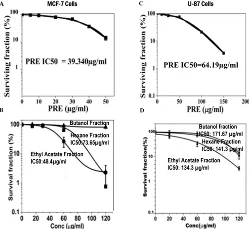
MCF-7 and U87 cells were treated with different concentrations of PRE, and EA-, Hex- and butane-extracted fractions. Whereas PRE, EA-fraction and Hex-fraction caused dose depen-dent reduction in the cloning efficiency in both cell lines (Fig 2A–2D) the butanol-fraction did not do so (Fig 2B). However, the degree of reduction in cloning efficiencies in both the cell lines was maximum with PRE. The value of inhibition concentration (IC50) deduced from the
Sigmoidal fit graph was 39.34μg/ml with PRE whereas for EA it was 48.4μg/ml in MCF-7
cells. For U87, these values were 64.2 and 134.3μg/ml (Fig 2A) with PRE and EA-fraction,
PRE and EA-fraction kill more cancer cells than normal cells
The frequencies of dead cells are more in the cancer cells than normal lymphocytes after treat-ment with PRE or EA-fraction (Table 1). Significant increase in sub-G1 cells in the cancer cell lines (measured by FACS) relative to the normal lymphocytes after the treatment with PRE or EA-fraction also point to the increased apoptosis in MCF-7 and U87 (Fig A inS1 File).
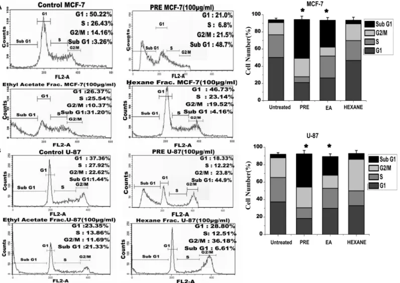
Flow cytometric analysis of cells after treatment with the extract
The distribution of cells in different phases of cell cycle was determined by flow cytometric analy-sis of DNA content in MCF-7 (Fig 3A) and U87 cells (Fig 3B). As shown in the figure, no signifi-cant accumulation of cells in any phase was observed in any of the treated cell lines. However the increased fraction of cells in sub-G1 phase was observed in samples treated with PRE (49% in MCF-7 and 45% in U87 cells) and EA-fraction (MCF-7: 31%, U87: 21%). The right panel of the
Fig 3A and 3Bshowed significant increase in the frequency of sub-G1 cells after the treatment with PRE and EA-fraction. Hexane-fraction did not alter the frequency in sub-G1 phase cells.
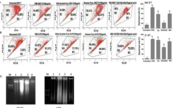
Analysis of the mitochondrial membrane potential by JC1 labelling revealed that percentage of polarized cells was significantly reduced in both MCF-7 and U87 cells after treatment with either PRE, EA or mixture of EC+GA+UA (Fig 4A and 4B; Fig B inS1 File). The reduction of polarized cells was lesser with Hex-fraction in both the cells. The untreated cells mostly Fig 2. Cells were treated for 24 h with various concentrations of crude extract ofP.fulgensroot (PRE) and its ethyl acetate, hexane andnbutanol soluble fractions.After the indicated time of treatment, cells were maintained without test materials for 10 days. At day 10, the grown colonies were counted and percentage of survival fraction was then calculated and shown in(A and B)in MCF-7 cells and(C and D)in U87 cells. Results were expressed as means±SD for three independent determinations.
exhibited red fluorescence, indicating an intact mitochondrial membrane potential. Upon treatment with PRE or EA or EC+GA+ UA the number of cells enhanced, as shown by green fluorescence. The right panel of the figure (Fig 4A and 4B) showed significant increase in the cells with mitochondrial membrane depolarization after the treatment with PRE, EA-fraction and the mixture of EC+GA+UA.
DNA fragmentation analysis
To validate the induction of apoptosis by PRE, EA or mixture of EC+GA+UA in both MCF-7 and U87 cells, DNA fragmentation analysis was performed (Fig 4C). It was observed that treat-ment with PRE and EA-fraction (100μg/ml) for 24 h showed DNA degradation in association
with DNA laddering in U87 cells (panel 3 and 4), however in MCF-7 cells partial and complete DNA degradation was observed in lane 3 and 4, respectively without DNA laddering. DNA degradation could not be observed in these cell lines after treatment with Hexane-fraction for 24 h. Taken together, the PRE and EA-soluble fraction induced DNA degradation in both U87 and MCF-7cells which is the biochemical hallmark of apoptotic cell death.
Detection of the PARP-1 proteolysis and expression level of apoptosis
inhibitors
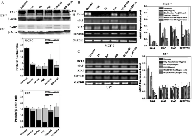
In order to confirm that the cell death induced by PRE, EA or mixture of EC+GA+UA in both MCF-7 and U87 cells was due to apoptosis, cleavage of PARP was measured in both the cell lines after 24 h treatment (Fig 5A). PARP-1 proteolysis was detected after the treatment with PRE, EA and mixture of EC+GA+UA in both the cells (Fig 5A). Consistent with this observa-tion, significant reduction in the expression of Bcl2 in both MCF-7 and U87 cells was also observed following treatments with PRE, EA fraction and the mix-compounds (Fig 5B and 5C). Treatment with Hex-fraction failed to reduce the level of Bcl2 in both the cell types.
Table 1. Trypan Blue Exclusion Assay in human lymphocytes and in cancer cell lines after treatment with PRE or EA-fraction.
Cell Type Percentage of Dead cells±SD
Primary Normal Cell
Untreated Lymphocytes 8.8±0.8
PRE (100μg/ml) treated 18.8±2.3 p = 0.06
PRE (150μg/ml) treated 24.6±2.2a
EA (100μg/ml) treated 14.2±1.4 p = 0.07
EA (150μg/ml) treated 22.9±2.2a
Cancer Cell lines
Untreated MCF-7 cells 5.5±0.9
PRE (100μg/ml) treated 49.1±1.8ap = 0.009
PRE (150μg/ml) treated 52.4±2.4a
EA (100μg/ml) treated 38.6±2.5ap = 0.02
EA (150μg/ml) treated 42.9±2.2a
Untreated U87 cells 4.9±0.8
PRE (100μg/ml) treated 42.6±2.5ap = 0.02
PRE (150μg/ml) treated 48.4±2.4a
EA (100μg/ml) treated 29.1±1.7ap = 0.01
EA (150μg/ml) treated 33.9±2.2a
Student’s t-test with respect to respective control value. aSigni
ficant in Paired t-test.
Besides Bcl2, the expression of inhibitors of apoptosis proteins was also reduced after these treatments in MCF-7 cells, whereas in U87 cells such reduction was not observed.
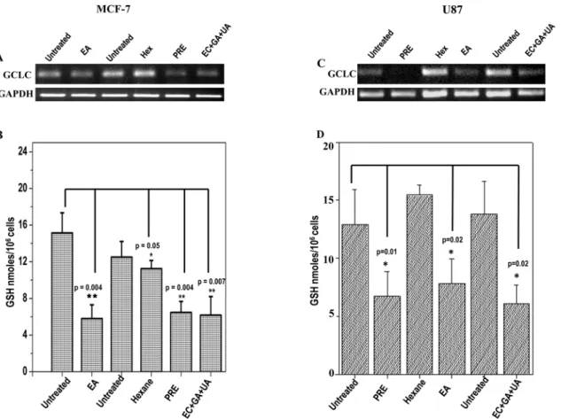
Level of reduced glutathione (GSH) and expression level of GCLC
Level of reduced GSH in MCF-7 and U87 cells is shown after the treatment with PRE, EA, Hex and EC+GA+UA (Fig 6B and 6D). The concentration of GSH was reduced significantly in both the cells after the treatment with PRE, EA and EC+GA+UA.
This reduction in GSH-level is accompanied by the lesser expression of GCLC in the treated MCF-7 and U87 cells (Fig 6A and 6C).
Discussion
P.fulgensroot-stock and whole herb are commonly used as folk medicine by natives of north-east India, Nepal and Bhutan for variety of ailments [8]. By using various analytical techniques Fig 3. Analysis of cell cycle (A) The flow cytometry analysis on cell cycle distribution of PI-labeled MCF-7 cells and (B) in U87 cells with and without treatment with PRE, ethyl acetate (EA) and hexane (Hex) soluble fractions ofP.fulgensroot extract.Right panel-The percentage of cells at different stages in both MCF7 and U87 cells with and without treatment. The values are the mean±SEM of three independent experiments.*p<0.05, Students’t-test as compared with untreated control.
with the crude extract it was shown the presence of triterpene acids and B-type flavan-3-ols in the extract which are generally known for various biological activities [11]. Solvent-solvent par-titioning of methanol extract yielded hexane, ethyl-acetate,nbutanol and aqueous extract. The present study demonstrated promising anticancer activity of the crude methanolic extract ofP.
fulgensroot and its EA-fraction against MCF-7 and U87 cells. The concentration used in this study for PRE, EA- and Hex-fraction was 100μg/ml, which was higher than the individual IC50
value. All these extracts were evaluated earlier for antioxidant and cytotoxic activities and the EA-fraction was found to be the most active [10]. In this study, it was observed that treatment with PRE or EA-fraction killed significantly more cancer cells than normal cells. Both PRE and EA-fraction caused dose-dependent reduction in the cloning efficiency in both cell lines, how-ever such reduction in cloning-efficiency was not observed with Hexane- andn butanol-frac-tions. Since EA-fraction showed most efficient growth inhibitory effect, it was further purified and tested for their anticancer activity.
Earlier nine compounds and some monomeric and dimeric flavan-3-ols were identified and characterized from the EA-fraction of the methanolic root extract ofP.fulgens[10]. From these nine compounds, three compounds viz., epicatechin (EC), gallic acid (GA) and ursolic acid (UA) were selected and mixed together. The selection of these compounds was made from three different sub-fractions and was based on their higher yield with respect to amount; GA (3.6%), UA (3.3%) and EC (2.7%) in crude methanol extract [10,11]. UA was found to be the most active compound among the isolated triterpene acids with IC505.3 and 7.8μM in DPPH•
and ABTS+•antioxidant assay [10,11].
Fig 4. Analysis of apoptosis in (A) MCF7 and (B) U87 cells by measuring the mitochondrial membrane potential after JC1 staining in untreated and treated with PRE, EA, Hex fractions and mixture of EC+GA+UA of EA fraction ofP.fulgensroot.The upper part indicates the percentage of cells show polarization of mitochondrial membrane and the lower part shows the percentage of cells having mitochondrial membrane depolarization. All these
experiments were repeated twice. Right panel shows percentage of depolarized cells after each treatment.*p<0.05, Students’t-test as compared with untreated control.(C)Effect of PRE, Hexane and ethyl acetate soluble fraction treatment on the DNA degradation and fragmentation in MCF7 and U87 cells. DNA samples are labeled as: (M) molecular weight marker, (1) untreated cells, (2) cells treated with hexane-fraction, (3) cells treated with PRE and (4) cells treated with ethyl acetate fraction. Results are representative of two independent experiments. The concentration was used 100μg/ml and the treatment was given for 24 h.
In this study, MCF-7 cells was more sensitive than U87 cells since significant reduction in cell growth was obtained by lower concentration of PRE and EA-fraction. Such inhibition in growth of these cell lines could be attributed due to either inhibition in cell proliferation or kill-ing the cells. The flow cytometric analysis demonstrated an increased fraction of cells in sub-G1 indicating cell death which could be due to apoptosis. To obtain additional evidence for apoptosis, we tested whether the dying cells exhibited other characteristics of programmed cell death. Flow cytometric analysis demonstrated significant reduction of polarized cells in mito-chondrial membrane whereas immunoblot results showed cleaved PARP-1 protein in the sam-ples treated with PRE, EA-fraction and the mixture of EC+GA+UA. PARP is activated at an intermediate stage of apoptosis and is inactivated by proteolytic cleavage at a late stage by cas-pase3 and caspase7. Such proteolytic cleavage of PARP is considered as hallmark of apoptosis [17]. Thus present results indicate that the crude extract ofP.fulgensroot as well as its EA-Fig 5. Protein expression analysis of MCF-7 and U87 cells by Western blotting after treatment with PRE, EA, Hex fractions and EC+GA+UA of EA fraction ofP.fulgensroot extracts in comparison with the untreated samples. (A)Determination of PARP cleavage. Lower panel shows quantitative densitometric analysis of the level of PARP with and without cleavage in the treated and untreated MCF-7 and U87 cells. The values are the mean±SEM of three independent experiments. Two independent untreated samples were used. The values are normalized to respectiveβ-actin values. Expression pattern of Bcl2, survivin, XIAP and cIAP using semiquantitative RT- PCR in(B)MCF-7 and(C)U87 cells treated with or without PRE, EA, Hex fractions and EC+GA +UA (two independent samples). Right-side panel shows quantitative densitometric analysis of the expression profile of genes mRNA level. The values are the mean±SEM of two independent experiments and are normalized to respective GAPDH values.
fraction induce apoptosis in these cancer cells and support the notion that some natural com-pounds including plants induce apoptosis that are blocked in cancer cells [18].
The apoptotic effect of PRE and EA-fraction was further confirmed by the DNA fragmenta-tion assay. These treatment extensively degraded DNA in U87 cells, suggesting that cells treated with PRE or EA-fraction underwent apoptosis. Normally, DNA fragmentation due to DNA degradation is considered a biochemical hallmark of apoptosis that involves cutting the inter-nucleosomal regions into small fragments of double-stranded DNA [19]. Thus, changing in the DNA characteristics after the treatment with PRE, EA-fraction or the mixture of EC +GA+UA confirmed cell death induction in U87 cells. Partial and adequate DNA degradation was also noticed in the MCF-7 cell line without DNA ladder formation which could be explained by the lack of caspase-3 in this cell line [20]. It was observed that MCF-7 cells can be killed by apoptotic stimuli, such as tumour necrosis factor and staurosporine, without DNA fragmentation [20]. Therefore, the potential of cells undergoing apoptosis by the treatment with PRE, EA-fraction and EC+GA+UA cannot be ruled out in these cell lines by showing PARP-clevage with or without DNA-fragmentation.
It is an established fact that the Bcl-2 family members either inhibitor (Bcl-2) or activator (Bax), play an important regulatory role in apoptosis [21]. The present data indicate that the induction of apoptosis was due to reduction in the expression of Bcl2, an anti-apoptotic Fig 6. Expression pattern of GCLC in (A) MCF-7 cells and in (C) U87 cells treated with or without PRE, EA, Hex fractions and mixture of EC+GA +UA.Two independent untreated samples were used in this study. Lower panel shows quantitative densitometric analysis of the expression profile of GCLC mRNA level. The values are the mean±SEM of two independent experiments and are normalized to respective GAPDH values. Levels of glutathione in(B) MCF-7 cells and(D)U87 cells treated with or without PRE, EA, Hex fractions and EC+GA+UA. Two independent untreated samples were used in this study. Values are mean±SEM of three different experiments.*p<0.05, Students’t-test as compared with untreated control.
protein, in both MCF-7 and U87 cells. Similar induction of apoptosis by down-regulating Bcl-2 expression in lymphocytic leukaemia cell line CEM-C7H2 was achieved after the treatment with PADMA 28 (a herbal mixture of 28 plants including aerial part ofPotentilla aurea)[22]. Moreover, in MCF-7 cells the induction of apoptosis was also achieved by down-regulating inhibitor of apoptosis proteins (IAPs) like cIAP, XIAP and survivin after the treatment with PRE, EA-fraction and EC+GA+UA. Such down-regulation of XIAP for the induction of apo-ptosis was reported earlier after the treatment with Isorhapontigenin, isolated from a Chinese herbGnetum cleistostachyum[23]. Kaempferol, a common flavonoid, present in many Poten-tilla sp, enhances TRAIL-induced apoptosis by down-regulation of IAPs in glioma cells [24]. However, the downregulation of IAPs was not seen in U87 cells in this study. Thus current data provide important insights into the understanding of the molecular mechanism(s) that are responsible for the anti-cancer effect showed by PRE and its EA-fraction.
A novel aspect of this study was the analysis of the level of endogenous GSH in MCF-7 and U87 cells after the treatment. GSH is the major cellular non-protein thiol antioxidant in normal mammalian cells and in many malignant cell types. Higher level of GSH is associated with a proliferative response [25]. Usually the level of GSH in tumor tends to be higher than normal tissues and thus malignant cells show multidrug or radiation resistance [26,27]. In this study, endogenous GSH was depleted significantly in both the cells after the treatment with PRE, EA-fraction and EC+GA+UA mixture. The level of GSH was also decreased in liver and in Dalton’s lymphoma cells of PRE treated tumor-bearing mice [28]. Research over the years has generated enough evidence to implicate that the depletion of GSH is considered to be an indicator of apo-ptosis induction in response to a number of proapoptotic stimuli [29,30]. Therefore, the pres-ent induction of apoptosis by EA-fraction in both the cancer cells supports the notion that depletion of GSH might favouring the apoptotic cell death. Interestingly, it was also observed in this study that such depletion of GSH in these cell lines was achieved by downregulating the expression ofγ-glutamyl-cysteine synthetase heavy/catalytic subunit (GCSC). This enzyme involves in GSH synthesis [31] and consists of a catalytic subunit performs enzyme activity and a modifier subunit that modulates GCSC activity [32]. It was demonstrated earlier that querce-tin and kaempferol increased the production of GSH in neuronal cells [33] and in COS-1 cells by increasing the expression of GCSC [34]. Therefore, natural plant products mediated GSH depletion or elevation by upregulating or downregulating GCSC is an important observation because earlier attempt to deplete the level of GSH by synthetic molecules have been limited due to its non-selective effects [35]. Moreover, it was noticed that those patients having higher level of GSH showed higher resistant to therapy and also managed to sustain the GSH-level than those lacking the synthetic ability [26].
the EA-soluble fraction of theP.fulgensroot extract in tumor cells opened up the possibility of a new therapeutic approach because these plant products are not harmful to normal cells and also regulating the tumor cellular response to different anticancer treatments [45]. Therefore, it would be interesting to examine efficacy of these plant products in human cancer treatment.
Supporting Information
S1 File. Extended experimental procedures.Flow cytometric analysis of cell death by PRE and EA-fraction in normal and cancer cells.Fig A. The flow cytometry analysis on cell cycle distribution of PI-labeled cells in human lymphocytes, MCF-7 cells and U87 cells with and without treatment with PRE, EA-fraction ofP.fulgensroot extract.Fig B.Histogram Plot showing Red fluorescent JC-1 aggregates (FL2-H) in MCF-7 & U-87 cells. Histograms showed decreased florescence intensity depicted by peak shift (M2) between untreated samples and treated samples indicating a decrease in the Mitochondrial membrane potential in the treated samples (Total 10000 cells were acquired for each sample).Table A.Primer sequences and the product sizes.
(DOCX)
Acknowledgments
This work was supported by DBT, Govt. of India (BT/59/NE/TBP/2010) to AC, UCB and IPS; DBT fellowship and DST-Inspire fellowship (IF 120044) to Alka Choudhary; DBT-fellowship to DT.
Author Contributions
Conceived and designed the experiments: A Chatterjee UCB IPS. Performed the experiments: DT A Choudhary A Chatterjee. Analyzed the data: DT A Chatterjee A Choudhary IPS. Con-tributed reagents/materials/analysis tools: A Chatterjee UCB IPS. Wrote the paper: A Chatter-jee UCB IPS. Obtained permission for use of human sample: A ChatterChatter-jee. Obtained
permission from forest officer: A Chatterjee.
References
1. Newman DJ (2008) Natural products as leads to potential drugs: an old process or the new hope for drug discovery? Journal of Medicinal Chemistry 51: 2589–2599. doi:10.1021/jm0704090PMID: 18393402
2. Coseri S (2009) Natural products and their analogues as efficient anticancer drugs. Mini-Reviews in Medicinal Chemistry 9: 560–571. PMID:19456286
3. Tomczyk M, Latté KP (2009)Potentilla—A review of its phytochemical and pharmaco-logical profile. Journal of Ethnopharmacol 122: 184–204.
4. Shushunov S, Balashov L, Kravtsova A, Krasnogorsky I, Latté KP, Vasiliev A (2009) Determination of acute toxicity of the aqueous extract ofPotentilla erecta(Tormentil) rhizomes in rats and mice. Journal of Medicinal Food 12: 1173–1176. doi:10.1089/jmf.2008.0281PMID:19857087
5. Tomczyk M, Pleszczyńska M, Wiater A (2010) Variation in total polyphenolics contents of aerial parts of
Potentillaspecies and their anticariogenic activity. Molecules 15: 4639–4651. doi:10.3390/ molecules15074639PMID:20657382
6. Zhao Y-L, Cai G-M, Hong X, Shan L-M, Xiao X-H (2008) Anti-hepatitis B virus activities of triterpenoid saponin compound fromPotentillaanserine L. Phytomedicine 15: 253–258. doi:10.1016/j.phymed. 2008.01.005PMID:18337074
8. Syiem D, Syngai G, Khup P, Khongwir B, Kharbuli B, Kayang H (2002) Hypoglycemic effects of Poten-tilla fulgensL. in normal and alloxan-induced diabetic mice. Journal of Ethnopharmacology 83: 55–61. PMID:12413707
9. Radhika M, Ghoshal N, Chatterjee A (2012) Comparison of effectiveness in antitumor activity between Flavonoids and polyphenols of the methanolic extract of roots ofPotentilla fulgensin breast cancer cells. Journal of Complementary and Integrative Medicine 9: 1–14.
10. Choudhary A, Mittal AK, Radhika M, Tripathy D, Chatterjee A, Banerjee UC, et al. (2013) Fulgic Acid A and B, two new Stereoisomeric antioxidant Triterpenes fromPotentilla fulgens. Fitoterpia 91C: 290– 297.
11. Choudhury A, Radhika M, Chatterjee A, Banerjee U, Singh IP (2015) Qualitative and quantitative analy-sis ofPotentilla fulgensroots by NMR, MALDI-TOF-MS, and HPLC-UV. Phytochemical Analysis 26: 161–170. doi:10.1002/pca.2547PMID:25515916
12. Lauterburg BH (2002) Analgesics and glutathione. American Journal of Therapeutics 9: 225–233. PMID:11941382
13. Franco R, Cidlowski JA (2009) Apoptosis and glutathione: beyond an antioxidant. Cell Death and Differ-entiation 16:1303–1314. doi:10.1038/cdd.2009.107PMID:19662025
14. Zamzami N, Larochette N, Kroemer G (2005) Mitochondrial permeability transition in apoptosis and necrosis. Cell Death and Differentiation 12: Suppl. 2 1478–1480. PMID:16247494
15. Akerboom TPS, Sies H (1981) Assay of glutathione, glutathione disulfide and glutathione mixed disul-fides in biological samples. Methods in Enzymology 77: 373–382. PMID:7329314
16. Chattopadhyay A, Deb S, Chatterjee A (1999) Modulation of the clastogenic activity ofγ-irradiation in buthionine sulfoximine-mediated glutathione depleted mammalian cells. International Journal of Radia-tion Biology 75: 1283–1291. PMID:10549605
17. Soldani C1, LazzèMC, Bottone MG, Tognon G, Biggiogera M, Pellicciari CE, Scovassi AI. (2001) Poly (ADP-ribose) polymerase cleavage during apoptosis: when and where? Experimental Cell Research. 269:193–201. PMID:11570811
18. Safarzadeh E, Sandoghchian Shotorbani S, Baradaran B (2014) Herbal Medicine as Inducers of Apo-ptosis in Cancer Treatment. Advances of Pharmaceutical Bulletin. 4 (Suppl 1): doi:10.5681/apb.2014. 062
19. Hanahan D, Weinberg RA (2000) The Hallmarks of Cancer. Cell. 100:57–70. PMID:10647931 20. Janicke RU, Sprengart ML, Wati MR, Porter AG (1998) Journal of Biological Chemistry, 273, 9357–
9360. PMID:9545256
21. Youle RJ, Strasser A (2008) The BCL-2 protein family: opposing activities that mediate cell death. Nature Review Molecular Cell Biology 9: 47–59.
22. Jenny M, Schwaiger W, Bernhard D, Wrulich OA, Cosaceanu D, Fuchs D, et al. (2005) Apoptosis induced by the Tibetan herbal remedy PADMA 28 in the T cell-derived lymphocytic leukaemia cell line CEM-C7H2. Journal of Carcinogenesis 4:15 doi:10.1186/1477-3163-4-15PMID:16138918 23. Fang Y, Yu Y, Hou Q, Zheng X, Zhang X, Zhang M, Zhang D, et al. (2012) The Chinese Herb Isolate
Isorhapontigenin Induces Apoptosis in Human Cancer Cells by Down-regulating Overexpression of Antiapoptotic Protein XIAP. Journal of Biological Chemistry 287: 35234–35243. doi:10.1074/jbc. M112.389494PMID:22896709
24. Siegelin MD, Reuss DE, Habel A, Herold-Mende C, Deimling AV (2008) The flavonoid kaempferol sen-sitizes human glioma cells to TRAIL-mediated apoptosis by proteasomal degradation of surviving. Molecular Cancer Therapy 7: 356–374.
25. Poot M, Teubert H, Rabinovitch PS, Kavanagh TJ (1995) De novo synthesis of glutathione is required for both entry into and progression through the cell cycle. Journal of Cell Physiology 163: 555–560. 26. Gamcsik MP, Kasibhatla MS, Teeter SD, Colvin MO (2012) Glutathione level in human tumors.
Bio-markers 17(8): 671–691. doi:10.3109/1354750X.2012.715672PMID:22900535
27. Estrela JM, Obrador E, Navarro J, Lasso De la Vega MC, Pellicer JA (1995) Elimination of Ehrlich tumours by ATP-induced growth inhibition, glutathione depletion and X-rays. Nature Medicine 1: 84– 88. PMID:7584960
28. Rosangkima G, Prasad SB (2004) Antitumour activity of some plants from Meghalaya and Mizoram against murine ascites Dalton's lymphoma. Indian Journal of Experimental Biology. 42: 981–988. PMID:15511001
29. Circu Magdalena L, Aw Tak Yee (2012) Glutathione and modulation of cell apoptosis. Biochimica et Biophysica Acta. 1823: 1767–1777. doi:10.1016/j.bbamcr.2012.06.019PMID:22732297
production and apoptosis in human leukemic cells. Biochemical and Biophysical Research Communi-cation 411: 1–6.
31. Griffith OW, Mulcahy RT (1999) The enzymes of glutathione synthesis: gamma-glutamyl- cysteine syn-thetase. Advances Enzymology Related Areas of Molecular Biolology 73: 209–267.
32. Franklin CC, Backos DS, Mohar I, White CC, Forman HJ, Kavanagh TJ (2008) Structure, Function, and Post-Transcriptional Regulation of the Catalytic and Modifier Subunits of Glutamate Cysteine Ligase. Molecular Aspects of Medicine doi:10.1016/j.mam.2008.08.009
33. Ishige K, Schubert D, Sagara Y (2001) Flavonoids protect neuronal cells from oxidative stress by three distinct mechanisms. Free Radical Biology and Medicine 30: 433–446. PMID:11182299
34. Myhrstad MCW, Carlsen H, Nordstrom O, Blomhoff R, Moskaug JO (2002) Flavonoids increase the intracellular glutathione level by transactivatin of the g-glutamylcysteine synthetase catalytical subunit promoter. Free Radical Biology and Medicine 22: 386–393.
35. Estrela JM, Ortega A, Obrador E (2006) Glutathione in cancer biology and therapy. Critical Review in Clinical Laboratory Sciences 43:143–181.
36. Verma S, Singh A, Mishra A (2013) Gallic acid: Molecular rival of cancer. Environmental Toxicology and Pharmacology 35: 473–485. doi:10.1016/j.etap.2013.02.011PMID:23501608
37. Lu Y, Jiang F, Jiang H, Wu Ka, Zheng X, Cai Y, et al. (2010) Gallic acid suppresses cell viability, prolifer-ation, invasion and angiogenesis in human glioma cells. Europian Journal of Pharmacology 641: 102– 107.
38. Jew S, AbuMweis SS, Jones PJH (2009) Evolution of the Human Diet: Linking Our Ancestral Diet to Modern Functional Foods as a Means of Chronic Disease Prevention. Journal of Medicinal Food 12: 925–934. doi:10.1089/jmf.2008.0268PMID:19857053
39. Elbaz HA, Lee I, Antwih DA, Liu J, Hüttemann M, Zielske SP (2014) Epicatechin Stimulates Mitochon-drial Activity and Selectively Sensitizes Cancer Cells to Radiation. PLoS One 9(2): e88322. doi:10. 1371/journal.pone.0088322PMID:24516636
40. De Angel RE, Smith SM, Glickman RD, Perkins SN, Hursting SD (2010) Antitumor effects of ursolic acid in a mouse model of postmenopausal breast cancer. Nutrition and Cancer 62: 1074–1086. doi: 10.1080/01635581.2010.492092PMID:21058195
41. Ramos AA, Pereira-Wilson C, Collins AR (2010) Protective effects of ursolic acid and luteolin against oxidative DNA damage include enhancement of DNA repair in Caco-2 cells. Mutation Research 692: 6–11, doi:10.1016/j.mrfmmm.2010.07.004PMID:20659486
42. Lauthier F, Taillet L, Trouillas P, Delage C, Simon A (2000) Ursolic acid triggers calcium-dependent apoptosis in human Daudi cells. Anti-Cancer Drugs 11: 737–745. PMID:11129737
43. Prasad S, Yadav VR, Kannappan R, Aggarwal BB (2011) Ursolic acid, a pentacyclin triterpene, potenti-ates TRAIL-induced apoptosis through p53-independent up-regulation of death receptors: evidence for the role of reactive oxygen species and JNK. Journal of Biological Chemistry 286: 5546–5557. doi:10. 1074/jbc.M110.183699PMID:21156789
44. Amin AR, Kucuk O, Khuri FR, Shin DM (2009) Perspectives for cancer prevention with natural com-pounds. Journal of Clinical Oncology 27:2712–2725. doi:10.1200/JCO.2008.20.6235PMID: 19414669