Universidade do Minho
Escola de Ciências
Ana Sofia Guerra Fontes Gonçalves
outubro de 2016
Molecular analysis of the response of Pinus
pinaster plants with differential behavior
towards the Pine Wood Nematode infection
Ana Sof ia Guerr a F ont es Gonçalv es Molecular anal ysis of t he response of Pinus pinas ter plants wit h dif
ferential behavior tow
ards t
he Pine W
ood Nematode infection
UMinho|20
Universidade do Minho
Escola de Ciências
Ana Sofia Guerra Fontes Gonçalves
outubro de 2016
Molecular analysis of the response of Pinus
pinaster plants with differential behavior
towards the Pine Wood Nematode infection
Trabalho realizado sob orientação da
Doutora Célia Miguel
e da
Professora Doutora Teresa Lino-Neto
Dissertação de Mestrado
Mestrado em Biologia Molecular, Biotecnologia
e Bioempreendedorismo em Plantas
iii
Acknowledgements
Finalmente dou por concluída esta etapa! Foi um período extremamente exigente a nível científico, mas sobretudo pessoal, e não teria chegado a este ponto sem o apoio incondicional de algumas pessoas às quais quero prestar o meu maior agradecimento nestas breves palavras:
Primeiramente à Doutora Célia Miguel que tão bem me recebeu no seu laboratório nestes últimos meses. Quero agradecer toda a sua disponibilidade, simpatia e apoio em todas as fases deste trabalho. Admiro a sua calma e atenção para com a pequena “família” que tem construído no seu laboratório, que é o reflexo da confiança que transmite nos outros.
À Professora Teresa Lino-Neto pela simpatia e atenção que sempre demonstrou para comigo e para com os meus colegas durante o ano curricular do mestrado. Agora na condição de co-orientadora da minha tese de mestrado tenho a agradecer a sua disponibilidade e preocupação no decorrer do meu trabalho.
À Doutora Isabel Carrasquinho por toda a ajuda no processo de inoculação, por proporcionar a realização deste trabalho ao fornecer o material biológico utilizado, pela sua simpatia e disponibilidade.
A todos os membros da “Forest Biotech Family” – Inês C., Andreia R., Andreia M., Inês M., Susana, Bruno, Sónia, Sofia L., Cirenia e Pedro – por me acolherem e apoiarem cientificamente durante estes meses, mas também por todos os momentos de descontração que me proporcionaram. Um agradecimento especial à Inês Modesto com a qual trabalhei lado a lado durante parte deste trabalho; obrigada por todo o apoio! E ao Bruno Costa pela enorme ajuda na componente bioinformática deste trabalho.
Às minhas amigas bioquímicas. Não interessa a distância ou o tempo que passa, “o que a Bioquímica juntou, ninguém irá separar”!
Ao Sérgio por todo o seu apoio e por estar sempre ao meu lado, incondicionalmente… À minha família, em especial aos meus pais. Não há palavras para descrever a gratidão que vos tenho por sempre me darem liberdade e apoio nas minhas decisões, por fazerem tudo para o meu bem-estar e do João, enfim… devo-vos em muito a pessoa que sou hoje e tenho ainda muito a aprender com o vosso bom exemplo. Esta tese é dedicada a vocês!
v
Abstract
The Pine Wood Nematode (PWN) Bursaphelenchus xylophilus is the causal agent of Pine Wilt Disease (PWD), which threatens several conifer species around the world leading to great ecological and economical losses. Pinus pinaster (maritime pine), one of the major forest species in Portugal, is susceptible to the PWN infection. The importance of controlling the evolution of the disease as well as its spreading to other countries motivated the initiation of a national breeding program to address this problem. Over five hundred adult trees have been selected as candidate PWN resistant trees for this program from a PWD highly affected area. In this study, seedlings derived from a candidate resistant tree were used in an inoculation experiment to assess susceptibility/tolerance to PWN infection. From the set of inoculated plants, some apparently tolerant plants were selected along with susceptible ones to conduct expression analyses of selected transcripts potentially involved in the response mechanisms against PWN infection, and that might explain the susceptibility or tolerance towards PWD.
Samples from the selected susceptible and tolerant P. pinaster plants were sent for small RNA and degradome next-generation sequencing. Differential expression analysis of the identified small RNAs between susceptible and tolerant plants was performed, and target transcripts for the differentially expressed small RNAs were predicted using computational approaches. Transcripts were also selected from a previous study that identified expressed sequence tags differentially expressed in P. pinaster susceptible plants versus plants from a tolerant species, Pinus pinea, inoculated with PWN. Expression analysis of small RNA targets and transcripts putatively involved in the response mechanism to PWD allowed the identification of genes with different expression profiles between susceptible and tolerant P. pinaster plants, that are apparently linked to the differential behavior of those plants to PWN infection. This study provides valuable information for future research to elucidate the response mechanisms to PWD, and to identify potential marker genes that, linked to a marker-assisted selection of trees, can be an effective approach to accumulate tolerance-related genes in a population or an individual, and contribute to the future establishment of pine forests tolerant to the PWD.
vii
Resumo
O Nemátode da Madeira do Pinheiro (NMP) Bursaphelenchus xylophilus é o agente causal da Doença da Murchidão do Pinheiro (DMP), que ameaça várias espécies de coníferas em todo o mundo levando a grandes perdas ecológicas e económicas. Pinus pinaster (pinheiro bravo), uma das principais espécies florestais em Portugal, é suscetível à infeção pelo NMP. A importância de controlar a evolução da doença, bem como a sua propagação para outros países motivaram o início de um programa de melhoramento nacional para fazer face a este problema. Mais de quinhentas árvores adultas foram selecionadas como árvores candidatas resistentes ao NMP, no âmbito deste programa, de uma área altamente afetada pela DMP. Neste trabalho, plantas jovens provenientes de uma árvore candidata resistente foram usadas num ensaio de inoculação para avaliar a suscetibilidade/tolerância à infeção com o NMP. Das plantas inoculadas, foram selecionadas algumas plantas aparentemente tolerantes à infeção, juntamente com algumas suscetíveis, para realizar análises de expressão de transcritos potencialmente envolvidos nos mecanismos de resposta à infeção com o NMP, e que poderão explicar a suscetibilidade ou tolerância à DMP.
Amostras das plantas de P. pinaster suscetíveis e tolerantes selecionadas foram enviadas para sequenciação de nova geração de pequenos RNAs e do degradoma. A análise da expressão diferencial dos pequenos RNAs identificados entre plantas suscetíveis e tolerantes foi efetuada, e transcritos-alvo dos pequenos RNAs diferencialmente expressos foram previstos usando abordagens computacionais. Também foram selecionados transcritos de um estudo anterior que identificou “expressed sequence tags” diferencialmente expressas em plantas suscetíveis de P. pinaster versus plantas de uma espécie tolerante, Pinus pinea, inoculadas com o NMP. A análise de expressão dos alvos de pequenos RNAs e transcritos putativamente envolvidos no mecanismo de resposta à DMP permitiu a identificação de genes com diferentes perfis de expressão entre plantas de P. pinaster suscetíveis e tolerantes, que estão aparentemente relacionados com o comportamento diferencial destas plantas face à infeção com o NMP. Este estudo fornece informação importante para futura investigação relativa aos mecanismos de resposta à DMP e para a identificação de potenciais genes marcadores que, ligada a uma seleção de árvores assistida por marcadores, poderá ser uma estratégia efetiva para acumular genes relacionados com a tolerância numa população ou indivíduo, e contribuir para o estabelecimento futuro de florestas de pinheiros tolerantes à DNP.
ix
Table of Contents
Acknowledgements ... iii Abstract ... v Resumo ... vii Table of Contents ... ixAbbreviations and acronyms ... xi
1. Introduction ... 1
1.1. Pine Wilt Disease ... 1
1.1.1. Historical Overview ... 1
1.1.2. The Pine Wood Nematode ... 2
1.1.3. The Vector Beetle ... 3
1.1.4. Transmission Biology of the Pine Wood Nematode ... 5
1.1.5. Pine Wilt Disease Development ... 7
1.2. Host Tree Response Mechanisms ... 9
1.3. miRNAs ...12
1.4. Objectives ...16
2. Materials and Methods ...18
2.1. Biological material ...18
2.1.1. Pinus pinaster ...18
2.1.2. Bursaphelenchus xylophilus ...18
2.2. Inoculation of Pinus pinaster plants with Bursaphelenchus xylophilus ...18
2.3. Total RNA extraction ...19
2.3.1. RNA samples purification and evaluation of RNA quality ...20
2.3.2. Total RNA and miRNA quantification using Qubit® fluorometer ...20
2.4. Small RNA and degradome sequencing ...21
2.5. Bioinformatic analyses of sequencing results ...22
2.5.1. Small RNA sequence processing ...22
2.5.2. Differential expression analysis of sequenced small RNAs ...24
2.5.3. Small RNA target gene prediction and validation ...25
2.6. Selection of small RNA target genes for expression analysis ...25
2.7. Selection of candidate PWD-related transcripts for expression analysis ...26
2.8. Two-step RT-qPCR ...26
2.8.1. cDNA synthesis ...26
x
2.8.2. Selection of reference genes for qPCR ...28
2.8.3. qPCR ...28
2.8.3.1. Experimental determination of qPCR conditions ...29
2.8.3.2. Primer’s efficiency calculations ...29
2.8.3.3. Expression stability of candidate reference genes ...30
2.8.3.4. Expression of predicted small RNA target transcripts ...31
2.8.3.5. Expression of PWD-related candidate genes...32
3. Results and discussion ...33
3.1. Total RNA isolation from Pinus pinaster plants with contrasting responses towards PWN infection ………….……….33
3.2. Small RNA and degradome sequencing ...34
3.3. Selection of small RNAs targets putatively involved in the response molecular mechanisms of Pinus pinaster plants affected with PWD ...35
3.3.1. Sequenced small RNAs and in silico evaluation of their expression profiles ...35
3.3.2. Target prediction for small RNAs differentially expressed between susceptible and tolerant plants……….39
3.4. Gene expression analyses of transcripts putatively involved in the differential behavior of Pinus pinaster plants towards the PWN infection ...42
3.4.1. qPCR experimental setup: determination of amplification efficiencies, and validation of reference genes for expression normalization ...43
3.4.1.1. qPCR amplification efficiencies of candidate reference genes ...43
3.4.1.2. Validation of reference genes for expression normalization ...44
3.4.2. Relative expression level quantification of candidate genes and selected small RNA targets………49
4. Concluding remarks and future perspectives ...61
5. References ...63
6. Appendices...76
Appendix A –Inoculation of Pinus pinaster plants with Bursaphelenchus xylophilus, and symptom observation…………. ...76
Appendix B –Agarose gel to check RNA integrit...77
Appendix C –miRPursuit configuration directory files………..78
Appendix D –Quality plots of small RNA sequencing libraries………..80
Appendix E –Size profile distribution of reads from small RNA sequencing libraries………..81
Appendix F –Small RNAs differentially expressed in susceptible and tolerant plants and their respective target transcript(s). ...81
Appendix G –Oligonucleotides used in gene expression analysis using qPCR………..84
Appendix H –Example of a melting curve obtained in real-time quantitative PCR experiments and agarose gel electrophoresis of PCR products. ...87
xi
Abbreviations and acronyms
µL – microliter
A(XXX) – absorbance at XXX nanometers of wavelength
BLAST – Basic Local Alignment Search Tool bp – base pair
C – cytosine
cDNA – complementary DNA cm – centimeters
Cp – crossing point
CTAB – hexadecyltrimethylammonium bromide DEPC – diethylpyrocarbonate
DNA – deoxyribonucleic acid DNAse – deoxyribonuclease dNTP – deoxynucleotide DTT – dithiothreitol
EDTA – ethylenediaminetetraacetic acid EST – expressed sequence tag
g – centrifuge force g – gram G – guanidine h – hour M – molar mg – milligrams MgCl2 - magnesium chloride min – minute miRNA – microRNA mL – milliliter mM – milimolar
mRNA – messenger RNA NaCl – sodium chloride NaOAc – sodium acetate
ng – nanogram nt – nucleotide ºC – degrees Celsius
PCR – polymerase chain reaction pg – picogram
PVP10 – polyvinyl pyrrolidone PWD – Pine Wilt Disease PWN – Pine Wood Nematode qPCR – real-time quantitative PCR RNA – ribonucleic acid
RNAse – ribonuclease RNA-seq – RNA sequencing rpm – revolutions per minute rRNA – ribosomal RNA RT – reverse transcription s – second
SDS – sodium dodecyl sulphate SEM – standard error of the mean siRNA – small interfering RNA ta-siRNA – transacting siRNA Tris-HCl – tris hydrochloride tRNA – transfer RNA U – enzyme unit U – uracil
USA – United States of America UV – ultraviolet
V – volt
w/v – weight per volume WB – workbench
1
1. Introduction
1.1. Pine Wilt Disease
1.1.1. Historical Overview
At the very beginning of the twentieth century, in the year of 1905, Japanese foresters began to notice a widespread mortality of pine trees at the port city Nagasaki. For several decades the mortality spread northward in the island and then to the mainland (from the 1900s up until the 1960s), and the cause of mortality was then thought to be the wood boring beetles that were prevalent in the dead trees (Zhao et al., 2008). It was only in 1971, after a series of inoculation experiments, that the pine wood nematode (PWN) Bursaphelenchus xylophilus was clearly identified as the causal agent of the pine wilt disease (Kiyohara and Tokushige, 1971) and Monochamus alternatus beetles as the vector for the PWN (Mamiya and Enda, 1972). Since then, intensive research on the biology and ecology of pine wilt took place in Japan. A few years later, PWNs were recovered from the wood of a dead pine in the USA and thereafter were found to be widely distributed throughout the country (Dropkin et al., 1971), yet only some exotic species suffered from the disease. After some investigation, it was found that the nematode was native to North America and was likely introduced into Japan in the early 1900s. Human activity has contributed intentional or accidently to the dissemination of some species away from their natural geographical distributions, and place a threat to the established ecosystems (Zhao et al., 2008). The introduction of the PWN into Japan and its subsequent spread to China (1982), Korea (1988) and more recently Portugal (Mota et al., 1999), Madeira Island (Fonseca et al., 2012) and Spain (Robertson et al., 2011) is a striking example of ecosystems being threatened by the establishment of an exotic organism (Zhao et al., 2008). The long-range spread of the PWN occurred as a result of human activities; frequently the nematodes and also their vectors are thought to be transported in timber used for the production of packing materials.
In Portugal, the forest sector is one of the greatest economic activities of the country with a 13.3% contribution to the industrial gross added value. It represents 2.1% of the national gross domestic product, about 10% of the Portuguese exports, and 3% of the national total employment. Wood for furniture and construction, wood for pulp, paper, and paperboard, the cork-based chain, chestnuts, umbrella pine nuts, resin and forest biomass for energy are the main forest-based chains of the Portuguese economy. Maritime pine forests (Pinus pinaster) of the center-north of
2
Portugal are one of the three main forest types of the country, alongside with the Mediterranean evergreen oaks (Quercus suber and Quercus rotundifolia) in the center and south, and Eucalyptus globulus plantations in the coastal northern part of the country (Reboredo, 2014). P. pinaster contributes to important industrial products, such as wood and resin, as well as coastal protection associated with sand dunes.
P. pinaster is known to be susceptible to the pine wilt disease (PWD), thus the introduction of PWN into Portugal has a tremendous economic and ecological impact. A national program for control of the PWN (PROLUNP) was implemented immediately after the nematode was discovered in Portugal, to primarily assess the extent of its distribution. Within a 30 km radius in the Setubal Peninsula, where the nematode was exclusively detected, all the symptomatic trees were felled (approximately 50,000 trees per year) (Rodrigues, 2008). Then, in 2007, a 3 km wide precautionary phytosanitary strip around the affected area devoid of P. pinaster was established, for the control and eventually the eradication of the nematode (Mota and Vieira, 2008). The clear-cut trees from the affected area were subjected to treatment that may include methyl bromide fumigation, high-temperature treatment of wood before use, or chipping and burial (Jones et al., 2008). Research on the bioecology of the nematode and its insect vector, new detection methods involving e.g. real-time PCR, tree ecology and pathology, and control methods has been ongoing since 1999 (Mota and Vieira, 2008).
1.1.2. The Pine Wood Nematode
Bursaphelenchus xylophilus (Steiner & Buhrer) Nickle is a migratory non-obligate endoparasite that infects mainly Pinus species, causing the PWD. Although parasitic in nature, the PWN can be easily maintained in the laboratory, usually by culture on the fungus Botrytis cinerea, completing its entire life cycle from fertilization to mature adult within 5 days at 25ºC (Hasegawa and Miwa, 2008). The PWN reproduces gonochoristically (male and female sexes), with a great number of offspring (about 100-800; Bolla and Boschert, 1993).
The PWN life cycle can progress in two different ways, the reproductive and the dispersal phases, where the nematode shows different feeding habits, phytophagous and mycophagous, which are characteristic of this species (Moens and Perry, 2009; Zhao et al., 2014). In each phase, its behavior, nutrition, reproduction, and distribution in the host tree are significantly influenced by cohabiting microorganisms (Futai and Mota 2008). Resembling insects, nematodes undergo several "molting" processes, progressing through four juvenile stages (J1 to J4) (Fig. 1).
3
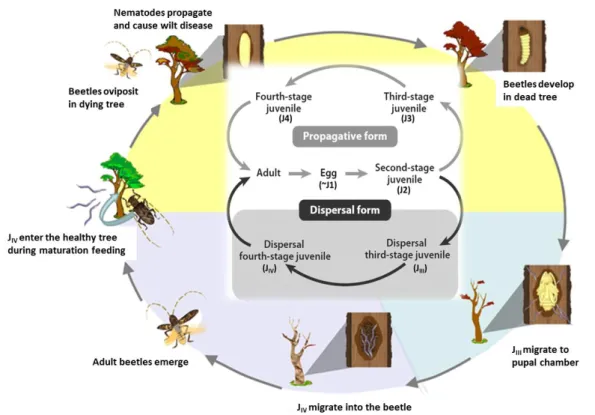
Figure 1 – The life cycle of the Pine Wood Nematode, Bursaphelenchus xylophilus. The life cycle includes
a propagative phase, in which the propagative form of the nematode arises when the conditions are appropriate for propagation, and a dispersal phase, that is induced by unfavorable conditions such as desiccation, food shortage, or environmental deterioration due to overpopulation. The two stages that constitute the dispersal form of the nematode (highlighted by the gray box) are in close relationship with the vector beetle. The gray arrows and black arrows show the propagation cycle in pine trees and that for transmission to new host trees, respectively. (Adapted from Futai, 2013)
In nature, the PWN invades a healthy host pine tree via feeding wounds made by a vector species, predominantly cerambycid beetles of the genus Monochamus, feeds initially on living tree tissues and later on fungi that colonize the dead tree. When food is unavailable, or in the presence of unfavorable conditions (e.g. cold temperatures), the nematode larva enters the dispersal third-stage (JIII), a survival stage that can adapt to adverse conditions (Ishibashi and Kondo, 1977; Kondo
and Ishibashi, 1978). Then, stimulated by the beetle pupa, JIII molts to become the dispersal
fourth-stage juvenile (JIV), which is the nematode form that is transported by the vector into a healthy pine
tree (Maehara and Futai, 1996), where it can enter the propagative adult stage and starts reproducing (Mamiya, 1975).
1.1.3. The Vector Beetle
PWN is vectored by cerambycid beetles of the genus Monochamus (Coleoptera: Cerambycidae), that include Monochamus alternatus in East Asia (Mamiya and Enda, 1972; Morimoto and Iwasaki, 1972; Lee et al., 1990; Yang, 2004), M. saltuarius in Japan (Sato et al., 1987), M. carolinensis in North America (Linit et al., 1983), and M. galloprovincialis in Portugal (Sousa et al., 2001). Among these vectors, M. alternatus is the most intensively investigated, especially in Japan, because it is the vector known for the longest time and due to its importance in PWD development in the economical and ecologically important pine trees. M. alternatus is
4
indigenous to Japan but its geographical distribution there was scarce up until the PWN introduction in the country. The understanding of the life-history traits of M. alternatus should be important to clarify the outbreak and PWN epidemics (Togashi, 2006).
M. alternatus is an ectothermic organism, which means that its development is affected by ambient temperature; when the ambient temperature is favorable for larvae to develop M. alternatus has a one-year life cycle, whereas in the presence of cold summer temperatures the larvae do not complete their development within one season and thus have a two-year life cycle (Togashi, 1989c). Reproductively immature adults emerge from dead host trees once a year, in late spring through summer, randomly disperse by flying, and feed on the bark of pine branches or other conifers for survival and sexual maturation (maturation feeding) (Togashi, 2006). Mature adults are then strongly attracted to volatiles emitted from dying or newly killed trees (Ikeda et al., 1980) since they cannot survive in live host trees due to oleoresin, the main defense agent in conifers. In weakened trees, they can, therefore, mate and oviposit successfully. Larvae develop through four instars and then make tunnels in the xylem in late summer through autumn (Togashi, 1989c). In the end of the tunnels, they make a pupal chamber where they overwinter (Fig. 2). Pupation occurs after overwintering, and then sclerotization of newly eclosed adults happens, and they are therefore ready to make their way out to the bark surface and leave the tree (Nakamura-Matori, 2008).
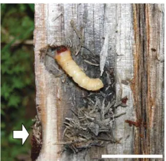
Figure 2 – Monochamus alternatus larva in the pupal chamber. The pupal chamber is observed in an infested
pine log that has been chopped vertically with a hatchet. The arrow points out the entrance of the tunnel bored into the xylem by the larva. (scale bar = 2 cm; Retrieved from Togashi, 2008)
5
After the PWN introduction in Japan, and since the nematode is highly pathogenic to Japanese pine trees like Pinus densiflora and Pinus thunbergii, the number of weakened and dying pine trees prominently improved, increasing the availability of resources for M. alternatus propagation. Thus, it can be inferred that a mutual relationship between the introduced PWN and the beetle could easily establish once the two organisms co-occurred (Nakamura-Matori, 2008). In Japan, PWN-killed trees are found in mid-summer through autumn, which coincides with the adult beetle flight season (Togashi, 1989a). So, trees killed from PWD are perfect places for beetle oviposition. Moreover, the trees release volatiles that seem to attract reproductively mature beetle adults and it is observed a positive, spatial association between trees diseased the previous year and those diseased in the early season of the current year (Togashi, 1991). Both of these evidences indicate that there is a well-established mutualistic relationship between the PWN and M. alternatus, which make it difficult to control the pathogenicity of PWD.
1.1.4. Transmission Biology of the Pine Wood Nematode
PWN is transmitted by cerambycid beetles from the genus Monochamus. M. alternatus, M. carolinensis, and M. galloprovincialis are the most important vector beetles for the PWN in East Asia, North America, and Portugal, respectively. Their conifer hosts belong to the family Pinaceae (Togashi, 2008). The PWD is characterized by a close relationship between the PWN and its vector beetle (Fig. 3). During summer, the PWN’s fourth-stage dispersal juveniles (JIV) are carried by the
vector beetles from dead to healthy host trees (Fig. 3), where they molt to adults and start to mate. The female adult then initiates oviposition (Fig. 3). PWN reproduction occurs through the four stages of the propagative form, which feed on parenchyma cells of the tree’s resin canals and later on fungi, ultimately leading to a dead tree (Fig. 3). Beetle females of the species M. alternatus lay eggs under the bark of those newly-killed trees (Fig. 3). Generally, the M. alternatus larvae stay in the inner bark in the first, second and third developmental stages (or instars), begin to bore tunnels into the xylem in the fourth instar, and then make pupal chambers in the xylem (Katsumi Togashi, 1989a; Katsumi Togashi, 1989b; Katsumi Togashi, 1991). The beetles overwinter as larvae and pupate the following year between late spring and early summer. Simultaneously to beetle development, reproduction of the nematode leads to increased population numbers in the pine trees. When the nematode population reaches a certain level, the third-stage dispersal juveniles (JIII) appear (Kiyohara and Suzuki, 1975). During winter, JIII aggregate around the pupal chambers
6
spring and early summer, JIII around the pupal chamber molt to JIV and then invade the beetle’s
body (Fig. 3) (Togashi, 2008). The numbers of nematodes that aggregate around the pupal chambers and that invade and are carried by the beetle are mostly determined by the fungal microflora present around the chamber (Maehara and Futai, 2002). The beetles then emerge from the killed host trees (Fig. 3) and move to young branches of nearby healthy trees, where they make feeding wounds (maturation feeding, Fig. 3) from which the JIV, after leaving the beetle’s body, can
invade the new host tree (Fig. 3). The process of leaving the beetle’s body is greatly influenced by the volatiles that the tree releases upon the beetle’s maturation feeding, that are perceived by the nematodes inside the beetles (Enda and Ikeda, 1983; Stamps and Linit, 1998).
Figure 3 – The relationships between Bursaphelenchus xylophilus life cycle and its transmission by the insect vector. In the center the life cycle of Bursaphelenchus xylophilus is schematically represented, showing its propagative and dispersal forms. The main events of the nematode life cycle that occur inside the host tree are illustrated in the outer cycle, in relation to the transmission of B. xylophilus by its insect vector. (Adapted from Shinya et al., 2013; Futai, 2013)
During unfavorable conditions for the development of PWD or in pine forests resistant to the PWN like the ones in the USA, the PWN populations can still thrive via alternative transmission pathways. It has been shown that female beetles can transmit PWNs to dying trees via oviposition wounds directly (Wingfield and Blanchette, 1983; Edwards and Linit, 1992), and male beetles searching for mates can also transmit the nematodes to dying trees via already existing wounds on
7
the bark (Arakawa and Togashi, 2002). Interestingly, PWNs sometimes move between beetles during the mating process before being transferred to the trees by either one of the sexes (Togashi and Arakawa, 2003).
1.1.5. Pine Wilt Disease Development
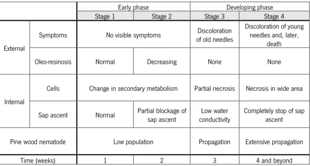
The artificial propagation of PWN and an inoculation procedure were first established by Mamiya (1980, 1984), which led to the first physiological measurements being conducted concerning PWD development (Tamura et al., 1987; 1988). During the 1980s, highly virulent PWN isolates were obtained from many cultures established from dead pines (Kiyohara and Dozono, 1986; Kiyohara and Bolla, 1990), and by the end of the following decade hundreds of reports on the phenomena related to trees’ inoculation with the PWN had been published in Japan (Fukuda, 1997; Yamada, 2006). In Table I is outlined the current knowledge about the PWD development, regarding the external symptoms and internal changes observed over time in pine wilt-susceptible saplings of P. thunbergii inoculated with the PWN.
Table I – Pine Wilt Disease development in susceptible pines inoculated with Bursaphelenchus xylophilus. External symptoms and internal changes observed in Pinus thunbergii seedlings inoculated with the pine wood nematode, B. xylophilus. (Adapted from Zhao et al., 2008)
Early phase Developing phase
Stage 1 Stage 2 Stage 3 Stage 4
External
Symptoms No visible symptoms of old needles Discoloration Discoloration of young needles and, later, death
Oleo-resinosis Normal Decreasing None None
Internal
Cells Change in secondary metabolism Partial necrosis Necrosis in wide area
Sap ascent Normal Partial blockage of sap ascent conductivity Low water Completely stop of sap ascent
Pine wood nematode Low population Propagation Extensive propagation
Time (weeks) 1 2 3 4 and beyond
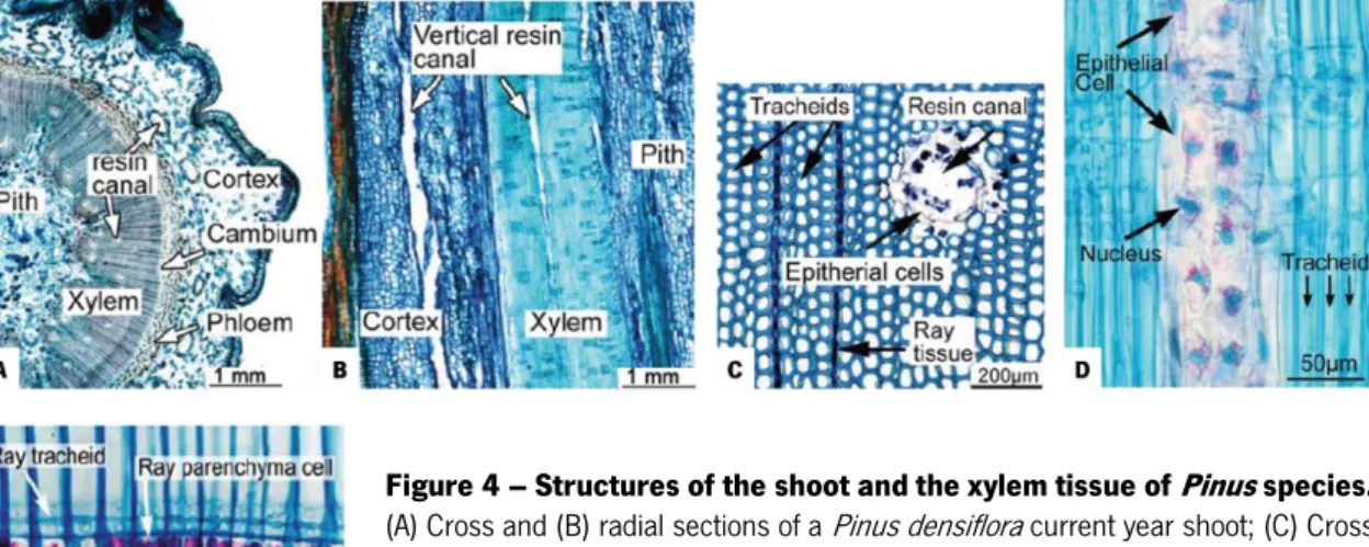
In the stems of conifers, most xylem tissue consists of elongated cells called tracheids, crossed orthogonally by ray tissue (Fig. 4C). In Pinus species, the ray tissue consists of ray tracheids and ray parenchyma cells (Fig. 4E). The resin canals are intercellular spaces formed by separation of parenchyma cells. The xylem of Pinus species contain vertical and horizontal resin
8
canals and their cortex contains vertical resin canals (Fig. 4B) (Evert, 2006). When pine tissue is injured mechanistically or by insect feeding, epithelial cells that surround the resin canals (Fig. 4C and D) immediately synthesize and exude resin that traps most of the PWNs hindering their invasion of the resin canals (Kuroda, 2008). Large numbers (3000-10000) of PWNs are then used in inoculation experiments because it is estimated that only about 10% of the inoculum successfully invades the tissue (unpublished data) (Kuroda, 2008). Although the resin effectively prevents the initial invasion of nematodes, it does not prevent the migration of PWNs in the tissue. PWNs indeed migrate very rapidly in the shoot and branches of a pine tree, 150 cm per day at maximum, readily invading the main stem. And since horizontal and vertical resin canals are distributed densely in the xylem tissue (Fig. 4A), PWNs can spread rapidly throughout the entire plant (Kuroda and Ito, 1992).
Figure 4 – Structures of the shoot and the xylem tissue of Pinus species.
(A) Cross and (B) radial sections of a Pinus densiflora current year shoot; (C) Cross section of Pinus thunbergii xylem tissue; (D) Radial section of a vertical resin canal in the xylem of P. densiflora (Note that the resin canal of P. densiflora is thinner than the P. thunbergii resin canal). (E) Ray parenchyma cells in the xylem of P. thunbergii. (Stained with nile blue; Adapted from Zhao et al., 2008)
In the early phase of PWD development, before the initiation of any visible symptoms (Stages 1 and 2 in Table I), the PWN population in pine stems is still low. At this time, a small number of PWNs are spreading through the host tree exclusively in the resin canals and significant internal changes have already started in the pine tissue. In the epithelium and ray parenchyma cells secondary metabolites, like terpenoids, phenolic compounds, and stilbenoids, are produced by the defense reaction (Hillis, 1987), and the subsequent decline and death of those cells were observed locally, in connection with the distribution of the PWNs. Another internal change that happens in this phase is the disturbance and partial blockage of the sap ascent that occurs in the xylem, as a
9
result of some tracheids that transport water from the roots to the shoot becoming dysfunctional (Kuroda et al., 1988; 1991). The dehydration of water conduits by gas (embolism) is a process that occurs every day even in healthy trees; however, the empty conduits are usually refilled with water, and water transport is reestablished (Sperry and Tyree, 1988). However, in PWD-susceptible trees the empty conduits do not refill so easily, leading to the formation of dry zones. The formation of such dehydrated areas is known as a defense reaction of plants to the infection by microorganisms. From 2 to 3 weeks after inoculation with PWNs, the dehydrated area in pine tissues increases, and thus there is a significant decrease in water conductivity (Kuroda et al., 1991; Ikeda and Suzaki, 1984) and in photosynthesis in the leaves, that begin to show some discoloration (Stage 3, Table I) (Fukuda et al., 1992; Fukuda, 1997). After the needles start to discolor, an increase in the PWN population is observed. PWNs spread from the resin canals to the cambial zone, and the immature cells around the cambium appear degraded, possibly by the action of hydrolytic enzymes exuded by the nematode (Kusunoki, 1987; Kuroda, 2008). In the second and third stages of the disease, oleoresin exudation from the wound decreases and stops (Table I). The decrease in the exudation of resin has been used to diagnose PWD just before the developing phase and symptom initiation (Kuroda, 2008). At the end of the developing phase (Stage 4, Table I), there is massive necrosis of parenchymal cells and PWN population is extensively propagated, which must be a physiological turning point for the tree, that possibly results in the termination of its defense reaction and ultimately in tree death.
The multitude of physiological and biochemical processes that constitute the early phase of the disease in a period preceding the appearance of symptoms must be the cause of the anatomical incidences and changes in metabolism occurring at the later developing phase of the disease. To detect the actual cause of the symptoms of the disease and to fully understand the molecular aspects of disease development, research must be conducted using samples obtained early after the inoculation of pines with the PWN.
1.2. Host Tree Response Mechanisms
The plant defense mechanisms that prevent or moderate the detrimental effects of a pathogen infection can be divided into two broad classes: resistance and tolerance. Resistance traits are roughly defined as host traits that reduce the extent of pathogen infection by preventing
10
infection or limiting pathogen growth and development within the host (Kover and Schaal, 2002; Horns and Hood, 2012), whereas tolerance traits do not inhibit the infection but, instead, reduce its negative effects on plant fitness (Roy and Kirchner, 2000; Miller et al., 2005). In this work, “resistance” is used as an absolute term to characterize a plant or species that has been shown to counteract the infection and almost “immunize” itself against the disease, and “tolerance” is used as a rather relative term to describe a plant or species that is able to withstand the infection better than another subject, called “susceptible”. Therefore, when the defense mechanism is not fully understood the term “tolerance” is applied, which does not mean that a plant or species is not effectively resistant to the disease.
The molecular response mechanisms of pine trees against PWN infection have not yet been elucidated. High-throughput screening procedures have been used to identify genes that are differentially expressed between susceptible and resistant pines, when they are available for a given species, and also between plants from different species that are considered either susceptible or tolerant/resistant. These studies have expanded the knowledge about the various molecular events that take place in pine trees following PWN infection. Shin et al. (2009) identified several upregulated genes in PWN-inoculated Japanese red pine (P. densiflora) related to plant biotic stress resistance, oxidative stress-related genes, water stress-responsive genes, pathogenesis-related (PR) proteins, secondary metabolism and posttranscriptional regulation. Nose and Shiraishi (2011) compared susceptible and resistant Japanese black pines (P. thunbergii) and found an upregulation of PR proteins, secondary metabolism genes and disease resistance genes in both susceptible and resistant pines; a downregulation of a growth regulator, a cell wall-loosening enzyme and a translation initiator factor in both susceptible and resistant pines; and an upregulation of secondary metabolism genes and certain PR proteins in susceptible pines. Santos et al. (2012) compared a susceptible species (P. pinaster) with a tolerant species (Pinus pinea) and identified several differentially expressed sequence tags (ESTs) related to PWN infection with roles in oxidative stress response, the production of lignin and ethylene, and the posttranscriptional regulation. Hirao et al. (2012) analyzed ESTs from PWN-inoculated P. thunbergii at different time points and identified temporal and quantitative differences between susceptible and resistant pines. PR proteins and microbial-related genes were rapidly induced in high levels after inoculation in susceptible plants. In tolerant pines, a moderate initial response mediated by PR proteins followed by a significant upregulation of cell wall-related genes induced by reactive oxygen species was found to be potentially related to an effective response against PWN infection. Xu et al. (2013)
11
analyzed ESTs from Masson pine (Pinus massoniana) at different time points and identified genes that were soon upregulated related to signal transduction, transcription and translation, and secondary metabolism; and stress response genes that were upregulated only later.
These studies can provide valuable information on the molecular mechanisms that control the responses of pine trees to PWN infection, and genes differentially expressed between susceptible and tolerant/resistant plants could serve as “markers” to assist resistance-oriented breeding programs. Selecting candidate resistant trees from a natural population or plantation severely affected by PWD represents the basis of resistance breeding. Clonal propagules or seedling progenies of selected candidates are often used in artificial inoculation experiments to assess their tolerance to PWN infection and the stability of the tolerance. Whenever possible, this assessment should be preferentially conducted under field conditions to account for environmental interactions (Carson and Carson, 1989). Since resistance to PWN is most probably determined by several genes, identification of marker genes linked to a marker-assisted selection (MAS) can be an effective way to accumulate different resistance-related genes in a population or an individual with long-term utility, reducing the scale and time of breeding compared to traditional breeding procedures (Paterson et al., 1991). Nevertheless, genetic diversity within a population must imperatively be maintained or else tree productivity, evolutionary potential, and resistance may be compromised (Carson and Carson, 1989). Additionally, pathogen population may also experience some genetic shift that can undermine the effect of resistance breeding (Burdon, 2001).
Recent studies with Japanese black and red pines have developed DNA markers (Lian et al., 2000), and, in the future, QTL analysis will be performed concerning PWD resistance with a linkage map built with the DNA markers (Isoda et al., 2007). Moreover, resistance genes were also identified by gene-expression profiling (ESTs) of PWN-inoculated Japanese black and red pines as already mentioned (Shin et al., 2009; Nose and Shiraishi, 2011; Hirao et al., 2012). Highly efficient breeding approaches employing MAS will lead to great progress in the achievement of PWD-resistant trees although resistance is not an absolute term. Resistant trees might still suffer some damage when infected with the PWN, especially if the environmental conditions are unfavorable for the trees. Tree age, virulence and pathogenicity of PWN populations, and the population density of the vector beetles are also factors that influence tree’s tolerance to PWN infection, and that should be taken into account if the objective is to maintain healthy pine forests (Nose and Shiraishi, 2008).
12
1.3. miRNAs
The interaction of plants with the diverse pathogenic organisms co-occurring in their native environment brings about many changes in the expression patterns of genes involved in plant-pathogen interaction. MicroRNAs (miRNAs) have been shown to play an important role in regulating genes involved in a multitude of plant stress responses, including biotic stress responses (Pareek et al., 2015). miRNAs are small (20–24 nucleotides), noncoding RNAs with important roles in the regulation of gene expression in processes that determine development and defense responses in plants (and animals) (reviewed in Jones-Rhoades et al., 2006; Carthew and Sontheimer, 2009; Ruiz-Ferrer and Voinnet, 2009; Rogers and Chen, 2013). Four main distinct classes of small RNAs have been described in plants: miRNAs, natural antisense small interfering RNAs (nat-siRNAs), transacting small interfering RNAs (ta-siRNAs), and nonclassified small interfering RNAs (siRNAs) (Hewezi et al., 2008). miRNAs represent a new class of noncoding endogenous small RNAs which act as negative regulators of gene expression. Initially, miRNAs have been identified simply by cloning and sequencing size-fractionated RNA molecules. After the advent of high-throughput sequencing technology, large numbers of miRNAs from different plant species and genetic materials have been identified (Meyers et al., 2006) and linked to the regulation of several biological processes. In particular, miRNAs were found to be implicated in plant–cyst nematode interactions in Arabidopsis thaliana, with traditional cloning and sequencing-based methods (Hewezi et al., 2008), and in soybean, by using high-throughput sequencing (Li et al., 2012). Besides their mentioned role in stress responses, they have a major role in regulating different aspects of plant growth and development, signal transduction, protein degradation, and response to environmental stresses (Jones-Rhoades et al., 2006; Khraiwesh et al., 2012; Kumar, 2014; Pareek et al., 2015). The other three classes of small RNAs differ from miRNAs in that they arise from long double-stranded RNAs that are processed into several small single-double-stranded RNA molecules, sometimes collectively called siRNAs; whereas miRNAs are the processing product of distinct genes (Hewezi et al., 2008).
13
Figure 5 – General features of primary miRNA (pri-miRNA). (Retrieved from Pareek et al., 2015)
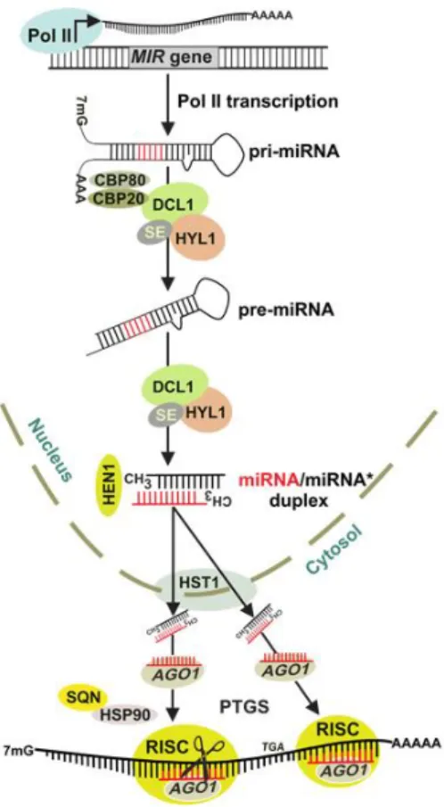
Generally, miRNA biogenesis starts with the transcription by RNA Polymerase II (Pol II) of noncoding nuclear miRNA (MIR) genes, mostly localized in inter and intragenic (intron) regions of the genome, and very few of them in the 5’- or 3’-untranslated (UTR) regions. The recruitment of Pol II is determined by the interaction between several transcriptional activators and various sequence motifs in MIR promoter regions (Rogers and Chen, 2013). The produced capped (7-methylguanosine cap; 7mG) and polyadenylated long primary miRNA transcripts (pri-miRNAs) (Fig. 5 and Fig. 6) then fold back into imperfect hairpin structures which are recognizable by the members of Dicer-like (DCL) family enzymes. Of the DCL family, DCL1 is known to be the mainly responsible for the processing of the pri-miRNA into a precursor-miRNA (pre-miRNA) in Arabidopsis, and for the subsequent cleavage of pre-miRNA to release a miRNA/miRNA* duplex (Axtell et al., 2011). DCL1 cleaves pri-miRNAs’ stem-loop structure mostly at the lower stem region, 16–17 bp from the single-strand–double-strand junction (Fig. 5). In a few cases, the terminal loop controls this processing (Pareek et al., 2015). According to the DCL family member that acts in miRNA biogenesis, the product miRNA length varies. The majority of plant miRNAs are 21 nucleotides (nt) long and are either processed by DCL1 or DCL4 proteins. DCL2 and 3 usually generate microRNAs that are 22 and 24 nt in length, respectively (Xie et al., 2004, 2005; Akbergenov et al., 2006; Deleris et al., 2006; Cuperus et al., 2010). These different lengths are proposed to be a consequence of the differences in DCL proteins’ molecular structures, in particular in the distance between the RNase III active site and PAZ domains (Macrae et al., 2006). The processing of pri-miRNAs to pre-pri-miRNAs also requires the double-stranded RNA-binding protein HYPONASTIC LEAVES1 (HYL1) and the C2H2 zinc finger protein SERRATE (SE), along with the cap-binding proteins (CBP) CBP20 and CBP80 (Kim et al., 2008; Pareek et al., 2015) (Fig. 6). Pre-miRNAs are unstable in the nucleus and are readily cleaved by DCL1 into a miRNA/miRNA* duplex, which is stabilized by the S-adenosyl methionine-dependent methyltransferase HUA ENHANCER 1 (HEN1)
14
(Fig. 6). Methyl groups placed on the 3’ terminal nucleotides of each of the duplex’s strand prevents their uridylation and consequent degradation by exonucleases (Li et al., 2005; Yang et al., 2006; Ramachandran and Chen, 2008). Then, miRNA/miRNA* is exported into the cytoplasm by HASTY (HST) (Park et al., 2005), where the duplex separates and the guide miRNA strand is loaded by Argonaute (AGO) proteins into the RNA-Induced Silencing Complex (RISC) (Fig. 6). What defines which strand of the duplex is the miRNA strand, and which is the miRNA* strand, is the thermodynamic stabilities of the 5’ ends linked to AGO. Most plant miRNAs carry a 5’ uridine that is usually bound by AGO1. So, the 5’ end of the miRNA strand has a lower thermodynamic stability than that of the miRNA* and is preferably loaded into RISC, whereas the miRNA* strand is typically excluded from this complex and is thought to be quickly degraded (Rogers and Chen, 2013; Pareek et al., 2015). The dissociation of the miRNA* strand from AGO1–miRNA/miRNA* complex does not require AGO1 endonuclease activity; instead, dissociation of the HEAT SHOCK PROTEIN90 (HSP90) and SQUINT (SQN) AGO1-associated proteins from the complex promotes miRNA* removal (Iki et al., 2010; Iki et al., 2012; Carbonell et al., 2012) (Fig. 6). The final product of miRNA biogenesis is then a small single-stranded RNA incorporated into a silencing complex. This fully assembled complex binds to its target through sequence complementarity with the miRNA strand, leading to posttranscriptional gene silencing (PTGS) (Fig. 6), mainly through mRNA cleavage or translational inhibition (Rogers and Chen, 2013) depending on the degree of their complementarity. If there is near-perfect complementarity between miRNA and its target, the result will be mRNA cleavage; if the mRNA does not have sufficient complementarity to the miRNA but does have a suitable clustering of miRNA-binding sites at its 3’-UTR, target gene expression will be regulated by miRNA-directed translational repression. Most plant microRNAs have single, extensive complementary target sites in mRNA open reading frames (ORFs), as was early on observed for evolutionarily conserved miRNAs from Arabidopsis (Rhoades et al., 2002). Target recognition is determined by the 5’ end domain of miRNAs, from nucleotide position 2 to 7, and the cleavage occurs at the 10th or 11th nucleotide independently of the miRNA length (Bartel, 2009; Pareek et al.,
2015). Extensive pairing was found to encompass nucleotides 9–11, suggesting a “slicing” mode of action similar to siRNA-directed silencing (Voinnet, 2009). Therefore, miRNA-directed target mRNA cleavage is the most common type of PTGS found in plants. Besides the PTGS mechanisms, some miRNA variants are also involved in DNA methylation, a well-known epigenetic regulation, and transcriptional gene silencing mechanism. AGO4, AGO6 and AGO9, all reported to be involved in RNA-directed DNA methylation (RdDM) in Arabidopsis, associate with 24-nt long small RNAs
15
generated from dsRNAs by the action of RNA-dependent RNA polymerase 2, RNA polymerase IV and DCL3 (Law and Jacobsen, 2010; Havecker et al., 2010; Pareek et al., 2015).
Figure 6 – Biogenesis of plant miRNAs. The primary miRNA
(pri-miRNA) transcript is transcribed from the microRNA (MIR) gene by RNA polymerase II (Pol II). Pri- to pre-miRNA conversion proceeds through splicing and further processing in nuclear dicing bodies mediated by HYL1 and SE and cap-binding proteins CBP20 and CBP80. The processed pre-miRNA yields a miRNA/miRNA* duplex that is stabilized by methylation by HEN1 and transported to the cytoplasm by HST1. The selected miRNA strand is incorporated and stabilized in AGO1, and the miRNA* strand removal is facilitated by HSP90 and SQN. miRNA-guided AGO1-containing RISC directs posttranscriptional gene silencing (PTGS) mechanisms, either mRNA cleave or translation inhibition of target transcripts. (Adapted from Khraiwesh et al., 2012)
Understanding the functional scope of plant miRNAs is considered to be one of the major current challenges in plant biology. The introduction of next-generation sequencing (NGS) in the past decade, along with powerful computational methods, has led to the genome or transcriptome-wide in silico identification of a massive amount of miRNA precursors in both model and non-model plants (Budak and Akpinar, 2015). Besides the identification of miRNAs and their precursors, miRNA targets are being confidently predicted through computational methods in plants as most miRNAs have high complementarity with their targets (Rhoades et al., 2002; Zhao et al., 2015). These bioinformatics tools are based on the three major distinctive features of miRNAs: hairpin-shaped stem-loop secondary structures, conserved highly complementary target regions, and high minimal folding free energy index (Kumar, 2014). Additionally, degradome sequencing data can complement these tools in the identification of biologically important and/or strongly regulated targets, establishing a direct link between a miRNA and its target(s) (Li et al., 2014). The degradome comprises the products of miRNA-directed cleavage. These cleavage products have a ligation-competent mRNA end with a 5’ phosphate that enables their distinction from other
16
degraded mRNAs isolated by standard methods, as the later are ligation incompetent during sequencing library preparation because they have a 5’ cap or lack the 5’ phosphate. By matching the 5’ end sequences of the cleavage products back to the corresponding genome, potential cleavage sites are identified and, by comparing the cleavage sites to the known or novel miRNA sequences, miRNA–target pairs can be established with confidence (German et al., 2008).
1.4. Objectives
Pine wilt disease, caused by the pine wood nematode Bursaphelenchus xylophilus, is one of the most serious and damaging diseases that has threatened pine forests worldwide and caused significant economic losses. Resistance against PWN infection seems to be linked to an early response that prevents the progression of the infection and stops the symptoms of the disease. While the physiological changes that occur throughout the disease progression have been characterized at the anatomical and biochemical levels, the molecular mechanisms that might be associated with susceptibility/tolerance of trees to PWN infection remain poorly understood. High-throughput screening procedures have been used to identify genes that are differentially expressed between tolerant/resistant and susceptible plants opening opportunities to unravel those mechanisms.
In recent years, microRNAs have been shown to be involved in plant response against pathogens, including nematodes, through sequence-specific silencing of plant transcripts that might have a role in the response to pathogen infection. Differentially expressed small RNAs and their targets, which provide defense mechanisms against pathogens, can be identified by small RNA sequencing and computational methods, and experimentally confirmed by high-throughput methods like real-time quantitative PCR (Kumar, 2014).
This Master project aims at the molecular analysis of Pinus pinaster plants exhibiting differential behavior (susceptibility and tolerance) towards PWN infection, focusing on the identification of miRNAs and their target genes, and transcripts highlighted in previous studies, putatively involved in the plant response mechanisms. P. pinaster plants used in this study were derived from seeds of a tree selected from a program initiated in 2009, which phenotypically identified over five hundred candidate resistant adult trees from an area with high incidence of PWD in Portugal (Ribeiro et al., 2012). In a previous study, seedlings obtained from the seeds of
17
ninety-six of those candidate trees (96 families) were inoculated with B. xylophilus to evaluate their tolerance to the infection, and genetic variability associated with the survival of half-sib seedlings was detected (Lisboa, 2016). Making use of this information, the plants used in this study belonged to one of those families in which a differential behavior (susceptibility or tolerance) towards the PWN infection was observed between individuals. After the inoculation of plants with the PWN, the objective was to select some plants apparently tolerant to the infection and some susceptible ones to conduct expression analyses of selected transcripts potentially involved in the response mechanisms against PWN infection, and that might explain the susceptibility or tolerance to PWD. Samples collected early after inoculation from the susceptible and tolerant P. pinaster plants were sent for small RNA and degradome next-generation sequencing in order to identify small RNAs differentially expressed between susceptible and tolerant samples, and to predict target transcripts for the differentially expressed small RNAs using computational approaches. Additionally, other transcripts were selected from a previous study that identified expressed sequence tags differentially expressed in P. pinaster susceptible plants versus plants from a tolerant species, Pinus pinea, inoculated with PWN.
Expression analysis of small RNA targets and transcripts putatively involved in the response mechanism to PWD should allow the identification of genes with different expression profiles between susceptible and tolerant P. pinaster plants, that might be linked to the differential behavior of those plants to PWN infection, and thus aid the elucidation of the response mechanisms against PWN infection by the identification of potential marker genes related to the disease.
18
2. Materials and Methods
2.1. Biological material2.1.1. Pinus pinaster
Fifty P. pinaster seedlings (2-year-old) were used in this study. The seedlings were kept in a greenhouse with a cooling system, with a relative humidity of 70%, and were watered every 2 days. They were provided by Dr. Isabel Carrasquinho from Instituto Nacional de Investigação Agrária e Veterinária, I. P. (INIAV, Oeiras) and were derived from seeds of a tree selected from a program initiated in 2009, aiming to phenotypically identify candidate resistant trees from an area with high incidence of PWD in Portugal (Ribeiro et al., 2012). Seedlings derived from the same candidate tree were previously used in a study that concluded that there is genetic variability associated with the survival of seedlings derived from the same tree to PWN infection (Lisboa, 2016).
2.1.2. Bursaphelenchus xylophilus
Bursaphelenchus xylophilus inoculum was prepared by collaborators in INIAV, Oeiras. B. xylophilus nematodes, originally isolated from infected pines, are maintained in the laboratory in culture media containing Botrytis cinerea at 25±1ºC, and are part of INIAV’s nematology collection. To prepare the inoculum, nematodes were extracted from the culture using the Baermann funnel technique (Southey, 1986). A water suspension of nematodes was adjusted to a concentration of 1000 nematodes/mL.
2.2. Inoculation of Pinus pinaster plants with Bursaphelenchus xylophilus
Fifty 2-year-old potted P. pinaster plants were inoculated, in collaboration with Dr. Isabel Carrasquinho (INIAV, Oeiras) and as part of the PhD work of Inês Modesto (Forest Biotechnology Lab, iBET, ITQB NOVA, Oeiras), following the method of Futai and Furuno (Futai and Furuno, 1979; Futai, 2003). Briefly, a small longitudinal wound was made in the stem approximately 10 cm from the apex and then covered with cotton. Afterwards, a 500 µL aliquot of the water suspension of 1000 nematodes/mL was pipetted into the cotton in the proximity of the wound and parafilm was wrapped around the inoculated regions to prevent drying of the inoculum (Appendix A). The same
19
procedures were performed in the control plants, which were inoculated with sterile water instead of the nematode suspension. Seventy-two hours after inoculation, a 2–4 cm portion of the stem (containing the inoculation site) and the needles immediately above that region were collected and stored at –80ºC. Part of the collected biological material of P. pinaster was used for all the studies performed in this work and described below.
2.3. Total RNA extraction
Based on the contrasting responses towards inoculation, as evaluated after at least 4 weeks following the inoculation, a set of the initial fifty inoculated plants were selected for further analysis. Three water-inoculated control plants, five susceptible plants, and five apparently tolerant plants were selected. Stems and needles from those plants were finely ground to powder in liquid nitrogen, and various RNA extraction procedures were tested. A protocol allowing the effective isolation of total RNA, including the small RNA fraction, from the stems and needles was established based on the CTAB method described by Chang et al. (1993). 70 mg of sample was homogenized in 1 mL of pre-warmed (65ºC) extraction buffer (300 mM Tris-HCl pH 8.0, 25 mM EDTA pH 8.0, 2 M NaCl, 2% CTAB, 2% PVP10, 0.05% Spermidine trihydrochloride and 2% β-mercaptoethanol) and the mixture vortexed and incubated for 10 min at 65ºC with agitation. 1 mL of ice-cold chloroform:isoamyl alcohol (24:1) was added twice to the homogenate in order to separate the aqueous upper phase (containing the RNA) from the organic phase. The mixture was shaken softly and centrifuged at 13,200 rpm for 10 min at 4ºC. The aqueous layer was transferred to another tube and mixed with 0.1 volume of 3 M NaOAc (pH 5.2) and 1 volume of ice-cold isopropanol. The mixture was stored at –80ºC for 3h. The nucleic acids (and any remaining carbohydrates) were pelleted by centrifuging at 13,200 rpm for 30 min at 4°C, and dissolved in 500 µL of Tris:EDTA (TE) buffer (pH 7.5). The solution was mixed with 1 volume of ice-cold isopropanol and 0.1% of SDS, and stored at –80ºC for 3h in order to precipitate the RNA. The precipitate was pelleted by centrifuging at 13,200 rpm for 30 min at 4ºC, washed with 1 mL of ice-cold absolute ethanol, air dried and dissolved in 40 µL of RNase-free water. RNA was stored at –80°C until use.
20
2.3.1. RNA samples purification and evaluation of RNA quality
RNA samples were treated twice with Ambion® TURBO DNA-free™ kit since one treatment was not sufficient for efficiently remove DNA from the RNA samples. Each treatment was performed in Biometra Tprofessional Thermocycler using 0.2 mL tubes. Briefly, to each 40 µL of sample 4 µL of 10x TURBO DNAse Buffer and 0.5 µL of TURBO DNAse (2 U/µL) were added. The samples were gently mixed and incubated at 37ºC for 30 min. Then another 0.5 µL of TURBO DNAse were added to each sample and tubes were incubated for another 30 min at 37ºC. 4 µL of DNase Inactivation Reagent was then added to stop the reaction and eliminate the DNAse and divalent cations. The samples were mixed and left at room temperature for 5 min with occasional agitation, and then centrifuged at 10,000 g for 2 min. The supernatant containing the purified RNA was placed in a new tube. RNA concentration and quality were estimated using Nanodrop ND-1000, and a 200 ng/uL aliquot of each sample was loaded in a 1.5% (w/v) agarose gel to check RNA integrity (Appendix B).
2.3.2. Total RNA and miRNA quantification using Qubit® fluorometer
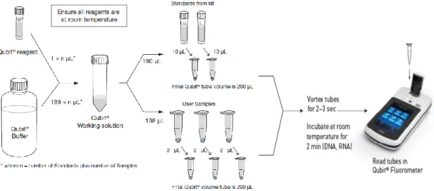
Total RNA concentration was measured using Qubit® fluorometer and Qubit® RNA BR (Broad Range; 1 ng/µL to 1 µg/µL) or HS (High Sensitivity; 250 pg/µL to 100 ng/µL.) (Invitrogen, USA), depending on the initially estimated sample concentrations. Qubit® fluorometer first needs to be calibrated using the two standards included in the assay kit. RNA samples to be quantified were diluted to perform this assay. 1 µL of each sample was diluted in 4 µL of water (1:5 dilution). Qubit® working solution must be added to standards and samples in specific Qubit® assay tubes. Qubit® working solution is prepared by diluting the Qubit® Reagent 1:200 in Qubit® Buffer (Fig. 7), at room temperature. The standards mixture was prepared by adding 10 µL of each standard to 190 µL of Qubit® working solution, and each sample tube was prepared by adding 2 µL of diluted RNA aliquot to 198 µL of Qubit® working solution (Fig. 7). All Qubit® assay tubes were vortexed 2-3 seconds and then incubated for at least 2 minutes at room temperature (Fig. 7). To read RNA concentration, the standard and sample tubes were inserted individually in the fluorometer (Fig. 7), and the concentrations in the original sample were determined taken into consideration the volume of sample added to the assay tube and the initial 1:5 dilution of the sample.
miRNA concentration was measured using Qubit® fluorometer and Qubit® miRNA Assay Kit (Invitrogen), using the same methodology used for total RNA concentration measurement but
21
using a different working solution (Qubit® microRNA Reagent diluted 1:200 in Qubit® microRNA Buffer) and two siRNA 21-mer standards (GAPDH siRNA) provided in the assay kit. This assay is accurate for initial small RNA sample concentrations ranging from 0.05 ng/µL to 100 ng/µL.
Figure 7 – Qubit quantitation assay kit workflow. Each assay kit provides concentrated Qubit reagent, dilution
buffer, and prediluted standards. Qubit working solution is prepared by diluting the Qubit reagent in Qubit buffer. The standards mixture was prepared by adding 10 µL of each standard to 190 µL of Qubit working solution, and each sample tube was prepared by adding 2 µL of diluted RNA aliquot to 198 µL of Qubit working solution. All tubes were vortexed 2-3 seconds and then incubated for at least 2 minutes at room temperature. RNA concentration in each sample tube is determined using the Qubit fluorometer and determined standards concentrations. (Adapted from lifetechnologies.com/qubit)
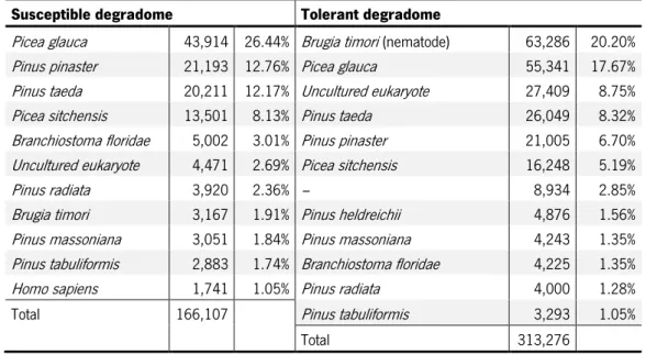
2.4. Small RNA and degradome sequencing
The RNA samples (from stems of three water-inoculated control plants, five susceptible plants, and five apparently tolerant plants) were sent for small RNA sequencing using Illumina HiSeq 2000 (LC Sciences, Houston, Texas – USA), along with two pools, one of them containing RNA from the stems of five susceptible plants and the other containing RNA from the stems of five tolerant plants, for degradome sequencing as a mean of identifying small RNA target transcripts. The samples were prepared to ship according to the company’s International Shipping Instructions (http://goo.gl/PcWIvj). Samples were prepared for small RNA and degradome sequencing by the service provider using the Illumina® TruSeq®Small RNA Sample Preparation protocol. Briefly, this protocol specifically binds Illumina adaptors to both the 5’ phosphate and the 3’ hydroxyl group leaved by the activity of Dicer or other RNA processing enzymes on mature miRNAs and other small RNAs. An RT reaction is then performed to generate single stranded cDNA molecules, which are
22
subsequently PCR amplified using a combination of a common primer and a primer containing one of the 48 possible index sequences (tags) for each sample. These tags distinguish different samples from one another in a single lane of a flow cell, allowing the samples to be processed in parallel through the RT-PCR amplification process. After PCR amplification, individual libraries with unique indexes obtained from separate samples can be pooled and gel purified together. Then, the libraries go through a quality control analysis and cluster generation before being sequenced.
2.5. Bioinformatic analyses of sequencing results
The bioinformatic analyses described in this section were performed with the support of Bruno Costa (Forest Biotechnology Lab, iBET, ITQB NOVA, Oeiras).
2.5.1. Small RNA sequence processing
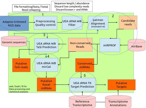
After removal of the adaptor sequences (“Adaptor trimmed NGS data”, Step 1 in Fig. 8), reads from each library were further processed individually using miRPursuit (Fig. 8) (Costa et al., 2016), a pipeline developed in the Forest Biotechnology lab (iBET, ITQB NOVA, Oeiras) constructed around several modules of the University of East Anglia small RNA workbench (UEA sRNA WB) (Stocks et al., 2012). In the initial “Preprocessing Quality control” and “UEA sRNA WB Filter” sequential steps (Step 1 in Fig. 8), low complexity (sequences containing less than 3 distinct nucleotides) and low abundant reads (less than 5) are removed, and only those with a length ranging from 18 to 26 bp and no match to plant rRNA/tRNAs are kept for further analysis (these parameters can be set in the wbench_filter.cfg configuration file – Appendix C.1). Quality plots and size profile distribution graphs before and after filtering were made for each library. In the following steps (Step 2, Fig. 8), the small RNAs were predicted by mapping the reads from step 1 to the setup genome file, with no mismatches allowed using PatMaN (Pattern Matching in Nucleotide databases; Prüfer et al., 2008) (“patman Alignment”, Step 2 Fig. 8). Due to the lack of a reference P. pinaster genome, the reference genome of Pinus taeda v1.01-masktrim was used as the setup genome file. The mapped reads were then mapped to the miRBase database release 21 (http://www.mirbase.org/) using miRProf (Step 2, Fig. 8), from the UEA sRNA WB, with no mismatches allowed to identify putative conserved miRNAs (the parameters are set in the wbench_mirprof.cfg configuration file – Appendix C.2). The small RNA reads that did not map to miRBase were classified as “Non-conserved reads” and put in a different file. The identified
23
conserved miRNAs were further grouped by match signature, organism, and miRNA family. Non-conserved reads were used to identify putative ta-siRNAs reads (“Putative tasi reads”, Fig. 8), with the aid of the TaSi Prediction tool on the UEA sRNA WB (Step 3, Fig. 8) and the genome of P. taeda to identify putative TAS genes from which phased 21-nt long small RNAs are generated (Khraiwesh et al., 2012) (the parameters used by the TaSi Prediction tool are set in the wbench_tasi.cfg configuration file – Appendix C.3). To predict “Putative Novel miRNAs”, using the miRCat tool in the UEA sRNA WB (Step 4, Fig. 8), the non-conserved reads were mapped to the setup genome file to find their respective precursor sequences (the parameters used by miRCat are set in the wbench_mircat.cfg configuration file – Annex C.4). The total number of sequence reads kept in each step of the miRPursuit workflow were calculated for each library.
Figure 8 – miRPursuit workflow. Step 1 – After removal of the sequencing adaptor from the reads (“Adaptor
trimmed NGS data”), reads from each library are further processed individually by “Preprocessing Quality control” and “UEA sRNA WB Filter” sequential steps, in which low complexity and low abundant reads are removed, and only 18 to 26 bp length and no match to plant rRNA/tRNAs reads are kept for further analysis. Step 2 – small RNAs are predicted using PatMaN (“patman Alignment”) by mapping the reads from step 1 to the setup genome file (Pinus taeda v1.01-masktrim), with no mismatches allowed; conserved miRNAs are identified using miRProf: reads are mapped to the miRBase with no mismatches allowed. Conserved miRNAs are further grouped by match signature, organism, and miRNA family. Step 3 – Non-conserved reads are used to identify putative ta-siRNA reads, using the TaSi Prediction tool and the setup genome, and also to identify putative novel miRNAs, using miRCat and the setup genome (to find their respective precursor RNAs – Step 4). Step 5 was not performed as described in the figure (see 2.5.3 for more information). (Retrieved from Costa et al., 2016)