UMinho | 2015
Universidade do Minho
Escola de Ciências
Bernardete Sofia da Cruz Fernandes
Outubro de 2015
Bernardete Sofia da Cruz Fernandes
Outubro de 2015
Dissertação de Mestrado
Mestrado em Biologia Molecular, Biotecnologia
e Bioempreendedorismo em Plantas
Trabalho efetuado sob a orientação da
Professora Doutora Mariana Sottomayor
e do
Professor Doutor Hernâni Gerós
Universidade do Minho
Escola de Ciências
Biotechnological approaches for yield
improvement of anticancer alkaloids
in the plant Catharanthus roseus
ii
DECLARAÇÃO
Nome: Bernardete Sofia da Cruz Fernandes
Endereço eletrónico: bernardetecruz@hotmail.com
Número do Cartão de Cidadão: 13235664
Título da Dissertação de Mestrado: Biotechnological approaches for yield improvement of anticancer alkaloids in the plant Catharanthus roseus
Orientadores:
Professora Doutora Mariana Sottomayor Professor Doutor Hernâni Gerós
Ano de conclusão: 2015
Designação do Mestrado:
Mestrado em Biologia Molecular, Biotecnologia e Bioempreendedorismo em Plantas
É AUTORIZADA A REPRODUÇÃO INTEGRAL DESTA DISSERTAÇÃO APENAS PARA EFEITOS DE INVESTIGAÇÃO, MEDIANTE DECLARAÇÃO ESCRITA DO INTERESSADO, QUE A TAL SE COMPROMETE.
Universidade do Minho, ___/___/______
iii
Agradecimentos
À professora Mariana Sottomayor por, antes de tudo, ter-me recebido no seu grupo de investigação, ter confiado em mim e ter sempre disponibilizado todas as condições para a realização do meu trabalho.
À Teré por tudo! Por me ter recebido e se ter disponibilizado para me ensinar com toda a paciência do mundo e por me acompanhar em todos os momentos deste projeto.
A todos os elementos do Bioactive Natural Products Group, por tudo o que me ensinaram e pela companhia diária no laboratório, em especial ao Diogo pelo apoio, que só quem está a passar pelo mesmo pode dar.
Aos amigos e à família, por estarem sempre presentes ajudando-me e incentivando-me durante todo este trajeto.
iv Biotechnological approaches for yield improvement of anticancer alkaloids in the plant Catharanthus roseus
Abstract
Catharanthus roseus (L.) G. Don produces an impressive number of specialized metabolites, over 130 terpenoid indole alkaloids (TIAs), among which the anticancer vinblastine (VLB) and vincristine (VCR). VLB and VCR were the first natural drugs used in cancer therapy, still holding an important position in the treatment of cancer nowadays. VLB and VCR accumulate in very low levels in C. roseus leaves that remain the only source of the anticancer TIAs, since efforts to obtain their entire chemical synthesis have been unfruitful. An intense research has been carried out over the last decades in C. roseus to unveil the biosynthesis of TIAs pathway, however, the characterization of several biosynthetic steps is still missing, and the membrane transport mechanisms are basically uncharacterized despite their importance for TIA accumulation.
Previous studies, in our group, allowed the identification of a number of very strong candidates to important functions within the TIA pathway, including MATE1, a strong candidate to TIA vacuolar accumulation, and CYP2, a strong candidate to two of the last steps of VLB and VCR biosynthesis. These genes are strong candidates for the possible manipulation of TIA levels in order to improve their accumulation.
The work developed here includes the sub-cloning of each gene into a plant binary vector followed by the genetic transformation of C. roseus hypocotyls using Agrobaterium tumefaciens. The induction of transgenic callus was optimized, leading to the successful establishment of transgenic callus lines confirmed by performing a Gus assay. The regeneration into a whole plant was actively pursued by changing an array of conditions but remained unsuccessful. Complementarily, downregulation of MATE1 expression was carried out using Virus Inducing Silencing Gene (VIGS), by sub-cloning a specific MATE1 fragment into a viral based plant vector followed by Agrobacterium mediated transformation of C. roseus plants. The putatively silenced plants showed significantly lower levels of the major alkaloids accumulated in C. roseus leaves, strongly supporting a role of MATE1 in their transtonoplast transport.
Overall, the work developed gives a significant contribution to the possibility of performing future biotechnological improvements of C. roseus for increased levels of the anticancer VLB and VCR.
v Abordagens biotecnológicas para a melhoria do rendimento dos alcaloides anticancerígenos presentes na planta Catharanthus roseus
Resumo
Catharanthus roseus (L.) G. Don produz um número impressionante de metabolitos secundários, mais de 130 alcalóides terpenóides indólicos (TIA), entre os quais, os anticancerígenos vinblastina (VLB) e vincristina (VCR). VLB e VCR foram os primeiros medicamentos de origem natural a serem utilizados no tratamento do cancro, mantendo ainda hoje uma posição relevante na terapia do cancro. VLB e VCR acumulam-se em níveis muito baixos nas folhas de C. roseus, que permanecem como a única fonte dos anticancerígenos, uma vez que, os esforços para os obter integralmente por síntese química têm sido infrutíferos. Ao longo das últimas décadas, C. roseus tem sido intensamente investigada com o intuito de desvendar a via de síntese dos TIA, no entanto, várias etapas ainda estão por caracterizar assim como os mecanismos de transporte membranar, apesar da sua importância para a acumulação dos TIAs.
Estudos anteriores, no nosso grupo, possibilitaram a identificação de genes candidatos a funções importantes dentro da via dos TIAs, incluindo MATE1 um forte candidato ao transporte dos TIAs para o vacúolo, e CYP2, um forte candidato a intervir num dos dois últimos passos da biossíntese de VLB e VCR. Estes genes são fortes candidatos para uma eventual manipulação dos níveis de TIA, a fim de aumentar a sua acumulação.
O presente trabalho inclui a sub-clonagem de cada um dos genes num vetor binário seguido pela transformação genética dos hipocótilos de C. roseus usando Agrobacterium tumefaciens. A indução de callus transgénico foi otimizada, alcançando-se com sucesso linhas de calli transgénicas, confirmadas através da realização de um ensaio de GUS. A regeneração para obter a planta foi arduamente perseguida alterando-se uma série de condições, mas até ao momento não foi alcançada. Complementarmente, o silenciamento da expressão do gene MATE1 foi levado a cabo usando Vírus Indutor de silenciamento do gene (VIGS), através da sub-clonagem de um fragmento específico de MATE1 num vetor viral modificado, seguindo-se a transformação das plantas mediada por Agrobacterium. As plantas putativamente silenciadas apresentaram níveis significativamente mais baixos dos principais alcalóides acumulados em folhas de C. roseus, reforçando fortemente o papel do MATE1 no seu transporte. No geral, o trabalho desenvolvido contribui significativamente para a possibilidade de no futuro realizarem-se melhorias biotecnológicas em C. roseus com vista a aumentar os níveis dos anticancerígenos VLB e VLC.
vi
Index
Agradecimentos ... iii Abstract ... iv Resumo ... v Index ... vi Abbreviations ... xList of Figures ... xiii
List of Tables ... xvii
1. Introduction ... 1
1.1 The medicinal plant Catharanthus roseus (L.) G. Don ... 1
1.2 C. roseus terpenoid indole alkaloids ... 2
1.3 The terpenoid indole alkaloid (TIA) pathway ... 3
1.4 Cytochromes P450 in the TIA pathaway ... 6
1.5 Cellular and sub-cellular compartmentation of the TIA pathway in C. roseus ... 6
1.6 MATE transporters in plants... 8
1.7 Virus Induced Gene Silencing (VIGS) ... 9
1.8 Plant biotechnology for genetic improvement ... 11
1.9 Genetic transformation of C. roseus ... 13
1.10 Agrobacterium-mediated transformation ... 12
1.11 Regeneration of C. roseus ... 14
1.12 Objectives ... 16
2. Materials and methods ... 17
2.1 Transformation and regeneration of Catharanthus roseus ... 17
2.1.1 Biological material ... 17
2.1.1.1 Plant material ... 17
vii
2.1.2. MATE1 and CYP2 genes ... 18
2.1.3 Primer design ... 18
2.1.4 Polymerase Chain Reaction (PCR) ... 19
2.1.5 Purification of PCR products ... 20
2.1.6 Molecular cloning ... 20
2.1.6.1 Digestion of the PCR products and of the expression vector ... 20
2.1.6.2 Ligation reaction ... 21
2.1.6.3 Transformation of E. coli ... 21
2.1.6.4 Selection of positive clones... 22
2.1.7 Transformation and regeneration of C. roseus explants ... 23
2.1.7.1 Seed sterilization and germination ... 23
2.1.7.2 Transformation of Agrobacterium ... 23
2.1.7.3 Transformation and co-cultivation of C. roseus explants ... 23
2.1.7.4 C. roseus regeneration ... 24
2.1.7.5 Analysis of putative transformed calli ... 26
2.2 Virus Induction Gene Silencing (VIGS) ... 26
2.2.1 Biological material ... 26
2.2.1.1 Plant Material ... 26
2.2.1.2 Bacterial strains and plasmids... 27
2.2.2 Primer design and cloning ... 28
2.2.3 PCR ... 28
2.2.4 Molecular cloning ... 29
2.2.4.1 Digestion and ligation of the PCR products and of the expression vector ... 29
2.2.4.2 Transformation of E. coli ... 30
2.2.4.3 Selection of positive clones... 30
viii
2.2.5.1 Transformation of Agrobacterium ... 32
2.2.5.2 Agrobacterium prick inoculation ... 33
2.2.5.3 Alkaloid extraction ... 34
3. Results ... 36
3.1 Optimization of engineering strategies of C. roseus for overexpression of MATE1 and CYP2 ... 36
3.1.1 Sub-cloning of C. roseus MATE1 and CYP2 into a plant binary vector ... 36
3.1.2 Transformation of C.roseus hypocotyl explants... 39
3.1.3 Generation of transgenic calli ... 40
3.1.4 Induction of Shoots ... 44
3.1.5 Analysis of GUS expression in transgenic calli ... 45
3.2 Virus-inducing ene silencing of MATE 1 in C. roseus plants... 46
3.2.1 Molecular sub-cloning of a MATE1 fragment into pTRV2:GFP ... 47
3.2.2 Agrobacterium prick inoculation of C.roseus plants ... 48
3.2.3 Analysis of the alkaloid profile and levels of the silenced plants ... 50
4. Discussion ... 53
4.1 Biotechnological approach ... 53
4.2 Agrobacterium mediated transformation of C. roseus hypocotyls ... 54
4.3 Callus induction in C.roseus hypocotyls ... 55
4.4 Regeneration attempts of C.roseus transgenic plants ... 57
4.5 Establishment of transgenic callus cultures ... 57
4.6 Silencing of MATE1 ... 58
5. Conclusions and Future Perspectives ... 60
6. Bibliography ... 61
x
Abbreviations
10HGO - 10-hydroxygeraniol oxidoreductase
16-OH OMT - 16-hydroxytabersonine-16-O-methyltransferase 2,4 –D - Ácido 2,4-diclorofenolacético
7DLH 7 - Deoxyloganic acid 7-hydroxylase AFLP – Amplified Fragment length polymorphism AVLB - Anhydrovinblastine
BA - 6-Benzylaminopurine bp - base-pair
CaMV 35S - Cauliflower Mosaic virus promoter CroPrx1 – C. roseus Peroxidase 1
D4H - Desacetoxyvindoline 4-hydroxylase DAT - Deacetylvindoline 4-O-acetyltransferase ddH2O - Bi-distillated H2O
DNA - Deoxyribonucleic acid dNTPs - Deoxynucleotide dsRNA - Double strand RNA
EDTA - Ethylenediamine tetraacetic acid EGFP - Enhanced green fluorescent protein EtBr - Ethidium bromide
Fwd - Forward
G10H - Geraniol-10-hydroxylase GC% - Guanine/Cytosine content GPPS - Geranyl diphosphate synthase
HPLC-DAD - Chromatography-Diode-Array Detection IDI - Isopentenyl Diphosphate Isomerase
IO - Iridoid oxidase
IPAP- Internal Phloem Associated Parenchyma kb/Kbp - kilo-base-pair
LB - Luria Bertrani
MATE - Multidrug and toxic compound extrusion MEP - 2-C-methyl-D-erythritol 4-phospate
xi mRNA - Messenger RNA
MS - Murashige & Skoog
MSCP - Murashige & Skoog Casein Proline NAA - 1-Naphthaleneacetic acid
NMT - 16-methoxy - 2,3-dihydro - 3-hydroxytabersonine - N-methyltransferase OMT - 16-hydoxytabersonine 16-O-methyltransferase
ON - Overnight
P450 - Cytochrome P450 monoxygenase PCR – Polymerase chain reaction
pDNA - Plamid DNA
PGR´s – Plant Growth Regulators pRi - Hairy root-inducing plasmid Prx1 – Peroxidase 1
PTGS - Post Transcriptional Gene Silencing pTi - Tumour-inducing plasmid
Rev - Reverse
RISC - RNA-induced silencing complex RNA - Ribonucleic acid
rpm - Revolutions per minute RT – Room temperature
SGD - Strictosidine -D-glucosidase siRNA - Small interfering RNA SLS - Secologanin synthase STR - Strictosidine synthase
T16H - Tabersonine 16-hydroxylase T3O - Tabersonine 3-oxygenase T3R - Tabersonine 3-reductase TAE buffer – Tris-acetate-EDTA buffer TDC - Tryptophan decarboxylase T-DNA - Transferred-DNA TDZ- Thidiazuron
TIA - Terpenoid Indole Alkaloids Tm - Melting temperature TRV - Tobacco rattle vírus
xii UV – Ultra-violet
v/v – volume/volume VCR - Vincristine
VIGS – Virus Inducing Gene Silencing VIR - Virulence
xiii
List of Figures
Figure 1 - Flowering plant of Catharanthus roseus cv. Little Bright Eye. ... 1
Figure 2 - Representative alkaloids of C. roseus. Adapted from O’Connor and Maresh, 2006... 2
Figure 3 - The TIA biosynthetic pathway in C. roseus. TDC, tryptophan decarboxylase; G10H, geraniol 10-hydroxylase; SLS, secologanin synthase; STR - strictosidine synthase, SGD - strictosidine -D-glucosidase, T16H - tabersonine 16-hydroxylase, OMT – 16-hydoxytabersonine-16-O-methyltransferase, NMT - 16-methoxy-2,3-dihydrotabersonine N-methyltransferase, T3O/T3R - tabersonine 3-oxygenase/ tabersonine 3-reductase D4H - desacetoxyvindoline 4-hydroxylase, DAT - deacetylvindoline 4-O-acetyltransferase, Prx1 – peroxidase 1. Adapted from Almagro, Fernández-Pérez and Pedreño, (2015). ... 4
Figure 4 - Cellular and subcellular organization of the biosynthetic pathway of TIAs in C. roseus leaves. Known single enzymatic steps in each cell type are indicated by grey arrows and abbreviation of enzyme names. Broken grey arrows and broken pink arrows indicate unknown enzymatic steps and metabolite translocation, respectively. DXS, 1-deoxy-D-xylulose-5-phosphate (DXP) synthase; DXR, DXP reductoisomerase; CMS, 4-(cytidine 5 ′-diphospho)-2C-methyl-D-erythritol (CM) synthase; CMK, CM kinase; MECS, 2C-methyl-D-′-diphospho)-2C-methyl-D-erythritol-2,4-cyclodiphosphate (MEC) synthase; HDS, hydroxymethylbutenyl 4-diphosphate (HD) synthase; HDR, HD reductase; IDI, isopentenyl diphosphate isomerase; GPPS, geranyl diphosphate synthase; GES, geraniol synthase; G10H (CYP76B6), geraniol 10-hydroxylase; CPR, cytochrome P450-reductase; 10HGO, 10-hydroxygeraniol oxidoreductase; IO, iridoid oxidase; IS, iridoid synthase; 7DLGT, 7-deoxyloganetic acid glucosyltransferase; 7DLH, 7-deoxyloganic acid 7-hydroxylase; LAMT, loganic acid O-methyltransferase; SLS (CYP72A1), secologanin synthase; TDC, tryptophan decarboxylase; STR, strictosidine synthase; SGD, strictosidine -glucosidase; T16H2 (CYP71D351), tabersonine 16-hydroxylase 2; 16OMT, 16-hydroxytabersonine O-methyltransferase; NMT, 16-methoxy-2,3-dihydrotabersonine N-methyltransferase; D4H, desacetoxyvindoline 4-hydroxylase; DAT, deacetylvindoline 4-O-acetyltransferase. DMAPP, dimethylallyl diphosphate; GAP, glyceraldehyde 3-phosphate; IPP, isopentenyl diphosphate (Dugé de Bernonville et al., 2015b) ... 7
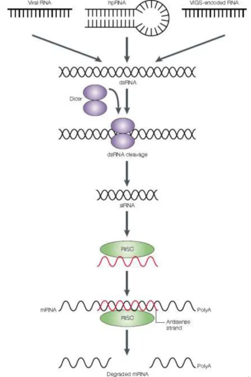
Figure 5 - The current model of RNA-mediated gene silencing in plants. This model is based on the results of in vitro studies of RNA-induced gene silencing, or RNA interference (RNAi), in animal
xiv extracts. RNAi is believed to operate in a similar manner in plants because small interfering RNAs (siRNAs) are found in silenced plants, and plants have homologues of the animal gene Dicer. Double-stranded RNA (dsRNA) from replicating viral RNA, viral-vector-derived (VIGS, or virus-induced gene silencing) RNA or hairpin RNA (hpRNA) transcribed from a transgene, is processed by a Dicer-containing complex to generate siRNAs. An endonuclease-containing complex (called the RNAi silencing complex, RISC), is guided by the antisense strand of the siRNA to cleave specific mRNAs, so promoting their degradation (Waterhouse and Helliwell, 2003). ... 10
Figure 6 - Schematic representation of pCAMBIA2301m expression vector. The figure was created using the bioinformatics tool SnapGene®. ... 18
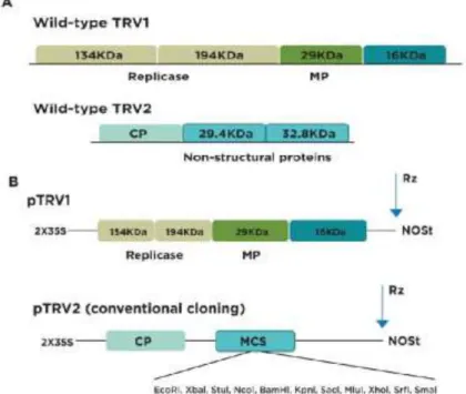
Figure 7 - Schematic representation of TRV1 and TRV2 (A), and of pTRV1 and pTRV2 vectors (B). Adapted from Senthil-Kumar and Mysore (2014). ... 27
Figure 8 - Schematic representation of pTRV2:GFP vector. Adapted from Tiang et al. (2013). ... 28
Figure 9 - Scheme of the VIGS protocol. ... 32
Figure 10 - C. roseus 9 week old plant used in the VIGS protocol. Red dots mark points of Agrobacterium prick inoculation. ... 34
Figure 11 - Agarose gel electrophoresis of PCR amplified CYP2 and MATE1 for sub-cloning into pCAMBIA2301m. a) CYP2, b) MATE1. M, Molecular weight marker, GeneRuler™ 1kb DNA Ladder (Thermo Scientific). The observed bands seem to possess the predicted size, near 1.5 kbp. .... 36
Figure 12 - Agarose gel electrophoresis of the colony PCR screening after transformation with a) CYP2-pCAMBIA2301m and b) MATE2-pCAMBIA2301m.,M, Molecular weight marker, GeneRuler™ 1kb DNA Ladder (Thermo Scientific). Positives colonies: a) 2, 8, 13 and b) 3, 4, 5, 7. ... 37
Figure 13 - Agarose gel electrophoresis of the restriction analysis of CYP2-pCAMBIA2301m and MATE1-pCAMBIA2301m selected clones: a) All the represented clones are positive as indicated by the presence of four band with 870 pb, 1043 pb, 2349 pb, and 9299 pb corresponding to the 3 restriction sites of Xho I in the plasmid and one restriction site in CYP2. b) The clone represented is positive, as indicated by the presence of four bands with, 870 pb, 1528 pb, 2349 pb, 10314 pb, corresponding to the 3 restriction sites of XhoI in the plasmid and one restriction site of XbaI in MATE1. M, Molecular weight marker, GeneRuler™ 1kb DNA Ladder (Thermo Scientific). ... 37
xv Figure 14 - Alignment between the nucleotide sequence of MATE1 with the MPGR gene ID cra_locus_1952 and the sequencing of MATE1 cloned in pCAMBIA2301m. ... 38
Figure 15 - Alignment between the nucleotide sequence of CYP2 with the MPGR gene ID cra_locus_4293 and the sequence of CYP2 cloned in pCAMBIA2301m. ... 39
Figure 16 - Steps to obtain the explants used in transformation protocols. ... 40
Figure 17 - Hypocotyls on callus induction medium surrounded by a clear overgrowth of Agrobacterium. ... 41
Figure 18 -- Hypocotyls on callus induction medium: on the left a hypocotyl after 14 days on A1 medium with 500 mg L-1 of carbenicillin, on the right a hypocotyl after 10 days on A2 medium with
500mg L-1 cefotaxime. Both hypocotyls show some signs of callus induction and a brownish
coloration. ... 41
Figure 19 - Hypocotyls after 9 days on callus induction media B to I, as detailed in table 7. All are non-transformed control hypocotyls. ... 42
Figure 20 - Application of the nurse culture procedure. In the middle of the Petri dish there is a portion of actively growing callus surrounded by hypocotyls. ... 43
Figure 21 - Callus proliferation in hypocotyls submitted to transformation using different constructs and procedures, together with non-transformed controls, after 12 days in medium I using a nurse culture. ... 44
Figure 22 - Calli in shoot induction medium showing the proliferation of calli upon successive subcultures along ca 15 weeks. ... 45
Figure 23 - Histochemical detection of GUS activity in callus transformed with the empty pCAMBIA2301m vector. ... 45
Figure 24 - Histochemical detection of GUS activity in callus transformed with CYP2-pCAMBIA2301m. ... 46
Figure 25 - Histochemical detection of GUS activity in callus transformed with MATE-pCAMBIA2301m. ... 46
xvi Figure 26 - Agarose gel electrophoresis of the PCR amplified MATE1 fragment with 413pb for sub-cloning into pTRV2:GFP. M, Molecular weight marker, GeneRuler™ 1kb DNA Ladder (Thermo Scientific). ... 47
Figure 27 - Agarose gel electrophoresis of the double digestion of MATE1-pTRV2 with EcoRI and XohI. M, Molecular weight marker, GeneRuler™ 1kb DNA Ladder (Thermo Scientific). Positive clones (arrows): 1, 3, 4. ... 48
Figure 28 - Alignment between the nucleotide sequence of the MATE1 fragment cloned in pTRV2:GFP with the original MATE1 sequence. ... 48
Figure 29 - C.roseus plants inoculated with the positive control pTRV2-ChlH. Plants 8 days after inoculation (left) do not present photo-bleaching, whereas plants 20 days after inoculation (right) display a clear photo-bleaching phenotype. ... 49
Figure 30 - C. roseus plants 20 days after inoculation with: a) positive control; b) negative control; c) MATE1-pTRV2:GFP; d) Mock treatment. ... 50
Figure 31 - Representative chromatogram of a leaf alkaloid extract registered at 260 nm. The three main peaks detected correspond to the papaverine, vindoline and catharanthine. ... 50
Figure 32 - Absorbance spectra of the peaks detected during HPLC-DAD analysis of leaf alkaloid extracts allowing the identification of papaverine, vindoline and catharanthine. ... 51
Figure 33 - Quantification of vindoline levels in the leaves of plants inoculated with the empty vector (pTRV2-MCS) and MATE1-pTRV2:GFP. Values are means of 10 plants. Asterisk indicate significant differences between vindoline production in plants inoculated with pTRV2-MCS and with MATE1-pTRV2:GFP, using Student’s t-test (P < 0.05) ... 51
Figure 34 - Quantification of catharanthine levels in the leaves of plants inoculated with the empty vector (pTRV2-MCS) and with MATE1-pTRV2:GFP. Values are the mean of 10 plants. Asterisk indicate significant differences between values of catharanthine production in plants inoculated with pTRV2-MCS and with MATE1-pTRV2:GFP using Student’s t-test (P < 0.05) ... 52
xvii
List of Tables
Table 1 - Primers used to amplify MATE1 and CYP2 to clone in pCAMBIA2301m. Endonuclease restriction sites are underlined. Additional nucleotides at the 5’ terminus to allow site recognition
by the restriction enzyme are in red. ... 19
Table 2 - Primer features as in OligoAnalyzer 3.1... 19
Table 3 - PCR optimized setup for amplification of the MATE1 and CYP2. ... 20
Table 4 - Double digestion reaction conditions. ... 20
Table 5 - Ligation reaction conditions. ... 21
Table 6 – Colony PCR optimized setup for amplification of the MATE1 and CYP. ... 22
Table 7 - MSCP1 basal medium composition. ... 24
Table 8 - Composition of the several media tested in the callus generation ... 25
Table 9 - MSCP2 basal medium composition. ... 25
Table 10 – Composition of the several media tested in the induction of shoots. ... 26
Table 11 - Primers used to amplify the MATE1 fragment to clone into modified pTRV2:GFP. Endonuclease restriction sites are underlined. Additional nucleotides at the 5’ terminus to allow site recognition by the restriction enzyme are in red. ... 28
Table 12 - PCR optimized setup for amplification of the MATE1 fragment ... 29
Table 13 - Double digestion reaction conditions. ... 29
Table 14 - Ligation reaction of MATE1 into pTRV2:GFP vector, using a 8:1 molar ratio. ... 30
Table 15 - Double digestion reaction conditions. ... 31
1
1.
Introduction
1.1 The medicinal plant
Catharanthus roseus
(L.) G. Don
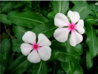
Catharanthus roseus (L.) G. Don commonly known as Madagascar periwinkle is a member of the Apocynaceae family that originates from Madagascar, but can nowadays be found as sub-spontaneous in many subtropical regions and also in gardens worldwide as an ornamental plant (Figure 1). This global distribution was namely caused by sailors, who took the plants on board during their travels, due to their medicinal properties. C. roseus is a perennial semi- shrub that grows about 80 cm high and blooms continuously year-round with pink, purple, or white flowers (Sottomayor and Barceló, 2006; van Der Heijden et al., 2004)
Figure 1 - Flowering plant of Catharanthus roseus cv. Little Bright Eye.
C. roseus plant has historically been used to treat a wide assortment of diseases. In several sub-tropical countries, it was used as a folk remedy for diabetes for centuries while in India, juice from the leaves was also used to treat wasp stings and other insect bites (Singh et al., 2001).
During the 1950’s, researchers from the Noble research group in Canada and from the Eli Lilly Pharmaceutical company in the USA discovered that the plant contains alkaloids useful to the treatment of cancer, vinblastine (VLB) and vincristine (VCR) (Noble, 1990; Sottomayor and Barceló, 2006). These alkaloids are produced only in C. roseus leaves, they were the first natural drugs used in cancer therapy, and still maintain an important position in the treatment of cancer nowadays (Costa et al., 2008; Suttipanta et al., 2011)
2
1.2
C. roseus
terpenoid indole alkaloids
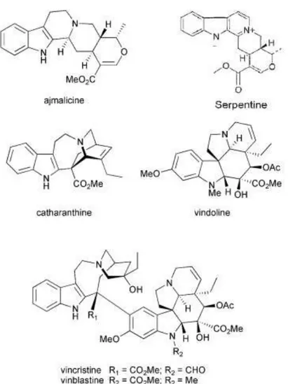
Alkaloids are a large group of specialized metabolites of low molecular weight containing nitrogen that share a characteristic toxicity and pharmacological activity (De Luca and St Pierre, 2000; Shitan and Yazaki, 2007). C. roseus is known to produce an impressive number of specialized metabolites, with over 130 terpenoid indole alkaloids (TIAs) isolated and identified, some of which are valuable therapeutic compounds (St-pierre, 2007; van Der Heijden et al., 2004): VLB and VCR are widely used as drugs for cancer chemotherapy, ajmalicine is used as an anti-hypertensive agent and serpentine is used as a sedative (Figure 2) (St-pierre, 2007; van Der Heijden et al., 2004).
Figure 2 - Representative alkaloids of C. roseus. Adapted from O’Connor and Maresh, 2006.
The anticancer VLB and VCR are highly valuable anticancer drugs, but are produced in very low amounts in the plant - it takes about 500 kg of dried periwinkle leaves to isolate 1 g of VLB, and their levels depend on the physiological and developmental stage of the plant (van Der
3 Heijden et al., 2004). Moreover, the majority of specialized metabolites with pharmaceutical activity are only obtained from cultivated plants, and despite all attempts made, their chemical synthesis in most cases is not possible or too expensive (Zárate and Verpoorte, 2007). In fact, the highly complex structures of alkaloids VLB and VCR impair their entire chemical synthesis. The more feasible process is the semi synthesis, using the natural monomeric precursors catharanthine and vindoline, which are present in greater amount and can be extracted from C. roseus leaves (Roepke et al., 2010). Hence, an extensive study of the TIA biosynthetic pathway has been carried out over the last three decades, making of C. roseus one of the most studied plants concerning specialized metabolism. The demand for the elucidation of their biosynthesis became critical: to either increase their production in planta or to engineer these pathways in microorganisms to allow industrial production of medicinally relevant compounds (El-Sayed and Verpoorte, 2007; O’Connor and Maresh, 2006).
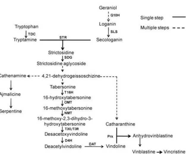
1.3 The terpenoid indole alkaloid (TIA) pathway
The research conducted over the last decades associated with the recent massive expansion of transcriptomic resources for C. roseus and the development of efficient approaches of virus-induced gene silencing led to a more accurate knowledge of genes involved in TIA production in C. roseus. In fact, the evolution of molecular cell biology techniques has also contributed to a major progress in deciphering the puzzling spatial organization of the TIA biosynthetic pathway (Courdavault et al., 2014). All the TIAs of C. roseus share a common precursor, strictosidine (O’Connor and Maresh, 2006). Strictosidine is formed by condensation of the indole precuror tryptamine with the terpenoid precursor secologanin through the catalytic action of strictosidine synthase (STR) (Figure 3). This coupling requires the combination of two metabolic pathways, the 2-C-methyl-D-erythritol 4-phospate pathway (MEP) through secaloganin, and the shikimate pathway through tryptamine (Murata and Luca, 2005; van Der Heijden et al., 2004).
4
Figure 3 - The TIA biosynthetic pathway in C. roseus. TDC, tryptophan decarboxylase; G10H, geraniol 10-hydroxylase;
SLS, secologanin synthase; STR - strictosidine synthase, SGD - strictosidine -D-glucosidase, T16H - tabersonine
16-hydroxylase, OMT – 16-hydoxytabersonine-16-O-methyltransferase, NMT - 16-methoxy-2,3-dihydrotabersonine
N-methyltransferase, T3O/T3R - tabersonine 3-oxygenase/ tabersonine 3-reductase D4H - desacetoxyvindoline
4-hydroxylase, DAT - deacetylvindoline 4-O-acetyltransferase, Prx1 – peroxidase 1.Adapted fromAlmagro,
Fernández-Pérez and Pedreño, (2015).
The biosynthesis of the indole moiety via the shikimate pathway yields tryptamine, which is derived from tryptophan by the action of tryptophan decarboxylase (TDC) (De Luca et al., 1989; St-Pierre et al., 1999). Whereas the assembly of the monoterpene secoiridoid moiety requires a total of ten enzymes to convert the MEP pathway-derived monoterpenoid skeleton into secologanin (Dugé de Bernonville et al., 2015b). Recently Miettinen et al., (2014) reported the identification and characterization of the last four enzymes that fill in the missing steps of the C. roseus secoiridoid pathway (Figure 4).
Strictosidine is subjected to deglucosylation catalysed by strictosidine -D-glucosidase
(SGD) yielding an unstable aglycone precursor required for the assembly of the corynanthe, iboga, and aspidosperma skeletons representing different ramifications of the TIA pathway, such as ajmalicine, catharanthine, and vindoline the latter two being the monomeric precursors of VLB and
5 VCR (Dugé de Bernonville et al., 2015b; Liscombe and O’Connor, 2011; Murata and Luca, 2005). The sequence of reactions since the labile aglycone until the formation of catharanthine or tabersonine remain poorly characterized. Nevertheless, the 6 steps leading to the production of vindoline from the precursor tabersonine are well understood at the molecular and biochemical levels (Figures 3 and 4) (Besseau et al., 2013; Murata and Luca, 2005). The first step in the conversion of tabersonine to vindoline is the hydroxylation by the enzyme tabersonine 16-hydroxylase (T16H) (Murata and Luca, 2005; St-Pierre and De Luca, 1995). The following step is the O-methylation of hydroxytabersonine to yield methoxytabersonine by the enzyme 16-hydroxytabersonine-16-O-methyltransferase (16-OH OMT). This is converted into 3-hydroxy-16-methoxy-2,3-dihydrotabersonine by tabersonine 3-oxygenase (T3O) and tabersonine 3-reductase (T3R) (Qu et al., 2015) followed by an N-methylation by the enzyme 16-methoxy - 2,dihydro - 3-hydroxytabersonine - N-methyltransferase (NMT), originating desacetoxyvindoline. Desacetoxyvindoline undergoes a hydroxylation by desacetoxyvindoline 4-hydroxylase (D4H) and the last step in vindoline biosynthesis is catalyzed by acetylcoenzyme A: deacetylvindoline 4-O-acetyltransferase (DAT) (Besseau et al., 2013; Guirimand et al., 2011; Liscombe and O’Connor, 2011; Murata and Luca, 2005)
The coupling of vindoline and catharanthine form the first dimeric TIA, -3’,4’ anhydrovinblastine (AVLB), which is the precursor of all dimeric TIAs, including the anticancer VLB and VCR. This dimerization step is believed to be performed by the major class III peroxidase localized in C. roseus mesophyll vacuole (Sottomayor and Barceló, 2006; Sottomayor and Ros Barceló, 2003; Sottomayor et al., 1998, Costa et al. 2008). Finally, the biosynthetic steps from AVLB to the anticancer alkaloids, VCR and VLB, are not characterized. In order to identify candidate enzymes to this steps, research was conducted in our lab including the differential transcription profiling of the specialized cells of C. roseus leaves, the idioblasts, where the late steps of VLB biosynthesis are believed to occur Carqueijeiro, (2013).
The screening data allowed to identify a number of very strong candidate genes to important functions within the TIA pathway, namely to the bottleneck hydroxylation of AVLB into VLB, to the transmembrane transport of TIA intermediates and to transcriptional regulation of the early and late steps of the TIA pathway (Carqueijeiro, 2013).
6
1.4 Cytochromes P450 in the TIA pathaway
Cytochrome P450 monooxygenases in plants are membrane-bound enzymes involved in a wide range of biosynthetic pathways, including in the TIA pathway (St-Pierre and De Luca, 1995).
In the TIA pathway of C. roseus, P450 monooxygenases have a crucial influence, until now, seven enzymes were identified and characterized: geraniol 10-hydroxylase (G10H, P450 CYP76B6), iridoid oxidase (IO, CYP76A26,) 7-deoxyloganic acid 7-hydroxylase (7DLH, CYP72A224), secologanin synthase (SLS, CYP72A1), tabersonine 16-hydroxylase 1 and 2 (T16H1 and 2, ) and tabersonine 3-oxygenase (T3O, CYP71D1V2) (Figure 4) (Besseau et al., 2013; Murata and Luca, 2005; Qu et al., 2015; St-Pierre and De Luca, 1995). All these P450 already identified in the C. roseus TIA pathway allow the prediction of a possible involvement of other P450s in several uncharacterized steps. In line with this assumption, previous studies in our laboratory revealed that several P450s are up-regulated in the TIA accumulating idioblasts. These are good candidates to the first reaction in the conversion of AVLB into VLB, and also in one of the predicted reactions in the transformation of VLB into VLC, which involves two oxidative steps: the oxidation of the alkyl group to an alcohol, and then the oxidation of the alcohol group to aldehyde. If the two step hypothesis is correct, two oxydireductases are required, one of them being a hydroxylase like a P450. Therefore, it is extremely important to study these candidate cytochrome P450s to decrypt the last steps of the TIA pathway.
1.5 Cellular and sub-cellular compartmentation of the TIA pathway in
C. roseus
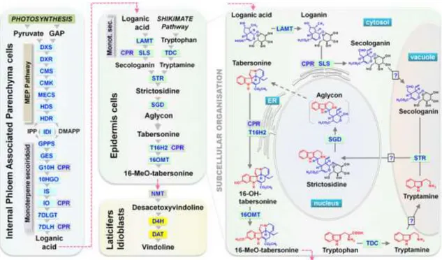
The characterization of the TIA pathway in C. roseus revealed a fascinating picture of cell-type specific compartmentation, involving at least four different cell cell-types and as many subcellular compartments with inherent intercellular and intracellular metabolite translocations. Over the last decade, several tools have been applied to unveil the architecture of cellular and sub-cellular compartmentation, with the mechanisms of inter-tissue mobility of compounds remaining largely unknown (Courdavault et al., 2014).
Recent research revealed that the internal phloem associated parenchyma (IPAP) cells harbour the whole Methyl Erythritol Phosphate (MEP) pathway and the eight first reactions of the monoterpene-secoiridoid pathway, from the synthesis of geranyl diphosphate synthase (GPPS) to its conversion into the iridoid loganic acid (Figure 4). At the subcellular level, the first 10 enzymatic
7 steps leading to geraniol synthesis are located in the plastid, not only localized in stroma of plastids but also in stromules. (Courdavault et al., 2014; Dugé de Bernonville et al., 2015b; Miettinen et al., 2014; Salim et al., 2014)
Figure 4 - Cellular and subcellular organization of the biosynthetic pathway of TIAs in C. roseus leaves. Known single enzymatic steps in each cell type are indicated by grey arrows and abbreviation of enzyme names. Broken grey arrows and broken pink arrows indicate unknown enzymatic steps and metabolite translocation, respectively. DXS,
1-deoxy-D-xylulose-5-phosphate (DXP) synthase; DXR, DXP reductoisomerase; CMS, 4-(cytidine 5
′-diphospho)-2C-methyl-D-erythritol (CM) synthase; CMK, CM kinase; MECS, 2C-methyl-D-′-diphospho)-2C-methyl-D-erythritol-2,4-cyclodiphosphate (MEC) synthase; HDS, hydroxymethylbutenyl 4-diphosphate (HD) synthase; HDR, HD reductase; IDI, isopentenyl diphosphate isomerase; GPPS, geranyl diphosphate synthase; GES, geraniol synthase; G10H (CYP76B6), geraniol 10-hydroxylase; CPR, cytochrome P450-reductase; 10HGO, 10-hydroxygeraniol oxidoreductase; IO, iridoid oxidase; IS, iridoid synthase; 7DLGT, 7-deoxyloganetic acid glucosyltransferase; 7DLH, 7-deoxyloganic acid 7-hydroxylase; LAMT, loganic acid O-methyltransferase; SLS (CYP72A1), secologanin synthase; TDC, tryptophan decarboxylase; STR, strictosidine synthase; SGD, strictosidine -glucosidase; T16H2 (CYP71D351), tabersonine hydroxylase 2; 16OMT, 16-hydroxytabersonine O-methyltransferase; NMT, 16-methoxy-2,3-dihydrotabersonine N-methyltransferase; D4H, desacetoxyvindoline 4-hydroxylase; DAT, deacetylvindoline 4-O-acetyltransferase. DMAPP, dimethylallyl diphosphate; GAP, glyceraldehyde 3-phosphate; IPP, isopentenyl diphosphate (Dugé de Bernonville et al., 2015b)
Loganic acid formation is the end of the IPAP-located steps of TIA biosynthesis, whereas 7DLH mRNA still was found in IPAP cells, the transcripts of the subsequent reaction were localized in leaf epidermis (Cordauvault et al., 2014). The leaf epidermis hosts a central bloc of the TIA biosynthetic pathway that begins with loganic acid, and whose subcellular compartmentalization is more complex than in IPAP cells, implicating at least four distinct organelles/compartments (Besseau et al., 2013; Guirimand et al., 2011; Cordauvault et al., 2014; Dugé de Bernonville et al., 2015b). The final steps leading to secologanin synthesis occur in the cytosol of epidermal cells and to achieve the strictodidine synthesis, the precursors (secologanin and tryptamine) appear to
8 be internalized into the vacuole where STR catalyses their condensation. To continue the pathway, strictosidine is exported from the vacuole and internalized into the nucleus giving rise to an aglycon that in turn is thought to leave the nucleus to the cytosol and suffer successive reactions leading to 16-methoxytabersonine. Finally, the two last steps of vindoline biosynthesis were shown to occur in laticifers and idioblasts (St. Pierre et al. 1999). Laticifers and idioblasts were long identified as the sites of alkaloid accumulation in C. roseus by Yoder and Mahlberg (1976) (Yoder et al., 2010). Idioblasts are identified by their distinctive blue autofluorescence and by their larger size and lower chloroplast content than the surrounding mesophyll cells (De Luca and St Pierre, 2000; Guirimand et al., 2011). The strong blue fluorescence of idioblasts and also of laticifers has been credited to the TIA serpentine (Brown et al., 1984). Taking advantage of this fluorescence properties, our laboratory has isolated protoplasts from leaf idioblasts by fluorescence activated cell sorting (FACS) and performed a differential transcriptomic screening that enabled the identification of several P450 candidates as mentioned above. In conclusion, the characterized TIA biosynthetic steps are marked by a complex subcellular distribution pattern (Figure 4) including soluble cytosolic enzymes (TDC, IS, 7DLGT, LAMT, D4H and DAT), endoplasmic reticulum anchored enzymes (G10H, IO, 7DLH, SLS, T16H1 and T16H2), plastidial enzymes (MEP pathway enzymes, GPPS, GES), vacuolar enzymes (STR and PEX1) and the nuclear SGD (Dugé de Bernonville et al., 2015b).
The complex subcellular compartmentation described above implies the existence of multiple steps of transmembrane transport of TIA intermediates, which requires transporter proteins poorly elucidated. So far, despite the existence of multiple intra and intercellular transports, only one transporter of the TIA pathway has been characterized to date, TPT2, that mediates the specific excretion of catharanthine at the leaf epidermis (Dugé de Bernonville et al., 2015a; Yu and De Luca, 2013). Particularly interesting is the case of transtonoplast transport, including several events along the pathway, namely the final accumulation in the vacuole, thought to be highly important in the regulation of the metabolic flux of the pathway. Meaningfully, the alkaloid nicotine was shown to be transported to the vacuole by a multidrug and toxic compound extrusion (MATE) family (Morita et al. 2009).
1.6 MATE transporters in plants
MATE transporters are H+ or Na+ antiports that take advantage of these cation gradients
9 seem to play several different physiological roles and display a high diversity including 58 different MATE transporter orthologs indentified in Arabidopsis, whereas the human genome contains only 2 MATE transporters (Moriyama et al., 2008; Omote et al., 2006). This family of transporters has been shown to play a major role in the vacuolar transport of specialized metabolites in plants, namely in the vacuolar sequestration of the alkaloid nicotine through a H+ antiport mechanism
(Morita et al. 2009, Shoji et al 2009). The likelihood of a MATE being involved in the vacuolar sequestration of another alkaloid, berberine, is very high, since it was demonstrated that a H+
antiport system is responsible for this process (Shitan and Yazaki, 2007; Shoji et al., 2008). Likewise, it was expectable that the alkaloids from C. roseus were also transported into the vacuole by a similar mechanism. In fact, our group recently demonstrated that the main TIAs from C. roseus leaves are accumulated in mesophyll vacuoles by a highly specific H+ antiport system, for which MATE transporters are chief candidates (Carqueijeiro et al., 2013). Being so, the knowledge of multidrug transporters presents in C. roseus will be preponderant to obtain a more reliable transporters network that may be combined with biosynthetic enzyme modules to establish effective metabolic engineering strategies to provide higher levels of plant products.
1.7 Virus Induced Gene Silencing (VIGS)
VIGS is a post transcriptional gene silencing (PTGS) method based on plant defense mechanisms against invading viruses that lead to the degradation of mRNA transcripts of a gene, or gene family. The gene to silence is targeted by a representative sequence inserted into a partially disarmed viral vector, that induces the silencing of transcript levels and the spreading through the plant, allowing the study of particular gene functions by the revealed phenotype (Liscombe and O’Connor, 2011; Lu, 2003; Senthil-kumar and Mysore, 2014). The transformed gene fragment is multiplied by the virus recruited replication machinery in the plant cell, and the transcripts are systemically spread throughout the plant. The silencing is achieved taking advantage of the viral systemic spreading in the plant and of its ability to stimulate the defense RNA silencing machinery (Figure 5) (Liscombe and O’Connor, 2011; Salim et al., 2013; Senthil-kumar and Mysore, 2014; Sung et al., 2014). The RNA-silencing process seems to be triggered by the production of double strand RNA (dsRNA) during the replication of single strand (ss) RNA virus (Matzke et al, 2001). The presence of dsRNA activates PTGs leading to dsRNA cleavage into small interfering RNA segments (siRNA) by Dicer-like enzyme-mediated cleavage. This siRNA binds to and activates the
RNA-10 induced silencing complex (RISC), which cleaves the viral RNA in a homology-dependent manner (Liscombe and O’Connor, 2011; Lu, 2003; Senthil-kumar and Mysore, 2014; Sung et al., 2014).
Figure 5 - The current model of RNA-mediated gene silencing in plants. This model is based on the results of in vitro
studies of RNA-induced gene silencing, or RNA interference (RNAi), in animal extracts. RNAi is believed to operate in a similar manner in plants because small interfering RNAs (siRNAs) are found in silenced plants, and plants have
homologues of the animal gene Dicer. Double-stranded RNA (dsRNA) from replicating viral RNA, viral-vector-derived
(VIGS, or virus-induced gene silencing) RNA or hairpin RNA (hpRNA) transcribed from a transgene, is processed by a Dicer-containing complex to generate siRNAs. An endonuclease-containing complex (called the RNAi silencing complex, RISC), is guided by the antisense strand of the siRNA to cleave specific mRNAs, so promoting their degradation (Waterhouse and Helliwell, 2003).
11 VIGS constitutes an efficient tool for gene function studies, gives fast results and avoids the need to produce transgenic plants, which is rather beneficial for recalcitrant species such as C. roseus (Dugé de Bernonville et al., 2015a). The success of the VIGS approach in functional analyses of C. roseus genes was first demonstrated by Liscombe and O’Connor, (2011), through the silencing of three known steps in vindoline biosynthesis (genes NMT, D4H and DAT). VIGS has been also successfully used to validate several candidate genes of the C. roseus TIA pathway selected through transcriptome resource analyses such as IS (Geu-Flores et al., 2012), CrTPT2 (Yu and De Luca, 2013), 7DLH (Salim et al., 2013) or IO (Salim et al., 2014), and candidates obtained via conventional homology-based PCR cloning, including 7DLGT (Asada et al., 2013) and a leaf-specific isoform of T16H (Besseau et al., 2013). Thus, VIGS has been used for functional validation of candidate genes and also to screen large sets of candidate genes identified being a key tool to fully clarify the TIA biosynthesis pathway.
1.8 Plant biotechnology for genetic improvement
Genetic manipulation of plants has been mainly conducted with models and with species of agronomic interest to improve specific crop traits. However, more recently, this approach has also been applied to medicinal plant species, in attempts to fully understand the specialized metabolite pathways and above all, to try to boost specialized metabolite yields of target plant species (Hansen, 1999; Zárate and Verpoorte, 2007). Genetic manipulation takes advantage of progress in plant molecular biology, in particular on the isolation and characterization of genes, together with development of DNA transfer techniques for plant cells, affording the breeding of an increasing number of genetically improved species (Pais, 2003). The process of manipulation requires an efficient gene transfer system, followed by regeneration of cells into fertile plants. Each transformation system has its advantages and applications, but also has some drawbacks (Zárate and Verpoorte, 2007). Direct transfer of foreign genetic material into plant cells is used in several transformation methods such as bombardment of plant cells with DNA-coated particles, protoplast fusion, electroporation and microinjection. When direct DNA transfer is used, transgenes tend to integrate as multiple copies, which are often fragmented and rearranged. In contrast, plant cell transformation using the bacterium Agrobacterium tumefaciens results mostly in single or low-copy integration of full-length T-DNA. However, Agrobacterium-mediated transformation is not efficient with all plant species. Especially monocotyledonous are less susceptible to Agrobacterium-mediated transformation, but some dicotyledonous are also recalcitrant (Van Der Fits et al., 2000).
12 Besides the ideal transformant should contain just a single copy of the transgene, the segregation should be mendelian and uniformly expressed from one generation to the next. Such characteristics are not easy to be found, are dependent on plant material and to some extent on the nature of the transgene (Hansen, 1999).
There is no reliable transformation process without a proper selection of transformants. In general, the gene of interest is co-inserted with a selectable marker to identify the growth of transformed cells. Selectable markers usually confer resistance to chemical agents, such as antibiotics or herbicides that inhibit various cellular functions. Scorable markers, such as GFP, are also used. Due to the increasing concerns about the use of antibiotic genes for selection, several approaches to replace selectable markers have been investigated (Hansen, 1999). The success of a transformation depends on an efficient and reliable tissue regeneration protocol for genetically transformed.
1.9 Agrobacterium-mediated transformation
Agrobacterium and A. rhizogenes are Gram negative soil bacteria belonging to the Rhizobiaceae family. These bacteria infect plants usually through a wound site, causing a plant disease known as crown gall tumors and hairy roots respectively. A stunning discovery was made four decades ago, that Agrobacterium has the ability to transfer a segment of its DNA, the T-DNA (transferred-DNA), to the host plant cells and to get it inserted into the plant nuclear genome. This skill involves bacteria-plant gene transfer and requires a highly specialized process involving a complex set of bacterial virulent proteins, which is not yet fully unraveled. Likewise, A. rhizogenes exhibits a similar gene transfer process (Hooykaas-Van Slogteren et al., 1984). The whole process is codified in a bacterial Ti (tumour inducing) plasmid, carrying the transferable T-DNA, which codifies a set of oncogenes and opine-catabolism genes, whose expression in plant cells leads to neoplastic growth of the transformed tissue and the production of opines, amino acid derivatives
used almost exclusively by the bacteria as a nitrogen source. The transport of the T-DNA into the
host cells and its random integration into a chromosome of plant cells is mediated by the products of the Ti plasmid virulence (vir) genes (Tzfira and Citovsky, 2006). Similarly, the T-DNA of the Ri (root inducing) plasmids possesses opine synthase genes and the rol genes, conferring the capacity to the transformed plants cells of differentiating into roots. Genes from the Ti plasmid that are
13 integrated in the plant chromosome are expressed at high levels in the plant (Hooykaas-Van Slogteren et al., 1984).
The manipulation of Agrobacterium natural functions allows the transfer of any foreign gene into plants. The Ti plasmid is genetically modified ("disarmed") by deleting the genes involved in the production of bacterial food and plant growth regulators, and inserting a gene of interest. A selectable marker, for the selection of transformed cells is also inserted. These genes generally code for proteins involved in breakdown of antibiotics, such as kanamycin. The deletion of the genes for tumor formation (disarmed Ti-Plasmid) were found to have no effect on the transfer efficiency from bacterium to plants (Swanberg and Dai, 2008). The Agrobacterium system is attractive because of the ease of the protocol coupled with minimal equipment costs. Moreover, transgenic plants obtained by this method often contain simple copy insertions. These advantages were a driving force to adapt this system to many different crops, including monocots. It is what has happened in the last decades, with a significant increase of reports on the successful Agrobacterium-mediated genetic transformation of many plant species, including, in C. roseus (Gelvin, 2003; Hooykaas-Van Slogteren et al., 1984).
1.10 Genetic transformation of
C. roseus
The main aim pursued with C. roseus transformation is the overexpression of genes encoding key components of the highly complex TIA biosynthetic pathway, expecting to achieve a higher amount of the anticancer alkaloids VLB and VCR. Bioengineering has created binary expression vectors for plants, harbouring promoters that enhance the expression of the gene target. One of the most used promoters for constitutive overexpression is the cauliflower mosaic virus 35S promoter (Goddijn et al., 1995; Pan et al., 2012). For C. roseus transformation exist mainly two vias: Agrobacterium mediated transformation and particle bombardment, both with proven efficacy. To date, most of bioengineering investigation in C. roseus has been made using transformed cell suspension and hairy root cultures, due to the difficulty of regenerating whole plants (Tzfira and Citovsky, 2006; Verma and Mathur, 2011). However, cell suspensions are limited by the absence of cytodifferentiation, which is necessary for the expression of all TIA pathway genes and enzymes, especially those involved in the latter steps of the pathway leading to the synthesis of vindoline. Hairy roots are also limited by the fact that vindoline and anticancer TIAs are only produced in leaves. Unfortunately, the recalcitrant nature of cultured cells and hairy roots of C. roseus have
14 hampered the regeneration into whole transgenic plants until very recently (Verma and Mathur, 2011; Wang et al., 2012; Dhandapani et al., 2008).
1.11 Regeneration of
C. roseus
So that the process of genetic transformation of plants is successful, it needs to be followed by an efficient regeneration protocol (Tzfira and Citovsky, 2006). An efficient protocol must obtain a whole plant from cells cultures or plant portions, using a suitable plants hormone scheme that will give rise to the differentiation of cultures/explants into a whole plant (roots, stems, leaves) (Pais, 2003). Plants can be regenerated from cell cultures via two methods, somatic embryogenesis and organogenesis (Hansen, 1999). Many factors, including genotype, medium, explant type, plant growth regulators (PGRs), and culture conditions, can affect plant regeneration (Swanberg and Dai, 2008). The somatic embryogenesis regeneration through embryogenic tissues is, in general, very efficient, allowing recovery of many transformants that are, in most cases, non-chimeric, because of the assumed single cell origin of somatic embryos. This is the tissue culture regeneration strategy usually chosen for monocots, because callus is easily initiated from the scutellum of immature embryos after exposure to auxin. The choice of the specific auxin and its concentration is related with genotype and the species. Auxin is then reduced or withdrawn and a cytokinin is initiated to allow the generation of shoots from callus for the subsequent recovery of plants. However, the use of embryogenic tissue can have some drawbacks. First, it is very labour intensive to establish and maintain the culture. Second, the recovery of plants can be a long process, with risk of resulting in morphological abnormalities and contamination. Third, this system requires a constant source of material to initiate new embryogenic cultures. These limitations have driven the use of mature embryos from seeds as an alternate, even though their response might not be very fruitful (Hansen, 1999).
Another alternative is organogenesis, the generation of organs from a variety of tissues. Organ regeneration in plants can be broadly categorised as direct or indirect. In the first case, shoots or roots are directly induced from tissue explants, whereas indirect organogenesis involves callus formation as an intermediate prior to shoot or root induction (Perianez-Rodriguez et al., 2014). Regeneration assays demonstrated that de novo shoot meristem formation requires elevated levels of both auxins and cytokinins, and that these hormones have antagonistic as well as synergistic roles. Both are required for cell division and play roles in meristem establishment
15 and activity, but differing cytokinin to auxin ratios favour development of either root or shoot meristems. In general, increasing the ratio of cytokinin to auxin results in a shift from root to shoot organogenesis (Hill and Schaller, 2013). Explants as cotyledons, leaf fragments, hypocotyls and scutella from embryos generally have the potential to generate shoots when placed on medium containing a proper balance between cytokinin and auxin. The advantage of this system is that shoots can usually form roots quickly (Hansen, 1999).
Regeneration of C. roseus into whole transgenic plants is still a challenge, especially through organogenesis using vegetative tissues. Over the years, numerous concentrations of auxins, cytokinins, TZD and other PGR´s as well as different cultivars and explant types, were been tested (Swanberg and Dai, 2008). C. roseus organogenesis was first reported in the late 1970s, however, the shoot regeneration rate was low. Much later, in Dhandapani et al. (2008), hypocotyls and cotyledons of the C. roseus cultivar ‘little bright eye’ generated whole plants via organogenesis, through balance between cytokinin and auxin, 6-benzylaminopurine (BA) and naphthaleneacetic acid (NAA) respectively. Mainly two factors, the type of plant growth regulators (combination and concentration), and the genotype specificity contributed to the final results. Still, in most of these reports, the formation of shoots and plantlets is rather infrequent and transient, besides requiring long duration. Recent research of Verma and Mathur, (2011) reported for the first time a protocol for high-frequency shoot bud regeneration from the leaves of C. roseus. The establishment of direct plant regeneration via adventitious shoot bud formation is crucial for application of genetic transformation techniques. Direct shoot bud organogenesis also helps in avoiding the interference of somaclonal variation in the transformation process when an intervening callus interface also becomes involved in the regeneration. However, the entire plant regeneration protocol from explants-to-whole plants took about 4 months (Verma and Mathur, 2011).
Wang et al., (2012) developed an Agrobacterium-mediated transformation and a stable regeneration system of C. roseus. In this protocol hypocotyls of C. roseus were used to generate an indirect organogenesis method to obtain transformed whole plants. To achieve the successful of the transformation and regeneration, it was checked several parameters, such as, Agrobacterium density and co-culture time, the effects of sonication and the influence of kanamycin concentration on plant regeneration. Besides obtaining the regenerated plants, this protocol also confirmed the efficiency of transformation system by biochemical assays, which until then had not been carried out (Wang et al., 2012).
16 Despite all the advances, the regeneration protocols accomplished for C. roseus still have several difficulties to be reproducible, and so, it is important to maintain the investigations in C. roseus regeneration.
1.12 Objectives
The main goal of this work was to obtain C. roseus plants and cell cultures genetically transformed and overexpressing genes presumably acting in the late steps of the TIA pathway, with the ultimate aim of obtaining an enriched source of anticancer TIAs. The genes used were: MATE1, whose protein product was previously identified in the vacuole proteome of C. roseus leaf cells and is candidate to the vacuolar accumulation of the major TIAs present in the leaves; and CYP2, previously identified in the TIA accumulating idioblast cells, and candidate to the late bottleneck reactions in the biosynthesis of the anticancer alkaloids VLB and VCR. A complementary goal was to investigate the functional role of MATE1 in TIA biosynthesis, by determining the impact of its VIGS-mediated silencing in TIA levels and profile.
To achieve the first goal, the work developed involved a step-by-step framework including: i) sub-cloning of MATE1 and CYP2 genes into a modified binary vector pCAMBIA2301 modified, ii) optimization of a protocol of Agrobacterium-mediated genetic transformation of C. roseus hypocotyls, iii) optimization attempts of a regeneration protocol from the hypocotyls into whole plants through indirect organogenesis, and of a protocol for the production of transgenic calli, iv) analysis of the transformed calli using the GUS assay.
The second goal was achieved by: i) sub-cloning the MATE1 gene into the viral vector pTRV2:GFP containing the tobacco rattle virus 2 genome, ii) inoculation of plants with the transformed Agrobacterium strain, iii) analysis and quantification of the alkaloid levels and profile present in inoculated and control plants.
17
2.
Materials and methods
2.1 Transformation and regeneration of
Catharanthus roseus
2.1.1 Biological material
2.1.1.1 Plant material
Seeds of Catharanthus roseus (L.) G. Don cv. Pacifica Cherry Red were gently offered by Panamerican Seed co. Plants were grown at 25 °C in a growth chamber, under a 16 h light and 8 h dark photoperiod, using white fluorescent light with a maximum intensity of 70 µmol m-2 s-1.
2.1.1.2 Bacterial strains and plasmid
In this work, Escherichia coli TOP10 was used for cloning procedures and Agrobacterium EHA 105 was used to perform plant transformation. Chemically competent E. coli cells were prepared according to a protocol adapted from Hanahan et al. (1991), described in Appendix 1, while Agrobacterium competent cells were chemically obtained following the protocol described in Appendix 2.
In order to generate the constructs to be used in plant transformation, MATE1 and CYP2 coding sequences were sub-cloned in the modified binary vector pCAMBIA2301 (pCAMBIA2301m, Figure 6). This vector harbours a GUS reporter gene and the selection marker gene NPTII (neomycin phosphotransferase gene conferring resistance to kanamycin). An intron is included in the GUS reporter sequence to ensure that glucuronidase activity occurs only in eukaryotic cells. The pCAMBIA2301m was kindly provided by Professor Pattanaik (Suttipanta et al., 2011) and it was obtained by replacing the original MCS by CaMV35S promoter-MCS-rbcS terminator cassette from pKYLX71 (http://www.uky.edu/~aghunt00/kylx.html). Unique restriction sites in the MCS are HindIII, BamHI, PstI, SacI and XbaI. This vector enables expression in plant systems.
18
Figure 6 - Schematic representation of pCAMBIA2301m expression vector. The figure was created using the
bioinformatics tool SnapGene®.
2.1.2. MATE1 and CYP2 genes
MATE1 (cra_locus_1952_iso_7_len_1704_ver_3, Appendix 3) and CYP2 (cra_locus_4293_iso_2_len_2076_ver_3, Appendix 4) coding sequences were obtained from the Medicinal Plant Genomic Resource database (MPGR). These genes were firstly reported at the Bioactive Natural Products (BioNatPro) lab by Carqueijeiro (2013), following the proteomic analysis of C. roseus vacuoles.
2.1.3 Primer design
The restriction analyses of MATE1 and CYP2 coding sequences were performed with NEBcutter V2.0 (http://tools.neb.com/NEBcutter2/). Primers used for the amplification and directional cloning of MATE1 and CYP2 into pCAMBIA2301m are shown in Table 1.
19
Table 1 - Primers used to amplify MATE1 and CYP2 to clone in pCAMBIA2301m. Endonuclease restriction sites are
underlined. Additional nucleotides at the 5’ terminus to allow site recognition by the restriction enzyme are in red.
Primer Sequence (5’→3’) Size (bp)
CYP2-Fwd-PstI GCCTGCAGATGGATGTTGATATTCTTCTTTCTC 33
CYP2-Rev-XbaI GGCGTCTAGAATCAGAGTTTAACAGGAATGAC 32
MATE1-Fwd-SacI CCGAGCTCATGGGTTCCAAACAAAACTATG 30
MATE1-Rev-XbaI GGCGTCTAGATTATTCATTGGACAAAGATTTTGGC 35
Primer features were evaluated using OligoAnalyzer 3.1 (http://eu.idtdna.com/analyzer/applications/oligoanalyzer/, IDT®) (Table 2).
Table 2 - Primer features as in OligoAnalyzer 3.1.
Primer Size (bp) Tm (ºC) GCs (%) Hairpin kcal/mole Homo-dimer
kcal/mole Hetero-dimer kcal/mole
CYP2-Fwd-PstI 33 60.3 42.4 -0.21 -10.24 -6.62 CYP2-Rev-XbaI 32 59.9 43.8 -1.62 -7.31 MATE1-Fwd-SacI 30 61.2 46.7 -0.55 -9.49 -8.91 MATE1-Rev-XbaI 35 60.8 40.0 -1.63 -7.31
2.1.4 Polymerase Chain Reaction (PCR)
MATE1 and CYP2 amplification was performed as described in Table 3. The PCR program consisted of one cycle at 95 ºC for 3 min, 35 cycles at 95 ºC for 30 s, 56 ºC for 30 s, 72 ºC 2.5 min and a final extension for 7 min. The reactions were performed in a T100™ thermal cycler (Bio-Rad). PCR products were ran in a 1% (w/v) agarose (Bio-rad) gel electrophoresis in 1x TAE buffer (40 mM Tris base, 10% (v/v) acetic acid and 10 mM EDTA) supplemented with 0.5 µg mL-1
ethidium bromide (BioRad) to allow visualization of the DNA under UV light. The GeneRuler™ 1kb DNA Ladder (Thermo Scientific) was used as molecular marker and the electrophoretic run was performed at 80-100 V (PowerPac Basic, BioRad). The DNA bands were observed using a UV transilluminator.
20
Table 3 - PCR optimized setup for amplification of the MATE1 and CYP2.
2.1.5 Purification of PCR products
PCR products were recovered and purified from the agarose gel using the GeneJET™ Gel extraction kit (Thermo Scientific), according to the manufacturer’s instructions.
2.1.6 Molecular cloning
2.1.6.1 Digestion of the PCR products and of the expression vector
In order to insert MATE1 and CYP2 coding sequences into pCAMBIA2301m, PCR products and plasmids were digested with the appropriate restriction enzymes. The double digestion reaction conditions (Table 4) were analysed in Double Digestion web tool (http://www.thermoscientificbio.com/webtools/doubledigest/).
Table 4 - Double digestion reaction conditions.
Double digestion for MATE1 Double digestion for CYP2
Reagents PCR product Volume (µl) Plasmid Volume (µl) Reagents PCR product Volume (µl) Plasmid Volume (µl) ddH2O 14.5 28 ddH2O 14.4 14 10x Tango™ Buffer 2 4 10X Tango™ Buffer 2 2 SacI 0.65 2.6 PstI 0.65 1.3 XbaI 0.35 1.4 XbaI 0.35 0.7 DNA 2.5 4 DNA 2.6 2 Total 20 40 Total 20 20 Reagents Volume (µl) ddH2O 36.8 10x DreamTaq Buffer 5 10mM dNTPs 2 10μM primer FWD 2 10μM primer REV 2 cDNA template 2
Dream Taq DNA polymerase 0.2
21 The digestion reactions were incubated at 37 ºC during 90 min. In order to avoid plasmid re-ligation, 1 µL of Thermosensitive Alkaline Phosphatase (FastAP, Thermo Scientific) was added to the reaction in the last 30 minutes of incubation. After this reaction time, both samples were incubated at 65 ºC for 20 min to inactivate the enzymes. The products of the digestion reactions were separated in a 1 % agarose gel, and the DNA of interest was recovered using the GeneJET™ Gel extraction kit.
2.1.6.2 Ligation reaction
Ligation reactions were performed using T4 DNA ligase (Thermo Scientific), as recommended by the manufacturer (Table 5). The insert:vector ratio used was 7:1 (v:v). Ligations were incubated at room temperature (RT) for 5 h and were heat inactivated at 65 ºC for 20 min.
Table 5 - Ligation reaction conditions.
MATE1- vector ligation CYP2- vector ligation
Reagents Volumes (µl) Reagents Volume (µl)
ddH2O 9 ddH2O 9
10x T4 DNA ligase Buffer 2 10x T4 DNA ligase Buffer 2
T4 DNA ligase 1 T4 DNA ligase 1
pCAMBIA2301m (SacI/XbaI) 1 pCAMBIA2301m (PstI/XbaI) 1
MATE1 (SacI/XbaI) 7 CYP2 (PstI/XbaI) 7
Total 20 Total 20
2.1.6.3 Transformation of E. coli
The heat shock method was used to transform chemically competent E. coli TOP10 cells. For each ligation, 50-100 μL of chemically competent TOP10 cells were mixed with 5 μL of the ligation products. After an incubation of 30 min on ice, the mixture was heat-shocked at 42 °C for 45 sec and placed back on ice for 2 min. One mL of Luria-Bertrani (LB) medium (10 g L-1 tryptone,
5 g L-1 yeast extract, 10 g L-1 NaCl) was added to the mixture and the cells were left to recover for
1 h at 37 ºC. The cell suspension was then centrifuged at 3000 rpm for 3 min at RT, and 900 μL of supernatant was removed. The cells were resuspended in the remainder volume, were plated onto LB-agar (LB with 1.5 % agar; Liofilchem) supplemented with 50 μg mL-1of kanamycin (Sigma)
22 2.1.6.4 Selection of positive clones
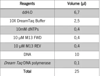
With the intention to test a correct insertion of MATE1 and CYP2 into expression vector a colony PCR was performed. Single isolated colonies were picked to transfer a small amount of bacteria into a PCR tube with 10 µL of ddH2O, and to re-plate onto LB-agar supplemented with
kanamycin 50 µg mL-1. MATE1 and CYP2 amplification was performed as described in Table 6.
The PCR program consisted of one cycle at 95 ºC for 3 min, 35 cycles at 95 ºC for 30 s, 55 ºC for 30 s, 72 ºC 5 min for MATE1 and 4 min for CYP2 and a final extension for 7 min. Positive clones were identified by gel electrophoresis as described above (section 2.1.5). Three positives colonies of each construct were picked to inoculate 5 mL of LB medium supplemented with 50 μg mL-1
kanamycin. Cultures were grown ON at 37 ºC with vigorous shaking (200 rpm). Plasmid DNA was recovered by the alkaline lysis miniprep method using the GeneJET Plasmid Miniprep Kit (Thermo Scientific) according to the manufacturer’s instructions. Cloning of MATE1 and CYP2 into pCAMBIA2301m was confirmed by restriction analysis of the miniprep DNA. Plasmid DNA was digested with XhoI plus XbaI for MATE1, and XhoI for CYP2. The positive clones were sent to be sequenced (STABVida). The analysis of sequence homology between the predicted sequences and the obtained sequences was performed using the multiple sequence alignment program MultiAlin (http://multalin.toulouse.inra.fr/multalin/).
Table 6 – Colony PCR optimized setup for amplification of the MATE1 and CYP.
Reagents Volume (µl) ddH2O 6,7 10X DreamTaq Buffer 2,5 10mM dNTPs 0,4 10 µM M13 FWD 0,4 10 µM M13 REV 0,4 DNA 10
Dream Taq DNA polymerase 0,1