Universidade do Minho
Escola de Ciências
João Pedro Cardoso Ribeiro
New Generation Antidiabetics: what are the
implications on spermatogenesis nutritional
support?
setembro de 2019 New Gen er at ion A nt idiabet ics : wh at are t he impl icat ion s on s pe rmat og en es is n ut rit ion al s upp ort ? Jo ão Pe dr o C ar do so R ibei ro UM inh o | 2019Instituição de Acolhimento: Unit for Multidisciplinary Research in Biomedicine (Instituto de Ciências Biomédicas Abel Salazar, Universidade do Porto)
Universidade do Minho
Escola de Ciências
setembro de 2019
João Pedro Cardoso Ribeiro
New Generation Antidiabetics: what are the
implications on spermatogenesis nutritional
support?
Dissertação de Mestrado
Bioquímica Aplicada
Especialização em Biomedicina
Trabalho efetuado sob a orientação do
Professor Doutor Pedro Fontes Oliveira
ii
DIREITOS DE AUTOR E CONDIÇÕES DE UTILIZAÇÃO DO TRABALHO POR TERCEIROS
Este é um trabalho académico que pode ser utilizado por terceiros desde que respeitadas as regras e boas práticas internacionalmente aceites, no que concerne aos direitos de autor e direitos conexos.
Assim, o presente trabalho pode ser utilizado nos termos previstos na licença abaixo indicada. Caso o utilizador necessite de permissão para poder fazer um uso do trabalho em condições não previstas no licenciamento indicado, deverá contactar o autor, através do RepositóriUM da Universidade do Minho.
Atribuição-NãoComercial-SemDerivações CC BY-NC-ND
iii AGRADECIMENTOS
A realização desta dissertação de mestrado foi suportada por contribuições e incentivos fundamentais sem os quais não seria possível a conclusão desta etapa académica e de crescimento pessoal. Assim sendo, tenho todo o gosto em expressar os meus sinceros agradecimentos a todos os que contribuíram direta ou indiretamente para a elaboração deste trabalho.
Ao meu orientador, Professor Doutor Pedro Fontes Oliveira, por me ter acolhido no seu grupo de trabalho, financiamento, conhecimento científico, paciência e disponibilidade demonstrada, bem como o acompanhamento e conselhos dados na duração deste percurso. Agradecer também todas as críticas, correções e sugestões que contribuíram para o melhoramento deste trabalho. Sem a sua orientação este trabalho não seria possível.
À minha coorientadora, Professora Sandra Paiva, pela total disponibilidade que demonstrou e por me ter guiado para o fantástico grupo de trabalho do Professor Doutor Pedro Oliveira e Professor Doutor Marco Alves.
Ao Professor Doutor Marco Alves pelo conhecimento científico, disponibilidade e motivação que me forneceu durante todo o percurso.
Aos colegas de laboratório que me ensinaram as rotinas do laboratório bem como as técnicas que foram realizadas neste trabalho, nomeadamente: Ana Martins, David Carrageta, Doutora Raquel Bernardino, Doutora Ivana Jarak, Ana Maria Silva e Doutor Romeu Videira. Obrigado pela simpatia, paciência e conhecimentos transmitidos.
Aos restantes colegas do grupo de trabalho: Doutor David Martín-Hidalgo, Doutor Tito Jesus, Susana Almeida, Luís Crisóstomo, Bruno Moreira, Sara Pereira, Cláudia Peixoto, Anette Veiga, Patrícia Braga e Cassandra Santos que me integraram no grupo e se mostraram sempre disponíveis para ajudar ao longo da realização da dissertação de mestrado.
A todos os meus amigos, em especial o Henrique, SóFres e Maazou que me motivam e me fazem rir mesmo nos piores momentos.
À Rita Peixoto pela paciência que tem comigo, por me motivar e me fazer melhorar todos os dias que passamos juntos.
A toda a minha família, em especial os meus pais, irmã, e avós por me motivarem e sustentarem para tornar este trabalho possível e acima de tudo, por me tornarem no que sou hoje.
iv DECLARAÇÃO DE INTEGRIDADE
Declaro ter atuado com integridade na elaboração do presente trabalho académico e confirmo que não recorri à prática de plágio nem a qualquer forma de utilização indevida ou falsificação de informações ou resultados em nenhuma das etapas conducente à sua elaboração.
Mais declaro que conheço e que respeitei o Código de Conduta Ética da Universidade do Minho.
v
ANTIDIABÉTICOS DE NOVA GERAÇÃO: QUAIS SÃO AS IMPLICAÇÕES NO SUPORTE NUTRICIONAL DA ESPERMATOGÉNESE?
A incidência de doenças metabólicas como Diabetes mellitus tipo 2 (TD2M) e obesidade tem aumentado entre crianças e jovens adultos. Estas doenças estão intimamente ligadas, portanto, novos agentes farmacológicos têm como objetivo controlar a glicémia enquanto provocam perda de peso. A recentemente implementada terapia combinada de dapagliflozina (inibidor do cotransportador de sódio-glucose) e exenatida (análogo do glucagon-like peptide 1) tem sido prescrita para tratar T2DM. Este tratamento influencia o metabolismo da glucose em todo o corpo, portanto, também podem afetar o metabolismo dos tecidos responsáveis pela fertilidade masculina. A espermatogénese é altamente dependente da cooperação metabólica estabelecida entre as células germinativas em desenvolvimento e as células de Sertoli. Estas usam glucose extracelular para produzir lactato, que as células germinativas usam como combustível.
Neste trabalho, avaliamos os efeitos da dapagliflozina e exenatida no metabolismo das células de Sertoli. Para tal, células de Sertoli TM4 de murganho foram tratadas na ausência (controlo) ou na presença de concentrações sub-farmacológicas, farmacológicas e supra-farmacológicas de dapagliflozina (50; 500; 5000 nM, respectivamente) ou exenatida (2,5; 25; 250 pM, respectivamente) ou ainda, com uma combinação das concentrações farmacológicas de ambas as drogas, durante 24 horas. A citotoxicidade destes tratamentos para as células de Sertoli foi avaliada através de ensaios de MTT, liberação da lactato desidrogenase (LDH) e SRB. O perfil glicolítico das células de Sertoli também foi determinado (espectroscopia de 1H-RMN), bem como
os níveis de expressão dos principais transportadores e enzimas como a LDH, transportadores de glucose 2 (GLUT2) e transportador de monocarboxilatos 4 (MCT4). Além disso, foi quantificado o conteúdo em glicogênio e as reservas lipídicas nas células de Sertoli TM4. A concentração farmacológica de dapagliflozina mostrou um leve efeito citotóxico que também foi observado no tratamento combinado. Além disso, a concentração farmacológica de dapagliflozina mostrou aumentar a eficiência glicolítica e aumentar a secreção de lactato pelas células de Sertoli, que é visto como um potencializador da espermatogénese. O tratamento combinado demonstrou que os diferentes fármacos parecem ter um efeito sinérgico para manter a homeostase metabólica das células de Sertoli. Assim, os nossos resultados sugerem que o tratamento com dapagliflozina, exenatida e o tratamento combinado de ambos os fármacos em concentrações farmacológicas podem ser adequados para homens na idade reprodutiva.
vi
NEW GENERATION ANTIDIABETICS: WHAT ARE THE IMPLICATIONS ON SPERMATOGENESIS NUTRITIONAL SUPPORT?
The incidence of metabolic diseases such as type 2 Diabetes mellitus (T2DM) and obesity has been increasing among children and young adults. Both conditions are tightly linked thus new pharmacological agents aim to control glycemia while provoking weight loss. Recently implemented combined therapy of dapagliflozin (sodium-glucose cotransporter inhibitor) and exenatide (glucagon-like peptide 1 analogue) has been prescribed against T2DM. This treatment influences whole-body glucose metabolism, and thus can also impact the metabolism of tissues responsible for male fertility. Spermatogenesis is highly dependent on the metabolic cooperation established between Sertoli cells and germ cells. The former uses extracellular glucose to produce lactate, which germ cells use as fuel.
In this work, we evaluated the effects of dapagliflozin and exenatide on Sertoli cells metabolism. For this purpose, mice TM4 Sertoli cells were treated in the absence (control) or presence of sub-pharmacologic, pharmacologic and supra-pharmacologic concentrations of dapagliflozin (50; 500; 5000 nM, respectively), or exenatide (2,5; 25; 250 pM, respectively), or with a combination of the pharmacological concentrations of both drugs, during 24 hours. The cytotoxicity of these compounds on Sertoli cells was evaluated by MTT, lactate dehydrogenase
(LDH) release, and SRB assays. The glycolytic profile of SCs was also determined (1H-NMR
spectroscopy), as well as the expression levels of key metabolite transporters and enzymes, such as LDH, glucose transporters 2 (GLUT2), and monocarboxylate transporter 4 (MCT4). To further pursue our aim, glycogen storage and lipid reserves were quantified in TM4 Sertoli cells. Pharmacological concentration of dapagliflozin showed a mild cytotoxic effect that was also seen in the combined treatment. In addition, the pharmacological concentration of dapagliflozin showed to enhance the glycolytic efficiency and increase lactate secretion by Sertoli cells, which is seen as a positive enhancer of spermatogenesis. The combined treatment demonstrated that the different drugs seem to have a synergic effect to maintain the metabolic homeostasis of Sertoli cells. Thus, our results suggest that treatment with dapagliflozin, exenatide and the combined treatment of both drugs at pharmacological concentrations may be suitable for males in reproductive age. Keywords: Dapagliflozin, Exenatide, Infertility, Metabolic diseases, Sertoli cells.
vii TABLE OF CONTENTS RESUMO ... v ABSTRACT ... vi LIST OF ABBREVIATIONS ... x LIST OF FIGURES ... xi
LIST OF TABLES ... xiii
1.INTRODUCTION ... 14
1.1. Diabetes mellitus ... 14
1.1.1. Type 2 Diabetes mellitus ... 15
1.2. Obesity and its role in the origin of T2DM ... 15
1.3. Sodium-glucose cotransporter 2 inhibitors ... 16
1.4. Glucagon-like peptide-1 receptor agonist ... 20
1.5. Combined therapy of exenatide plus dapagliflozin ... 23
1.6. Male reproductive function and spermatogenesis ... 25
1.6.1. Sertoli cells and Spermatogenesis ... 29
1.7. Negative influence of metabolic diseases on male fertility ... 32
1.8. Exenatide and Dapagliflozin impact on male fertility ... 34
2.OBJECTIVES ... 37
3.METHODS ... 39
3.1. Chemicals ... 39
3.2. Mouse Sertoli cell line TM4 culture ... 39
3.3. Experimental groups ... 39
3.4. Evaluation of cytotoxic profile of the compounds ... 40
3.4.1. SRB cytotoxic assay ... 40
3.4.2. MTT cytotoxic assay ... 41
3.4.3. LDH release assay ... 41
viii
3.6. Intracellular LDH activity assay ... 42
3.7. Nuclear Magnetic Resonance spectroscopy ... 42
3.8. Reverse Transcriptase Polymerase Chain Reaction ... 43
3.9. Western Blot ... 44
3.10. Colorimetric Method for Glycogen Quantification ... 44
3.11. Oil Red O Staining ... 45
3.12. Mitochondrial membrane potential ... 46
3.13. Statistical analysis ... 46
4.RESULTS ... 48
4.1. GLP-1 receptor and SGLT2 are expressed in Sertoli cells ... 48
4.2. Exposure of TM4 Sertoli cells to exenatide ... 48
4.2.1. Exenatide decreased Sertoli cell membrane integrity and metabolic viability, without affecting cell proliferation ... 48
4.2.2. Exenatide maintained glucose consumption by Sertoli cells while increasing the expression of GLUT2, when at pharmacological concentrations ... 50
4.2.3. Exenatide treatment maintained the secretion of monocarboxylates and alanine in TM4 Sertoli cells... 51
4.2.4. Sub-pharmacological concentration of exenatide decreases mitochondrial membrane potential in TM4 Sertoli cells ... 54
4.2.5. Incubation with exenatide maintained glycogen and lipid droplet reserves on TM4 Sertoli cells ... 56
4.3. Exposure of TM4 Sertoli cells to dapagliflozin ... 57
4.3.1. Dapagliflozin decreases Sertoli cell proliferation, membrane integrity, and metabolic viability when at pharmacologic concentrations ... 57
4.3.2. Dapagliflozin does not alter glucose consumption but increases GLUT2 expression in TM4 Sertoli cells ... 59
4.3.3. Pharmacological concentration of dapagliflozin increases the secretion of lactate and alanine by TM4 Sertoli cells ... 60
ix
4.3.4. Supra-pharmacological concentration of dapagliflozin decreases the amount of lipid
droplets on TM4 Sertoli cells... 65
4.3.5. Dapagliflozin maintains the mitochondrial membrane potential of Sertoli cells ... 66
4.4. Exposure of TM4 Sertoli cells to exenatide plus dapagliflozin... 67
4.4.1. Combined treatment with exenatide plus dapagliflozin decreases Sertoli cells proliferation and metabolic viability without decreasing membrane integrity ... 67
4.4.2. Combined treatment with exenatide plus dapagliflozin maintains the glucose consumption and GLUT2 expression on TM4 Sertoli cells ... 68
4.4.3. Combined treatment with exenatide plus dapagliflozin maintained the production of monocarboxylates and alanine on TM4 Sertoli cells ... 69
4.4.4. Glycogen and intracellular lipid contents were maintained in Sertoli cells after the combined treatment with exenatide plus dapagliflozin ... 71
4.4.5. Combined treatment with exenatide plus dapagliflozin did not alter mitochondrial membrane potential of TM4 Sertoli cells ... 71
5.DISCUSSION ... 74
6.CONCLUSION ... 81
x LIST OF ABBREVIATIONS
ATP Adenosine triphosphate BTB Blood-testis barrier
cAMP Cyclic adenosine monophosphate DM Diabetes mellitus
EDTA Ethylenediaminetetraacetic acid FBS Fetal Bovine Serum
FSH Follicle-stimulating hormone GLP-1 Glucagon-like peptide 1 GLUT’s Glucose transporters
GnRH Gonadotrophin-releasing hormone LDH Lactate dehydrogenase
LH Luteinizing hormone
MCT’s Monocarboxylate transporters NMR Nuclear molecular resonance PBS Phosphate-buffered saline PCR Polymerase chain reaction PKA Protein kinase A
RIPA Radioimmunoprecipitation assay ROS Reactive oxygen species
SGLT’s Sodium glucose co-transporters SRB Sulforhodamine B
T1DM Type 1 Diabetes mellitus T2DM Type 2 Diabetes mellitus TCA Tricarboxylic acid
xi LIST OF FIGURES
Figure 1: Schematic representation of glucose reabsorption by sodium-glucose cotransporter 2 (SGLT2) in proximal tubule.. ... 17 Figure 2: Scheme of the mechanism of glucagon-like peptide 1 (GLP-1) release from L-cells and insulin secretion pathway activation. In the presence of different nutrients, L-cells secrete GLP-1 though different metabolic pathways.. ... 21 Figure 3: GLP-1 amino acid sequence and the differences in the sequence (amino acids in yellow) that allow exenatide increased half-life time. ... 22 Figure 4: Scheme of the different stages of spermatogenesis. ... 27 Figure 5: Schematic representation of the blood-testis barrier (BTB) and the different stages of spermatogenesis. ... 28 Figure 6: Mechanisms of Sertoli cells glucose metabolism and its metabolic cooperation with the germ cells... ... 31 Figure 7: Confirmation of the expression of GLP-1 receptor (A) and SGLT2 (B) in TM4 Sertoli cells by reverse transcriptase polymerase chain reaction. ... 48 Figure 8: Evaluation of the cytotoxic profile of the different concentration of exenatide on TM4 Sertoli cells. ... 49 Figure 9: Effect of exenatide at 2,5 pM, 25 pM, and 250 pM on glucose consumption by TM4 Sertoli cells.. ... 50 Figure 10: Effect of exenatide concentrations (2,5 pM, 25 pM, and 250 pM) on lactate production in TM4 Sertoli cells.. ... 52 Figure 11: Effect of exenatide concentrations (2,5 pM, 25 pM, and 250 pM) on metabolite production and secretion in TM4 Sertoli cells. ... 54 Figure 12: Effect of exenatide treatments (2,5 pM, 25 pM, and 250 pM) on the mitochondrial function of TM4 Sertoli cells. ... 55 Figure 13: Effect of exenatide treatments (2,5 pM, 25 pM, and 250 pM) on carbon storage by TM4 Sertoli cells. ... 57 Figure 14: Evaluation of the cytotoxic profile of the different concentration of dapagliflozin on TM4 Sertoli cells. ... 59 Figure 15: Effect of dapagliflozin (50 nM, 500 nM, and 5000 nM) on glucose consumption by TM4 Sertoli cells. ... 60
xii
Figure 16: Effect of dapagliflozin concentrations (50 nM, 500 nM, and 5000 nM) on lactate production in TM4 Sertoli cells. ... 62 Figure 17: Effect of dapagliflozin concentrations (50 nM, 500 nM, and 5000 nM) on acetate and alanine production in TM4 Sertoli cells. ... 64 Figure 18: Effect of dapagliflozin treatments (50 nM, 500 nM, and 5000 nM) on carbon storage by TM4 Sertoli cells. ... 66 Figure 19: Effect of dapagliflozin (50 nM, 500 nM, and 5000 nM) on the mitochondrial function of TM4 Sertoli cell line. ... 67 Figure 20: Evaluation of the cytotoxicity profile of the combined treatment (E+D) (25pM exenatide plus 500 nM dapagliflozin) on TM4 Sertoli cells. ... 68 Figure 21: Effect of the combined treatment (E+D) (25pM exenatide plus 500 nM dapagliflozin) on glucose consumption by TM4 Sertoli cells. ... 69 Figure 22: Effect of the combined treatment (E+D) (25 pM exenatide plus 500 nM dapagliflozin) on monocarboxylates (lactate and acetate) and alanine production in TM4 Sertoli cells. ... 70 Figure 23: Effect of the combined treatment (E+D) (25pM exenatide plus 500 nM dapagliflozin) on carbon storage by TM4 Sertoli cells. ... 71 Figure 24: Effect of the combined treatment (E+D) (25pM exenatide plus 500 nM dapagliflozin) on the mitochondrial function of TM4 Sertoli cells. ... 72
xiii LIST OF TABLES
Table 1: Genes, oligonucleotide sequence and respective conditions for polymerase chain reaction amplification of GLP1-receptor and SGLT2 genes. ... 43
13
Chapter I
14 1. INTRODUCTION
1.1. Diabetes mellitus
Diabetes mellitus (DM) is a chronic disease that has reached pandemic proportions and its incidence has been increasing dramatically over the last decades. Moreover, it is expected a growing trend in the number of individuals suffering from this metabolic disease. It has been estimated that a worrisome 693 million people will develop DM by 2045 (1). DM consists of multiple conditions related to deficient carbohydrate, lipid and protein metabolism. This dysregulation impacts multiple organs and systems such as the kidney, retina, circulatory system, and nervous system (2). Hence, acute complications in long-term DM patients can result in blindness, macrovascular and microvascular complications, and sexual dysfunction, among others (3). Moreover, DM increases the chance of strokes and coronary artery disease in undiagnosed or untreated individuals (4), which could result in death. Impaired glucose homeostasis results in hyperglycemia which is caused by hypoinsulinemia or insulin resistance (5).
Insulin is a hormone that is secreted into circulation after a meal rich in carbohydrates. Glucose levels are sensed with the participation of specific glucose transporters together with neural stimuli and incretin signaling. This leads to the activation of insulin secretion on β-cells in the pancreas. Insulin will act on other cells increasing their glucose uptake and decreasing glycemia levels (6). Besides, insulin also mediates meal-time and has a role in cell growth and differentiation (7). The plasmatic insulin is insufficient to maintain glucose homeostasis in DM patients, which can be caused by autoimmune β-cell elimination, known as type 1 diabetes mellitus (T1DM); or gradual loss of insulin sensitivity, classified as type 2 diabetes mellitus (T2DM) (8).
Patients with T1DM are increasing among children and has been reported that this growth in incidence does not only depend on genetic factors (9) but also in environmental factors (10) such as enterovirus infection or nutritional factors (e.g. vitamin D deficiency or premature contact with cow’s milk proteins) (11). These factors can lead to the destruction of insulin-producing β-cells by T lymphocytes in an autoimmune manner. Additionally, nearly one-third of patients with T1DM suffer from diabetic ketoacidosis (8). This condition is a consequence of diminished insulin/glucagon ratio presence in circulation that activates ketogenic machinery in liver cells to produce glucose, but also ketones from long-chain fatty acids (12). Ketones such as acetoacetate and β-hydroxybutyrate increase in plasma and this result in metabolic acidosis (13) that in severe cases can lead to coma (14). Thus, to work around this condition the patients suffering from T1DM will depend on exogenous insulin for life (15), forcing them to continuous health care.
15 1.1.1. Type 2 Diabetes mellitus
T2DM is the most prevalent type of DM accounting approximately 90% of all occurrences (16) and is tightly linked with obesity, sedentary lifestyle (17), but also genetic factors (18). Usually, the risk of developing this condition increases with age (19). However, this epidemy is affecting an increasing number of individuals from younger group ages, which intensifies the health, sociologic, and economic problems associated with T2DM (20). This type of chronic hyperglycemia results from diminished insulin bioactivity, which can be due to anomalous insulin secretion, decreased insulin sensitivity, or a combination of both (21, 22). In a scenario with high glucose intake, a healthy individual would secrete proportional insulin to normalize glycemia levels. However, diabetic patients insulin secretion do not compensate the subnormal hormone effect on its receptors of skeletal muscle, pancreas, adipose tissue, and liver (23), promoting the increase of glucose levels on plasma thus, causing hyperglycemia.
Lifestyle modifications are essential to counter the effects of T2DM, including changing diet and exercise habits. Multiple drugs were also developed to counteract hyperglycemia and its associated comorbidities. However, a complex study is needed to find the most suitable antidiabetic therapy for each patient situation (20).
1.2. Obesity and its role in the origin of T2DM
According to the World Health Organization, a person is considered overweight when its Body Mass Index (BMI) score is above 25 and is considered obese when the score is superior or equal to 30. BMI is a crude measure for studying obesity in a population and can be calculated by dividing a person weight (kilograms) with the square of the person height (meters) (24). Through the years, BMI has increased in developed countries possibly by their easier access to high caloric meals. This excessive caloric intake is contributing to pandemic obesity never seen before. Most victims suffering from obesity also develop T2DM, since obesity already induces some degree of insulin resistance (25). In obese humans, the bioactivity of the circulating insulin is diminished by the excessive abdominal fat. Thus, hyperglycemia will aggravate and this causes more insulin secretion, increasing its concentration on plasma which worsens insulin resistance and promotes high blood pressure (26). Furthermore, fat cells can penetrate the pancreatic islets and influence insulin secretion in more advanced obese patients (26). Obesity is also known to dysregulate the secretion of hormones responsible for the all-body energy balance, mainly ghrelin, leptin, glucagon-like peptide (GLP-1), and insulin, being the former already discussed above. Ghrelin is a hormone
16
secreted by the endocrine cells of the gastrointestinal tract and it is known as the hunger hormone since it is related to the origin of appetite. Ghrelin levels in obese people plasma are reduced in relation to the ones of a healthy individual (27). Leptin, however, is released by white adipose tissue and its levels are increased in obese persons when compared with healthy individuals. It is believed that leptin plays a role in reducing appetite and together with ghrelin they help in the regulation of energy homeostasis, glucose metabolism, and reproductive function (28).
Weight-loss should be the main goal to control glycemia in obese T2DM individuals, even though this can be the most challenging aspect of the treatment (29). In 2015, Whitmore stated that managing weight in T2DM patients is comparable to a “juggling act” with balanced energy intake/output, glycemic control, hypoglycemia, medication and the negative sides that those factors can generate (30). DM treatment is constantly evolving, and new approaches are being developed to improve the health of the patients and their lifestyle (30). Some of the antidiabetic drugs used to regulate T2DM also have anti-obesogenic properties which provoke weight-loss in the patient, namely: low affinity/high capacity sodium-glucose cotransporter 2 (SGLT2) inhibitors (e.g. dapagliflozin) and GLP-1 receptor agonist (e.g. exenatide) (20). Both these pharmacological agents were already used separately or in combination with other drugs. Combined therapy of dapagliflozin (10 mg once daily) plus exenatide (2 mg injection once weekly) is being studied in the form of phase III clinical trial to compare the results of this novel therapy with more standardized therapies (31).
1.3. Sodium-glucose cotransporter 2 inhibitors
Kidneys role in glucose homeostasis starts when Malpighi glomerulus filters plasmatic glucose without restrains, followed by its reabsorption in the proximal tubule into circulation by specific glucose transport proteins. In individuals with chronic hyperglycemia, the renal threshold (180 mg/dL) is not enough to reabsorb all plasmatic glucose, forcing the remaining glucose to be excreted through the urine (32). This event can be explained by the SGLT2 activity and expression in the proximal tubule. This transporter is responsible for glucose resorption and its activity is insulin-dependent (33). In addition, kidneys are also involved in glucose metabolism homeostasis by performing gluconeogenesis, when humans are fasted for more than 14 hours, contributing with around 20% to 25% of total plasmatic glucose after that period (34).
SGLT2 is mainly found in the initial part of renal convoluted proximal tubules, more specifically in the apical membrane. This transporter is responsible for the reabsorption of approximately 90%
17
of glucose in the kidneys (35). The reabsorption is established when SGLT2 imports one sodium ion and one glucose molecule into the cytosol finally, glucose is then transported into the bloodstream through the action of glucose transporter 2 (GLUT2) (36) (Figure 1). Interestingly, patients with T2DM have increased expression levels of SGLT2 in proximal tubules cells. This fact suggests that T2DM patients have an above normal renal threshold (37). Thus, more glucose is reabsorbed into the bloodstream which will only aggravate hyperglycemia (32). Considering this, compounds capable of inhibiting SGLT2 could be a convenient way of controlling glycemic levels, blocking the reabsorption of this hexose into circulation, increasing its excretion through the urine, and diminishing the overall caloric intake of the individual.
Dapagliflozin favors this management of glucose homeostasis. Thus, the European Union approved dapagliflozin for the treatment of T2DM in 2012 (38). Dapagliflozin is chemically described as (2S,3R,4R,5S,6R)-2-[4-chloro 3-(4-ethoxybenzyl) phenyl]-6-(hydroxymethyl) tetrahydro-2H-pyran-3,4,5-triol). Its molecular formula is C21H25ClO6 which translates in a molecular
Figure 1: Schematic representation of glucose reabsorption by sodium-glucose cotransporter 2 (SGLT2) in the proximal tubule. Glucose and sodium ion are transported from the lumen of the tubule to the cytoplasm through the SGLT2. After this, glucose transporters 2 (GLUT2) transport the glucose into the circulation.
18
weight of 408.87 g/mol (38). Dapagliflozin structure is organized in one glucose molecule which is linked to an aglycone component through a carbon-carbon bond with the purpose of increasing its metabolic stability against glucosidase enzymes (39). This fact allows dapagliflozin to be prescribed for oral administration and can be found in tablets of 5 mg or 10 mg (39). It needs only approximately 1 hour (tmax) after the first dosage of 5 mg or 10 mg dapagliflozin to be absorbed.
Dapagliflozin is also rapidly absorbed even when the treatment is made after a meal, due to its high aqueous solubility and great intestinal absorption (40). Its maximal plasma concentration can reach up to approximately 70 ng/ml (5 mg treatment) and 200 ng/ml (10 mg treatment) (38, 41) moreover, dapagliflozin half-life time (t1/2) is approximately 17 hours (42, 43).
The efficiency and safety of dapagliflozin 10 mg once-daily therapy for obese with T2DM patients has already been studied in multiple clinical trials. DURATION-8 was a 28-week long phase III clinical trial in adults with inadequate glycemic control (HbA1c 8-12%). During this clinical trial,
19% dapagliflozin treated group had a decreased on HbA1cscore by 7%, 20% of the group lost more than 5% of their weight, and systolic blood pressure decreased 1,8 mm Hg in relation to the baseline values at the beginning of the trial (44). In another phase III clinical trial with similar dosage, dapagliflozin showed to improve HbA1c score and weight loss by a difference of -0,90% and -1.38 Kg, respectively, in comparison to individuals from the placebo group at the end of the 24-week trial duration (45). Moreover, 10 mg once daily therapy of dapagliflozin can be also used as a complementary agent to exogenous insulin treatment in patients with T1DM (46). In relation to the placebo group, patients treated with dapagliflozin improved their HbA1c score by -0,45% in the first 4 weeks of the trial, maintaining that difference in the remaining time of the study. In addition, dapagliflozin (10 mg/day) treatment allowed the reduction of the exogenous insulin dosage by 9,8% per kilogram of bodyweight at week 24 of the study. Similarly, body weight also decreased by 3.72% in relation to the individuals from the placebo group, most of this reduction occurred in the first 8 weeks of the trial and was maintained until the end of the experiment (46).
As above demonstrated, dapagliflozin decreases hyperglycemia, body weight and blood pressure in DM patients, being that these are the key factors responsible for the most deleterious effects of DM, making this pharmacological agent a promising drug for the treatment of this condition and increasing the life quality of the user. However, like any drug in the market, dapagliflozin entails some negative side effects to the user. The main concern about this drug is its influence on renal and cardiovascular function of treated DM and obese patients. T2DM patients with normal or moderately impaired renal function were subjected to dapagliflozin therapy. Their
19
estimated glomerular filtration rate was calculated to access the implications that dapagliflozin could generate to patients’ renal function. Kohan and coworkers analyzed twelve clinical trials and stated that dapagliflozin (10 mg/day) decreased estimated glomerular filtration rate by -4,13 mL/min/1,73m2 in relation to the baseline value in the first week. Nevertheless, this value returned
to baseline by week 24 and remained stable until the end of the experiment (47). Blood urea nitrogen values were significantly increased in relation to placebo at the end of the experiment which is justified by the osmotic diuretic effect that the drug entails (47) which also allows the reduction of blood pressure (48). Regarding cardiovascular function, a randomized clinical trial with 28 weeks of duration was performed. Dapagliflozin (10mg/day) was administered to patients with a high risk of cardiovascular problems and the results showed a greater occurrence of hypotension and dehydration in relation to the placebo group, with the usual improvement in glycemic levels and body weight (49). These results could be justified by a large number of plus 65 years old patients that participated in this study and by the fact that the patients already had high risk of cardiovascular problems. However, another study described that dapagliflozin treatment increased plasmatic levels of high-density lipoprotein 2-cholesterol, which are regarded as an advantageous cardiometabolic marker (50). On the other hand, low-density lipoproteins, which are considered a strong marker of possible cardiovascular complications, did not change between the dapagliflozin treated group and the placebo group (50). Moreover, SGLT2 inhibitors such as dapagliflozin have been also associated with risks of inducing diabetic ketoacidosis. This could be due to the increase of glucagon/insulin ratio levels in circulation which promote hepatic ketogenesis when in dapagliflozin therapy. Furthermore, treatment for T2DM with SGLT2 inhibitor combined with exogenous insulin increases the chances of developing diabetic ketoacidosis due to the need of lowering exogenous insulin dosage to prevent hypoglycemia, which only aggravates the glucagon/insulin ratio. This happens due to enhanced lipolysis caused by the lack of insulin, increasing ketones levels on plasma (51). SGLT2 inhibitors, such as dapagliflozin, are also linked with genitourinary tract infections caused by the enhanced glucose concentration in the urine (52).
Besides that, studies suggest that dapagliflozin is a relatively safe pharmacological agent and even entails some advantages to the patient, such as the ability to protect pancreatic β-cells of hyperglycemia adverse effect, conserving their function (53). Moreover, this drug is associated with weight loss which results from the enhanced glucose excretion, maximizing caloric loss yet, this fact can originate an increase in appetite (54). In addition, a study stated that dapagliflozin (5mg/day) treatment for six months decreased total body weight, without significantly changing
20
skeletal muscle mass and soft lean mass in relation to the placebo group (55). Dapagliflozin also minimizes the chances of hypoglycemia since SGLT2 inhibitors do not interfere directly with insulin secretion (56), unlike other antidiabetic drugs. Thus, dapagliflozin can be complemented and used simultaneously with other antihyperglycemic drugs capable of modulating insulin secretion (44), such as exenatide which belongs in the GLP-1 receptor agonist class of antidiabetic drugs.
1.4. Glucagon-like peptide-1 receptor agonist
GLP-1 is an incretin derived from a different path of post-transcriptional modification of proglucagon in enteroendocrine L-cells, which can be found along the gastrointestinal tract with greater abundance in the distal gut, ileum, and colon (57). Oral ingestion of nutrients is the major physiologic stimuli for the secretion of GLP-1 by L-cells, although its concentration in the bloodstream depends on the composition of the meal. Meals richer in carbohydrates and fats stimulate greater GLP-1 secretion than meals rich in proteins (58). It is believed that GLP-1 secretion is mediated by direct and indirect sensing since GLP-1 levels rise in circulation within minutes. Normally, this time is insufficient for the food to reach the L-cells in the distal gut. Thus, it is believed that some of these cells are situated in the upper gut to sense the nutrients ingested and share that information with the richest L-cells regions for a rapid and efficient GLP-1 release into circulation (57). After this, and when it binds to its G-protein coupled receptors on pancreatic β-cells, GLP-1 stimulates insulin secretion by activating the second messenger cyclic adenosine monophosphate (cAMP) which, in its turn, activates the insulin effectors protein kinase A (PKA) (59) (Figure 2). Interestingly, as the incretin effects are proportional to the glucose intake, also insulin secretion will be mediated by the ingested glucose (57). The ability of such peptide to modulate insulin secretion and suppress excessive glucagon secretion (reducing hepatic gluconeogenesis) (60), shows that GLP-1 receptors agonists could be an interesting approach to control glycemia in individuals with T2DM (61).
Exenatide is an example of one GLP-1 receptor agonist composed of 39 amino acids with a molar mass of 4186.6 g/mol (62), originally extracted from the salivary excretion of the Heloderma suspectum (Gila monster) and commonly named exadin-4 (63). When compared with GLP-1, exenatide shares 53% of sequence homology (64), has a 20 to 30-fold optimized half-life and 5500 times greater potency in decreasing glycemic levels in T2DM patients (65). This significant difference in half-life time is due to the enzyme DPP-4 present in circulation, which cleaves GLP-1 in the second amino acid residue (alanine). In exenatide, this alanine was altered to a glycine
21
residue (Figure 3) to enhance the biodisponibility of the drug (58). This GLP-1 receptor agonist is administered by subcutaneous injection in dosages of 5 µg or 10 µg of exenatide twice daily (66) or 2 mg once weekly and has a maximal concentration found in plasma around 90 pg/ml (for dosage of 5 µg) or 210 pg/ml (for dosage of 10 µg and 2mg) (67-69). Moreover, when is prescribed the long action exenatide microspheres, the drug diffusion lasts for approximately 2 weeks and drug clearance around 7 weeks since the injection (67).
Figure 2: Scheme of the mechanism of glucagon-like peptide 1 (GLP-1) release from L-cells and insulin secretion pathway activation. In the presence of different nutrients, L-cells secrete GLP-1 through different metabolic pathways. After, entering in circulation, GLP-1 binds to its receptor on pancreatic β-cells and activate the pathway of insulin secretion. FFA, free fatty acids; AA, amino acids; PKCϚ, protein kinase CϚ; cAMP, cyclic adenine monophosphate; PKA, protein kinase A.
22
When comparing the two major dosage schedules of exenatide treatment (exenatide 10 µL twice daily or exenatide 2 mg once weekly), a study referred that exenatide 2 mg once weekly demonstrated the best results in lowering glycemic levels, and a significantly lower incidence in adverse effects in relation to the twice-daily dose in a 28-week long study (70). With this in mind, and together with a more convenient dosage schedule, we prioritized studies that evaluated the impact of the treatment of exenatide 2 mg once weekly. DURATION-8 clinical trial studied the effect of exenatide 2 mg once weekly on glycemic control, weight, and blood pressure in a similar manner to dapagliflozin treatment. The results showed that exenatide decreased the baseline score of HbA1c by 7% in 27% of the individuals. Some patients (14%) loss more than 5% of their total body mass and also decreased the systolic blood pressure by -1,2 mm Hg in the 28 weeks of the trial in the individuals treated with exenatide (44). In another study, exenatide 2 mg once weekly therapy was compared to a therapy of another GLP-1 receptor agonist (liraglutide) in a single daily administration of 1.8 mg. Exenatide was less efficient in regulating HbA1c score, weight loss and systolic blood pressure (-0,36 %; -0,90 kg; and -0,87 mm Hg, respectively), when compared with liraglutide therapy. Still, the occurrence of adverse effects on exenatide treated group was less common in almost all categories (71).
Metabolic diseases, such as DM and obesity, already entail cardiovascular complications and have been described that some antidiabetic drugs could enhance the probability of those complications (20). However, studies have suggested that this is not the case for exenatide. In fact, exenatide showed to decrease arterial stiffness in T2DM patients averaging 64 years of age and improved diastolic function in relation to the placebo group. Unfortunately, this did not result in a significant increase in exercise capacity of the patients. Still, exenatide showed to have a protective Figure 3: GLP-1 amino acid sequence and the differences in the sequence (amino acids in yellow) that allow exenatide increased half-life time.
23
effect on cardiac function, which is welcome on T2DM obese patients (72). On the other hand, GLP-1 receptor agonists have been known for causing side effects of gastrointestinal nature, such as moderate nausea, diarrhea, and vomiting. These adverse effects are dependent on the dosage and normally attenuate over time (73). This class of antihyperglycemic drugs has also been associated with higher risks of pancreatitis incidence and pancreatic and thyroid cancer, but no significant relation was found in relation to the treatment with other antidiabetic drugs (74). Another potential problem associated with this drug is the appearance of antibodies against exenatide in patients plasma, yet, no difference was shown in the glycemic control in patients with or without exenatide antibodies (58).
The anti-obesogenic properties present GLP-1 receptor agonists such as exenatide are another advantage for overweight T2DM patients. The weight loss is provoked by the ability of exenatide to slow gastric emptying, thus reducing intestinal nutrient absorption, as well as reducing glucose and lipids levels in postprandial plasma (75). Gastric emptying is slowed by the inhibition of small intestine motor function and gastric acid secretion interfering with nutrient absorption and decreasing appetite, possibly by an interaction between exenatide and the enteric nervous system (76) not yet fully understood. On the other hand, studies have shown that exenatide (0,05 µg/100 nL) injection on ventral tegmental area decreased palatable high-fat diet intake of Sprague-Dawley rats by decreasing the amount of food ingested per meal. The authors of this study stated that the activation of the GLP-1 receptor modulates the activity of the dopaminergic neurons in the ventral tegmental area. Thus, the injection of exenatide increased dopamine secretion, which is also associated with an anorectic effect (77).
1.5. Combined therapy of exenatide plus dapagliflozin
As mentioned above, dapagliflozin (10 mg once daily) plus exenatide (2 mg injection once weekly) combination treatment for obese patients with insulin resistance is being evaluated and compared with more standardized therapies (31). Is worth noticing that the combined therapy of both drugs seems complementary to each other on their effects in the body. Dapagliflozin can increase appetite yet, exenatide had shown to have anorectic effect in the patients. Exenatide stimulates insulin secretion in a direct way, while dapagliflozin does not interfere with hormone secretion in a direct manner. This, together with the fact that both have antidiabetic and anti-obesogenic properties, evidences that the combined therapy of these two pharmacological agents seem logical, promising and directed for T2DM obese patients. With this in mind, other clinical
24
trials are currently recruiting to further elucidate the effects of this combined therapy on human metabolism. For example, the influence of this combined therapy on food-related stimuli will be assessed in a phase IV randomized trial in obese T2DM patients (78). Despite that, both classes of drugs are considered safe and even seem to have some beneficial impacts on metabolism such as provoking weight loss with low risks of hypoglycemia and protective effects in heart and kidney tissue (79). In addition, other clinical trials with this therapy have already been performed and showed promising results.
In a recent study, Lundkvist and co-workers examined the anti-obesogenic potential of the combined treatment of dapagliflozin plus exenatide, on obese people without DM (80). Of 50 participants, 73.5% had prediabetes which is a major risk factor for DM and is characterized by impaired glucose metabolism but with HbA1c scores below the ones seen in DM (81). The results showed an average reduction on body weight of 4,13 Kg in relation to the placebo group, mainly in visceral adipose tissue, in a 24-week long study, as well as a reduction on prediabetes cases within the participants (34,8% versus 85% for the placebo group). The authors also proposed an additive effect in the combined therapy since the weight loss was greater than with each monotherapy (80). In another study, the same authors continued the project for 52 weeks and noticed that the decrease in body weight, prediabetes percentage, and systolic blood pressure were attenuated after the 24 weeks until 52-weeks of treatment. In the first 24 weeks, the average weight decreased 4,5 Kg, while in the next 28 weeks the difference was -1,2 Kg, of which 1,5 L were total adipose tissue volume. The proportion of individual in the study with prediabetes by the 52-week mark also increased by a thin margin of 0,5%. However, the systolic blood pressure decreased 12 mm Hg in the 52 weeks in relation to the baseline value. This shows that dapagliflozin plus exenatide improve multiple cardiometabolic risk factors, ideal in the treatment of metabolic diseases such as DM and obesity (82). Is also worth noticing that in this trial, as well as in the DURATION-8 trial, the subgroups with greater baseline values were the ones that also had a more accentuated decrease of body weight and systolic blood pressure (83). In DURATION-8 trial the safety of the administration of exenatide plus dapagliflozin was compared with that of exenatide plus placebo, and dapagliflozin plus placebo efficiency. In all groups the more common adverse effects were of gastrointestinal nature, varying from 19,9% (exenatide plus placebo) to 14,2% (dapagliflozin plus placebo) in incidence, with these adverse effects occurring in 17,7% of the individual in the combined treatment. In addition, not major hypoglycemic events occur in all trial
25
duration. A maximum of 0,4% of individual suffered cardiovascular problems (84) which confirms the notion that this therapy is relatively safe and without major side effects.
Tatarkiewicz and colleagues determined that exenatide (0,03 mg/kg/day) plus dapagliflozin (1 mg/kg/day) combined therapy optimized kidney function by reducing creatinine on plasma and improving glomerular filtration rate in obese diabetic mice (85). For the noted increase in cardioprotection, Ferrannini et al hypothesized that moderate but constant levels of ketones (β-hydroxybutyrate) in circulation are oxidized by the myocardium instead of fatty acids and this can yield better oxygen consumption, improving myocardium function. The hyperketonemia highlighted above can be observed in treatments with SGLT2 inhibitors, showing that this class of antihyperglycemic drugs can provide heart protection however, hyperketonemia can have serious implications (86). In a different study, Charokopou stated that treatment with dapagliflozin and exenatide has shown to be cost-effective (within established United Kingdom parameters) for individuals that made an unsuccessful treatment with metformin (87).
Although there is still a small number of authors evaluating the impact of this drug combination, all of them stated that dapagliflozin and exenatide in complementary treatment seem viable and have advantages for T2DM patients, especially in obese subjects. However, in all the studies, there were also some undesired side effects, especially of gastrointestinal nature. Scarce studies were published describing the effect of dapagliflozin or exenatide in male reproductive tissues (88) and none assessing the effect of combined therapy on spermatogenesis. The study of the impact of antidiabetic and anti-obesogenic drugs on male reproductive function is imperative once both classes of drugs modulate glucose metabolism, which is essential for normal spermatogenesis, hence for male fertility. The effect of other antidiabetic drugs, such as metformin, has already been studied and the results obtained showed that metformin seems to protect spermatogenesis on diabetic individuals. However, in healthy individuals, metformin seems to influence male fertility in a negative way (89, 90). Thus, it seems convenient to evaluate the effect of the novel therapy on spermatogenesis and enlighten if this therapy can worsen or increase the fertility rates in males.
1.6. Male reproductive function and spermatogenesis
Male reproductive tract is constituted by the testes, efferent ducts, epididymis and vas deferens. These heterogeneous tissues are protected by the scrotum, which helps maintain the ideal temperature for the formation of gametes. Inside the scrotum resides the testes. Testes are the organ responsible for the creation of the microenvironment that allows the spermatogenesis
26
(91). These organs are encapsulated by the tunica albuginea, which forms septations separating the human testis into lobules. Besides coating the testes, tunica albuginea also regulates the interstitial pressure and the blood flow through the testes (92). The multiple conical lobules include interstitial tissue (tissue between the tubules) and coiled seminiferous tubules, being the later responsible for 60-80% of the total volume of the testis (93). The end of all seminiferous tubules converges to the rete testis, where the secretions of the tubules are collected and then sent through the epididymis tubule (94). Inside the seminiferous tubules, we can find the blood-testis barrier (BTB), which is formed by adjacent Sertoli cells. The interstitial tissue consists of Leydig cells, leukocyte, fibroblasts and blood vessels (95). Leydig cells are capable of performing steroidogenesis, being the ones responsible for testosterone synthesis, which is essential for the occurrence of spermatogenesis (96). Is worth noting that all processes of spermatogenesis occur inside the seminiferous tubules. Spermatozoa is then guided from rete testis into the efferent ductules, which lead into the epididymis. Finally, spermatozoa enter the vas deferens and are forced into the ejaculatory duct by peristaltic movements (97).
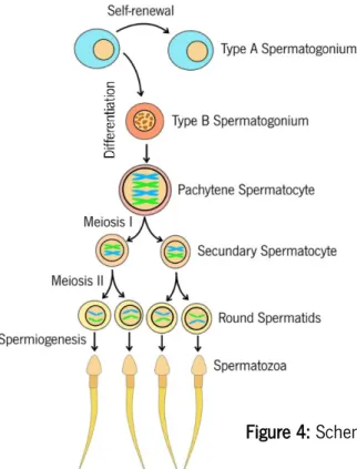
Spermatogenesis can be defined as a set of complex and coordinated processes that allow the transmission of genetic information to the next generation (98). About 40 millions of spermatozoa are formed daily in a fertile man and a complete spermatogenic cycle is concluded in 74-76 days (99). Four major phases are involved in the preparation of genetic information and each phase is controlled by endocrine, paracrine, and autocrine factors. The first phase (mitotic phase) involves a series of spermatogonia type A and type B stems cells divisions (100). Spermatogonium is a diploid cell that suffers mitotic divisions in the periphery of seminiferous tubules to originate spermatocytes and can be divided into three categories: type A Dark and type A Pale that does not exhibit heterochromatin; and its successor (type B) that presents packed chromatin (101). Spermatogonia type A Dark are the least differentiated from the stem cells and are capable of dividing into other spermatogonia type A Dark or differentiate into a type A Pale spermatogonium, which will start a new process of spermatogenesis. Then, the type A Pale is differentiated into type B spermatogonia giving place to the second phase (meiotic phase). This phase consists of the maturation of type B spermatogonia into pre-leptotene and then into leptotene spermatocytes. Spermatocyte goes through two consecutive meiotic cycles, where it is transformed into zygotene and pachytene spermatocytes (102). In these sub-phases, homologous recombination is established and after the meiotic processes, the result is a haploid round spermatid. The third phase (spermiogenesis), round spermatids suffer major morphologic changes, being transformed
27
into elongated spermatids (98), which will develop into highly specialized and functional sperm cells (102). Even though the genetic information is ready to be passed on in rounded spermatids, in natural conditions, it is not sufficient for oocyte fertilization (103). To accomplish natural fertilization, round spermatids have to suffer morphological changes in their transformation into spermatozoa, including condensation and elongation of the nucleus, development of the flagellum and acrosome, and loss of cytoplasm to facilitate the mobility and fertilization (104). The development of the acrosome has the purpose to support the final stage of fertilization. In the moment of contact between sperm cell and oocyte, the acrosome binds to the oocyte and secrete enzymes to allow the fecundation (105). Furthermore, the reorganization of mitochondria in the spermatozoa midpiece allows adenosine triphosphate (ATP) synthesis through the oxidative phosphorylation, which is essential for the flagellum movement (106). After this morphologic specialization, Sertoli cells release the fully developed spermatozoa (Figure 4) into the epididymis to continue the maturation, in a process called spermiation, which is the fourth and final phase of spermatogenesis (104). In the epididymis, immature sperm cells suffer a change in their membrane proteomics providing them with motility and more fertilization competency however, this process is not yet fully understood (107). In addition, the spermatozoa are stored in the epididymis tail before entering the vas deferens to be ejaculated. Mobility and mitochondrial function in spermatozoa are essential to male fertility (108). All these processes take place inside
28
the seminiferous tubules that provide the conditions to enhance all phases of spermatogenesis. These conditions are ensured by Sertoli cells, which form the BTB.
The BTB supports the development of the germ cells. BTB can be divided into basal and apical section (109). Cells in different phases of spermatogenesis can be found in different sections of the barrier. While the basal section supports the mitosis and differentiation of spermatogonia, which is more exposed to the circulatory and lymphatic systems, the apical section supports the meiosis and the morphologic specialization of spermatocytes and spermatids, respectively (110). This process takes place away from circulatory and lymphatic systems creating an immune free micro-environment (Figure 5). This physical barrier together with immuno-suppressors creates a privileged environment able to protect the haploid cells from the immune system that could identify them as strange cells and destroy them (111).
BTB (or Sertoli cell barrier) is primarily formed by Sertoli cells, with small contributions by other cells like peritubular myoid and endothelial cells (112). Specialized junctions, such as tight junctions, basal ectoplasmic specializations, gap junctions, basal tubular complex, and desmosome-like junctions can be found between adjacent Sertoli cells forming the barrier (112, 113). Tight junctions are known to allow the transport of biomolecules depending on the size of its pores (113). Ectoplasmic specializations are highly packed filaments of actin and can be found between spermatids and Sertoli cells (apical section), which function to anchor the spermatids Figure 5: Schematic representation of the blood-testis barrier (BTB) and the different stages of spermatogenesis.
29
during maturation. Ectoplasmic specializations are also found together with gap junctions, tight junctions, and desmosomes-like junctions to create the already mentioned immune free environment unique to BTB (114). For the occurrence of the different phases of spermatogenesis, the BTB structure must suffer some changes to allow the developing germ cells to transit from basal to apical sections, to be released as viable spermatozoa (110). It will also need the decoupling of BTB in the spermiation (115). All these processes take place without compromising the structure and function of the BTB (110).
BTB is also impermeable to several drugs, preventing their action in the developing germ cells. Thus, possibly interfering in the treatment of some infertility conditions, decreasing drug efficiency and detain the development of contraceptive pills for men.
1.6.1. Sertoli cells and spermatogenesis
Spermatogenesis depends on Sertoli cells which occupy up to 40% of the volume of seminiferous epithelium in humans (116). Hence, a subnormal number of Sertoli cells is related to subnormal testis size and a diminished function and efficiency of Sertoli cells will result in subnormal number of viable germ cells (117). Sertoli cells are responsible for the support and nourish of all the cells during the processes of spermatogenesis. One man at reproductive age has about 800 to 1200 million of Sertoli cells (118). These cells are also involved in the transportation of spermatids into the lumen of seminiferous tubules and are the mediators and producers of endocrine and paracrine factors, which modulate the development of germ cells.
Sertoli cells are able to supply the germ cells with their nutritional requirements for proper development. In the different stages of spermatogenesis, the metabolism of the developing germ cells will differ (119). In more detail, the main pathway utilized by spermatogonia to obtain energy is glycolysis and pentose phosphate pathway. Contrastingly, spermatocytes and spermatids obtain their energy mostly from the substrates like lactate and pyruvate. Finally, spermatozoa utilizes glucose/fructose to produce ATP in a similar manner as spermatogonia (120).
Hence, for spermatids to develop properly, Sertoli cells metabolism need to perform a “Warburg-like” metabolism to supply the developing germ cells with their required substrates (121), establishing what has been named as the testicular metabolic cooperation. These somatic cells use a less cost-effective metabolism by using the glycolytic pathway which transforms a glucose molecule into 4 ATPs molecules and lactate, instead of directing the resultant pyruvate through the TCA cycle which would result into a max of 36 ATPs molecules (121). Thus, β-oxidation
30
of lipids followed by TCA cycle is the main metabolic pathway used by Sertoli cells to produce ATP to itself once almost all glucose is used to produce lactate, which is the preferred substrate for energy synthesis in developing germ cells (119).
To internalize the glucose, cells rely on two different major families of membrane glucose transporters, GLUTs, and SGLTs. SGLTs expression has already been identified on testes tissue (122) and multiple isoforms of GLUTs were already described on Sertoli cells, namely: GLUT1 (123), GLUT2 (124), and GLUT3 (125). Once the glucose molecule enters Sertoli cell cytoplasm, the majority of it metabolized into two molecules of pyruvate through glycolysis (119), followed by the metabolization (approximately 95%) of pyruvate into lactate for the developing germ cells. The lactate is secreted through specific monocarboxylate transporters (MCTs) to germ cells that use it as their main energy source. However, Sertoli cells can utilize other substrates in a glucose-free environment to continue the production of lactate, energy, and ATP (119). In fact, to maintain the high levels of ATP and lactate needed to ensure spermatogenesis in the absence of glucose, Sertoli cells can metabolize lipids and glycogen (119). Fatty acids and amino acids are metabolized into energy by the TCA cycle, and the majority of the resulting ketone bodies are also transformed into energy by this metabolic pathway (112). Moreover, glycogen can also be mobilized and suffer glycolysis originating pyruvate, which is used to produce ATP through TCA cycle (119) or is metabolized into lactate by lactate dehydrogenase (LDH) (126). The developing germ cells consume the lactate with specialized transporters in the plasmatic membrane (MCT 2), which internalize the lactate into the cytoplasm. Therein, a specific LDH isoform (LDH-C4) metabolizes lactate into pyruvate to be transferred to the mitochondrion to start a new TCA cycle (via acetyl coenzyme A) (Figure 6) to produce ATP (127). Besides being the preferred energy source for the developing germ cells, lactate can also modulate gene expression (126), modify the expression of important proteins for spermiogenesis in spermatids (128), and has been reported to have anti-apoptotic properties for the developing germ cells (129). To support, nourish, and facilitate the cooperation with all the developing germ cells, Sertoli cells cytoplasm is stretched enough to reach fully developed germ cells. This allows direct communication and better development management of germ cell development (130).
Sertoli cells also produce and secret glycoproteins with different functionalities. The ones with the highest secretion rate are transporting enzymes, such as transferrin and androgen binding protein. Proteases and proteases inhibitors, important for the morphologic changes of germ cells, as well as insulin-like growth factor (131). This cellular type also secretes into circulation inhibin B
31
(132). This protein is a marker of Sertoli cell activity and is important in the regulation of gonadotrophic hormones.
Spermatogenesis and Sertoli cell metabolism are tightly regulated by the hypothalamus-pituitary-testis axis. Specialized neurons in the hypothalamus area create secretion impulses of gonadotropin-releasing hormone (GnRH), which influences the anterior pituitary to produce follicle-stimulating hormone (FSH) and luteinizing hormone (LH) (133). Both hormones are secreted by the pituitary and have an impact on spermatogenesis (134). Sertoli cells are regulated by FSH and Leydig cells by LH (135) nevertheless, both cell types regulate each other activity (136). LH influences the secretion of androgens by Leydig cells to control spermatogenesis (135). Without testosterone or androgen receptors in Sertoli cells, spermatogenesis is not possible due to the inability of sperm cells develop past the meiosis phase, unsuccessful spermiation, and BTB structural problems (137). Moreover, testosterone can be also converted into estrogens by aromatase present in Leydig cells. It has been shown that this hormone has an important role in the regulation of testosterone and LH secretion (138). On the other hand, FSH plays a role in Sertoli cells proliferation during fetus development and also regulates Sertoli cells differentiation Figure 6: Mechanisms of Sertoli cells glucose metabolism and its metabolic cooperation with the germ cells. Sertoli cells can use multiple substrates to produce energy, but the main subtract used is glucose. Glycolysis uses glucose to produce pyruvate that can be transformed in lactate, alanine or in acetyl-CoA to start the tricarboxylic acid (TCA) cycle. Lactate and acetate are transported out of Sertoli cells and into the germ cells cytoplasm by monocarboxylate transporters (MCT4 and MCT2, respectively). Thicker arrow indicates prioritized pathway by Sertoli cells and germ cells.
32
after puberty (139). Androgens and estrogens, such as 5α-dihydrotestosterone and 17β-estradiol, respectively, also affect Sertoli cells metabolism. It has been reported that these hormones affect the glycolytic flux and the production of acetate (via acetyl coenzyme A) in a different manner. While 5α-dihydrotestosterone decreases acetate production, 17β-estradiol increases acetate production (140). Acetate is an important substrate for Sertoli cells once it is described that is involved in the formation of lipids by the Sertoli cells for the formation of new sperm cells (141).
In sum, Sertoli cell metabolism is tightly regulated by endocrine, paracrine and autocrine factors, and these will contribute ultimately to the formation of new sperm cells and hence to the establishment of male reproductive potential. Metabolic diseases such as obesity and DM and what they entail are able to dysregulate the balance of this system and impact male fertility. In the next chapter, the main factors that negatively influence male fertility will be further discussed.
1.7. Negative influence of metabolic diseases on male fertility
Infertility can be defined ha a disease of the reproductive system which prevents a couple of achieving clinical pregnancy and can be diagnosed after one year of regular sexual intercourse without contraceptive measures (142). Around the globe, approximately one in six couples encounter some form of infertility (143). In western countries, about half of those cases, the hurdle comes from the male part, either by himself or in combination with the female partner (144).
Alterations in three major features of the semen are frequently associated with male infertility: the concentration (145), the motility, and the morphology of spermatozoa (146). Obese males are more susceptible to these alterations due to anatomical features, dysfunctional metabolism and impaired hormone secretion (147, 148). An obese male has more fat deposits in suprapubic and upper thigh regions which can difficult testicular cooling and so, obese male testis are more likely to have a superior scrotal temperature when compared with a male with normal BMI (148). This factor can cause heat stress on the testis and influence gene expression of germ cells as well as DNA repair (149), production of free reactive oxygen species (ROS), and apoptosis (150). The changes in gene expression profile and DNA repair can ultimately lead to a different proteomic profile in the spermatocyte. Some of the up-regulated genes are translated into proteins with specific roles in cellular apoptosis. However, a study described that this up-regulation stops after the testes reach normal temperature for a few hours to prevent the elimination of all sperm cells, in mice models (151). In a different manner, the increased ROS levels also increase apoptosis risk
33
by oxidation of DNA and lipids (150). Moreover, DNA polymerase beta and DNA ligase III have reduced expression in hyperthermia, which compromises DNA repair (150).
In another perspective, obese males excessive adipose tissue transforms androgens into estrogens via aromatization at a higher rate than lean males. This process, similar to the one that occurs in the testis, often results in free testosterone reduction and the dysregulation of the hypothalamic-pituitary negative feedback, which compromises spermatogenesis (152). Thus, testosterone deficiency can result in diminished testes volume and functionality (147). In addition, the continuous formation of new adipose tissue characteristic of obese males enhances the release of cytokines (ILs and TNF-α), resulting in a constant inflammation status, which recruits leucocytes that enhance the production of ROS (148). This chronic inflammatory status and the secretion of TNF- α observed on long term obese males also has a negative impact on rodent Leydig cells steroidogenesis (153).
White adipose tissue in excess will secrete an abnormal quantity of leptin, which is the principal hormone secreted by white adipose tissue (154). It was described that this hormone plays a role in the regulation of appetite, glucose metabolism, and has an impact on reproductive and immune systems (155). Reports described that leptin is associated with increased LDH activity and GLUT2 expression levels in human Sertoli cells (156). This metabolic effect was corroborated by another study that stated that leptin can modulate Sertoli cells metabolism by conditioning cellular glucose transport (141) and the production of acetate in human Sertoli cells (141). In addition, the excess of adipocytes and consequent increase of leptin secretion was described to diminish free androgen levels (157) and consequently increase the apoptosis in germ cells (158). The dysfunctional androgen response is due to a subnormal transition of 17-OH-progesterone to testosterone (157) and the increase in apoptosis is due to the production of ROS and TNF- α. In fact, increased ROS levels can still be found in seminal fluid (152).
Ghrelin is another hormone which secretions are impacted by the BMI of the person. A higher BMI usually indicates diminished ghrelin levels. This 28-amino acid peptide is secreted by the stomach and is known to promote food consumption in humans. Its secretion is more accentuated in the fasting state when the person is in food deprivation. In fact, ghrelin has an important role in regulating energy expenditure, which directly and indirectly modulates Sertoli cell metabolism and spermatogenesis (159). Tena-Sempere and co-workers studied the effect of ghrelin on testosterone secretion by rat Leydig cells. This group of work discovered that ghrelin at concentrations of 10-7 M
34
responsible for steroidogenesis (160). A distinct study described that ghrelin could upregulate apoptosis in germ cells. This increase in apoptosis rates is associated with the increased translation of apoptosis regulator Bcl-2-associated X protein and decrease in the expression of the proliferating cell nuclear antigen (161).
On the other hand, a defective insulin secretion also impacts Sertoli cell metabolism (162). Oliveira and co-workers reported that Sertoli cells deprived of insulin showed decreased glucose and pyruvate consumption. They also described that when these cells were subjected to longer periods of insulin deprivation, lactate production was also affected (163). These results were explained by the ability of insulin to enhance some GLUTs and MCTs activity, modulating glucose and lactate transport, respectively. In a different study performed by the same group, insulin deprivation diminished acetate production by Sertoli cells. Thus, insulin deprivation affects Sertoli cells metabolism and energy synthesis, which could ultimately influence spermatogenesis (140). Furthermore, Sertoli cells apoptotic pathways dependent on caspases. This class of proteins is enhanced under insulin absence, promoting apoptosis of Sertoli cells (164) and concurrently diminishing germ cell numbers (132).
1.8. Exenatide and Dapagliflozin impact on male fertility
Exenatide and dapagliflozin, like the other pharmacological agents prescribed for the treatment of DM and obesity, modulate energy expenditure and the glucose metabolism in multiple tissues and cellular systems. Still, almost no data is available regarding the effects of each of these two drugs on male fertility. In addition, no study has been made to evaluate the effect of the combined therapy of exenatide plus dapagliflozin on male reproductive function.
Unfortunately, not only the impact of exenatide and GLP-1 receptors agonists on male fertility have been overlooked, but also that of the incretin GLP-1. One of the few studies available indicated that GLP-1 interfered with the hypothalamic-pituitary-gonadal axis, which resulted in decreased testosterone secretion (165). This reduced testosterone production can result in arrested germ cell development. However, in a more recent study, exenatide treatment (1 nmol/kg) showed to protect spermatogenesis from ROS by increasing antioxidant enzyme activity (166). Furthermore, exenatide showed the ability to increase testosterone levels in aging mice, expanding their fertile period. In another study, exenatide influence on testis inflammation of induced obese mice was accessed (167). In this study, exenatide seemed to decrease the expression of pro-inflammatory cytokines. However, these results were attributed to the ability of exenatide to reduce weight, hence