IDENTIFICATION OF S-NITROSYLATED PROTEINS IN
INJURY-INDUCED NEUROGENESIS
Ana Sofia Maximiano Leitão Ribeiro Lourenço
Tese para obtenção do Grau de Doutor em Ciências Biomédicas
Trabalho efectuado sob a orientação da Professora Doutora Inês Maria Pombinho de Araújo
IDENTIFICATION OF S-NITROSYLATED PROTEINS
IN INJURY-INDUCED NEUROGENESIS
Declaração de autoria de trabalho
Declaro ser a autora deste trabalho, que é original e inédito. Autores e trabalhos consultados estão devidamente citados no texto e constam da listagem de referências incluída.
____________________________________________________
Copyright Ana Sofia Maximiano Leitão Ribeiro Lourenço. A Universidade do Algarve tem o direito, perpétuo e sem limites geográficos, de arquivar e publicitar este trabalho através de exemplares impressos reproduzidos em papel ou de forma digital, ou por qualquer outro meio conhecido ou que venha a ser inventado, de o divulgar através de repositórios científicos e de admitir a sua cópia e distribuição com objectivos educacionais ou de investigação, não comerciais, desde que seja dado crédito ao autor e editor.
Agradecimentos
A realização deste trabalho só foi possível devido à ajuda e colaboração de várias pessoas, às quais não posso deixar de agradecer.
Em primeiro lugar, à minha orientadora, a Professora Doutora Inês Araújo, pela oportunidade concedida e por todos os ensinamentos e conhecimentos partilhados ao longo destes anos.
Ao Centro de Neurociências e Biologia Celular (CNC), Centro de Biomedicina Regenerativa (CBMR) e ao Programa Doutoral em Ciências Biomédicas pela oportunidade, e à Fundação para a Ciência e a Tecnologia pelo financiamento.
À Professora Doutora Caetana Carvalho e Professor Doutor Eduardo Melo pelo acompanhamento e sugestões a este trabalho.
Ao Professor Doutor Paulo Martel pela disponibilidade e colaboração na análise e representação da estrutura tridimensional da PEBP-1.
Ao Professor Doutor Fred Gage por ter cedido as células estaminais de hipocampo.
A todos os colegas de laboratório ao longo deste tempo. Bruno e Inês por tudo o que me ensinaram no laboratório. Ana e Vanessa, por todos os momentos que passámos e tudo o que aprendemos e partilhámos quando embarcámos juntas nesta aventura. Ana, obrigada por confiares que eu poderia dar continuação ao teu trabalho. Vanessa, obrigada por estares presente e me ajudares nos momentos difíceis a manter a calma. À Daniela, Dorinda e Sónia que também participaram neste projecto. Sónia, obrigada pela companhia e os bons momentos, e também pelos conhecimentos partilhados. À Joana pela boa disposição e companheirismo.
A todos os outros colegas que de algum modo me ajudaram, nem que fosse com uma palavra amiga; vocês são muitos, mas sabem quem são.
A todos os técnicos do CNC e CBMR que de algum modo contribuíram. Pela possibilidade de utilização das unidades de cultura celular (obrigada Ana Luísa), microscopia (obrigada Cláudia) e biotério.
Abstract
In the adult mammalian brain, new neurons can be generated (adult neurogenesis) due to the presence of neural stem cells (NSC). Adult neurogenesis can be affected by several factors and is particularly altered in pathological conditions, which can increase NSC proliferation, migration and differentiation into neurons. However, neurogenesis after brain injury is not efficient due to low survival of new neurons. Brain insults trigger neuroinflammation with activation of microglia, which release several molecules, such as nitric oxide (NO). NO from inflammatory origin enhances adult neurogenesis through ERK/MAPK pathway signaling, but the exact mechanism is unknown. NO can directly modify protein function through S-nitrosylation, the addition of S-nitrosothiol to a cysteine thiol group. In this work, we aimed to validate new putative targets of S-nitrosylation by NO in NSC that are involved in the ERK/MAPK pathway, which were recently identified by our group. Here, we describe the S-nitrosylation of these new targets in conditions of increased post-injury neurogenesis mediated by NO and study in more detail phosphatidylethanolamine binding protein 1 (PEBP-1). PEBP-1 inhibits the ERK/MAPK pathway by binding to c-Raf and is the most promising target for neurogenesis. We show that S-nitrosylation of cysteine 133 of PEBP-1 is necessary for ERK phosphorylation induced by NO in NSC, which can be a mechanism involved in the release of PEBP-1 inhibitory function in the ERK/MAPK pathway. Moreover, in a model of post-injury neurogenesis we show that 14-3-3, 14-3-3 , hnRNP K and PEBP-1 are transiently S-nitrosylated following seizures, according to their role in the ERK/MAPK pathway, preceding the onset of proliferation of NSC. Overall, our data shows that these proteins may be important for regulation of post-injury neurogenesis and suggests that they could be good candidates for regulation, in order to enhance NSC proliferation and neuronal replacement efficiency following brain injury.
Keywords: Adult neurogenesis, nitric oxide, S-nitrosylation, neural stem cells, brain injury, hippocampus
Resumo
No cérebro adulto dos mamíferos, são produzidos novos neurónios (neurogénese) devido à presença de células estaminais neurais (NSC). A neurogénese pode ser afectada por diversos factores e está particularmente alterada em condições patológicas, o que pode aumentar a proliferação de NSC, migração e diferenciação em neurónios. No entanto, a neurogénese após lesão cerebral não é eficiente devido à baixa sobrevivência dos novos neurónios. Diversas lesões cerebrais desencadeiam neuroinflamação com activação da microglia, que liberta diversas moléculas, tal como o óxido nítrico (NO). O NO de origem inflamatória melhora a neurogénese adulta através de sinalização pela via da ERK/MAPK, mas o mecanismo exacto é desconhecido. O NO pode modificar directamente a função de proteínas através de S-nitrosilação, pela adição de S-nitrosotiol ao grupo tiol de uma cisteína. Neste trabalho, tivemos como objectivo validar novos possíveis alvos de S-nitrosilação pelo NO envolvidos na via da ERK/MAPK em NSC, e que foram recentemente identificados pelo nosso grupo. Aqui, descrevemos a S-nitrosilação destes novos alvos em condições de neurogénese pós-lesão aumentada pelo NO e estudamos em mais detalhe a proteína de ligação a fosfatidiletanolamina 1 (PEBP-1). A PEBP-1 inibe a via da ERK/MAPK ao ligar-se à c-Raf e é o alvo mais promissor para a neurogénese. Mostramos que a S-nitrosilação da cisteína 133 da PEBP-1 é necessária para a fosforilação da ERK induzida pelo NO em NSC, o que pode ser um mecanismo envolvido em ultrapassar a função inibitória da PEBP-1 na via da ERK/MAPK. Além disso, mostramos que num modelo de neurogénese pós-lesão as proteínas 14-3-3, 14-3-3 , hnRNP K e PEBP-1 estão S-nitrosiladas de forma transiente após convulsões, de acordo com a sua função na via da ERK/MAPK, e precedendo o início da proliferação de NSC. No geral, este trabalho mostra que estas proteínas poderão ser importantes para a regulação da neurogénese pós-lesão e sugere que possam ser boas candidatas para serem reguladas, de forma a melhorar a proliferação de NSC e a eficiência da substituição neuronal após lesão cerebral.
Palavras chave: Neurogénese, óxido nítrico, S-nitrosilação, células estaminais neurais, lesão cerebral, hipocampo
Table of contents
Abbreviations ... 1
Chapter 1. General introduction ... 3
1.1. Adult neurogenesis ... 5
1.1.1. Neurogenic niches ... 6
1.1.2. Regulation of neurogenesis ... 8
1.1.3. Neurogenesis in pathological conditions ... 10
1.1.3.1. Neuroinflammation ... 12 1.2. Nitric oxide ... 13 1.2.1. Biology of NO ... 13 1.2.2. NO signaling ... 15 1.2.2.1. Classical NO signaling ... 15 1.2.2.2. Non-classical NO signaling ... 16 1.2.2.2.1. S-nitrosylation ... 16 1.2.2.2.2. S-glutathionylation ... 17 1.2.2.2.3. Tyrosine nitration ... 18 1.2.3. NO and neurogenesis ... 18
1.2.3.1. Identification of new putative targets of S-nitrosylation by NO in NSC . 19 1.2.3.1.1. Phosphatidylethanolamine-binding protein 1 (PEBP-1) ... 22
1.3. Objectives ... 25
Chapter 2. Methods and materials ... 27
2.1. Methods ... 29
2.1.1. Animals ... 29
2.1.2. Genotyping ... 29
2.1.3. SVZ-derived NSC cultures ... 30
2.1.4. Hippocampal stem cell cultures ... 30
2.1.5. Mutagenesis of PEBP-1 ... 31
2.1.6. PEBP-1 overexpression ... 32
2.1.7. Synthesis of S-nitrosocysteine ... 32
2.1.8. Experimental treatments ... 32
2.1.9. Evaluation of cell proliferation by incorporation of EdU... 32
2.1.10. Immunocytochemistry ... 33
2.1.11. Western Blot analysis ... 34
2.1.12. Preparation of cell protein lysates for fluorescence switch and biotin switch assays ... 34
2.1.14. Mouse model of kainic acid-induced seizures ... 36
2.1.15. Preparation of tissue protein lysates for biotin switch assay ... 37
2.1.16. Biotin switch assay ... 37
2.1.17. Statistical analysis ... 40
2.2. Materials ... 41
Chapter 3. Results ... 43
3.1. PEBP-1 as a target of S-nitrosylation ... 45
3.1.1. S-nitrosylation of PEBP-1 in SVZ-derived NSC ... 45
3.1.1.1. PEBP-1 is S-nitrosylated by NO in NSC ... 45 3.1.1.2. Mutagenesis of PEBP-1... 46 3.1.1.2.1. Construct C13S ... 48 3.1.1.2.2. Construct C133S ... 49 3.1.1.2.3. Construct C13S/C133S ... 50 3.1.1.2.4. Construct C168S ... 51 3.1.1.3. Overexpression of PEBP-1 in NSC ... 52
3.1.1.4. S-nitrosylation of PEBP-1 mutants ... 53
3.1.1.5. PEBP-1 cysteine 133 is necessary for NO-dependent activation of ERK ... 56
3.1.1.6. PEBP-1 overexpression inhibits NSC proliferation ... 59
3.1.2. S-nitrosylation of PEBP-1 in hippocampus after seizures ... 61
3.1.2.1. PEBP-1 is S-nitrosylated by NO in the DG after seizures ... 61
3.2. S-nitrosylation targets of NO after brain injury ... 64
3.2.1. hnRNP K, 14-3-3 and 14-3-3 are S-nitrosylated after KA-induced seizures ... 64
3.2.2. hnRNP K, PCNA, 14-3-3 and EF-1 are S-nitrosylated in hippocampal stem cells ... 66
Chapter 4. Discussion ... 69
Chapter 5. Conclusions ... 77
Abbreviations
ANOVA - analysis of variance
BDNF - brain-derived neurotrophic factor bFGF - basic fibroblast growth factor BSA - bovine serum albumin
cAMP - cyclic adenosine monophosphate cGMP - cyclic guanosine monophosphate CREB - cAMP response element-binding CysNO - S-nitrosocysteine
DEA/NO - diethylamine NONOate DG - dentate gyrus
D-MEM/F-12 - Dulbecco’s modified eagle medium: nutrient mixture F-12 DTT - dithiothreitol
EDTA - ethylenediaminetetraacetic acid EdU - 5-ethynyl-2’-deoxyuridine
EF - elongation factor
EGF - epidermal growth factor
EGFR - epidermal growth factor receptor eNOS - endothelial nitric oxide synthase ERK - extracellular signal-regulated kinase GRK2 - G protein-coupled receptor kinase 2
HEPES - 4-(2-hydroxyethyl)-1-piperazineethanesulfonic acid hnRNP K - heterogeneous nuclear ribonucleoprotein K IGF-1 - insulin-like growth factor 1
iNOS - inducible nitric oxide synthase KA - kainic acid
KO - Knockout
MAPK - mitogen-activated protein kinase MEK - mitogen-activated protein kinase kinase
NF-B - Nuclear factor kappa-light-chain-enhancer of activated B cells nNOS - neuronal nitric oxide synthase
NOC-18 - DETA NONOate NOS - nitric oxide synthase NSC - neural stem cells
PBS - phosphate-buffered saline PCNA - proliferating cell antigen
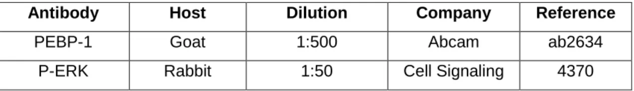
PEBP-1 - phosphatidylethanolamine-binding protein 1 P-ERK - phospho-ERK
PenStrep - Penicillin/Streptomycin PKC - protein kinase C
PKG - cGMP-dependent protein kinases PTM - post-translational modification PVDF - polyvinylidene difluoride RMS - rostral migratory stream RT - room temperature
SDS - sodium dodecyl sulfate Ser - serine
sGC - soluble guanylyl cyclase SGZ - subgranular zone SVZ - subventricular zone TBI - traumatic brain injury
TBS-T - Tris-buffered saline with tween 20 VEGF - vascular endothelial growth factor Wnt - Wingless-type
1.1. Adult neurogenesis
The formation of new neurons (neurogenesis) in the brain was thought for a long time to only occur during embryogenesis. Ramón y Cajal in 1894 described for the first time the complex structure of brain neuronal networks and speculated that they were fixed and irreplaceable. This led to the idea that, contrary to other tissues, there was no way to replace neurons that are lost in the adult brain due to injury or aging. However, in 1965, Altman and Das observed proliferation of cells in rat hippocampus (Altman & Das 1965), contradicting the previous established paradigm. Since then, adult neurogenesis has been an intense subject of investigation and, nowadays, it is well established that there is formation of new neurons during the adult life of mammals, both in physiological and pathological conditions.
Neurogenesis in the adult occurs to maintain brain structure and plasticity, and can increase in response to harmful stimuli. It is present in different animal species, ranging from invertebrates (Tanaka & Reddien 2011, Koizumi & Bode 1991, Fernandez-Hernandez et al. 2013) to non-mammalian (Chapouton et al. 2007, Alunni & Bally-Cuif 2016) and mammalian (Brus et al. 2013) vertebrates. With the discovery of adult neurogenesis in humans (Eriksson et al. 1998) emerged the possibility of brain regeneration taking advantage of neural stem cells (NSC) for transplantation or enhancement of endogenous neurogenesis, in situations detrimental to normal brain functioning. Neurogenesis is a complex process, comprising proliferation, migration, differentiation and survival of the newly formed neurons, with functional integration in the existing neuronal circuits. Despite its importance, neurogenesis in mammals is restricted to certain areas of the adult brain, due to the presence of pools of NSC in specific regions. In the subventricular zone (SVZ) and in the hippocampus, new neurons are generated during adulthood. Neurogenesis in the SVZ is important for replacement of neurons of the olfactory bulb (OB), which is crucial for odor discrimination and processing of sensory information. Unlike several species, in humans there are few new OB neurons originating from neurogenesis in the SVZ, and OB neurogenesis seems to have little relevance (Bergmann et al. 2012). Interestingly, humans present more new neurons in the adult striatum than other mammals, but it is still elusive if these new neurons originate in the SVZ (Ernst et al. 2014). Hippocampal neurogenesis is critical for learning, memory and cognition and is conserved across mammals, including in humans (Gould et al. 1999, Spalding et al. 2013).
1.1.1. Neurogenic niches
In the adult mammalian brain, the SVZ that lines the lateral walls of the lateral ventricles (Fig. 1.1A) and the subgranular zone (SGZ) of the dentate gyrus (DG) of the hippocampus (Fig. 1.1D) are the main regions that contain NSC. These cells have the ability of self-renewal, slowly proliferating by either symmetric or asymmetric divisions (Morrison & Kimble 2006), and are multipotent, having the ability to originate neurons, astrocytes and oligodendrocytes (Lois & Alvarez-Buylla 1993).
In the SVZ, ependymal cells line the ventricles and separate the SVZ from the cerebrospinal fluid (Fig. 1.1B). NSC can contact the cerebrospinal fluid extending an apical process between rosettes of ciliated ependymal cells (Lim & Alvarez-Buylla 2016). Blood vessels also provide the necessary nutritional support to NSC, as well as access to regulatory and signaling molecules (Mercier et al. 2002). NSC (B cells) give rise to transient amplifying progenitors (C cells), rapidly proliferative cells with limited self-renewal capacity, which in turn originate neuroblasts (A cells, immature neurons) (Doetsch et al. 1997). Neuroblasts migrate long distances in chains along the rostral migratory stream (RMS) towards the OB (Lois & Alvarez-Buylla 1994, Lois et al. 1996), where they differentiate into granular and periglomerular neurons and integrate in the existing neuronal circuits as inhibitory interneurons (Alvarez-Buylla & Garcia-Verdugo 2002). Additional paths of migration have been described in mouse brain, with neuroblasts derived from the SVZ migrating towards forebrain regions such as cortex, striatum, and nucleus accumbens (Inta et al. 2008). In humans, neurogenesis in the OB is limited and the presence of a RMS is controversial (Curtis et al. 2007, Sanai et al. 2011), but neuroblasts derived from the SVZ may migrate to striatum (Ernst et al. 2014). The SGZ (Fig. 1.1E), located between the hilus and the granule cell layer of the hippocampus, is also a vascularized region (Palmer et al. 2000). There, radial glia-like cells (type-1 cells) proliferate and originate transient amplifying progenitors (type-2a and 2b cells) (Kempermann et al. 2004), which proliferate rapidly and give rise to neuroblasts (type-3 cells) (Doetsch 2003). Neuroblasts migrate short distances towards the granule cell layer of the DG and differentiate into functional granular neurons that extend their axons into the CA3 region (van Praag et al. 2002).
In the neurogenic niches, NSC are maintained in a stem state due to the presence of several factors and interaction with different cells. NSC transplanted to different regions of the brain differentiate into neurons or glia depending on the transplanted site, and regardless of the origin of NSC (Shihabuddin et al. 2000, Seidenfaden et al. 2006). Therefore, environmental cues are necessary for the maintenance of NSC properties and fate commitment. The different types of cells present in the niches can be distinguished and identified according to specific cell markers (Fig. 1.1C,F). NSC are
from a glial lineage (Kriegstein & Alvarez-Buylla 2009) and therefore express glial fibrillary acidic protein, which is absent in neural progenitor cells that in turn express sex determining region Y-box 2 (Sox2) and nestin (both also present in NSC) (Filippov et al. 2003, Zhang & Jiao 2015). Upon commitment towards a neuronal fate, neuroblasts express doublecortin, but not nestin (Kronenberg et al. 2003). Fully mature neurons can be identified by expression of Neuronal nuclei (NeuN) and -III-tubulin. Neurogenesis and its regulation can be studied using cultures of NSC. They can be isolated and cultured in vitro both as an adherent monolayer or as floating aggregates (neurospheres), being maintained as multipotent by epidermal growth factor (EGF) and/or basic fibroblast growth factor (bFGF) (Weiss et al. 1996, Kuhn et al. 1997).
Figure 1.1 - Neurogenic niches in the adult mammalian brain. Representation of the
neurogenic niches of the SVZ (A-C) and the SGZ of the DG (D-F). The SVZ lines the lateral wall of the lateral ventricles (A), where NSC (B cells) contact the cerebrospinal fluid through ciliated ependymal cells (E) and give rise to highly proliferative transient amplifying progenitors (A cells), which in turn originate neuroblasts (C cells) (B). In the SGZ of the DGof the hippocampus (D), radial glia-like cells (type-1) give rise to transient amplifying progenitors (type-2a and 2b), which originate neuroblasts (type-3) that migrate and differentiate into DG neurons (E). The different types of cells can be distinguished by expression of different markers (C,F). LV, lateral ventricle; BV, blood vessel; GFAP, glial fibrillary acidic protein; Sox2, sex determining region Y-box 2; DCX, doublecortin; NeuN, Neuronal nuclei.
1.1.2. Regulation of neurogenesis
Neurogenesis is a highly-regulated process, being affected by several components of the neurogenic niches, such as intrinsic factors of NSC, immune cells, extracellular matrix components, vascular system and signaling pathways (Christie & Turnley 2012). EGF promotes NSC proliferation in the SVZ and glial differentiation in both the SVZ and SGZ (Kuhn et al. 1997). bFGF increases NSC proliferation and neuronal differentiation in the SVZ (Kuhn et al. 1997), and is necessary for neurogenesis in the SGZ (Kang & Hebert 2015). Insulin-like growth factor 1 (IGF-1) promotes migration of neuroblasts to the OB and neuronal differentiation (McCurdy et al. 2005, Hurtado-Chong et al. 2009) and, in the SGZ, it enhances NSC proliferation and increases cell survival (Aberg et al. 2000, Lichtenwalner et al. 2006). Notch signaling is important for the self-renewal capacity and maintenance of NSC pool in the SVZ (Imayoshi et al. 2010). In the SGZ, Notch is necessary for neurogenesis, affecting proliferation, differentiation and survival of new neurons (Breunig et al. 2007). Sonic hedgehog signaling is necessary for maintenance of NSC pool in both the SVZ and SGZ (Machold et al. 2003) and migration of neuroblasts in the SVZ (Balordi & Fishell 2007). Canonical Wingless-type (Wnt) signaling in the SVZ increases NSC proliferation (Adachi et al. 2007) and post-injury neurogenesis (Shruster et al. 2012), while in the SGZ it is important for maintenance of NSC pool (Wexler et al. 2009) and for increase in neurogenesis (Lie et al. 2005). Non-canonical Wnt signaling influences maturation of new neurons in the OB (Pino et al. 2011) and DG (Varela-Nallar et al. 2010). Bone morphogenetic protein signaling in the SVZ decreases neurogenesis by increasing glial differentiation, which is prevented by Noggin (Lim et al. 2000). Nuclear factor kappa-light-chain-enhancer of activated B cells (NF-B) signaling is necessary for differentiation of new hippocampal neurons (Rolls et al. 2007).
Factors outside the niches also regulate neurogenesis. Hormonal differences between males and females affect neurogenesis. In female rats, estrogen during proestrous increases proliferation of NSC and enhances neurogenesis in the hippocampus (Tanapat et al. 1999). Pregnancy hormones also increase proliferation of NSC, in mouse and rat SVZ (Shingo et al. 2003, Furuta & Bridges 2005). Testosterone affects neurogenesis, so male castration of certain mouse strains leads to increase in proliferating cells in the SVZ (Tatar et al. 2013).
Physical exercise, particularly running, enhances hippocampal neurogenesis (van Praag et al. 1999b, Naylor et al. 2008, Bednarczyk et al. 2011) and improves cognition (van Praag et al. 1999a). This effect is mediated by increase in brain-derived neurotrophic factor (BDNF) (Farmer et al. 2004) and presence of serotonin (Klempin et al. 2013). Exercise induces angiogenesis (Black et al. 1990, Swain et al. 2003), releasing factors
that also increase neurogenesis, such as vascular endothelial growth factor (VEGF) (Jin et al. 2002).
Environmental enrichment is the improvement in housing conditions of laboratory animals, to increase cognitive stimulation (Baumans & Van Loo 2013). Some of the strategies used are bigger cages, new objects and social interaction by contacting with other animals. Environmental enrichment leads to increased survival of mice and rat hippocampal newborn granular neurons (Kempermann et al. 1997, Kempermann et al. 2002, Segovia et al. 2006). Animals with more neurogenesis also display better performance in cognitive tests that evaluate spatial memory, highlighting the association between the amount of new neurons and ability to learn (Sisti et al. 2007). This cognitive stimulation in adulthood helps prevent a decline in neurogenesis with aging, by maintaining the pool of NSC (reviewed in Kempermann 2008). Environmental enrichment also increases the number of neuronal branches and dendritic spines (Rampon et al. 2000), which improves synaptic activity. These effects are mediated by an increase in transcription factors, such as cyclic adenosine monophosphate (cAMP) response element-binding (CREB) (Williams et al. 2001), in proteins involved in synaptogenesis (Frick & Fernandez 2003, Nithianantharajah et al. 2004) and in neurotrophic factors, such as BDNF (Zhang et al. 2016). Running wheels are usually a part of environmental enrichment, so some studies evaluated the contribution of exercise for the effect in neurogenesis observed with environmental enrichment, concluding that increase in BDNF and proliferation of hippocampal NSC results mainly from physical exercise (Kobilo et al. 2011, Bechara & Kelly 2013). Therefore, physical exercise is responsible for increasing the proliferation of NSC, and environmental enrichment is responsible for increasing the survival of new neurons (reviewed in Olson et al. 2006). Environmental enrichment is able to reduce stress in mice (Olsson et al. 1994, Sztainberg et al. 2010). Chronic stress impairs hippocampal neurogenesis, due to increase in glucocorticoids that inhibit the proliferation of NSC and/or differentiation into neurons (Ridder et al. 2005, Kronenberg et al. 2009, Hodes et al. 2012, Anacker et al. 2013). High levels of glucocorticoids also lead to decreased levels of BDNF (Smith et al. 1995, Gourley et al. 2009). Astrocytes are important for neurogenesis (Song et al. 2002, Sultan et al. 2015) and may have a major role in modulating neurogenesis in conditions of stress (reviewed in Luarte et al. 2017).
Aging impairs all steps of neurogenesis. In aged brains, NSC enter a more quiescent state, which leads to a decrease in proliferating NSC both in the SVZ and SGZ (Bouab et al. 2011, Lugert et al. 2010). Fate specification is also altered with aging, resulting in increased glial differentiation and decreased neuronal differentiation (Encinas et al. 2011). The environment of neurogenic niches is a main contributor to the differences
observed in neurogenesis of young and aged mice (Villeda et al. 2011, Katsimpardi et al. 2014). Alterations in Wnt signaling (Piccin et al. 2014), and in the factors IGF-1, bFGF, VEGF (Shetty et al. 2005) and EGF (Enwere et al. 2004) have been described to play a role in decreasing neurogenesis in the aged brain. In humans, neurogenesis in the SVZ declines earlier (Sanai et al. 2011) than hippocampal neurogenesis (Spalding et al. 2013).
1.1.3. Neurogenesis in pathological conditions
Adult neurogenesis is affected in pathological conditions, such as stroke, traumatic brain injury (TBI), epilepsy and neurodegenerative diseases. In animal models of both transient global brain ischemia and focal brain ischemia, neurogenesis is increased in the SVZ (Tonchev et al. 2005, Jin et al. 2001) and SGZ (Kee et al. 2001, Arvidsson et al. 2001). Proliferation of NSC is increased following ischemic stroke (Takagi et al. 1999), and cultured NSC derived from ischemic brain show more proliferative capacity and differentiation into neurons (Deierborg et al. 2010). Moreover, neuroblasts derived from proliferation of NSC in the SVZ switch their migration route from the RMS towards the site of lesion in the striatum (Arvidsson et al. 2002, Zhang et al. 2004). There is also increased angiogenesis in the SVZ (Zhang et al. 2014) and in the dorsomedial striatum near the SVZ, so the new blood vessels provide a scaffold for migration of new neuroblasts (Thored et al. 2007), like in the RMS in physiological conditions. Factors of the neurogenic niches, such as bFGF (Lin et al. 1997) and IGF-1 (Yan et al. 2006) increase in the ischemic cortex following stroke, and their absence prevents enhancement of neurogenesis (Yoshimura et al. 2001, Yan et al. 2006). Despite the enhancement of neurogenesis, in ischemic stroke conditions the new neurons have low long-term survival (Arvidsson et al. 2002, Thored et al. 2006), and aberrant morphologies that lead to maladaptive integration in the existing circuits and memory impairment (Niv et al. 2012, Woitke et al. 2017).
In animal models of TBI, there is increase in NSC proliferation in the SVZ and SGZ (Chirumamilla et al. 2002, Gao et al. 2009). Moreover, there is enhancement of neuronal differentiation in some models (Dash et al. 2001, Rice et al. 2003, Villasana et al. 2014), whereas in others there is only increase in glial differentiation (Bye et al. 2011, Gao & Chen 2013), which can be modulated by age (Sun et al. 2005). VEGF mediates increased hippocampal neurogenesis following TBI, by promoting survival of the new granule neurons (Lee & Agoston 2010). In the SVZ, Eph receptor B3, which is necessary for negative regulation of cell proliferation and survival by EphrinB3 (Ricard et al. 2006), is transiently reduced following TBI and allows proliferation and survival of NSC (Theus
et al. 2010). Other factors involved in the increase in neurogenesis and survival of new neurons following TBI are bFGF (Sun et al. 2009b), EGF (Sun et al. 2010), IGF-1 (Carlson et al. 2014) and BDNF (Gao & Chen 2009).
Epilepsy is characterized by spontaneous recurrent seizures, which can disrupt adult neurogenesis. Several days following seizures, NSC proliferation is increased both in the SVZ (Parent et al. 2002) and SGZ (Gray & Sundstrom 1998), returning to physiological levels after a few weeks (Parent et al. 1997, Bonde et al. 2006). Most newborn cells differentiate into granule neurons (Jessberger et al. 2007). Notch (Sibbe et al. 2012), Sonic hedgehog (Banerjee et al. 2005) and Wnt (Jang et al. 2013) signaling pathways are involved in increasing neurogenesis after seizures. Expression of factors such as BDNF (Isackson et al. 1991) and VEGF (Newton et al. 2003) are altered after seizures and may also affect proliferation of NSC. Many hippocampal granule neurons born after seizures present abnormal morphologies, presenting hilar basal dendrites (Ribak et al. 2000, Dashtipour et al. 2003, Shapiro et al. 2005) and mossy fiber sprouting (Kron et al. 2010). Changes in the glial scaffold may be involved in this process (Shapiro et al. 2005). Nevertheless, functional integration occurs, with establishment of recurrent excitatory networks, of the new abnormal hippocampal granule neurons (Jessberger et al. 2007), and seems to be accelerated by seizures (Overstreet-Wadiche et al. 2006). Migration is also altered after seizures. In the SVZ, some neuroblasts leave the RMS towards non-olfactory forebrain regions, where they are not able to survive (Parent et al. 2002). In the DG, some granule cells migrate ectopically to the hilus and the border hilus/CA3 and integrate the existing circuits (Parent et al. 1997). However, this integration is abnormal and the new ectopic granule neurons burst in synchrony with CA3 pyramidal cells (Scharfman et al. 2000). Loss of reelin signaling (Gong et al. 2007) and excessive mechanistic target of rapamycin (mTOR) signaling (Pun et al. 2012) may be involved in this abnormal migratory behavior. Despite the initial increase in neurogenesis, the potential for neurogenesis is decreased several weeks following seizures (Hattiangady et al. 2004, Kralic et al. 2005), due to a shift to symmetrical divisions, differentiation into reactive astrocytes and depletion of NSC pool (Sierra et al. 2015).
In neurodegenerative diseases such as Alzheimer’s disease, Huntington’s disease and Parkinson’s disease neurogenesis is also altered. Studies with animal models of Alzheimer’s disease show contradictory effects on neurogenesis, due to a large variability of factors between studied models. A single mutation of amyloid precursor protein transgene results in decreased neurogenesis (Donovan et al. 2006), while double and triple mutations lead to increased proliferation and survival of new neurons (Haughey et al. 2002, Mirochnic et al. 2009). A triple transgenic mouse model, containing
mutation of amyloid precursor protein, presenilin1 and tau genes, also results in impaired neurogenesis, associated with presence of β-amyloid plaques (Rodriguez et al. 2008). Imbalance between hippocampal GABAergic and glutamatergic neurotransmission caused by β-amyloid leads to decrease in neurogenesis (Sun et al. 2009a); and apolipoprotein E4, a risk factor for developing Alzheimer’s disease, also has a detrimental effect in neurogenesis (Li et al. 2009). In animal models of Huntington’s disease, NSC proliferation in the SGZ is decreased (Lazic et al. 2004, Gil et al. 2005), and there are less neuroblasts and immature neurons (Fedele et al. 2011). Quiescence of NSC pool is increased, CREB signaling is decreased and transforming growth factor-β signaling seems to be involved in modulating neurogenesis (Kandasamy et al. 2010). In the SVZ, proliferation of NSC is maintained, but there are less new OB neurons (Phillips et al. 2005, Kohl et al. 2010). Local environment seems to be critical for survival of new neurons in the striatum, with contribution of BDNF and Noggin (Cho et al. 2007). In animal models of Parkinson’s disease, neurogenesis is decreased in the SVZ and DG due to presence of -synuclein. NSC proliferation is impaired in some models (Crews et al. 2008, Kohl et al. 2012), while in others there is decrease in survival and integration of new neurons (Winner et al. 2004, Nuber et al. 2008, Winner et al. 2012). Alterations in CREB (Winner et al. 2012), BDNF and glial cell-derived neurotrophic factor (Kohl et al. 2012) may contribute to the impairment of neurogenesis observed in Parkinson’s disease.
1.1.3.1. Neuroinflammation
A common factor in brain pathological conditions is neuroinflammation. Inflammation in the brain is characterized by activation of microglia, the main immune cells of the brain, release of inflammatory cytokines and disruption of the blood brain barrier, further exacerbating the inflammatory response (Russo et al. 2011). In physiological conditions, microglia present a ramified morphology and are continuously monitoring the central nervous system environment (Nimmerjahn et al. 2005). Upon harmful stimuli, microglia become activated and release high amounts of free radicals and cytokines, which can be either pro or anti-inflammatory. When activated by lipopolysaccharide, microglia release reactive oxygen and nitrogen species, such as nitric oxide (NO), and pro-inflammatory cytokines, such as interleukin (IL)-1, IL-6 and tumor necrosis factor-, in order to protect the organism from foreign pathogens. However, this pro-inflammatory activation also affects healthy neurons. Another way of microglial activation involves supporting tissue repair and angiogenesis, by releasing anti-inflammatory cytokines, such as IL-4, IL-10 and transforming growth factor-. The balance between these two
types of activation is critical for the outcome of the inflammatory process and differently affects neurogenesis (reviewed in Ekdahl 2012). Acute neuroinflammation appears to be detrimental to neurogenesis. Inflammation caused by activation of microglia with lipopolysaccharide impairs neurogenesis in rat DG both in normal and injured brain due to pro-inflammatory cytokines, such as IL-6 (Ekdahl et al. 2003, Monje et al. 2003). This effect results from decreased survival of new neurons, and can be prevented using minocycline, which suppresses microglia activation, or indomethacin, a non-steroidal inflammatory drug (Ekdahl et al. 2003, Monje et al. 2003). Survival of new neurons in the OB is also impaired by microglial activation, and can be rescued using minocycline (Lazarini et al. 2012). In a model of stroke, inflammation alters proliferation of NSC from the SVZ and survival of new neurons, which can be prevented by treatment with indomethacin (Hoehn et al. 2005). On the other hand, activation of microglia by low levels of interferon- stimulates differentiation of NSC into neurons (Butovsky et al. 2006). Inflammation can also be important for functional integration of new neurons in the hippocampus (Jakubs et al. 2008).
The presence of neuroinflammation in pathological conditions that increase neurogenesis may also decrease the survival of new neurons and lead to low efficiency of neuronal replacement. It is necessary to understand how cells in different stages of neuronal formation/differentiation are affected by inflammatory factors. Strategies to overcome this problem are needed, in order to enhance endogenous neurogenesis for brain repair. Enhancing the proliferation of NSC, the amount of potential new neurons that can survive and functionally integrate in the neuronal circuits increases. Of the molecules released by microglia during inflammation, NO has emerged as an important regulator of neurogenesis, so its role during this process deserves further investigation.
1.2. Nitric oxide 1.2.1. Biology of NO
NO is a free radical that is synthesized by the enzyme nitric oxide synthase (NOS) from the conversion of L-arginine into L-citrulline (Fig. 1.2). There are three isoforms of NOS. Neuronal NOS (nNOS), mainly present in neurons, and endothelial NOS (eNOS), mainly present in endothelial cells, are constitutively expressed and are regulated by calcium levels and binding of calmodulin (Alderton et al. 2001). In the brain, inducible NOS (iNOS) expression in microglia and astrocytes is triggered by insults to the central nervous system and its regulation is independent of calcium-calmodulin, being active once expressed (Alderton et al. 2001). nNOS and eNOS release low levels of NO (picomolar to nanomolar range), while iNOS releases higher amounts of NO (micromolar
range). Levels of NO released and the kinetics of NO synthesis and consumption in vivo and in vitro are critical to the outcome of the biological response induced by NO (Hall & Garthwaite 2009).
The use of NO donors, carriers of NO that release it over time, is a common strategy to induce NO release in in vitro studies. In this work, three NO donors were used to release NO. Diazeniumdiolates (NONOates) spontaneously decompose in solution, at physiological temperature and pH (Morley & Keefer 1993). There are several NONOates available, with different half-lives. Diethylamine NONOate (DEA/NO) has a short half-life of 2 min at 37C, while the half-life of DETA NONOate (NOC-18) is 20 h at 37C (Fitzhugh & Keefer 2000). S-nitrosothiols, such as S-nitrosocysteine (CysNO), contain a single chemical bond between a thiol group (R-SH) and the NO moiety (Zhang & Hogg 2005). They release NO under certain biological conditions, or directly transfer the nitroso group (R-NO) to other thiol groups (transnitrosylation) (Singh et al. 1996). In aqueous solutions, CysNO has a half-life shorter than 2 min, at 37C (Mathews & Kerr 1993).
NO is a gaseous molecule with a short half-life (0.64 s in rat cortex) and is highly diffusible (diffusion coefficient in rat cortex is 2.2×10⁻⁵ cm2/s) (Santos et al. 2011), being able to rapidly reach several biological targets at certain limited range. Due to its high reactivity, NO lacks a specific molecular target and interacts with several molecules. So, it is important for many different processes in the organism, such as vasodilation and smooth muscle relaxation, synaptic plasticity, inflammation and apoptosis, as well as neurogenesis. The effects of NO are produced through different signaling mechanisms, as described next.
Figure 1.2 - NO is synthesized by NOS enzymes and acts in several pathways. NO is synthesized from L-arginine and oxygen by calcium/calmodulin (CaM)-dependent (nNOS and eNOS) and calcium-independent (iNOS) isoforms of NOS. It is important for several cellular processes and acts by different signaling mechanisms.
1.2.2. NO signaling
1.2.2.1. Classical NO signaling
Guanylyl cyclases are enzymes that synthesize cyclic guanosine monophosphate (cGMP) and exist in two forms: membrane-bound and soluble. Membrane-bound guanylyl cyclases are plasma membrane receptors that form homodimers and have an extracellular ligand binding domain, a short transmembrane region, and an intracellular region with the catalytic domain (Potter 2011). Soluble guanylyl cyclase (sGC) is homologous to the catalytic domains of the membrane-bound forms of guanylyl cyclase and is a receptor of NO (Fig. 1.3). sGC is a heterodimeric enzyme composed by two homologous subunits: an α subunit (α1 or α2) and a β subunit (β1) (Friebe & Koesling 2009). The α2 subunit interacts with many proteins and allows the α2β1 isoform localization to the plasma membrane, while the β1 subunit, which contains a ferrous (Fe(II)) heme, is essential for the dimerization and catalytic activity of sGC. At nanomolar concentrations, NO binds to sGC and promotes the rupture of the His–Fe(II) bond within the heme, leading to a conformational change in the His ligand (Russwurm & Koesling 2004, Rodriguez-Juarez et al. 2007). This increases the conversion of guanosine triphosphate into cGMP by sGC. Submicromolar concentrations of cGMP activate cGMP-dependent protein kinases (PKGs), homodimeric serine-threonine kinases. This interaction leads to a change in the conformation of PKGs and releases autoinhibitory contacts. Phosphodiesterases, particularly 5, 6 and 9, stop sGC/cGMP/PKG signaling by degrading cGMP (Lugnier 2006).
Figure 1.3 - Classical NO signaling. NO binds to the heme group of sGC, which produces cGMP from conversion of guanosine triphosphate. cGMP activates PKG, and can be degraded by phosphodiesterases (PDE).
1.2.2.2. Non-classical NO signaling
NO can produce its effects in a way that is independent of cGMP and that does not involve binding to metal centers, by inducing protein post-translational modifications (PTM), such as S-nitrosylation, S-glutathionylation or tyrosine nitration (Fig. 1.4).
Figure 1.4 - Non-classical NO signaling. NO can directly modify protein function by inducing PTM, such as nitrosylation (A), glutathionylation (B) and tyrosine nitration (C). (A, S-nitrosylation) NO, in the potent oxidant form N2O3 (1, 2), can react with free thiol groups (R-SH) of cysteines leading to the formation of a S-nitrosothiol (R-SNO) (3). (B, S-glutathionylation) Glutathione (GSH) can react with S-nitrosylated proteins and lead to the formation of a mixed disulfide bond with those cysteines (SSG) (1). Alternatively, NO can react with glutathione, originating S-nitrosoglutathione (GSNO) that can react with free thiols, and lead to formation of the mixed disulfide bond (2). (C, tyrosine nitration) When in high amounts, NO reacts with superoxide anion and originates peroxynitrite (ONOO-) (1), which can react with protein 3-position tyrosine residues and form 3-nitrotyrosine (Tyr-NO2) (2).
1.2.2.2.1. S-nitrosylation
S-nitrosylation, also called S-nitrosation (Martinez-Ruiz & Lamas 2004), is a reversible PTM in which a nitroso group (R-NO) is covalently attached to a cysteine thiol (R-SH), forming a S-nitrosothiol (R-SNO) (Martinez-Ruiz et al. 2011). Formation and breakage of S-nitrosylation does not rely completely on enzymes because the specificity of this
reaction does not depend on the recognition of a target by an enzyme. Instead, S-nitrosylation is dependent on the reactivity between a nitrosylating agent and a target. There are three crucial regulatory factors for increasing the specificity and selectivity of S-nitrosylation for particular proteins and avoid broad reactivity: subcellular compartmentalization, site specificity and denitrosylation specificity (Derakhshan et al. 2007). Subcellular compartmentalization ensures that higher concentrations of the nitrosylating agent around selected cysteine residues lead to increased specificity. S-nitrosylation is considered a short-range signaling mechanism (Martinez-Ruiz et al. 2013). Since it is not able to directly react with cysteine thiols (unless in a thiyl radical form), high concentrations of NO are needed to induce formation of reactive nitrosylating species. S-nitrosothiols can be formed by different mechanisms (Guikema et al. 2005), such as the reaction of NO and O2 that results in N2O3, a very potent nitrosylating species. Site specificity involves the reactivity of individual cysteine residues in certain protein microenvironments, being more susceptible to being S-nitrosylated. Accordingly, in physiological conditions only certain residues are modified, while very high concentrations of nitrosylating agents may be able to modify slower-reacting cysteines. Levels of cellular S-nitrosothiols are usually low, which suggests that denitrosylation is a highly active mechanism. The rate of denitrosylation is another critical factor for S-nitrosylation specificity (Benhar et al. 2009). Of the different mechanisms of denitrosylation, two are similar to the pathways used to reduce thiols with other oxidative PTM. On the one hand, reaction of S-nitrosothiols with glutathione leads to either protein glutathionylation or transnitrosylation. On the other hand, thioredoxin can directly act on protein S-nitrosothiols (Lillig & Holmgren 2007).
1.2.2.2.2. S-glutathionylation
S-glutathionylation is the addition of glutathione to a protein by formation of a mixed disulfide bridge with a cysteine residue (Klatt & Lamas 2000, Mieyal et al. 2008). S-glutathionylation is a stable mechanism of redox signaling controlled by several enzymatic mechanisms, and is not dependent on NO, but can be induced by NO and reactive nitrogen species. So, protein S-glutathionylation can be induced by peroxynitrite (Okamoto et al. 2001, Adachi et al. 2004). Moreover, it can result from the reaction of a free thiol and a nitrosothiol, so that glutathione can react with a cysteine of a S-nitrosylated protein, or S-nitrosoglutathione can be formed and react with the cysteine thiol. S-glutathionylation has been suggested as being a mechanism to protect the cell against thiol oxidation to sulfinic or sulfonic acid (Klatt & Lamas 2000).
1.2.2.2.3. Tyrosine nitration
Tyrosine nitration consists of the addition of a nitro group (-NO2) to the phenolic ring of tyrosine residues, resulting in the formation of a 3-nitrotyrosine residue (Martinez-Ruiz et al. 2011). It is an irreversible modification, thermodynamically stable under physiological conditions. Peroxynitrite, which results from the reaction of NO with superoxide, is one of the main nitrating agents, though other reactive species have been described (Radi 2004, Souza et al. 2008). There is selectivity for increased nitration at individual proteins (Souza et al. 1999, Ischiropoulos 2003), but the factors that regulate it are still being investigated (Abello et al. 2009).
1.2.3. NO and neurogenesis
NO has been established as a regulator of adult neurogenesis. Several studies show that physiological levels of NO, released by nNOS, decrease neurogenesis both in the SVZ and DG. Abolishment of NO release, in nNOS knockout mice and by pharmacological inhibition of rat NOS, increases NSC proliferation in the SVZ and in the DG (Packer et al. 2003). Selective inhibition of nNOS increases NSC proliferation in the SVZ and causes a delay in neuronal differentiation (Moreno-Lopez et al. 2004). The anti-proliferative effect of NO released by nNOS is also observed in cultures of NSC derived from the SVZ (Matarredona et al. 2004). This effect is mediated by inhibition of epidermal growth factor receptor (EGFR) and phosphoinositide-3-kinase/Akt pathway (Torroglosa et al. 2007), through S-nitrosylation of EGFR (Murillo-Carretero et al. 2009). In the DG of nNOS knockout mice or after selective inhibition of nNOS, there is increased NSC proliferation and survival of newborn cells (Zhu et al. 2006, Fritzen et al. 2007).
On the other hand, NO released by iNOS during inflammation seems to promote neurogenesis. In a model of stroke, there is an increase in the proliferation of NSC in rat DG, which is prevented by inhibition of iNOS, and in iNOS knockout mice (Zhu et al. 2003). Our group has been extensively studying the role of NO from inflammatory origin in neurogenesis. In SVZ-derived NSC treated with concentrations of a NO donor (NOC-18) within the pathological range (1, 10 and 100 M), NO has a dual role in the proliferation of NSC, dependent on its concentration and time of exposure (Carreira et al. 2010). Exposure to high levels of NO for a long period of time decreases the proliferation of NSC. In mixed cultures of SVZ-derived NSC and microglia, NO released by microglia also decreases NSC proliferation, which is prevented abolishing NO release by using iNOS-/- microglia (Carreira et al. 2014). This anti-proliferative effect of NO from inflammatory origin results from inhibition of the extracellular signal-regulated kinase (ERK)/mitogen-activated protein kinase (MAPK) pathway due to nitration of EGFR by
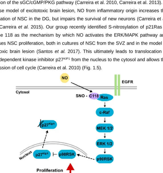
peroxynitrite, which prevents EGFR phosphorylation and its consequent activation (Carreira et al. 2014). Exposure of SVZ-derived NSC to low levels of NO (10 M) for a short period of time increases NSC proliferation in a biphasic manner (Carreira et al. 2010, Carreira et al. 2013). In an initial stage, the proliferative effect of NO is mediated by activation of the ERK/MAPK pathway, while at a later stage the effect is mediated by activation of the sGC/cGMP/PKG pathway (Carreira et al. 2010, Carreira et al. 2013). In a mouse model of excitotoxic brain lesion, NO from inflammatory origin increases the proliferation of NSC in the DG, but impairs the survival of new neurons (Carreira et al. 2010, Carreira et al. 2015). Our group recently identified S-nitrosylation of p21Ras in cysteine 118 as the mechanism by which NO activates the ERK/MAPK pathway and increases NSC proliferation, both in cultures of NSC from the SVZ and in the model of excitotoxic brain lesion (Santos et al. 2017). This ultimately leads to translocation of cyclin-dependent kinase inhibitor p27KIP1 from the nucleus to the cytosol and allows the progression of cell cycle (Carreira et al. 2010) (Fig. 1.5).
Figure 1.5 - NO increases NSC proliferation through ERK/MAPK pathway signaling. NO bypasses the EGFR and S-nitrosylates cysteine 118 of p21Ras, which activates the downstream signaling cascade of protein kinases, ultimately leading to translocation of p27KIP1 to cytosol and its degradation, allowing cell proliferation to occur.
1.2.3.1. Identification of new putative targets of S-nitrosylation by NO in NSC
The ERK/MAPK pathway is crucial for the proliferative effect of NO, which can regulate protein function by S-nitrosylation. Involvement of S-nitrosylation of some proteins in neurogenesis has been described (reviewed in Santos et al. 2015). S-nitrosylation of
myocyte enhancer factor 2 transcription factors acts as a redox switch to inhibit both neurogenesis and neuronal survival (Okamoto et al. 2014). On the contrary, S-nitrosylation of histone deacetylase 2 in embryonic cortical neurons regulates dendritic growth and branching, due to chromatin remodeling and activation of CREB-dependent genes involved in neuronal development, promoted by neurotrophic factors (Nott et al. 2008). Moreover, S-nitrosylation of histone deacetylase 2 also regulates neuronal radial migration during cortical development (Nott et al. 2013).
Due to the low efficiency of post-injury neurogenesis, the identification of new proteins that could be targets of S-nitrosylation by NO and increase neurogenesis is of great interest. Therefore, our group identified S-nitrosylated proteins in NSC, in the presence of NO (Ana I. Santos, Ana S. Lourenço and Inês Araújo, submitted). SVZ-derived NSC were treated with a S-nitrosothiol (CysNO), and proteins were separated by bidimensional electrophoresis. Using a technique that allows fluorescent labeling of oxidized cysteines (fluorescence switch), there were several spots that appeared on the gel of NSC treated with CysNO, but not on the control (non-treated NSC). Those spots corresponded to oxidized proteins, which were then collected and analyzed by mass spectrometry. Several proteins were identified and, after researching their different biological functions, the involvement of some in the ERK/MAPK pathway regulation and cell proliferation was evident (Fig. 1.6). The ERK/MAPK pathway is involved in several cellular processes such as cell proliferation, differentiation, survival and apoptosis (Kolch 2005). Through binding of extracellular molecules, such as growth factors and hormones, to cell surface receptors, signals are transmitted by different effectors, which ultimately alters gene expression and affects cellular behavior. Receptor tyrosine kinases span the cell membrane and have an extracellular ligand binding domain and an intracellular tyrosine kinase domain, existing as inactive monomers. Ligand binding induces dimerization and receptor activation by autophosphorylation. This leads to activation of the GTPase p21Ras that binds and activates Raf kinase. Raf phosphorylates and activates mitogen-activated protein kinase kinase (MEK), which in turn phosphorylates and activates ERK. ERK can either interact with other effectors that can translocate to the nucleus, or translocate to the nucleus itself. In NSC, ERK/MAPK signaling is involved in cell proliferation mainly through binding of EGF and bFGF to their receptors (Sutterlin et al. 2013).
S-nitrosylation of those proteins of interest was assessed, and some were shown to be S-nitrosylated: 14-3-3 (particularly 14-3-3 ), proliferating cell antigen (PCNA), heterogeneous nuclear ribonucleoprotein K (hnRNP K), elongation factor (EF)-1 and phosphatidylethanolamine-binding protein 1 (PEBP-1).
14-3-3 proteins are a family of 7 isoforms of adaptor proteins that regulate cell signaling pathways and are highly expressed in the brain (Cornell & Toyo-Oka 2017). During postnatal formation of the rat brain, 14-3-3 proteins are differentially expressed and have a role in neuronal development and synaptogenesis (Umahara et al. 2011). In the adult, they are necessary for hippocampal long-term potentiation and associative learning and memory (Qiao et al. 2014). 14-3-3 can activate the ERK/MAPK pathway, which is involved in the proliferative effect of NO in NSC (Carreira et al. 2010), by interacting with c-Raf (Chang & Rubin 1997, Roberts et al. 1997). The isoform 14-3-3 is highly expressed in the human hippocampus and is upregulated in temporal lobe epilepsy (Schindler et al. 2006). Moreover, 14-3-3 has an important role in neurogenesis and neuronal migration (Toyo-oka et al. 2014), and has been described to be S-nitrosylated in mesangial cells (Kuncewicz et al. 2003).
PCNA is necessary for DNA replication and is, therefore, essential for cell proliferation. Its regulation by PTM is essential for inducing a different set of cellular functions during DNA replication (Choe & Moldovan 2017). In response to EGFR signaling, which is involved in NSC proliferation (Gritti et al. 1995), PCNA is phosphorylated and protected from degradation (Wang et al. 2006). PCNA can be S-nitrosylated in cysteine 81 (Lam et al. 2010), which blocks its interaction with caspase-9 and promotes apoptosis in a human neuroblastoma cell line (Yin et al. 2015).
hnRNP K belongs to a family of proteins that bind RNA and are involved in nucleic acid metabolism (Dreyfuss et al. 1993). hnRNP K is essential for axonogenesis in Xenopus (Liu & Szaro 2011) and its inactivation, coupled with activation of the neuronal Hu-p21 pathway, may be essential to the switch from proliferation to differentiation in NSC (Yano et al. 2005). The activity of hnRNP K is regulated by several PTM (reviewed in Lu & Gao 2017), but S-nitrosylation is still yet to be described.
EF-1 and EF-1 are part of the EF 1 complex, which is responsible for translation elongation. EF-1 delivers aa-tRNAs to the A site of the ribosome (Carvalho et al. 1984) and EF-1 replaces EF-1-bound GDP for GTP (Pittman et al. 2006). EF-1 and its regulation are essential for the function of EF-1. Although a role in neurogenesis is not described, EF-1 can interact with and regulate Akt (Pecorari et al. 2009, Khwanraj et al. 2016), a pathway involved in the proliferation of NSC (Peltier et al. 2007, Le Belle et
al. 2011). EF-1 can be regulated by phosphorylation (Venema et al. 1991), but its
S-nitrosylation has not been previously described.
PEBP-1 regulates the ERK/MAPK pathway, preventing MEK phosphorylation by binding to c-Raf (Yeung et al. 1999), and its overexpression increases neuronal differentiation, both in a human neuroblastoma cell line (Hellmann et al. 2010) and in adult rat
hippocampal progenitor cells (Sagisaka et al. 2010). PEBP-1 is regulated by phosphorylation (Corbit et al. 2003), but its S-nitrosylation has not been described. Its role in neurogenesis is therefore interesting to study in more detail, so we focused more our study in this protein.
Figure 1.6 - ERK/MAPK pathway signaling through EGFR. Upon ligand binding (EGF), its receptor (EGFR) dimerizes and becomes activated by autophosphorylation. p21Ras is activated by adaptor proteins (not represented) and the downstream protein kinases are sequentially activated by phosphorylation, namely c-Raf, MEK and ERK. ERK can be translocated to the nucleus or interact with other proteins that are then translocated. In the nucleus, gene expression can be altered and lead to cell proliferation. 14-3-3 , PCNA, hnRNP K and PEBP-1 are involved in different steps of this signaling pathway.
1.2.3.1.1. Phosphatidylethanolamine-binding protein 1 (PEBP-1)
PEBP-1 belongs to a family of proteins characterized by having a conserved region that was a binding site for the phospholipid O-phosphatidylethanolamine (Serre et al. 1998). It is a highly conserved family of proteins, existing in organisms that range from bacteria to mammals. PEBP-1 gene is composed of 4 exons localized in mouse chromosome 5, and human and rat chromosome 12. Knockout mice for PEBP-1 revealed that PEBP-1 is mainly expressed in testis and in the brain, mostly in structures of the limbic system, including the hippocampus (Theroux et al. 2007). These animals are viable but develop olfactory deficits. The protein has 187 aminoacids, of which in mouse 3 are cysteines (13, 133 and 168). Human and rat PEBP-1 have 2 cysteines (133 and 168). Cleavage
of the N-terminal of PEBP-1 originates the hippocampal cholinergic neurostimulating peptide, which corresponds to PEBP-1 aminoacids 2-12, and increases production of choline acetyltransferase in the hippocampus (Ojika et al. 2000).
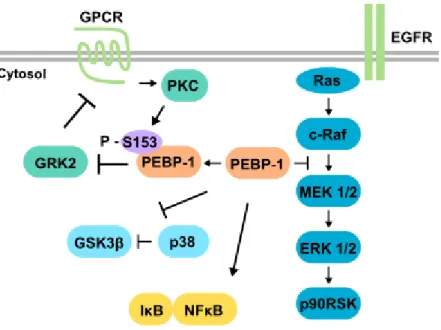
PEBP-1 is involved in the regulation of different signaling pathways (Fig. 1.7). It is also called raf kinase inhibitory protein (RKIP) because it regulates the ERK/MAPK pathway by binding to c-Raf and MEK, preventing their association and phosphorylation of MEK, which results in inhibition of the signaling pathway (Yeung et al. 1999, Yeung et al. 2000). Phosphorylation of PEBP-1 serine 153 by protein kinase C (PKC) releases the association between PEBP-1 and c-Raf (Corbit et al. 2003) and triggers dimerization of PEBP-1, increasing its specificity for binding and inhibiting G protein-coupled receptor kinase 2 (GRK2) ability to phosphorylate G protein-coupled receptors (Lorenz et al. 2003, Deiss et al. 2012). PEBP-1 also regulates the NFB pathway by interacting with several kinases, acting as a scaffold protein, and both its overexpression and downregulation impair the degradation of inhibitory B upon stimulation by IL-1β (Yeung
et al. 2001, Tang et al. 2010). Glycogen synthase kinase 3 signaling can be directly
activated by PEBP-1, through prevention of its inhibition by phosphorylation (Al-Mulla et al. 2011).
In the brain, PEBP-1 has different functions, such as mediating PKC-dependent MAPK activation during cerebellar long-term depression (Yamamoto et al. 2012). Several studies show that PEBP-1 is important for neurogenesis. Hippocampal progenitor cells secrete PEBP-1 to culture medium (Dahl et al. 2003). Moreover, PEBP-1 is involved in directing differentiation of hippocampal progenitor cells and neuroblastoma cells towards a neuronal lineage (Sagisaka et al. 2010, Hellmann et al. 2010) through enhanced crosstalk of PKC and ERK/MAPK and enhancement of G protein-coupled receptor signaling (Hellmann et al. 2010). In neuroblastoma cells, long-term ethanol exposure impairs neuronal differentiation, which is accompanied by downregulation of PEBP-1 and PKC, and by decreased activation of the ERK/MAPK pathway by BDNF (Hellmann et al. 2009). In an ischemic model, PEBP-1 regulates differentiation towards a neuronal lineage and decreases differentiation into astrocytes, in the hippocampus (Toyoda et al. 2012).
Figure 1.7 - PEBP-1 regulates several signaling pathways. PEBP-1 inhibits ERK/MAPK pathway signaling by binding to Raf and MEK, and by preventing phosphorylation of MEK by c-Raf. Upon activation through G protein-coupled receptor (GPCR), PKC phosphorylates serine 153 of PEBP-1. This induces the release of PEBP-1 inhibitory effect in the ERK/MAPK pathway, and prevents the inhibitory effect of GRK2 in GPCR signaling. PEBP-1 can also regulate NFB signaling by preventing degradation of inhibitory B, and can activate glycogen synthase kinase 3 (GSK3) by preventing its inhibitory phosphorylation.
1.3. Objectives
Post-injury neurogenesis is not an efficient process, which impairs the potential of brain regeneration. NO is an important regulator of adult neurogenesis, being able to enhance the proliferation of NSC in conditions of neuroinflammation. One of the ways by which NO can produce its effects is by inducing S-nitrosylation of proteins by reacting with thiol groups of cysteine residues. A mechanism of activation of the ERK/MAPK pathway through S-nitrosylation by NO and enhancement of proliferation of NSC has been described by our group (Santos et al. 2017). Moreover, our group also recently identified a group of proteins that are S-nitrosylated by NO in NSC (Ana I. Santos, unpublished results). Some of these proteins are involved in cell proliferation by interacting with the ERK/MAPK pathway. PEBP-1, one of the identified targets, is a regulator of cell proliferation by means of c-Raf inhibition, being a promising target to study in detail. In this work, we propose to investigate the role of PEBP-1 S-nitrosylation by NO in neurogenesis, particularly in the proliferation of NSC. In addition, we also propose to investigate the biological role of the other newly identified targets of S-nitrosylation of NO in post-injury neurogenesis.
The methods used in this work are described in detail in chapter 2.
In chapter 3.1, the role of S-nitrosylation of PEBP-1 by NO in proliferation was evaluated in SVZ-derived NSC using normal or NO-insensitive forms of PEBP-1, lacking specific cysteines. The contribution of each cysteine for S-nitrosylation of PEBP-1, and its effect on ERK/MAPK signaling and cell proliferation was evaluated, after treatment with the S-nitrosothiol CysNO or the NO donor NOC-18. Moreover, S-nitrosylation of PEBP-1 in post-injury neurogenesis was evaluated using a mouse model of excitotoxic brain lesion associated with increased hippocampal neurogenesis mediated by NO (Carreira et al. 2015). S-nitrosylation of PEBP-1 was assessed in the DG of wild type mice and mice lacking iNOS, and also in the stem cells of the DG, using cultured hippocampal stem cells treated with CysNO.
In chapter 3.2, the role of S-nitrosylation of the other identified proteins by NO in post-injury neurogenesis was evaluated using the mouse model described above. S-nitrosylation of 14-3-3, hnRNP K, PCNA and EF-1 was assessed in mouse DG, and in hippocampal stem cells treated with CysNO.
Chapter 4 includes an integrated discussion of the results presented in this work, and chapter 5 presents the conclusions.
2.1. Methods 2.1.1. Animals
C57Bl6 male mice were purchased from Charles River (Barcelona, Spain), and B6.129P2- Nos2tm1Lau/J (knockout for iNOS, iNOS KO) male mice were purchased from
The Jackson Laboratory (Bar Harbor, ME, USA). The animals were kept in the animal facilities of Center for Neuroscience and Cell Biology (Coimbra) and Center for Biomedical Research (University of Algarve, Faro) with food and water ad libitum in a 12-hour dark:light cycle. All experiments were performed in accordance with institutional and European guidelines (2010/63/EU and DL 113/2013) for the care and use of laboratory animals, under the project license 0421/000/000/2013.
2.1.2. Genotyping
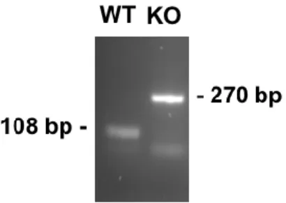
The genotype of B6.129P2-Nos2tm1Lau/J mice was confirmed by polymerase chain reaction, using the primers recommended by The Jackson Laboratory (Table 2.1) and NZYTaq 2x Green Master Mix. DNA was obtained from the tip of the tail, after purification with NZY Tissue gDNA Isolation kit. After denaturation at 94C for 5 min, 35 cycles of 94C, 55C and 72C for 1 min each were performed, followed by a final extension at 72C for 10 min.
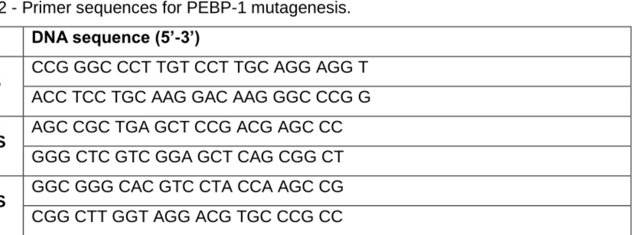
Table 2.1 - Primer sequences for genotyping.
DNA sequence (5’-3’)
iNOS
ACA TGC AGA ATG AGT ACC GG TCA ACA TCT CCT GGT GGA AC AAT ATG CGA AGT GGA CCT CG
The amplicons were run in a gel electrophoresis (2% agarose with GreenSafe Premium), and were detected using ChemiDoc XRS+ (BioRad Laboratories Inc., Hercules, CA, USA), resulting in a product of 108 base pairs for the wild type allele, and a product of 270 base pairs for the recombinant allele (Fig. 2.1).
Figure 2.1 - Genotyping profile of WT and iNOS KO mice. Representative image of PCR products of wild type (WT) and iNOS KO mice genotyping.
2.1.3. SVZ-derived NSC cultures
NSC were isolated from the SVZ of P0-3 C57Bl6 mice as previously described (Morte et al. 2013). Briefly, mice were decapitated, the brain and meninges were removed, and slices of 1 mm were cut coronally. Tissue lining the lateral walls of the lateral ventricles was dissected and enzymatically digested in 0.025% Trypsin/0.265 mM ethylenediaminetetraacetic acid (EDTA) in Hanks' Balanced Salt Solution, 15 min at 37C. Single cells were counted using 0.1% trypan blue exclusion assay and plated in uncoated T-25 flasks at a density of 100,000 cells/ml. NSC were allowed to grow as floating aggregates (neurospheres) in proliferation medium (Dulbecco’s modified eagle medium: nutrient mixture F-12 (D-MEM/F-12) with 2 mM GlutaMAXTM-I (L-Ala-L-Gln) supplemented with 1% B27, 1% antibiotic (PenStrep, 10,000 units/ml of penicillin, 10 mg/ml streptomycin), 5 ng/ml bFGF and 10 ng/ml EGF, in 95% air/5% CO2 humidified atmosphere at 37C. Approximately 5 days later, NSC were mechanically dissociated and resuspended in fresh proliferation medium (passage). After at least 2 passages, NSC were plated on 0.1 mg/ml poly-L-lysine-coated multiwells or coverslips, and maintained with proliferation medium until the desired confluency was achieved. Each culture was used until reaching a maximum of 12 passages.
2.1.4. Hippocampal stem cell cultures
Hippocampal stem cells were kindly provided by Dr. Fred H. Gage (Salk institute, USA). These cells were isolated from the hippocampus of adult Fischer 344 rats, as described by Gage et al. (1995), and their capacity for self-renewal and multipotency was characterized by Palmer et al. (1997). For maintenance of the cell line, hippocampal stem cells were cultured adherent on plates coated with polyornithine (10 µg/ml) and laminin (10 µg/ml), in D-MEM/F-12 with 2 mM GlutaMAXTM-I (L-Ala-L-Gln) supplemented with 1% PenStrep, 1% N2 and 20 ng/ml bFGF, and in 95% air/5% CO2 humidified atmosphere at 37C. Culture medium was replaced every 3-4 days and, when confluent,