Universidade de Lisboa
Faculdade de Ciências
Departamento de Biologia AnimalC
C
C
C
OMPARATIVE STUDY OF
OMPARATIVE STUDY OF
OMPARATIVE STUDY OF
OMPARATIVE STUDY OF THE CYTOTOXIC EFFE
THE CYTOTOXIC EFFE
THE CYTOTOXIC EFFE
THE CYTOTOXIC EFFECTS OF
CTS OF
CTS OF
CTS OF
MICROCYSTIN
MICROCYSTIN
MICROCYSTIN
MICROCYSTIN
----LR
LR
LR
LR
IN MAMMALIAN CELL LI
IN MAMMALIAN CELL LI
IN MAMMALIAN CELL LI
IN MAMMALIAN CELL LINES
NES
NES
NES
::::
V
V
V
V
ERO
ERO
ERO
ERO
,,,,
H
H
H
H
EP
EP
EP
EP
G2,
G2,
G2,
G2,
C
C
C
C
ACO
ACO
ACO
ACO
2
2
2
2
AND
AND
AND
AND
MDCK.
MDCK.
MDCK.
MDCK.
Carina Alexandra Gomes Menezes
Mestrado em Biologia Humana e Ambiente 2009
Universidade de Lisboa
Faculdade de Ciências
Departamento de Biologia AnimalC
C
C
C
OMPARATIVE STUDY OF
OMPARATIVE STUDY OF
OMPARATIVE STUDY OF
OMPARATIVE STUDY OF THE CYTOTOXIC EFFECT
THE CYTOTOXIC EFFECT
THE CYTOTOXIC EFFECT
THE CYTOTOXIC EFFECTS OF
S OF
S OF
S OF
MICROCYSTIN
MICROCYSTIN
MICROCYSTIN
MICROCYSTIN
----LR
LR
LR
LR
IN MAMMALIAN CELL LI
IN MAMMALIAN CELL LI
IN MAMMALIAN CELL LI
IN MAMMALIAN CELL LINES
NES
NES
NES
::::
V
V
V
V
ERO
ERO
ERO
ERO
,,,,
H
H
H
H
EP
EP
EP
EP
G2,
G2,
G2,
G2,
C
C
C
C
ACO
ACO
ACO
ACO
2
2
2
2
AND
AND
AND
AND
MDCK.
MDCK.
MDCK.
MDCK.
Carina Alexandra Gomes Menezes
Tese orientada por:
Na instituição que confere o grau: Professora Doutora Ana Amorim
Professora Auxiliar da Faculdade de Ciências da Universidade de Lisboa
Na instituição de Acolhimento: Doutora Elsa Alverca
Investigadora do Laboratório de Biologia e Ecotoxicologia, Departamento de Saúde Ambiental, Instituto Nacional de Saúde Dr. Ricardo Jorge.
Mestrado em Biologia Humana e Ambiente 2009
I
TABLE OF CONTENTS
LIST OF FIGURES ... III LIST OF TABLES ... V LIST OF ABBREVIATIONS ... VII ACKNOWLEDGMENTS ... IX ABSTRACT ... XI RESUMO ALARGADO ... XIII KEYWORDS/PALAVRAS-CHAVE... XVII
INTRODUCTION ... 1 1. Cyanobacteria ... 3 1.1. Characterization ... 3 1.2. Blooms ... 3 2. Cyanotoxins ... 5 3. Microcystins ... 6 3.1. Chemical structure ... 6
3.2. Occurrence in the environment ... 7
3.3. Human intoxication ... 8
3.4. Microcystin-LR ... 8
3.4.1. Chemical properties. Toxin uptake, distribution and elimination. ... 8
3.4.2. Regulatory aspects ... 9
3.4.3. Carcinogenic potential ... 9
3.4.4. Cellular targets and mechanisms of toxicity ... 10
3.4.5. Effects of MCLR in non-liver cells ... 11
4. Objectives ... 12
MATERIALS AND METHODS ... 13
1. MCLR: sources and preparation of stock solutions... 15
1.1. Semi-purified extracts from M. aeruginosa ... 15
1.2. Pure commercial Microcystin-LR ... 16
2. Mammalian cell lines and culture conditions ... 16
2.1. Cell lines maintenance ... 16
II
3. Evaluation of MCLR effects ... 18
3.1. Cytotoxicity assays ... 18
3.2. Specific labelings of cellular organelles ... 19
3.2.1. Acridine Orange ... 19
3.2.2. Rhodamine-123 ... 19
3.2.3. Phalloidin... 20
3.3. Protein analysis ... 20
3.3.1. Analysis of LC3B and GRP94 proteins by immunofluorescence ... 20
3.3.2. Analysis of GRP94 expression by Western Blot ... 21
3.4. Transmission Electron Microscopy ... 22
RESULTS ... 23
1. Effects of MCLR on cell viability ... 25
2. Effects of MCLR on cellular organelles ... 27
2.1. Lysosomes ... 27
2.2. Mitochondria ... 27
2.3. Microfilaments... 32
2.4. Autophagosomes (LC3B protein) ... 32
2.5. Endoplasmic reticulum (GRP94 protein) ... 33
3. Effects of MCLR on GRP94 expression ... 36
4. Effects of MCLR on cellular ultrastructure ... 36
DISCUSSION ... 43
CONCLUSIONS... 55
III
L
IST OFF
IGURESFigure 1 - Chemical structure of microcystins. ... 7
Figure 2 - Microcystins worldwide occurrence in freshwater environments ... 7
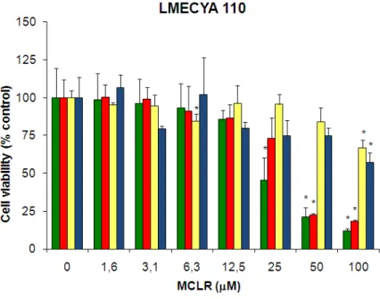
Figure 3 - Viability of HepG2, Vero, MDCK and CaCo2 cells exposed to MCLR-containing LMECYA 110 extract evaluated by the NR assay ... 25
Figure 4 - Viability of HepG2, Vero, MDCK and CaCo2 cells exposed to pure MCLR and a non-toxic LMECYA 127 extract evaluated by the NR assay. ... 26
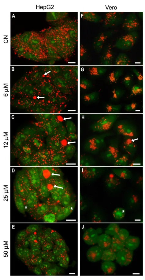
Figure 5 – HepG2 and Vero cells stained with AO after MCLR exposure ... 29
Figure 6 - Confocal fluorescence images of HepG2 and Vero cells stained with rh-123 after exposure to MCLR ... 30
Figure 7 - Actin cytoskeleton of HepG2 and Vero cells exposed to MCLR ... 31
Figure 8 - Immunolabeling of LC3B protein in HepG2 and Vero cells exposed to MCLR ... 34
Figure 9 - Immunolabeling of GRP94 protein in HepG2 and Vero cells treated with MCLR ... 35
Figure 10 - Expression of the GRP94 protein in HepG2 and Vero cells treated with MCLR ... 36
Figure 11 - Micrographs of the ultrastructural organization of HepG2 cells after treatement with MCLR ... 38
Figure 12 – Micrographs of the ultrastructural organization of Vero cells exposed to MCLR. .... 39
Figure 13 – Micrographs of the ultrastructural organization of MDCK cells after treatment with MCLR ... 40
V
L
IST OF TABLESTable 1 - Relevant dates and events in toxic cyanobacteria history ... 4
Table 2 - Types of cyanotoxins, toxic cyanobacterial genera and acute toxicity ... 5
VII
L
IST OFA
BBREVIATIONSAbDil – Antibody Dilution Solution
ADDA-(2S,35’,8S,9S)-3-amino-9-methoxy-2,6,8-trimethyl-10-phenyl-(4E),(6E)-decadienoic acid
AO – Acridine Orange
ATCC – American Type Culture Collection Bcl-2 – anti-apoptotic protein
CaCo2 – Human colon adenocarcinoma cell line CBS – Cytoskeleton Buffer with sucrose
CCD – Cooled camera device
DAPI – 4′-6-diamidino-2-phenylindole
DMEM – Dulbecco´s Modified Eagle Medium DMSO – Dimethyl sulfoxide
DSMZ – Deutsche Sammlung von Mikroorganismen und Zellkulturen GmbH (German Collection of Microorganisms and Cell Cultures)
EDTA – Ethylene Diamine Tetraacetic acid ECL – Enhanced Chemical Luminescence ER – Endoplasmic reticulum
FBS – Fetal Bovine Serum
GRP94 – Glucose-regulated protein 94 GSH - Glutatione
HepG2 – Human hepatoma cell line
HPLC – High Pressure Liquid Chromatography
INSA - Instituto Nacional de Saúde (National Health Institute) ISO – International Organization for Standardization
LBE – Laboratory of Biology and Ecotoxicology, National Health Institute Dr. Ricardo Jorge LC3B – Light chain 3B
LC3B-I – Light chain 3B, form 1 (cytosolic) LC3B-II – Light chain 3B, form 2 (membranous) LD50 – Lethal dose to 50% of the treated animals
VIII
LDH – Lactate dehydrogenase
LMECYA 110– Microcystis aeruginosa toxin producer strain kept in culture in LBE LMECYA 127 - M. aeruginosa non-toxin producer strain kept in culture in LBE LMP – Lysosome membrane potential
MDCK – Dog kidney-derived cell line MCLR – Microcystin-LR
MEM – Modified Eagle Medium
MMP – Mitochondrial membrane potential
MPT – Mitochondrial permeability transition state
MTT – 3-(4,5-dimethylthiazol-2yl)-2,5-diphenyl tetrazolium bromide NR – Neutral Red
OATP – Organic anion transporting polypeptide PBS – Phosphate buffered saline
PBS/T – Phosphate buffered saline with 0,02% Tween PFA – Paraformaldehyde
PP1 – Protein Ser/Thr Phosphatase type 1 PP2A - Protein Ser/Thr Phosphatase type 2A PVDF – Polyvinylidene Fluoride
Rh-123 – Rhodamine-123 ROS – Reactive oxygen species rt – room temperature
SDS-PAGE - sodium dodecyl sulfate polyacrylamide gel electrophoresis TEM – Transmission Electron Microscopy
TBS - Tris-buffered saline
TBS/T – Tris-buffered saline with 0,02% Tween
Vero – African green monkey Cercopithecus aethiops kidney-derived cell line WHO – World Health Organization
IX
A
CKNOWLEDGMENTS/A
GRADECIMENTOSEm primeiro lugar quero agradecer à Prof. Ana Amorim pela simpatia e entusiasmo com que me acolheu no seu mundo das microalgas. O seu contributo para este trabalho foi muito importante e sem as suas críticas, apoio e orientação, este trabalho não se realizaria. Quero agradecer também o apoio financeiro prestado através do projecto HABCOL Nº PDCT/MAR/60086/2004.
À Doutora Elsa Alverca, a minha guia através do mundo da microscopia. Quero agradecer a simpatia, orientação, apoio, críticas e paciência, especialmente com a minha ignorância na análise de ultrastrutura celular e cepticismo geral.
À Doutora Elsa Dias, pela partilha do seu vasto conhecimento na área da cultura celular e da microcistina-LR (para além de muitos outros temas!). Muito obrigado pela orientação, críticas construtivas, disponibilidade, companheirismo e apoio constantes.
À Doutora Maria João Silva, pelo uso da sua sala de culturas do seu laboratório; ao laboratório de Parasitologia, pelo uso do agitador de placas e da incubadora a 37oC; à
Doutora Paula Alvito, pelas células CaCo2; ao Bruno Silva pelas células HepG2; ao Doutor Paulo Matos pelo uso do microscópio confocal e pelas células MDCK; à Doutora Sónia Moniz pela ajuda preciosa nas técnicas de SDS-PAGE e Western Blot.
E sem nunca esquecer, quero agradecer profundamente a todos no Laboratório de Biologia e Ecotoxicologia. Neste laboratório encontrei um grupo de pessoas extraordinárias que transformaram este ano não só numa experiência profissional proveitosa mas também levaram a um crescimento pessoal. Ao Doutor Paulo Pereira, por me ter aceite no seu laboratório e por me ter purificado a toxina. À Albertina Amaral, pela ajuda com os reagentes e soluções e por me ensinar (com muita paciência) a pesar nas balanças ultra-modernas do laboratório; à Catarina Churro, pela ajuda com as culturas de cianobactérias; à Filomena Sam-Bento, pela paciência com que cortou e contrastou inúmeros blocos para o TEM; ao Sérgio Paulino, por me introduzir ao mundo das cianobactérias e da astronomia, e à Stela Tomé, por me mostrar como funciona o laboratório.
A todos que contribuiram de alguma forma para a realização deste trabalho, o meu profundo agradecimento.
XI
A
BSTRACTMicrocystin-LR (MCLR) is a natural occurring freshwater cyanotoxin, recognized as one of the most toxic microcystin variants. It is thought to be responsible for cases of livestock and human intoxication due to consumption of toxic cyanobacteria-contaminated water. Although considered a hepatotoxin, MCLR also targets other organs such as the kidneys and intestines. In spite the cellular mechanisms associated with the toxicity of MCLR are still unclear, a previous work in a monkey kidney cell line suggested that the endoplasmic reticulum was an early target of MCLR toxicity and that autophagy was triggered as a cell defense mechanism at subcytotoxic concentrations of MCLR.
In the present work, cytotoxic, morphological and ultrastructural effects of MCLR were compared in HepG2 (human liver), Vero (monkey kidney), MDCK (dog kidney) and Caco2 (human intestine) cell lines. MCLR induced a concentration-dependent decrease in cell viability by the NR assay in all cell lines, with HepG2 and Vero showing the lowest cytotoxic thresholds of 25 and 50 μM MCLR, respectively. In these cells, MCLR exposure induced lysosomal damages previously to mitochondrial disruption, reinforcing the role of lysosomes in MCLR-induced toxicity. Immunolabelling and ultrastructural visualization of autophagosomes, showed that autophagy was a response transversal to both cell lines, triggered at subcytotoxic MCLR concentrations, confirming its importance as a defense mechanism to early damages inflicted by the toxin. The analysis of GRP94, an ER stress protein, did not undoubtedly demonstrate that MCLR targets the ER. However, together with the ultrastructural data, suggested that in both HepG2 and Vero cells, the ER has a role in autophagy induction. Additionally, in HepG2 cells, GRP94 down-regulation with increasing MCLR concentrations supported the ER role in the triggering of apoptosis. At high toxin concentrations, ultrastructural alterations consistent with apoptosis were observed for all four cell lines, proving that this is a general MCLR-induced mechanism.
XIII
R
ESUMOA
LARGADOAs cianobactérias são organismos procariotas com origem há cerca de 2,8 mil milhões de anos e que ainda hoje proliferam em todos os ecossistemas da Terra. O aparecimento de florescências de cianobactérias é um fenómeno potenciado pela acção do homem nos ecossistemas através da eutrofização das massas de águas e que acarreta a potencial libertação de toxinas produzidas por estes organismos (cianotoxinas). Estes fenómenos são mais comuns em sistemas de água doce pelo que a possibilidade de intoxicação por cianotoxinas em águas de consumo ou de recreio é actualmente uma preocupação em Saúde Pública, em particular em regiões com elevada dependência de reservas de água superfícial (e.g.rios, albufeiras, lagos) como é o caso de Portugal .
As microcistinas são as cianotoxinas mais frequentes, sendo detectadas na maior parte das florescências de cianobactérias. São também, as cianotoxinas mais estudadas e melhor caracterizadas em relação à estrutura, actividade e toxicologia. De entre as 70 variantes conhecidas de microcistinas, a microcistina-LR (MCLR) é uma das mais comuns e mais tóxicas. Alguns casos humanos de intoxicação aguda por microcistinas foram já descritos e apresentaram como principais manifestações clínicas vómitos, diarreia, hemorragia hepática e, nos casos mais graves, a morte dos indivíduos.
Actualmente, a MCLR é reconhecida sobretudo como uma hepatotoxina, uma vez que o seu órgão-alvo principal é o fígado. Devido à natureza hidrofílica da MCLR, a sua entrada nas células é mediada pelos transportadores dos ácidos biliares (OATP). Estes estão presentes em grande quantidade no fígado e em menor extensão noutros órgãos como o cérebro, rins e intestinos. Desta forma, a maioria dos estudos sobre os efeitos da MCLR têm sido realizados principalmente em células hepáticas in vivo ou in vitro. No entanto, estudos recentes têm demonstrado efeitos tóxicos da MCLR em células renais e de intestino.
Um dos mecanismos de toxicidade da MCLR melhor documentados é a inibição das fosfatases proteicas 1 e 2A. Estas são importantes reguladoras de proteínas implicadas em inúmeros processos celulares tais como o metabolismo, a manutenção do citosqueleto e a divisão celular. O efeito inibitório da MCLR sobre as fosfatases PP1 e PP2A conduz à despolimerização e agregação dos componentes do citosqueleto, levando ao colapso da estrutura dos hepatócitos.
XIV
Por outro lado, grande parte dos artigos científicos publicados descreve também a mitocôndria como um organelo-alvo da toxicidade da MCLR, designadamente o seu papel mediador no processo apoptótico induzido pela MCLR. Este processo, por sua vez, parece ser desencadeado pela indução da produção de espécies reactivas de oxigénio pela MCLR. Mais recentemente, outros organelos celulares começaram a ser também identificados como alvos intracelulares da MCLR.
Em trabalhos prévios realizados no Laboratório de Biologia e Ecotoxicologia, INSA com a linha celular renal de macaco verde africano Vero-E6 (Alverca et al., 2009), foi demonstrado que antecedendo o efeito apoptótico, a MCLR induz a autofagia e que estes dois processos envolvem não só o citosqueleto e as mitocôndrias, como também outros organelos celulares tais como os lisossomas e o retículo endoplasmático. O presente estudo, surge na sequência deste trabalho e procurou estudar se os efeitos observados na linha renal Vero-E6 também ocorrem noutros tipos celulares. Neste contexto, os efeitos citotóxicos, morfológicos e ultrastruturais da MCLR foram comparados em quatro linhas celulares: 1) HepG2 (hepatoma humano); 2) Vero-E6 (rim de macaco); 3) MDCK (rim de cão); 4) CaCo2 (adenocarcinoma do cólon humano). Estas linhas celulares permanentes representam, neste estudo, os vários órgãos alvo da MCLR.
A viabilidade celular dos diferentes tipos celulares expostos à MCLR foi avaliada através do teste de citotoxicidade do Vermelho Neutro, que se baseia na integridade dos lisossomas. Em todas as linhas celulares estudadas houve um decréscimo da viabilidade celular de uma forma dependente da concentração de toxina, embora estas tenham apresentado sensibilidades diferentes. O decréscimo de viabilidade foi significativo aos 25 μM de MCLR para as células HepG2, aos 50 μM, para as células Vero e aos 100 μM para as células MDCK e CaCo2. Estes dados foram consistentes com as alterações ultrastruturais observadas, embora nas células MDCK se tenham observado alterações estruturais nas mitocôndrias a concentrações de toxina mais baixas (25 μM de MCLR). Os limiares de citotoxicidade obtidos reflectem a situação de intoxicação com MCLR in vivo, em que o fígado é o orgão mais afectado, seguido dos rins e intestinos. Assim, é também demonstrada a boa representabilidade destas linhas celulares enquanto modelos de órgãos-alvo da MCLR.
Face à maior sensibilidade das células HepG2 e Vero à MCLR, apenas estas duas linhas celulares foram usadas para a avaliação dos efeitos da MCLR ao nível dos organelos
XV e processos celulares. Foram efectuadas marcações específicas de lisossomas, mitocôndrias e filamentos de actina através do uso de fluorocromos específicos (laranja de acridine, rhodamina-123 e faloidina) e marcação com anticorpos para uma proteína do retículo endoplasmático (GRP94) e para uma proteína dos autofagossomas (LC3B). Os resultados obtidos nas duas linhas celulares mostram que a perturbação dos organelos celulares é dependente da concentração de MCLR. De facto, e para ambas as linhas celulares, concentrações relativamente baixas de toxina (12 μM MCLR) induziram a diminuição do número de lisosomas, mas o aumento do seu tamanho, enquanto que a concentrações mais elevadas (25 μM) a MCLR induziu a ruptura destes organelos. As alterações ao nível mitocondrial e nos filamentos de actina foram observadas de forma generalizada, apenas a concentrações de toxina superiores (50 μM MCLR). Assim, nestas linhas celulares, e ao contrário do que é tradicionalmente descrito, os danos nos lisossomas antecedem os das mitocôndrias, sugerindo que terão um papel importante na acção tóxica da MCLR.
A proteína LC3B associa-se à membrana dos autofagossomas, sendo um bom marcador da autofagia. A imunolocalização desta proteína e a análise ultrastructural, demonstrou que existe indução de autofagia a concentrações baixas de toxina (6 e 12 μM MCLR) para as duas linhas celulares confirmando os resultados obtidos em estudos prévios para as células Vero. No entanto, esta resposta autofágica foi mais pronunciada nas células HepG2 do que nas células Vero. De facto, a análise ultrastrutural e de fluorescência, permitiram identificar inúmeros autofagossomas de grande tamanho nas células HepG2. Este aspecto estará provavelmente relacionado com as funções de destoxificação fundamentais nas células hepáticas. Embora a autofagia pareça ser um importante mecanismo de defesa contra danos celulares precoces induzidos pela MCLR, como indicam os resultados das células HepG2 e Vero, não é um processo geral de resposta celular à toxina, já que no estudo ultrastrutural não foi detectada nas linhas celulares MDCK e CaCo2.
A proteína GRP94 existe no retículo endoplasmático de células fisiologicamente normais. No entanto, está também envolvida em processos de reparação associados com o stress do retículo e apresenta propriedades anti-apoptóticas. Os resultados da imunolocalização desta proteína revelaram que o seu padrão de distribuição é dependente da linha celular. Por outro lado, também a expressão da GRP94 foi afectada diferencialmente nas duas linhas celulares estudadas. Assim, por western blot verificou-se
XVI
que a expressão desta proteína diminui com o aumento da concentração de MCLR indiciando que na linha hepática a activação de vias apoptóticas parece ser mediada pelo retículo endoplasmático. Por outro lado, na linha celular renal a expressão da proteína GRP94 manteve-se inalterada, independentemente da concentração de MCLR. Isto sugere que nas células Vero a apoptose induzida pela MCLR não será activada pelo retículo endoplasmático, mas sim por outros organelos como sejam as mitocôndrias ou os lisossomas. A concentrações elevadas de toxina, observaram-se alterações ultrastruturais consistentes com a apoptose nas quatro linhas celulares em estudo, comprovando que este é um mecanismo geral de resposta celular à MCLR.
XVII
K
EYWORDS/P
ALAVRAS-
CHAVEKeywords Microcystin-LR Mammalian cell lines Cytotoxicity Cellular organelles Autophagy Apoptosis Ultrastructure Palavras-chave Microcistina-LR
Linhas celulares de mamífero Citotoxicidade
Organelos celulares Autofagia
Apoptose Ultrastrutura
1
Comparative study of the cytotoxic effects of MCLR in mammalian cell lines
3 1.
1. 1.
1. CyanobacteriaCyanobacteriaCyanobacteria Cyanobacteria
1.1. 1.1. 1.1.
1.1. CharacterizationCharacterizationCharacterizationCharacterization
The Cyanobacteria or Cyanoprokaryota, commonly known as blue-green algae, are a group of ancient photosynthetic organisms present on Earth since 2.7 billion years ago (Badger and Price, 2003). This group is divided into the four orders Chroococcales, Oscillatoriales, Nostocales and Stigonematales, including 2000 species grouped in 150 genera (Chorus and Bartram, 1999). These organisms can be found in a wide variety of environmental conditions such as soil, rocks, desert sand, volcanic ash, cold or hot water springs and salt, brackish or freshwaters, in different forms such as primitive single-celled, colonial or filamentous (Chorus and Bartram, 1999). They are characterized by the absence of a nucleus and membrane-bound organelles and have a cell wall composed of peptidoglycan and lipopolysaccharides (Oberholster et al., 2004). These aerobic photoautotrophic organisms possess chlorophyll a and accessory pigments (phicobilipigments such as phycocyanin, allophycocyanin and phycoerythrin), acquiring blue-green to violet-red cell colors and the ability to perform water-splitting, oxygenic photosynthesis (Chorus and Bartram, 1999; Chorus et al., 2000).
The successful adaptation of several planktonic forms to the aquatic environment may be explained by the presence of cellular specializations in some species. Heterocysts are cells specialized in nitrogen fixation, while akinetes are resistance cysts that ensure survival under unfavorable environmental conditions. Additionally, intracellular gas vesicles named aerotopes, enable movement along the water column favoring better light and oxygen conditions. This ability is termed buoyancy and is achieved by means of a balance between carbohydrate and gas volume content in the aerotopes (Chorus et al., 2000).
1.2. 1.2. 1.2.
1.2. BBBBloomsloomslooms looms
The occurrence of favorable freshwater environmental conditions may trigger the development of cyanobacterial blooms. These are massive developments of cyanobacteria, usually characterized by a slimy scum at the water surface (Falconer, 2005). Blooms generally occur in late summer or early fall when the temperatures are usually between 15 and 30°C. Also, eutrophic or hyper-eutrophic bodies of water containing adequate levels of essential inorganic nutrients such as nitrogen and phosphorus and pH levels between 6 and 9 may potentiate bloom occurrence providing optimal bloom conditions (WHO, 1998). A large number of blooms have been reported worldwide over the years (Chorus and Bartram, 1999; Falconer, 1999; Chorus et al., 2000; Figueiredo et al., 2004; Galvão et al., 2008) accompanying the increasing water alterations, such as eutrophication, consequence of
Introduction
4 Table Table Table
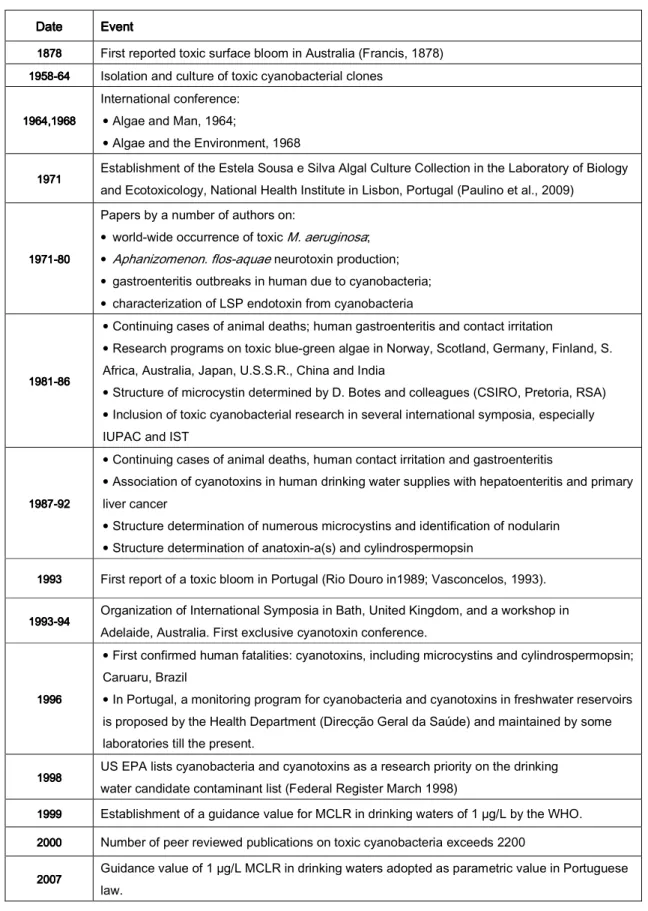
Table 1111 ---- Relevant dates and events in toxic cyanobacteria history (Adapted from Carmichael, 2002).
Date Date Date
Date EventEventEventEvent
1878 1878 1878
1878 First reported toxic surface bloom in Australia (Francis, 1878)
1958 19581958
1958----646464 64 Isolation and culture of toxic cyanobacterial clones
1964,1968 1964,19681964,1968 1964,1968
International conference: • Algae and Man, 1964;
• Algae and the Environment, 1968
1971 1971 1971
1971 Establishment of the Estela Sousa e Silva Algal Culture Collection in the Laboratory of Biology
and Ecotoxicology, National Health Institute in Lisbon, Portugal (Paulino et al., 2009)
1971 19711971 1971----808080 80
Papers by a number of authors on:
• world-wide occurrence of toxic M. aeruginosa; • Aphanizomenon. flos-aquae neurotoxin production; • gastroenteritis outbreaks in human due to cyanobacteria; • characterization of LSP endotoxin from cyanobacteria
1981 19811981 1981----868686 86
• Continuing cases of animal deaths; human gastroenteritis and contact irritation
• Research programs on toxic blue-green algae in Norway, Scotland, Germany, Finland, S. Africa, Australia, Japan, U.S.S.R., China and India
• Structure of microcystin determined by D. Botes and colleagues (CSIRO, Pretoria, RSA) • Inclusion of toxic cyanobacterial research in several international symposia, especially IUPAC and IST
1987 19871987 1987----929292 92
• Continuing cases of animal deaths, human contact irritation and gastroenteritis
• Association of cyanotoxins in human drinking water supplies with hepatoenteritis and primary liver cancer
• Structure determination of numerous microcystins and identification of nodularin • Structure determination of anatoxin-a(s) and cylindrospermopsin
1993 1993 1993
1993 First report of a toxic bloom in Portugal (Rio Douro in1989; Vasconcelos, 1993).
1993 19931993
1993----949494 94 Organization of International Symposia in Bath, United Kingdom, and a workshop in
Adelaide, Australia. First exclusive cyanotoxin conference.
1996 1996 1996 1996
• First confirmed human fatalities: cyanotoxins, including microcystins and cylindrospermopsin; Caruaru, Brazil
• In Portugal, a monitoring program for cyanobacteria and cyanotoxins in freshwater reservoirs is proposed by the Health Department (Direcção Geral da Saúde) and maintained by some laboratories till the present.
1998 1998 1998
1998 US EPA lists cyanobacteria and cyanotoxins as a research priority on the drinking
water candidate contaminant list (Federal Register March 1998)
1999 1999 1999
1999 Establishment of a guidance value for MCLR in drinking waters of 1 µg/L by the WHO.
2000 2000 2000
2000 Number of peer reviewed publications on toxic cyanobacteria exceeds 2200
2007 2007 2007
2007 Guidance value of 1 µg/L MCLR in drinking waters adopted as parametric value in Portuguese
Comparative study of the cytotoxic effects of MCLR in mammalian cell lines
5 human activity. This demonstrates the cyanobacterial remarkable capacity of adaptation to environmental modifications.
So far, as many as 40 species of cyanobacteria are known to be toxin producers. Therefore bloom formation is often accompanied by the release of toxins when toxic cyanobacterial species are present (Carmichael, 1992; Chorus and Bartram, 1999; Falconer, 2005).
Table 1 summarizes some relevant dates concerning toxic cyanobacteria history.
2. 2. 2.
2. CyCyCyanotoxinsCyanotoxinsanotoxinsanotoxins
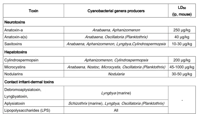
Cyanotoxins are secondary metabolites produced by freshwater cyanobacteria that can be classified functionally into neurotoxins, hepatotoxins and dermotoxins or accordingly to its chemical structure into alkaloids, cyclic peptides and lipopolysaccharides (Carmichael, 1992; Hitzfield et al., 2000; Codd, 2000; Chorus et al., 2000; Oberholster et al., 2004; Falconer, 2005). Cyanotoxins are produced by a variety of cyanobacterial genera/species and have distinct toxicities as summarized in table 2.
Table Table Table
Table 2222 ---- Types of cyanotoxins, toxic cyanobacterial genera and acute toxicity (Adapted from Chorus and Bartram, 1999; Chorus et al., 2000; Carmichael, 2002).
Neurotoxins are produced by species of Anabaena, Aphanizomenon, Oscillatoria and
Lyngbya and include the alkaloids anatoxin-a and saxitoxin and also the naturally occurring organophosphate anatoxin-a(s) (Carmichael, 1992). Anatoxin-a is an acetylcholine agonist and anatoxin-a(s) is an inhibitor of acetylcholinesterase and both induce continuous muscle
Toxin Toxin Toxin
Toxin Cyanobacterial genera producersCyanobacterial genera producers Cyanobacterial genera producersCyanobacterial genera producers LDLDLDLD50 50 50 50 (ip, mouse) (ip, mouse)(ip, mouse) (ip, mouse) Neurotoxins
NeurotoxinsNeurotoxins Neurotoxins
Anatoxin-a Anabaena, Aphanizomenon 250 μg/kg Anatoxin-a(s) Anabaena, Oscillatoria (Planktothrix) 40 μg/kg Saxitoxins Anabaena, Aphanizomenon, Lyngbya,Cylindrospermopsis 10-30 μg/kg Hepatotoxins
HepatotoxinsHepatotoxins Hepatotoxins
Cylindrospermopsin Aphanizomenon, Cylindrospermopsis 200 μg/kg Microcystins Anabaena, Nostoc, Microcystis, Oscillatoria (Planktothrix) 45-1000 μg/kg
Nodularins Nodularia 30-50 μg/kg
Contact irritant Contact irritantContact irritant
Contact irritant----dermal toxinsdermal toxinsdermal toxins dermal toxins Debromoaplysiatoxin,
Lyngbyatoxin, Lyngbya (marine)
-
Aplysiatoxin Schizothrix (marine), Lyngbya, Oscillatoria (Planktothrix)
Introduction
6
stimulation leading to paralysis and death within minutes or hours due to respiratory arrest (Vasconcelos, 2001). Saxitoxin is a sodium ion channels blocker that leads to the inhibition of skeletal muscle and peripheral nerves stimulation with consequent death by respiratory arrest (Vasconcelos, 2001; Carmichael, 1992).
Lipopolysaccharides are components of the cellular wall outer layers of all cyanobacterial cells and may act as dermotoxins producing irritations when in direct contact with the skin (Codd, 2000).
Hepatotoxins include nodularin produced by Nodularia, cylindrospermopsin produced by Cylindrospermopsis and microcystins produced by Microcystis, Anabaena, Oscillatoria
and Nostoc (Chorus and Bartram, 1999; Codd, 2000). Microcystins are the most frequently occurring cyanotoxins and these cyclic heptapeptides were first isolated from Microcystis aeruginosa from which the toxins take their name (Carmichael et al., 1988; Chorus and Bartram, 1999).
Cyanotoxins are produced inside the cyanobacterial cells and the release into the surrounding water occurs mainly during cell senescence, cell death or algicide application. Once in the water, they can persist for several days to weeks, constituting a threat even after the bloom as passed (Duy et al., 2000). However, photochemical breakdown in full sunlight can occur with varying rates. Anatoxins are rapidly degraded while cylindrospermopsins and microcystins are rapidly degraded in the presence of cyanobacterial pigments (Chorus and Bartram, 1999). Additionally, aquatic bacteria commonly found in the environment are also responsible for microcystins degradation and clearance from the environment (Jones et al., 1993; Chorus and Bartram, 1999).
3. 3. 3.
3. MicrocystinsMicrocystinsMicrocystinsMicrocystins 3.1.
3.1. 3.1.
3.1. Chemical structureChemical structureChemical structure Chemical structure
Microcystins are heptapeptides with a general structure composed of five amino acids, D-Alanine, D-Methylaspartic acid, D-Glutamic acid, N-Methyldehydroalanine and a side chain with the specific ADDA amino acid ((2S,35’,8S,9S)-3-amino-9-methoxy-2,6,8-trimethyl-10-phenyl-(4E),(6E)-decadienoic acid) (positions 1, 3, 6, 7 and 5 in figure 1A respectively), responsible for the biological activity of these cyclic peptides (Carmichael, 1992). The two variable amino acids are responsible for the multiplicity of microcystins variants (X and Z in positions 2 and 4 in figure 1A). To date 70 microcystin variants are known with different hydrophobic/hydrophilic properties as well as different degrees of toxicity, with a LD50 (lethal dose to 50% of the treated animals) in intraperitoneal (ip)
administered mice of 45-1000 μg.kg-1 body weight (Carmichael, 1992; WHO, 1998; Chorus et
Comparative study of the cytotoxic effects of MCLR in mammalian cell lines
7 Figure
Figure Figure
Figure 1111 ---- Chemical structure of microcystins (adapted from Zurawell et al., 2004). 3.2. O
3.2. O 3.2. O
3.2. Occurrenceccurrenceccurrence in the environmentccurrencein the environmentin the environmentin the environment
Microcystins have attracted attention because of their health effects, wide occurrence and persistence in the environment (Figure 2). From the cyanobacterial samples investigated worldwide, it was estimated that 75% contained toxins, of which microcystin-LR (MCLR) was the most commonly found (WHO, 1998). In Portugal, analysis of cyanobacterial samples collected between 1989 and 1992 concluded that MCLR was the most common microcystin variant and its proportion in each sample ranged from 45.5% to 99.8% of the total microcystin contents (Vasconcelos et al., 1996). In a recent Portuguese study (Galvão et al., 2008), microcystins were detected in 23% of the water samples analyzed (n=51).
Figure Figure Figure
Introduction
8
3.3. Human 3.3. Human 3.3. Human
3.3. Human intoxication intoxication intoxication intoxication
Human intoxication can occur through several forms: inhalation of cyanobacterial cells, ingestion of contaminated water from a drinking source or during recreational activities, ingestion of contaminated aquatic organisms, direct skin contact with cyanobacterial blooms and hemodialysis (Chorus et al., 2000; Hitzfield et al. 2000; Benson et al., 2005). The consumption of contaminated food supplies is possible due to the accumulation and transfer along the food chain. In fact, microcystins are known to be transferred through filter-feeding mollusks (such as the mussels), crayfish and fish used for human consumption (Vasconcelos, 1999).
Once ingested, microcystins are transported through the gastrointestinal tract (cell linings of the small intestine) by a specific bile acid transport system to the liver, the primary target of action (Falconer, 1999). The symptoms of acute microcystins intoxication are vomiting and diarrhea, severe liver damage which is characterized by a disruption of liver cell structure, loss of sinusoidal structure, increase in liver weight due to intra-hepatic hemorrhage, heart failure and death (Chorus and Bartram, 1999; Falconer, 2005). Some of these signs can be commonly mistaken with food poisoning, making it difficult to assess microcystins intoxication. When high quantities of toxin are ingested, renal failure may also occur that, in most severe cases, can lead to death (Chorus et al., 2000). One of the most studied and recognized case of human intoxication by cyanotoxins was the Caruaru syndrome which occurred in Brazil in 1996. This incident was caused by the use of cyanotoxins-contaminated water in a dialysis clinic and resulted in 76 deaths, of which 52 were attributed to cyanotoxins (Jochimsen et al., 1998; Charmichael et al., 2001; Azevedo et al., 2002). However, reported cases of livestock and human acute intoxications have multiplied over the past few years (Griffiths and Saker, 2003; Dittman and Wiegand, 2006).
3.4. 3.4. 3.4.
3.4. MicrocystinMicrocystinMicrocystinMicrocystin----LRLRLRLR
3.4.1. Chemical p 3.4.1. Chemical p 3.4.1. Chemical p
3.4.1. Chemical properties. Toxin uptake, distribution and elimination.roperties. Toxin uptake, distribution and elimination.roperties. Toxin uptake, distribution and elimination.roperties. Toxin uptake, distribution and elimination.
MCLR is the most extensive studied and characterized microcystin. It is the most common and toxic variant, exhibiting an i.p. LD50 of 50 μg.kg-1 body weight in mice (WHO,
1998). The two variable amino acids in MCLR are leucine (L) and arginine (R) in positions 2 and 4 respectively (Figure 1). These amino acids provide hydrophilic properties to the molecule which is extremely stable at high temperatures and resistant to hydrolysis (Oberholster et al., 2004). This last property enables the toxin to cross through the peptidases in the stomach and the passage of significative amounts of MCLR to the blood stream in an oral intoxication scenario (Chorus and Bartram, 1999). Due to MCLR large size (1000 dalton) and hydrophilic nature, organic anion transporting polypeptides (OATPs) are
Comparative study of the cytotoxic effects of MCLR in mammalian cell lines
9 required for active uptake of MCLR into the cells (Fisher et al., 2005; Feurstein et al., 2009). Distribution studies of MCLR injected ip in mice revealed a major accumulation of this toxin in the liver (50-70%) followed by the kidneys and intestine responsible for MCLR excretion (Rao et al., 2005). These are considered as the major targets of MCLR toxicity (Wang et al., 2008). 3.4.2. Regulatory aspects 3.4.2. Regulatory aspects 3.4.2. Regulatory aspects 3.4.2. Regulatory aspects
A reference value of 1 μg.L-1 of MCLR in drinking water was proposed by WHO in
1998, calculated from the 0.04 μg/kg of body weight TDI value (Tolerable Daily Intake) obtained from sub-chronic studies of oral administration of MCLR in mice (Fawell et al., 1994). This provisional guideline was adopted by many countries as a parametric reference value in their national laws on water. In Portugal this value is included in Decreto-Lei 306/2007 de 27 de Agosto. Although this is a measure that may prevent acute human exposure to MCLR, the implications of low-level chronic exposure to microcystins are not yet known. Nevertheless, the International Agency for Cancer Research classified MCLR as a Group 2 compound (i.e. probably carcinogenic to humans; IARC, 2006).
3.4.3. 3.4.3. 3.4.3.
3.4.3. CarcinogenCarcinogenCarcinogenic potCarcinogenic potic potentialic potentialentialential
Epidemiological studies associated the exposure to microcystins-contaminated water and the occurrence of primary liver cancer (Ueno et al., 1996) or colorectal cancer (Zhou et al., 2002). Besides, evidences of tumour promotion activity induced by MCLR were reported in the liver (Hu et al., 2008), skin (Falconer, 1991) and colon (Humpage et al., 2000) of rodents previously treated with tumour initiators, although the exact mechanism in not known so far. Also, the formation of neoplastic nodules in mouse liver without the previous exposition to an initiator agent has been reported in mice (Ito et al., 1997) suggesting a possible carcinogenic action of MCLR. Furthermore, the ability of MCLR to induce DNA alterations appears to be dependent on the cell type and the toxin exposure concentration, making the genotoxicity properties of MCLR somewhat controversial. DNA damages have been reported in vivo in the liver (Rao e Bhattacharya, 1996; Rao et al., 1998; Gaudin et al, 2008), in cultured hepatocytes (Ding et al., 1999; Žegura et al, 2003, 2004, 2006; Nong et al., 2007) and in other cell types (Lankoff et al., 2004; Žegura et al, 2008). However, DNA adducts formation, considered as an indicator of pre-mutagenic lesion, was not induced by MCLR exposure in rat hepatocytes (Bouaïcha et al., 2005). This suggests the existence of an indirect genotoxic mechanism such as the MCLR-induced oxidative stress, rather than direct genotoxic action (Žegura et al., 2003, 2004, 2006 2008; Nong et al., 2007). Additionally, Lankoff et al. (2004) suggested that DNA fragmentation could be a consequence of apoptosis rather than a genotoxic effect.
Introduction
10
3.4.4. 3.4.4. 3.4.4.
3.4.4. Cellular Cellular Cellular targets and Cellular targets and targets and targets and mechanisms mechanisms mechanisms mechanisms of toxicityof toxicityof toxicityof toxicity
Microcystins hepatotoxicity is mediated through the inhibition of protein phosphatases 1 and 2A (Honkanen et al., 1990; Mackintosh et al., 1990). These protein phosphatases are responsible for the phosphorylation of key proteins and their inhibition can lead to the hyperphosphorylation of cytoskeletal proteins with the consequent hepatocyte deformation, membrane blebbing, cell shrinkage and rounding, chromatin condensation, and organelle redistribution, classical signs of an apoptotic process (Fladmark et al., 1999; Batista et al., 2003; Herfindal and Selheim, 2006). Necrosis may occur as well (Khan et al., 1995; Hitzfield et al, 2000; Trinkle-Mulcahy and Lamond, 2006).
Traditionally, mitochondria are considered as the main intracellular target of MCLR and several reports have considered it as a central executioner of MCLR-induced apoptosis in hepatic cells (Ding et al., 1998, 2000a, 2000b; Ding and Ong, 2003). The events that take place in this mitochondrial apoptotic pathway include the formation of reactive oxygen species (ROS), decrease of the mitochondrial membrane potential (MMP), membrane depolarization and mitochondrial permeability transition (MPT) pore aperture, with the release of Ca2+ and citochrome-c to the cytoplasm (Ding et al., 1998, 2001; Ding and Ong,
2003; Žegura et al., 2004; Nong et al., 2007; Weng et al., 2007). Other mechanisms such as caspases and calpain activation (Fladmark et al., 1999), alterations in the expression of pro-apoptotic and anti-pro-apoptotic Bcl-2 family proteins and p53 gene are also involved in MCLR-induced apoptotic pathway (Fu et al., 2005; Weng et al., 2007; Billam et al., 2008). The involvement of oxidative stress in MCLR-mediated toxicity have been demonstrated by the decrease of reduced glutathione (GSH) and also of antioxidant enzymes such as glutathione peroxidase, glutathione reductase, superoxide dismutase and catalase, as well by the increase of lipid peroxidation in response to MCLR exposure (Moreno et al., 2005; Andrinolo et al., 2008). GSH is a major antioxidant that can act as a free radical scavenger or as an important conjugate of MCLR in detoxifying pathways (Ding et al., 2000a). Its depletion is often accompanied by ROS generation and increased cell susceptibility to MCLR-induced cytotoxicity (Ding and Ong, 2003). These effects altogether are relatively well studied in mice liver in vivo (Fawell et al., 1994; Yoshida et al., 1997), in primary cultured rat hepatocytes (Khan et al., 1995; Toivola et al., 1997; Ding et al., 2000b; Mankiewicz et al., 2001; Moreno et al., 2005), human hepatocytes (Batista et al., 2003) and fish hepatocytes (Fisher et al., 2000; Malbrouck et al., 2003; Boaru et al., 2006). However, sensitivity differences to MCLR are reported to exist between rat and human primary cell hepatocytes cultures (Batista et al., 2003). Additionally, comparison of the sensitivity of primary and permanent cell lines to MCLR revealed a 100-fold difference in the concentrations of MCLR necessary to obtain similar effects (Khan et al., 1995; Wickstrom et al., 1995; McDermott et al., 1998).
Comparative study of the cytotoxic effects of MCLR in mammalian cell lines
11 Furthermore, there is a higher sensitivity to MCLR in hepatic permanent cell lines than non-hepatic permanent cell lines (Chong et al., 2000)
This may be due to the fact that non-liver cells and permanent cell lines exhibit a lower expression, or even loss, of OATP during the process of cell culture. In fact, the transfection of OATPs genes into MCLR insensible permanent cell lines induced cytotoxic effects after toxin exposure (Monks et al., 2007).
3.4.5. Effects of MCLR in non 3.4.5. Effects of MCLR in non 3.4.5. Effects of MCLR in non
3.4.5. Effects of MCLR in non----liver celiver celiver celiver cellsllslls lls
Although MCLR is considered primarily as a hepatotoxin, there have been also recognized effects in non-hepatic cells. However, these appear to be less severe than in hepatocytes, as reported in kidney and intestine cells (Ito et al., 2001; Atencio et al., 2008; Wang et al., 2008).
Nephrotoxicity has been suggested to be a consequence of MCLR accumulation (Wang et al., 2008) and elimination (Nobre et al., 1999) in the kidneys. In fact, the kidneys are responsible for the partial excretion of microcystins in the organism (Robinson et al., 1991) and expression of OATPs was demonstrated in kidney in vivo (Hagenbuch et al., 2003). The nephrotoxic activity of MCLR was characterized by alterations in renal function and antioxidant enzyme activity reported in vivo in rodents (Nobre et al., 1999; Milutinović et al., 2003; Moreno et al., 2005; Andrinolo et al., 2008). Additionally, Milutinović et al (2003) detected renal alterations equivalent to hepatotoxicity damages (cytoskeletal disruption, apoptosis and necrosis) in kidneys of rats treated with low concentrations of MCLR for 8 months.
Enterotoxicity is also a possible effect of MCLR. The entero-hepatic recirculation of MCLR leads to a reintroduction of the toxin into the small intestine after being transported to the liver (Falconer et al., 1992). Following MCLR exposure, intestinal secretion was observed in rats (Nobre et al., 2004) and enterocyte injury in chickens (Falconer et al., 1992). Additionally, enterocyte apoptosis is reported as well as the presence of MCLR in the villi of mice treated with 75% LD50 MCLR for 24h (Botha et al., 2004b). These observations may
account for the gastroenteritis symptoms observed in human intoxications by MCLR and confirm that the intestine is indeed a target of MCLR.
The mechanisms mentioned above (section 3.4.4) for liver cells were also described in the kidney in vivo (Moreno et al., 2005), in kidney mitochondria isolates (La-Sallete et al., 2008), human lymphocytes (Mankiewicz et al., 2001; Lankoff et al., 2004) and some permanent cell lines (McDermott et al., 1998; Lankoff et al., 2003; Žegura et al., 2008).
In a previous study with the monkey kidney Vero-E6 cell line it was suggested that the ER could be a primary target of MCLR, and that it seemed to be involved in the triggering of autophagy at subcytotoxic MCLR concentrations (Alverca et al., 2009). The same authors
Introduction
12
also suggested that this was a cell survival mechanism adopted to eliminate the toxin or/and the MCLR-induced cellular damages. Additionally, lysosomal injuries were shown to occur previously to mitochondrial impairment, contrary to what is the most frequently reported in the literature. In this context, and to better understand the underlying mechanisms of MCLR toxicity, it is of extreme importance to establish if the toxin effects and cellular response mechanisms reported, are transversal to cells from other MCLR-target organs, or on the contrary, are specific to this cell line.
4. 4. 4.
4. ObjectivesObjectivesObjectives Objectives
The cellular mechanisms by which MCLR induces its effects are still not completely understood, particularly in non-liver cells. This study follows the previous work performed in the Vero cell line and tests the hypothesis of an organelle cross-talk responsible for the effects of MCLR, involving the ER, lysosomes and autophagy. For that purpose, in vitro
models of the main target organs of toxin accumulation/elimination were chosen for comparison of toxic responses: HepG2 (human liver), Vero-E6 (monkey kidney), MDCK (dog kidney) and CaCo2 (human intestine).
The primary aims of this work are:
1) Compare the sensitivity of the permanent cell lines HepG2 (liver), Vero-E6 and MDCK (kidney) and CaCo2 (intestine) to MCLR.
2) Evaluate cytotoxic and morphological MCLR-induced damages and establish a dose/response relation.
3) Detect intracellular targets of MCLR through fluorescence and transmission electronic microscopy (TEM) observation of lysosomes, mitochondria, nucleus, plasma membrane, endoplasmic reticulum and cytoskeleton.
M
M
M
Comparative study of the cytotoxic effects of MCLR in mammalian cell lines
15 1.
1. 1.
1. MCLR: sources and preparation of stock solutionsMCLR: sources and preparation of stock solutionsMCLR: sources and preparation of stock solutionsMCLR: sources and preparation of stock solutions
Two sources of MCLR were used in this work: semi-purified extracts from a MCLR-producerstrain of Microcystis aeruginosa (Kützing) and commercial MCLR (named hereafter as pure MCLR). A study of MCLR effects at the concentrations tested in the present work implies the use of large amounts of toxin with a very high cost. For this reason, MCLR semi-purified extract obtained from a M. aeruginosa strain was used in all experiments. In parallel, cytotoxity assays with pure MCLR and an extract from a non-toxic M. aeruginosa strain were used as positive and negative controls, respectively.
1.1. 1.1. 1.1.
1.1. SemiSemiSemi----puriSemipuripuripurified extractsfied extracts from fied extractsfied extractsfrom from from M. aeruginosaM. aeruginosaM. aeruginosaM. aeruginosa
Strains of Microcystis aeruginosa used in this study were isolated in 2000 from Montargil reservoir (Portugal) and have since then been successfully maintained in laboratory conditions (Valério et al., 2009a), within the “Estela Sousa e Silva Algal Culture Collection” of the Laboratory of Biology and Ecotoxicology, National Health Institute Dr. Ricardo Jorge. MCLR was purified from extracts of large-scale cultures of a strain of
Microcystis aeruginosa (LMECYA 110) characterized as a producer of the MCLR variant of microcystins (Valério et al., 2009b). A non-toxic M. aeruginosa strain (LMECYA 127) was also used with biomass1 concentration equivalent to the biomass concentration of LMECYA
110 strain. This non-toxic LMECYA 127 extract was prepared with the same protocol used for LMECYA 110 extract and provided a negative control. Cultures of LMECYA 110 and LMECYA 127 were grown in plankton light reactors (Aqua-Medic, Bissendorf, Germany) with 2.5L of Z8 medium (Skulberg and Skulberg, 1990) under continuous aeration, at a light intensity of 40 µE.m-2.s-1 and in a 14h/10h L/D cycle at 22 ± 1oC.
Cells harvested during late exponential growth phase were lyophilized in a freeze drier (Micromodul Y10, Savant, NY, USA) and extracted with 75% methanol (10 mL/100 mg dry weight) overnight at 4oC under magnetic stirring. The extracts were further sonicated with
an ultrasonic probe (Sonics Vibra-Cell CV33, Sonics & Materials Inc., CA, USA), centrifuged and submitted to rotary evaporation at 35oC (Buchi-R, Flawil, Switzerland) to eliminate the
alcoholic fraction. The resulting aqueous extracts were cleaned-up by solid phase extraction on Sep-Pak C18 cartridges (500 mg Waters, Massachusetts, USA) previously conditioned with 20 mL of ethanol and equilibrated with 20 mL of distilled water. The microcystin-LR containing fraction (and the correspondent fraction of the LMECYA 127 extract) was eluted with methanol at 80% (v/v) and evaporated to dryness. The solid residue was ressuspended in 25 mM acetic acid and manually injected into a Bio-Gel P2 (40–90 mm, Bio-Rad Inc., CA,
1
Biomass is considered as the total mass of cyanobacterial cells lyophilized present in the extract and is expressed as mg of dry weight per mL.
Materials and Methods
16
USA) packed preparative column (Amersham Biosciences, XK 26/40, i.d./length). The mobile phase consisted of 25 mM acetic acid, and the flow rate was set at 1 mL.min-1 (Knauer
Well-Chrom K-120 pumps, Germany). Elution fractions (5 mL) were collected on a fraction collector (Bio-Rad Mod. 2110, CA, USA) and analyzed by HPLC-DAD according to the ISO standard method 20179. The concentration of MCLR on the extracts was determined by fitting the area of the corresponding HPLC-DAD peak to the MCLR calibration curve. The later was constructed by analyzing commercially available MCLR standards (Alexis Biochemicals, CA, SA). MCLR-containing fractions (and the correspondent LMECYA 127 fractions) were evaporated to dryness and ressuspended in bi-distilled water. The final extracts were sterilized by filtration on 0.22 µm syringe filters and kept at -20oC until use. The
concentration of MCLR on the final extracts was reanalyzed by HPLC-DAD to contemplate eventual toxin losses during ressuspension and filtration procedures. Extract-work solutions were prepared by serial dilutions of stock extracts in fresh cell line culture medium.
1.2. 1.2. 1.2.
1.2. Pure Pure Pure Pure commercial commercial commercial commercial MicrocystinMicrocystinMicrocystinMicrocystin----LRLRLRLR
Microcystin-LR was purchased from Sigma–Aldrich (CAS Number 101043-37-2) as a white solid film (purity 95%, by HPLC). A stock solution of pure MCLR (1 mM) was prepared by dissolving the toxin in growth medium without Fetal Bovine Serum (FBS) and kept at -20
oC until use.
2. 2. 2.
2. Mammalian cell liMammalian cell liMammalian cell liMammalian cell lines and culture conditions nes and culture conditions nes and culture conditions nes and culture conditions
2.1. 2.1. 2.1.
2.1. Cell lines maintenance Cell lines maintenance Cell lines maintenance Cell lines maintenance
In this work four mammalian cell lines were used derived from liver, kidney and intestine. The human hepatoma HepG2 cell line (DSMZ ACC 180) has an epithelial morphology and was grown in Dulbecco´s Modified Eagle Medium (DMEM) supplemented with 10% FBS. The Vero-E6 cell line, is a clone of kidney epithelial cells from the African green monkey Cercopithecus aethiops (ATCC-CRL 1586) that has fibroblast morphology and was maintained in Modified Eagle Medium (MEM) supplemented with 10% FBS, 1 mM sodium pyruvate and 0.1mM non-essencial aminoacids. The MDCK cell line (ATCC CCL-34) was isolated from a cocker spaniel dog kidney, has an epithelial morphology and was maintained in MEM supplemented with 10% FBS, 1mM sodium pyruvate and 0.1 mM non-essencial aminoacids. The CaCo2 permanent cell line was established from a human intestine carcinoma (ATCC HTB-37). It has an epithelial morphology and it was maintained in RPMI 1640 medium supplemented with 10% FBS.
These cell lines are anchorage-dependent and their growth is inhibited by cell contact. Cell propagation was performed twice a week to dilute the cells into new cell culture
Comparative study of the cytotoxic effects of MCLR in mammalian cell lines
17 flasks. Cell monolayers with a degree of about 70% confluence were trypsinized (0.05% trypsin-EDTA) for a few minutes and ressuspended in fresh growth medium pre-warmed to 37oC to prevent temperature shock. A small aliquot of the cellular suspension was
transferred to new flasks (Orange Scientific, Belgium) containing fresh growth medium at 37oC. In CaCo2 cells, growth medium was replaced between propagations. All cell lines were
maintained in a 5% CO2 humidified incubator at 37ºC.
Frozen stocks of the four cell types were maintained throughout this study. For this purpose, cells in exponential growth phase, were trypsinized and ressuspended in fresh growthmedium at 37oC. After centrifugation, the supernatant was discarded and the cellular
pellet ressuspended in 1-1,5 mL of freezing medium (FBS + 10% DMSO) and transferred to criotubes (Nunc, Roskilde, Denmark). These were maintained at -80oC. All media and
supplements were purchased from Invitrogen (Paisley, UK).
2.2. 2.2. 2.2.
2.2. Cell inoculationCell inoculationCell inoculationCell inoculation and exposure to MCLRand exposure to MCLRand exposure to MCLR and exposure to MCLR
The effects of MCLR on cell viability and cellular ultrastructure were evaluated in all four cell lines. Due to time constrains, and according to the sensitivity of each cell line to the toxin (see section 3.1.) all the remaining procedures were conducted only in HepG2 and Vero cell lines. Cells in exponential growth phase were trypsinized and counted in a haemocytometer by the trypan blue exclusion method (Philips, 1973). HepG2, Vero, MDCK and Caco2 cells were seeded in 96 well plates (Sarstedt, Newton, USA) for the cytotoxicity assay (1x10⁴ cells per well) and in 6 well plates (Nunc, Roskilde, Denmark) for electron
microscopy analysis (1x10⁵ cells per well). For fluorescence microscopy studies HepG2 and
Vero cells (3.5x10⁵) were seeded in 4 well plates (Nunc, Roskilde, Denmark) over sterile
coverslips (Marienfeld, Lauda-Königshofen, Germany). For Western Blot analysis HepG2 (2x10⁶) and Vero (1x10⁶) cells were inoculated in 6-well plates.
After 24h incubation for cell adherence and growth, the growth medium was removed and serial dilutions of pure MCLR and MCLR extract in fresh medium containing 1.6 to 100 µM of MCLR were added to each cell line culture for cytotoxicity tests. TEM analysis was performed with MCLR extract in the concentrations of 6 to 100 µM of MCLR. In parallel, similar biomass dilutions of LMECYA127 extract (0.25 to 16 mg dw.ml-1) were also tested by
both methods to exclude any cyanobacterial matrix effects. For the fluorescence microscopy and Western Blot experiences, MCLR extract concentrations used were 6, 12, 25 and 50 µM. For each assay, the negative control consisted of cells growing in the corresponding growth medium.
Materials and Methods
18 Table Table Table
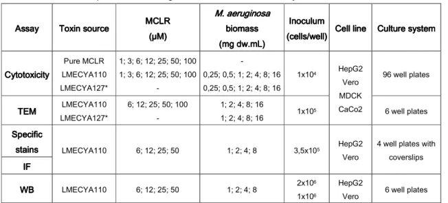
Table 3333 ---- Outline of experimental setting conditions used in this study. Assay
AssayAssay
Assay ToxinToxin sourceToxinToxinsourcesource source MCLRMCLRMCLRMCLR (µM) (µM)(µM) (µM) M. aerug M. aerug M. aerug M. aeruginosa inosa inosa inosa
bbbbiomassiomassiomass iomass (mg dw.mL) (mg dw.mL)(mg dw.mL) (mg dw.mL) Inoculum Inoculum Inoculum Inoculum (cells/well) (cells/well) (cells/well)
(cells/well) Cell lineCell line Culture systemCell lineCell line Culture systemCulture systemCulture system
Cytotoxicity Cytotoxicity Cytotoxicity Cytotoxicity Pure MCLR LMECYA110 LMECYA127* 1; 3; 6; 12; 25; 50; 100 1; 3; 6; 12; 25; 50; 100 - - 0,25; 0,5; 1; 2; 4; 8; 16 0,25; 0,5; 1; 2; 4; 8; 16 1x10⁴ HepG2 Vero MDCK CaCo2 96 well plates TEM TEMTEM TEM LMECYA110 LMECYA127* 6; 12; 25; 50; 100 - 1; 2; 4; 8; 16 1; 2; 4; 8; 16 1x10⁵ 6 well plates Specific SpecificSpecific Specific stains stains stains
stains LMECYA110 6; 12; 25; 50 1; 2; 4; 8 3,5x10⁵ HepG2
Vero
4 well plates with coverslips IF IF IF IF WB WBWB WB LMECYA110 6; 12; 25; 50 1; 2; 4; 8 2x10⁶ 1x10⁶ HepG2
Vero 6 well plates
*LMECYA127 was used as a negative control of LMECYA110. 3.
3. 3.
3. Evaluation of MCLR effectsEvaluation of MCLR effectsEvaluation of MCLR effectsEvaluation of MCLR effects
3.1. 3.1. 3.1.
3.1. Cytotoxicity assaysCytotoxicity assaysCytotoxicity assaysCytotoxicity assays
The Neutral Red (NR) cell viability assay was performed in order to compare the sensitivity of the different mammalian cell lines to MCLR, to access the respective cytotoxic thresholds and the toxin concentration range used in further experiments, in order to contemplate non-cytotoxic, low and moderately cytotoxic concentrations. The NR test was chosen because it was previously demonstrated as one of the most sensitive viability assays to evaluate the MCLR toxicity in in vitro cell cultures (Pichardo et al., 2005; Bouaru et al, 2006; Alverca et al., 2009; Dias et al., 2009). This assay measures the selective intake and retention of the NR dye by the lysossomes of viable cells by opposition to non-viable cells that cannot retain the NR due to lysosomal membrane damage (Borenfreud and Puerner, 1985). The degree of absorbance measured is correlated with cell viability.
The assay was performed according to the protocol that follows (adapted from Borenfreud and Puerner, 1985).
Neutral Red Assay Protocol:
1. Replacement of growth medium by fresh medium (100 µL).
2. Addition of 10 µL of NR solution (at 50 µg.ml-1, Merk, Darmstad, Germany) and incubation
for 3h at 37oC.
3. Washing 2x with PBS.
4. Addition of a solution of ethanol:acetic acid:water (50:1:49) to each well to extract the NR from the lysosomes.
Comparative study of the cytotoxic effects of MCLR in mammalian cell lines
19 5. Shaking for 15´ and absorbance reading at 540nm (Multiscan Ascent spectrophotometer,
Labsystems, Helsinki, Finland).
The cell viability assays were performed in triplicate and the results are presented as the mean ± standard deviation (%) of the three replicates relative to the control. An arbitrary threshold of 50% cell viability was considered as an indicator of a marked cytotoxic effect. Values were analyzed with the t-student´s test; p <0.05 was considered as a statistically significant difference.
3.2. 3.2.3.2.
3.2. Specific Labelings of cellular organellesSpecific Labelings of cellular organellesSpecific Labelings of cellular organellesSpecific Labelings of cellular organelles
3.2.1. 3.2.1.3.2.1.
3.2.1. Acridine OrangeAcridine OrangeAcridine OrangeAcridine Orange
Acridine Orange (AO) is a metachromatic dye and a week base that, at low concentrations easily enters the lysosomal membrane of live cells, where it accumulates as it becomes protonated. The interaction with the acidic content results in a red coloration visible at the fluorescence microscope under blue light (Lovelace e Cahill, 2007). To assess the toxic effect induced by MCLR in the lysosomes, HepG2 and Vero cells were stained with AO accordingly to the protocol described underneath. HepG2 cells were observed in a confocal microscope (Leica TCS-SPE) while observations of Vero cells were performed under an Olympus BX60 fluorescence microscope (λ=487 nm) coupled with a CCD camera (Olympus DP11).
Acridine Orange staining protocol:
1. Removal of growth medium and washing for 5’ with PBS (pH 7.4) pre-warmed at 37ºC. 2. Incubation of coverslips with AO solution (10 μM in growth medium; Invitrogen), 10’, in the
dark at 37ºC.
3. Rinsing vigorously for 5’ with PBS, 37ºC. 4. Mounting between coverslip and slide.
3.2.2. 3.2.2.3.2.2.
3.2.2. RhodamineRhodamineRhodamineRhodamine----123123123123
Rhodamine-123 (Rh-123) is commonly used to specifically stain mitochondria of live cells. This fluorescent probe has a cationic nature that interacts with the electronegative potential of the mitochondrial membrane conferring a green coloration to the mitochondria (Johnson et al, 1980). The toxic effects induced in the mitochondria of HepG2 and Vero cells treated with MCLR were assessed by staining with Rh-123 according to the protocol showed bellow. Observation was achieved in a confocal microscope (Leica TCS-SPE).
Materials and Methods
20
Rhodamine-123 staining protocol:
1. Removal of the growth medium and washing with fresh growth medium pre-warmed at 37ºC for 5’.
2. Incubation of the coverslips in Rh-123 solution (15 μg/ml in culture medium; Invitrogen), 10’, in the dark at 37ºC.
3. Washing vigorously with growth medium at 4ºC. 4. Mounting between coverslip and slide.
3.2.3. 3.2.3.3.2.3.
3.2.3. Phalloidin Phalloidin Phalloidin Phalloidin
For the visualization of the MCLR-induced cytoskeletal toxic effects, phalloidin was used as a fluorescence probe for actin cytoskeleton in HepG2 and Vero cells. This mycotoxin binds specifically to the junctions between the subunits of F-actin (Cooper, 1987) and visualization is achieved through conjugation with Alexa Fluor 488® (Invitrogen). The actin filaments staining was performed accordingly to the protocol described below and observation was performed in a confocal microscope (Leica TCS-SPE).
Phalloidin staining protocol:
1. Removal of the growth medium and washing for 5’ with PBS (pH 7.4) pre-warmed to 37ºC.
2. Fixation with 3.7% PFA in CBS (Cytoskeleton Buffer with 0,32M sucrose, pH 6.1), 15’, rt. 3. Washing 3X 5’ with PBS (pH 7.4), rt.
4. Permeabilization with 0.5% Triton X-100 in PBS, 3’. 5. Incubation with AbDil solution, 10’, rt.
6. Incubation with 25µM phalloidin diluted in AbDil, 1 h, in the dark, rt. 7. Washing for 5’ with 0.1% Triton X-100 in PBS.
8. Labeling with DAPI (0.5µg/ml), 5’, in the dark, rt. 9. Washing for 5’ with PBS, rt.
10. Mounting between coverslip and slide with Vectashield and sealing with nail polish.
3.3. 3.3.3.3.
3.3. Protein analysisProtein analysisProtein analysisProtein analysis 3.3.1.
3.3.1.3.3.1.
3.3.1. AnalysAnalysAnalysAnalysis of LC3B and GRPis of LC3B and GRPis of LC3B and GRPis of LC3B and GRP94 proteins by immunofluorescence94 proteins by immunofluorescence94 proteins by immunofluorescence 94 proteins by immunofluorescence
In this study, it was used an antibody against LC3B, a key protein of autophagy, for the visualization and study of the MCLR-induced autophagy. This protein is implicated in autophagosome formation and can occur in two forms: I, which is cytosolic and LC3B-II, associated with the inner and outer autophagosomal membrane (Kabeya et al., 2000).