UNIVERSIDADE DA BEIRA INTERIOR
Ciências da Saúde
Clinical Characterization of Parkinson's Disease
Patients Followed at CHCB
Comparison with a control group
Patrícia Valério dos Santos
Dissertação para obtenção do Grau de Mestre em
Medicina
(ciclo de estudos integrado)
Orientador: Prof. Doutor Graça Maria Fernandes Baltazar
Coorientador: Prof. Doutora Maria Luiza Rosado
Comparison with a control group
Acknowledgments
Apesar da solidão que caracteriza a redação de uma dissertação de mestrado, há contributos, ao longo do meu percurso, que não podem ser esquecidos.
À Profª. Doutora Graça Maria Fernandes Baltazar, minha orientadora, pelo acompanhamento e organização deste trabalho, pela disponibilidade e dedicação reveladas, sem os quais esta investigação não seria possível.
À Profª. Doutora Luiza Rosado, minha coorientadora, a quem agradeço sinceramente a disponibilidade, dedicação e apoio, essenciais à concretização deste trabalho.
À Dra. Andreia Monteiro, Dra. Débora Rodrigues e restantes profissionais do Laboratório de Patologia Clínica, cujo contributo foi indispensável.
À equipa médica de Neurologia do Centro Hospitalar da Cova da Beira, que contribuíram para o crescimento e enriquecimento deste trabalho.
Aos profissionais do sector 1 da Consulta Externa do Centro Hospitalar da Cova da Beira, pela disponibilidade e apoio prestados.
À equipa médica de Urologia e ao Dr. Humberto Gonçalves, pela disponibilidade e apoio concedidos à minha proposta.
À Rita Videira e restantes estudantes e profissionais do CICS, pelo incansável apoio numa área tão desconhecida para mim.
À Profª. Mafalda Fonseca e Profª. Olga Lourenço, pelo apoio crucial a este projeto.
Ao Mauro Sousa, por em todos os momentos me inspirar e motivar a ser e fazer sempre melhor.
À Vânia Pinto, pelos momentos passados juntos na construção do nosso futuro. À Mélina Lopes, pelo apoio incondicional nos momentos de maior desalento. Às minhas amigas, por acreditarem em mim.
À minha família pela motivação constante. A todos os participantes deste estudo.
Comparison with a control group
Abstract
Parkinson’s disease is the second most common neurodegenerative disease, characterized by four motor symptoms: rest tremor, bradykinesia, rigidity and postural instability. Diagnosis is made through strict clinical criteria, but clinical diagnosis is difficult in the early disease state and frequently does not allow a definitive diagnose. One hallmark of Parkinson’s disease is the death of the pigmented dopaminergic neurons of midbrain substantia nigra, with consequent loss of the neurotransmitter dopamine in the corpus striatum. The exact mechanisms underlying are still not understood and current theories include neuroinflammation and oxidative stress. Evidences suggest the presence of chronically activated microglia and several studies detected higher levels of their markers, namely cytokines and oxidative stress products. The main objectives of this study were to characterize CHCB’s Parkinson population, to evaluate inflammatory and oxidative markers and immunological status.
Medical records were evaluated for patients (n=38) and healthy controls (n=32). Patient’s motor symptoms at onset and at evaluation were determined. The inflammatory markers measured were C reactive protein, erythrocyte sedimentation rate and serum cytokines (interleukins 1β, 8, 6, 10, 12p70 and Tumor Necrosis Factor). Immunological status was accessed through leukogram. Urate was our oxidative marker.
The cytokines measurement reveals values below the lower limits of detection for all. C reactive protein was higher in patients, but erythrocyte sedimentation rate was similar between both groups. In leukogram analysis, we detected higher values of neutrophils in patients. However, lymphocytes were the only that decrease throughout the temporal evolution of the disease and in patients using higher doses of L-DOPA. We attempted to clarify the real cause of lymphocytes decrease. Although there was a tendency to observe a lower number of lymphocytes for higher doses of L-DOPA in all phases of temporal evolution of the disease, the difference was not statistically significant. In relation to urate, we obtained lower values in patients and the decrease was marked in the higher stages of the disease and longer disease duration.
Regarding inflammatory aspects of the disease, our study showed that cytokine pattern is not sensitive or specific to assess disease progression. On the other hand, the antioxidant urate presents us as a putative marker of disease severity.
Keywords
Biomarkers Cytokines Lymphocytes Oxidative stress Parkinson’s diseaseComparison with a control group
Resumo
A doença de Parkinson é a segunda doença neurodegenerativa mais comum, caracterizando-se por quatro sintomas motores: tremor de repouso, bradicinesia, rigidez e instabilidade postural. O diagnóstico é feito através de rigorosos critérios clínicos, mas o diagnóstico clínico é difícil na fase inicial da doença e, frequentemente, não permite o diagnóstico definitivo. A Doença de Parkinson consiste na morte dos neurónios dopaminérgicos pigmentados da
substantia nigra do mesencéfalo, com consequente perda do neurotransmissor dopamina no
corpo estriado. Os mecanismos exatos subjacentes ainda não são compreendidos e as teorias atuais incluem mecanismos inflamatórios e de stress oxidativo. Evidências sugerem a presença de microglia cronicamente ativada, com diversos estudos mostrando níveis elevados de marcadores da microglia, nomeadamente citoquinas e produtos de stress oxidativo. Os principais objetivos deste estudo foram caracterizar a população de doentes do CHCB, avaliar os seus marcadores inflamatórios e oxidativos e o seu estado imunológico.
Os processos clínicos foram avaliados para os doentes (n = 38) e controlos (n = 32). Os sintomas motores dos doentes no início da doença e no dia da avaliação foram registados. Os marcadores inflamatórios medidos foram a proteína-C-reativa, a velocidade de hemossedimentação e citoquinas séricas (interleucinas 1β, 8, 6, 10, 12p70 e Fator de Necrose Tumoral). O sistema imunológico foi avaliado através do leucograma e o ácido úrico foi o marcador oxidativo utilizado.
A medição das citoquinas revelou valores abaixo dos limites inferiores de deteção. A proteína-C-reativa foi superior nos doentes, mas a velocidade de hemossedimentação foi semelhante entre os grupos. Na análise do leucograma, detetamos valores superiores de neutrófilos nos doentes. No entanto, os linfócitos foram os únicos que diminuíram com a evolução temporal da doença e nos indivíduos que utilizavam maiores doses de L-DOPA. Na tentativa de esclarecer a verdadeira causa dessa diminuição, detetámos que, embora houvesse uma tendência a se observar um menor número de linfócitos para altas doses de L-DOPA em todas as fases de evolução temporal da doença, a diferença não foi estatisticamente significativa. Em relação ao ácido úrico, obtiveram-se valores menores nos doentes, sendo a diminuição superior nos estágios mais elevados e na maior duração da doença.
Relativamente aos aspetos inflamatórios da doença, os nossos resultados mostraram que o padrão de citoquinas não é sensível ou específico para avaliação da progressão da doença. Por outro lado, o ácido úrico apresenta-se como um possível marcador da severidade da doença.
Palavras-chave
Biomarcadores Citoquinas Doença de Parkinson Linfócitos Stress oxidativoComparison with a control group
Resumo alargado
A doença de Parkinson é a segunda doença neurodegenerativa mais comum, caracterizando-se por quatro sintomas motores: tremor de repouso, bradicinesia, rigidez e instabilidade postural. A existência de uma fase pré-motora deve orientar a busca de biomarcadores específicos e identificação de fatores de risco e de proteção. O diagnóstico é feito através de rigorosos critérios clínicos, mas o diagnóstico clínico é difícil na fase inicial da doença. A doença de Parkinson consiste na morte dos neurónios dopaminérgicos pigmentados da
substantia nigra pars compacta do mesencéfalo, com consequente perda do neurotransmissor
dopamina no corpo estriado, um dos núcleos subcorticais envolvidas no controlo do movimento. Os mecanismos exatos subjacentes ainda não são compreendidos e as teorias atuais incluem mecanismos inflamatórios e de stress oxidativo. Evidências sugerem a presença de microglia cronicamente ativada, com diversos estudos mostrando níveis elevados de marcadores da microglia, nomeadamente citoquinas e produtos de stress oxidativo. Os principais objetivos deste estudo foram caracterizar a população de doentes do CHCB, avaliar os seus marcadores inflamatórios e oxidativos e o seu estado imunológico.
Para todos os doentes de Parkinson (n=38), relativamente à clínica, foram registadas as seguintes variáveis: idade de início da doença, gravidade da doença (através da escala modificada de Hoehn & Yahr para estadiamento e dos anos de duração da doença), dose de L- DOPA de libertação imediata atualmente em utilização, sintomas inaugurais da doença e sintomas no dia da avaliação. Os processos clínicos foram avaliados para os doentes e controlos (n = 32). Os marcadores inflamatórios medidos foram a proteína-C-reativa, a velocidade de hemossedimentação e citoquinas séricas (interleucinas 1β, 8, 6, 10, 12p70 e Fator de Necrose Tumoral). O sistema imunológico foi avaliado através do leucograma e o ácido úrico foi o marcador oxidativo utilizado. O hemograma também foi incluído para uma melhor caracterização da população do estudo. As citoquinas no soro foram medidas através do kit BD™ CBA Human Inflammatory Cytokines, através de citometria de fluxo, no laboratório do Centro de Investigação em Ciências da Saúde (CICS), da Universidade da Beira Interior. Os restantes parâmetros analíticos foram obtidos através das análises de rotina prescritos pelo médico responsável pelo respetivo doente, no Hospital Pêro da Covilhã.
A nossa população de doentes é composta maioritariamente por homens, com média de idade de início da doença de 73,8 anos, sendo uma população com mais co morbilidades associadas, em comparação com os controlos. A maioria dos doentes apresentou tremor como sintoma inicial, mas a rigidez foi o mais prevalente na avaliação. A medição citoquinas revelou valores abaixo dos limites inferiores de deteção. A proteína-C-reativa foi superior nos doentes, mas a velocidade de hemossedimentação foi semelhante entre os grupos. Na análise do leucograma,
detetamos valores superiores de neutrófilos nos doentes. No entanto, os linfócitos foram os únicos que diminuíram com a evolução temporal da doença e nos indivíduos que utilizavam maiores doses de L-DOPA. Na tentativa de esclarecer a verdadeira causa dessa diminuição, detetámos que, embora houvesse uma tendência a se observar um menor número de linfócitos para altas doses de L-DOPA em todas as fases de evolução temporal da doença, a diferença não foi estatisticamente significativa. Em relação ao ácido úrico, obtiveram-se valores menores nos doentes, especialmente para a população masculina, sendo a diminuição superior nos estágios mais elevados e na maior duração da doença. Por último, o hemograma mostrou níveis de hemoglobina mais baixos para os doentes, principalmente na população feminina.
Relativamente aos aspetos inflamatórios da doença, os nossos resultados mostraram que o padrão de citoquinas não é o sensível ou específico para avaliação da progressão da doença, sendo os resultados muito inconsistentes. Por outro lado, o ácido úrico apresenta-se como um possível marcador da severidade da doença.
Comparison with a control group
Index
Acknowledgment ... iii Abstract... v Resumo ... vii Resumo alargado ... ixList of Figures ... xiii
List of Tables ... xv
List of Acronyms ... xvii
1. Introduction ... 1
1.1. Neuroinflammation ... 2
1.2. Oxidatve stress ... 2
1.3. Objetives ... 3
2. Materials and Methods ... 5
2.1. Patients ... 5 2.2. Controls ... 7 2.3. Ethical Approval ... 7 2.4. Exclusion Criteria ... 7 2.5. Sample collection ... 8 2.6. Sample analysis ... 8 2.7. Statistical analysis ... 9 2.8. Data Analysis ... 10 3. Results ... 11 3.1. Patient recruitment ... 11
3.2. Demographic characteristics of study population... 11
3.3. Clinical characteristics of PD patients ... 11
3.4. Inflammatory parameters ... 14
3.4.1. Cytokine measurement with BDCBAHICK ... 14
3.4.2. Biochemical parameters ... 15
3.5. Leukogram parameters ... 16
3.6. Oxidative parameters ... 18
3.7. Hemogram parameters ... 20
4. Discussion ... 21
References ... Erro! Marcador não definido. Appendix ... 31
Comparison with a control group
List of Figures
Figure 1 – Hypothesized disease course of PD. ... 1
Figure 2 – United Kingdom Parkinson’s Disease Society Brain Bank Criteria (UKPDBBC) for PD diagnosis. ... 6
Figure 3 - Modified Hoehn & Yahr staging scale (mH&Yss). ... 6
Graph 1 - Cardinal symptoms at onset.. ... 13
Graph 2 - Cardinal symptoms at evaluation ... 13
Graph 3 – mH&Yss distribution of PD patients. ... 14
Graph 4 - eGFR from PD and HCgroups: females (a) and males (b) – scatter plot graphs. .... 16
Graph 5 - Neutrophils from PD and HC groups – scatter plot graphs. ... 17
Comparison with a control group
List of Tables
Table 1 - Sample analysis performed. ... 9
Table 2 - Variables investigated. ... 10
Table 3 – Gender distribution of study population. ... 11
Table 4 – Age distributions of study population. ... 11
Table 5 – PD group medical history, with main comorbidities represented... 12
Table 6 - Clinical features of PD patients. ... 12
Table 7 - LLOD for each cytokine, according with BDCBAHICK Instruction Manual. ... 15
Table 8 - Median FI obtained for PD and HC groups. All cytokines measured were below the LLOD. ... 15
Table 9 - Inflammatory parameters of PD and HC groups. ... 15
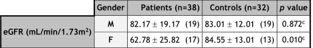
Table 10 – eGFR of PD and HC groups, according with gender. ... 16
Table 11 - Leukogram parameters of PD and HC groups. ... 17
Table 12 - Analysis of lymphocyte number in patients, according with Disease Duration. .... 18
Table 13 - Analysis of lymphocyte number in patients, according with L-DOPA Dose. ... 18
Table 14 - Crossed Disease Duration and L-DOPA Dose subgroups: comparison between L-DOPA Dose subgroups lymphocytes, according to disease duration. ... 18
Table 15 - Urate values of PD and HC groups, according with gender. ... 19
Table 16 - Comparison between urate levels of male and females from PD group, according with Disease Stage. ... 19
Table 17 - Comparison between urate levels of male and females from PD group, according with Disease Duration. ... 19
Table 18 - Comparison between urate levels of male and females from Early Stage PD subgroup and HC group. ... 19
Comparison with a control group
List of Acronyms
AC Absolute countBDCBAHICK BD™ CBA Human Inflammatory Cytokines Kit
CBA Cytometric Bead Array
CDK-EPI Chronic Kidney Disease Epidemiology Collaboration
CHCB Centro Hospitalar da Cova da Beira
CICS Centro de Investigação em Ciências da Saúde
CKD Chronic kidney disease
CNS Central nervous system
COMT Catechol-O-methyl transferase
Cr Serum creatinine
CRP C reactive protein
CSF Cerebrospinal fluid
DM Diabetes Mellitus
DN Dopaminergic neurons
EDTA Ethylenediamine tetraacetic acid
eGFR Estimate glomerular filtrate rate
ER Extended release
ESR Erythrocyte sedimentation rate
F Female FI Fluorescence intensity Hb Hemoglobin HC Healthy controls HTA Hypertension IL Interleukin IR Immediate release LBs Lewy bodies
LLOD Low limit of detection
LNs Lewy neurites
M Male
MAO Monoamine oxidase
MCHC Mean corpuscular hemoglobin concentration
MCV Mean corpuscular volume
mH&Yss Modified Hoehn & Yahr staging scale
NMDA N-methyl-D-aspartate
NSAIDs Non-steroids anti-inflammatory drugs
PD Parkinson’s disease
RDW Red Cell Distribution Width
ROS Reactive oxygen species
SN Substantia nigra
SNpc Substantia nigra pars compacta
TNF Tumor necrosis factor
Comparison with a control group
1. Introduction
Parkinson’s Disease (PD) is the second most common neurodegenerative disease (1, 2). PD, classically a motor disorder, is now recognized as a complex condition that includes neuropsychiatric and nonmotor symptoms1 (3, 4). The four cardinal features of PD are the
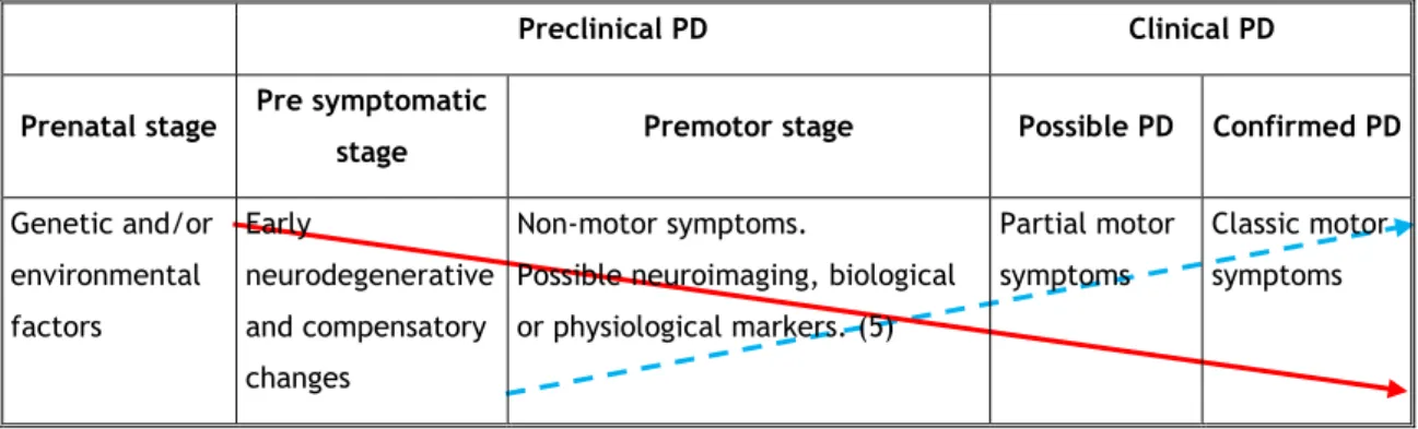
following motor symptoms: rest tremor, bradykinesia, rigidity and postural instability (3). The existing data suggest that PD starts years before the appearance of the cardinal motor symptoms (preclinical stage), when several premotor symptoms may already be present (premotor stage) (Figure 1) (4, 5). Evidences suggests that premotor stage could extend from 3 to 20 years and its existence should guide the search for PD biomarkers and the identification of risk and protective factors (6).
Preclinical PD Clinical PD
Prenatal stage Pre symptomatic
stage Premotor stage Possible PD Confirmed PD
Genetic and/or environmental factors Early neurodegenerative and compensatory changes Non-motor symptoms.
Possible neuroimaging, biological or physiological markers. (5)
Partial motor symptoms
Classic motor symptoms
Figure 1 – Hypothesized disease course of PD (4).
Dopaminergic neurons (DN); Clinical rating.
Nowadays PD diagnosis is made through strict clinical criteria. National Institute for Heath and Care Excellence – NICE current guidelines (7) recommend United Kingdom Parkinson’s Disease Society Brain Bank Criteria (UKPDSBBC) (Figure 2) (8). However, clinical diagnosis is difficult in the early disease state and frequently does not allow a definitive diagnose (8). PD etiology remains largely unknown (1, 2, 9). About 5-10% of cases have genetic origin, with inheritable genetic mutations (2, 9). These are mainly atypical, with early onset and low prevalence of tremor (10). 95% are of idiopathic origin, although several risk factors have been identified2 (2, 9).
PD results from the death of the pigmented dopaminergic neurons (DN) of midbrain
substantia nigra (SN) pars compacta (SNpc), with consequent loss of the neurotransmitter
dopamine in the corpus striatum, one of the subcortical nuclei involved in control of movement (2, 7, 9). Clinical symptoms emerge when approximately 50% of SNpc DN and
1 They include olfactory dysfunction; sleep disturbances; mood disorders; psychosis and hallucinations; dysautonomia; fatigue; cognitive dysfunction/dementia; pain and sensory disturbances.
2 Such as, age (2, 9), genetic predisposition, environmental toxins, neuronal injury (such as traumatic brain injury or stroke) and bacterial or viral infections (9).
80% of the striatal dopamine were lost (2, 4), which begins during the preclinical stage (4). The nigral damage spreads to other brain regions like lower brainstem, olfactory bulb, autonomic nervous system and, in the severest cases, neocortex (11).
Another PD’s hallmark is the presence of Lewy neurites (LNs) in cellular processes and Lewy bodies (LBs) in neuronal perikarya (11).
The exact mechanisms underlying DN degeneration are still not understood. Current theories include neuroinflammation, oxidative stress, mitochondrial dysfunction3 and disturbances of
intracellular ions homeostasis. (12) These are compatible with the evidences that SN is very susceptible to microglia neurotoxicity and oxidative stress: SN has high density of microglia (9), high levels of iron (13), reduced level of intracellular antioxidant glutathione (14) and accumulates dopamine, which induces reactive oxygen species (ROS) through oxidation reactions (13, 15).
1.1.
Neuroinflammation
Post-mortem studies revealed the first evidences of the presence of chronically activated microglia in SN (9, 16).
Microglia, the resident immune cells of central nervous system (CNS), is sensitive to minor microenvironment disturbances, becoming readily activated. Activated microglia has functional plasticity and they transform into a macrophage-like phenotype, which is maintained with continuous stimulation. Microglia activation mechanisms are not clearly understood. It could be indirectly through a positive feedback from degenerating neurons (reactive microgliosis), or directly due to a toxin, pathogen or endogenous protein4 (9).
Inflammatory markers from activated microglia, such as cytokines, are increased and they potentiate microglial activation (9, 12). Several studies detected higher levels of pro- and anti-inflammatory cytokines in PD patients’ (cerebrospinal fluid) CSF (9) and peripheral blood than in healthy controls (HC) (9, 12, 17-19).
1.2.
Oxidative stress
Post mortem and in vivo studies showed that PD brains present high levels of oxidative stress products (13).
Oxidative stress can be both a trigger and a maintainer of DN degeneration (13). Activated microglia further contributes to oxidative stress (13, 20), starting several responses to eliminate the source of inflammatory signals through it (13). On the other hand, ROS could stimulate production of pro-inflammatory cytokines by microglia (12, 13).
3 Particularly membrane lipid peroxidation.
4 Such as -synuclein-aggregates, neuromelanin (9, 17), adenosine triphosphate and matrix metalloproteinase-3 (9).
Comparison with a control group
1.3.
Objetives
The development of a sensitive biomarker for PD diagnosis is an ambitious goal that could contribute to the identification of individuals at risk, earlier and accurate diagnosis, discrimination from atypical parkinsonism and tracking disease progression (4, 21). Currently many research groups are trying to identify reliable markers.
In this study, we proposed to develop a demographic and clinical characterization of PD population of Centro Hospitalar da Cova da Beira (CHCB) and, then, to evaluate several parameters presented by patients in different stages of the disease and to compare with HC, aiming to focus on the initial phases of the disease.
Inflammation markers, such as serum cytokines [interleukin (IL)-1β, IL-8, IL-6, IL-10, Tumor Necrosis Factor (TNF), and IL-12p70], C reactive protein (CRP) and Erythrocyte sedimentation rate (ESR) were measured to determinate the magnitude of inflammation intervention in the disease. Immunological and oxidative status were accessed through leukogram and urate values, respectively. The haematological profile was also analysed.
Comparison with a control group
2. Materials and Methods
2.1.
Patients
The study was carried out with 38 PD patients, 21 males (55,3%) and 17 females (44,7%), with age between 55 and 86 years.
PD patients group were recruited from appointment list of CHCB (Covilhã - Portugal) Neurology sector, between January 2013 and December 2013.
Patients with clinical diagnosis of PD established by the UKPDBBC (Figure 2) (8) and modified Hoehn & Yahr staging scale (mH&Yss) (Figure 3) between 1 and 4 (22, 23) were included. Patients with recent and previous diagnosis (people who were followed for several years due to DP) were recruited.
All patients underwent an interview with a questionnaire (Annex 1), neurological examination performed by a neurologist, laboratory tests and medical record evaluation. A neurologist performed a neurological examination to apply mH&Yss (23) and to provide clinical data, by evaluating PD cardinal signs5. Interview and medical record evaluation allowed to obtain
demographic and medical information including: duration of PD, early motor symptoms, actual motor symptoms, presence or absence of non-motor symptoms, mean daily dosage of L-DOPA, actual and previous PD medications, copies of brain imaging exams, comorbidities, additional medications and Emergency appointments.
Participants with Diabetes mellitus (DM), hypertension (HTA), coronary and heart disease, hyperlipidemia and thyroid disease were included, if treated with the appropriate medication and if there was no evidence of exacerbations or complications in the clinical records, in the past 6 months.
All documents were identified by a study number to maintain confidentiality.
5 Tremor is involuntary, rhythmic and, mainly, at rest. In limbs, when patient relaxes with hands on the lap it is possible evaluate tremor. It could also affect legs, lips, jaw or tongue. (24) Rigidity is characterized by increased resistance to passive movement about a joint, decreased arm swing with walking and the typical stooped posture (24). Bradykinesia is a generalized slowness of movement or even absence of movement (akinesia). It’s evaluated through observation of facial hypomimia and limb movement, including speed, amplitude and rhythm of finger tapping, hand gripping, pronation-supination hand movements and heel or toe taping. Gait freezing and festination may develop in later disease. (24) “Pull” test evaluates postural instability: the examiner stands behind the patients and pulls the patients by his shoulders. Patients with postural instability take multiple steps backwards or could fall (positive “pull” test). Disease progression could lead to a festinant gait or confine the patient in a wheelchair, when postural reflexes are lost. (24)24. Chou KL, Hurtig, H. I., D ashe, J. F. C linic al manifestations of Parkinson disease.2012 May, 2013
Step 1 - Diagnosis of parkinsonian Syndrome Bradykinesia
At least one of the following: muscular rigidity 4-6 Hz rest tremor
postural instability not caused by primary visual, vestibular, cerebellar, or proprioceptive dysfunction
Step 2 – Exclusion criteria for Parkinson’s disease History of:
repeated strokes with stepwise progression of parkinsonian features repeated head injury
definite encephalitis oculogyric crises
neuroleptic treatment at onset of symptoms more than one affected relative
sustained remission
strictly unilateral features after 3 years supranuclear gaze palsy
cerebellar signs
early severe autonomic involvement
early severe dementia with disturbances of memory, language, and praxis Babinski sign
presence of cerebral tumor or communication hydrocephalus on imaging study negative response to large doses of L-DOPA in absence of malabsorption MPTP exposure
Step 3 - Supportive prospective positive criteria for Parkinson’s disease Three or more required for diagnosis of definite PD, in combination with step one: unilateral onset
rest tremor present progressive disorder
persistent asymmetry affecting side of onset most excellent response (70-100%) to L-DOPA
severe L-DOPA-induced chorea L-DOPA response for 5 years or more clinical course of ten years or more
Figure 2 – United Kingdom Parkinson’s Disease Society Brain Bank Criteria for PD diagnosis (8).
1,0: Unilateral involvement only 1,5: Unilateral and axial involvement
2,0: Bilateral involvement without impairment of balance 2,5: Mild bilateral disease with recovery on pull test
3,0: Mild to moderate bilateral disease; some postural instability; physically independent 4,0: Severe disability; still able to walk or stand unassisted
5,0: Wheelchair bound or bedridden unless aided Figure 3 - Modified Hoehn & Yahr staging scale (mH&Yss) (23).
Comparison with a control group
2.2.
Controls
The HC group had 32 participants, 19 males (59,4%) and 13 females (40,6%), with age between 55 and 86 years. They were all volunteers from CHCB. Most of them were recruited from Urology and Gynecology sectors.
All volunteers underwent an interview with a questionnaire (Annex 2), neurological and physical exam, laboratory tests and medical records evaluation. A summary neurological examination was performed6 to exclude neurological symptoms and cardiac and pulmonary
evaluations provided additional clinical information. Interview and medical record evaluation allowed to obtain comorbidities and actual medications.
All documents were identified by study number to maintain confidentiality.
2.3.
Ethical Approval
Patients and HC had given their informed consent to the study (Annex 3). The research protocol was approved by the Ethical Committee of CHCB.
2.4.
Exclusion Criteria
The exclusion criteria for PD patients are those included in UKPDBBC.
HC taking the following medications were excluded: urate synthesis inhibitors; anticoagulants; antiplatelets; vitamin supplements (except D vitamin supplements); PPAR
agonists (glitazones); serotonin-noradrenaline reuptake inhibitors; β2-agonists; antipsychotics; dopamine antagonists and agonists; catechol-O-methyl transferase (COMT) inhibitors; Monoamine oxidase (MAO) inhibitors; N-methyl-D-aspartate (NMDA) inhibitors; and acetylcholine agonists and antagonists. Likewise, active smokers, individuals with personal history of secondary parkinsonism, neurodegenerative diseases, stroke or chronic kidney disease (CKD), and those with head injury, myocardial infarction or surgery in the last three months were also excluded from HC group.
We also excluded the patients and controls with inflammatory and immune diseases7, acute or
chronic infectious disease, hepatic failure, alcohol addiction, myocardial infarction and surgery within the last 6 months and actual treatment with non-steroids anti-inflammatory drugs (NSAIDs), glucocorticoids [except intranasal8 (25)], immunosuppressors, systemic
6 We evaluated the following. Cranial nerves number II (visual fields); III, IV and VI (ocular motility); VII-motor (wrinkle forehead, close eyes tight, show teeth); and XII (stick out tongue and move it side to side). Tremor (patient’s arms are held outstretched and fingers extended), hand rapid alternating movements (finger tapping), finger-to-nose, muscular tone (elbow, wrist and knee passive movements), diadochokinesia (alternating pronate and supinate movements), osteotendinous reflexes (biceps, finger flexors and patellar) and plantar reflex.
7 Except one of the controls, who has Myasthenia Gravis, but inflammatory markers were normal. 8 Systemic bioavailability less than 2%.
hormonal therapy (except nonsteroidal), antibiothics, and cytostatics9. Those who had
estimate glomerular filtrate rate (eGFR) < 60 mL/min/1.73m2 during more than one year were
also excluded; e-GFR was calculated through Chronic Kidney Disease Epidemiology Collaboration (CDK-EPI) equation10.
2.5.
Sample collection
Venipuncture was performed in the morning and immediately after the clinical evaluations, without fasting. The responsible physician prescribed routine analysis and, at the same time, we collected peripheral blood samples to a tube without anticoagulant. Within an hour, that tube was transported to Clinical Pathology Laboratory of CHCB where it was centrifuged and kept at 4 degrees. Within 5 hours the tube was transported to the Health Science Research Center
(
CICS) at the Universidade da Beira Interior where the serum was divided in micro-tubes and stored at -70ºC.In our study, we used some of the values from routine analyses prescribed by the responsible physician. Peripheral blood samples were collected to one tube without anticoagulant and to one ethylenediamine tetraacetic acid (EDTA) tube. Within an hour, the tubes were transported and they were immediately analyzed in Clinical Pathology Laboratory of CHCB.
2.6.
Sample analysis
The sample analysis included cytokines measurement, CRP, ESR, serum creatinine (Cr), leukogram, urate and hemogram (Table 1).
One of the micro-tubes of serum was used in the cytometric bead array analysis - BD™ CBA Human Inflammatory Cytokines Kit (BDCBAHICK), which allows the measurement of 8, IL-1β, IL-6, IL-10, TNF, and IL-12p70 protein levels in a single sample, using beads of known size and fluorescence. Each capture bead has been conjugated with a specific antibody, enabling the detection of a set of analytes using flow cytometry.
The entire procedure is explained in Annex 4. All collected samples were analyzed in the same day.
9 Except one of the PD patients, who was using hydroxyurea to Policitemia Vera.
10 eGFR = 141 X min(Cr/κ,1) X max (Cr/κ,1)-1.209 X 0.993Age X 1.018 [if female] X 1.159 [if black], where Cr is serum creatinine (mg/dL), κ is 0.7 for females and 0.9 for males, is –0.329 for females and –0.411 for males, min indicates the minimum of Cr/κ or 1, and max indicates the maximum of Cr/κ or 1.
Comparison with a control group Table 1 - Sample analysis performed. BD™ CBA Human Inflammatory Cytokines Kit (BDCBAHICK).
(a) Performed at Clinical Pathology Laboratory of CHCB. (b) Performed at CICS laboratory.
Tube Values to determinate Objective Method
Without anticoagulant
Cr(a) Renal function Kinetic colorimetric assay based on Jaffé method
Urate(a) Antioxidant Enzymatic colorimetric assay CRP(a) Inflammatory status Immunoturbidimetry Cytokines(b) BDCBAHICK EDTA
ESR(a) Modified Westergren method
Hemogram(a) Immunological
status Cytometry
2.7.
Statistical analysis
The variables studied are present in Table 2.
In order to explore the disease variables, PD patients were divided:
- Into 2 subgroups according with mH&Yss: Early Stage (mH&Yss < 2,0) and Late Stage (mH&Yss 2,0) – Disease Stage subgroups;
- Into 3 subgroups according with disease duration: Short Duration (< 5 years), Middle Duration ([5-10[ years), and Long Duration ( 10 years) – Disease Duration subgroups; - Into 2 subgroups according with immediate release L-DOPA11 (26) dose: Low Dose
(<300 mg/day) and High Dose (300 mg/day) – L-DOPADose subgroups.
11 Extended release (ER) L-DOPA was converted for immediate release (IR). Conversion from ER to IR is the following: 200mg ER = 350 mg/day IR; 300 mg ER 12h/12h or 200 mg ER 8h/8h = 550 mg/day IR; 800 mg ER 8h/8h = 750 mg/day IR; 1000 ER 8/8h = 950 mg/day IR (26).
Table 2 - Variables investigated.
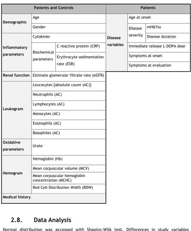
Patients and Controls Patients
Demographic Age Disease variables Age at onset Gender Disease severity mH&Yss Inflammatory parameters
Cytokines Disease duration
Biochemical parameters
C reactive protein (CRP) Immediate release L-DOPA dose Erythrocyte sedimentation
rate (ESR)
Symptoms at onset Symptoms at evaluation Renal function Estimate glomerular filtrate rate (eGFR)
Leukogram
Leucocytes [absolute count (AC)] Neutrophils (AC) Lymphocytes (AC) Monocytes (AC) Eosinophils (AC) Basophiles (AC) Oxidative parameters Urate Hemogram Hemoglobin (Hb)
Mean corpuscular volume (MCV) Mean corpuscular hemoglobin concentration (MCHC)
Red Cell Distribution Width (RDW) Medical history
2.8.
Data Analysis
Normal distribution was accessed with Shapiro-Wilk test. Differences in study variables between PD and HC groups were assessed by analysis of variance, unpaired T-test and Mann-Whitney U test for continuous measures, and Chi-square test was used for categorical variables. Correlation was used to assess the relationship between variables.
A p value of less than 0.05 was regarded as statistically significant.
Data are presented as means standard deviation for continuous measures and percentages and counts (in parenthesis) for categorical variables.
Statistical analysis was performed using GraphPad Prism version 5 and IBM SPSS Statistics version 20.
Comparison with a control group
3. Results
3.1.
Patient recruitment
Patients were recruited from appointment list of CHCB Neurology sector, between January 2013 and December 2013. 39 patients were identified with PD diagnosis established by the UKPDBBC (8) 35 potential HC were identified according with the previous criteria. In the appointment day, we proceed with clinical evaluation and blood sample collection. At the end of the study 38 patients and 32 HC were validated (one patient was excluded because the blood sample was not collected and three HC were excluded because they presented stage 3 CKD). None of the patients presented 5 on mH&Yss.
3.2.
Demographic characteristics of study population
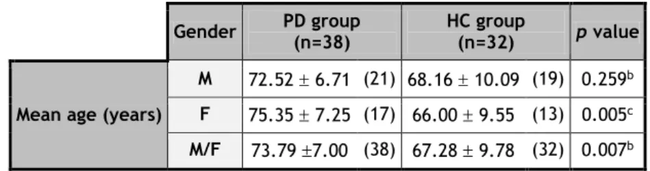
We start by analyzing the demographic characteristics of study population. PD and HC groups are similar in gender distribution. However, PD group, mainly women, is older than HC group. (Tables 3 and 4)
Table 3 – Gender distribution of study population. Male (M). Female (F). aChi-square test.
Gender PD group (n=38) HC group (n=32) p value
M 55.25% (21) 59.38% (19) 0.729a F 44.74% (17) 40.63% (13) Table 4 – Age distributions of study population.
b Mann-Whitney U test; cT-test.
Gender PD group (n=38) HC group (n=32) p value
Mean age (years)
M 72.52 6.71 (21) 68.16 10.09 (19) 0.259b F 75.35 7.25 (17) 66.00 9.55 (13) 0.005c M/F 73.79 7.00 (38) 67.28 9.78 (32) 0.007b
3.3.
Clinical characteristics of PD patients
To further investigation of our patients, we analyzed their clinical characteristics, focusing on medical history and symptomatology (Tables 5 and 6; Graphs 1-3). All PD participants present comorbidities, being HTA the most prevalent (Table 5).
Table 5 – PD group medical history, with main comorbidities represented.
Comorbidities PD group (n=38)
HTA 65.79% (25)
Musculoskeletal/ rheumatologic problems 36.84% (14)
Type 2 DM 28.95% (11)
Hyperlipidemia 18.42% (7)
Profound venous thrombosis/ Venous insufficiency 13.16% (5)
Hypothyroidism (on treatment) 10.53% (4)
Stroke/ Transitory ischemic accident history 10.53% (4)
Congestive heart failure 10.53% (4)
Benign prostatic hyperplasia 10.53% (4)
Obstructive sleep apnea 10.53% (4)
No comorbidities 0% (0)
Table 6 - Clinical features of PD patients. Positive familiar history is considered positive when patients have at least 2 relatives with PD.
Patients (n=38) Mean age at PD onset (years) 66.86 8.21 Mean PD duration (years) 6.93 4.24 Mean stage in mH&Yss 1.90 1.00 Mean dose of L-DOPA (mg/day)12 430.20 220.70
Positive familiar history 2.63% (1)
Symptoms at onset and at evaluation were accessed, in order to compare the progression of the disease symptomatology. The majority of patients presented tremor as initial symptom. Two of them presented a non motor symptom: widespread pain and paresthesia in the right arm. (Graph 1)
At evaluation, rigidity was the most frequent symptom (Graph 2).
Comparison with a control group 32 4 3 2 0 5 10 15 20 25 30 35
Tremor Rigidity Bradikynesia Other
Graph 1 - Cardinal symptoms at onset. The variable “Other” represents the PD patients with non motor symptoms.
31 28 27 4 1 0 5 10 15 20 25 30 35
Rigidity Bradikynesia Tremor Postural instability
No symptoms Graph 2 - Cardinal symptoms at evaluation
We determinate the mH&Yss at evaluation and most patients present early stage disease (mH&Yss < 2,0) (Graph 3). 84.21%; 10.53%; 7.89%; 5.26%; 81.58%; 73.68%; 71.05%; 10.53%; 2.63%;
Graph 3 – mH&Yss distribution of PD patients. Each color represents a stage of mH&Yss of PD population.
Patients with a more prolonged disease presented higher mH&Yss value and they take the higher doses of L-DOPA.
Patients on late stage, taking lower doses of L-DOPA, presented more tremor at evaluation than those taking higher doses. However, this is not observed for others symptoms.
3.4.
Inflammatory parameters
In order to explore the inflammatory side of PD, we evaluated the serum concentration of cytokines that were previously proposed to be associated with the disease. Besides, we analyzed the values of unspecific systemic inflammatory markers routinely used in clinic (CRP and ESR).
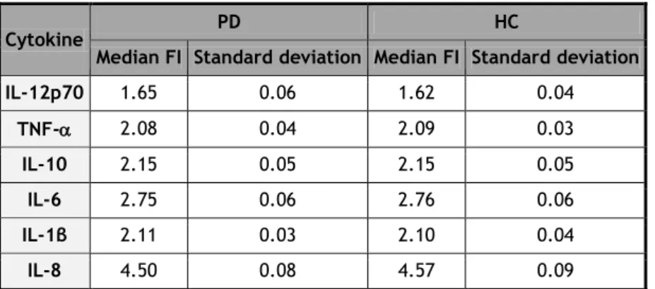
3.4.1. Cytokine measurement with BDCBAHICK
In the 38 PD patients and 32 HC, all cytokines measured (8, 1β, 6, 10, TNF, and IL-12p70) are below the lower limits of detection (LLOD) (see Table 7).
Comparison with a control group Table 7 – Lowe limits of detection (LLOD) for each cytokine, according with BDCBAHICK Instruction Manual. Cytokine LLOD (pg/mL) IL-12p70 2,5 TNF 3,0 IL-10 3,0 IL-6 3,9 IL-1β 2,9 IL-8 5,4
In Annex 5, the fluorescence intensity (FI) presented by each sample is displayed. The median and standard deviation of FI are presented on Table 8.
Table 8 - Median FI obtained for PD and HC groups. All cytokines measured were below the LLOD.
Cytokine PD HC
Median FI Standard deviation Median FI Standard deviation
IL-12p70 1.65 0.06 1.62 0.04 TNF- 2.08 0.04 2.09 0.03 IL-10 2.15 0.05 2.15 0.05 IL-6 2.75 0.06 2.76 0.06 IL-1β 2.11 0.03 2.10 0.04 IL-8 4.50 0.08 4.57 0.09
The calibration curves for each cytokine are presented on Annex 6.
3.4.2. Biochemical parameters
CRP and ESR values for patients and controls are presented on Table 9. CRP is higher in PD patients.
Table 9 - Inflammatory parameters of PD and HC groups. For the variables that depend on gender values for males and females are presented. Due to errors in blood analysis requisition, verified at the time of the sample collection, some parameters were not analyzed in all the patients. For CRP and ESR analysis, we excluded patients on actual treatment with NSAIDs, glucocorticoids (except intranasal) and immunosuppressors. Normal ranges are on Annex 7. Male (M). Female (F).
b Mann-Whitney U test; c T-test.
Gender Patients (n=38) Controls (n=32) p value CRP (mg/dL) M/F 0,30 0,17 (21) 0,26 0,17 (26) 0.050c ESR (mm/H) M 8.67 6.93 (14) 8.24 5.13 (17) 0.970
b F 27.14 17.93 (7) 14.64 8.49 (11) 0.126b
All inflammatory parameters are within the reference values for both groups (see Annex 7). For CRP and ESR measurements, participants with eGFR <60 mL/min/1.73m2 on evaluation
day were not included (Table 10).
Table 10 – eGFR of PD and HC groups, according with gender. Due to errors in blood analysis requisition, verified at the time of the sample collection, some parameters were not analyzed in all the patients. Male (M). Female (F).
cT-test.
Gender Patients (n=38) Controls (n=32) p value eGFR (mL/min/1.73m2) M 82.17 19.17 (19) 83.01 12.01 (19) 0.872
c F 62.78 25.82 (17) 84.55 13.01 (13) 0.010c
eGFR is within the normal range for both groups (see Annex 7). Although there are no significant differences in the values obtained for the male groups, the PD female patients present eGFR levels significantly lower than females in the HC group (Graph 4).
3.5.
Leukogram parameters
We considered leukogram parameters so that we can have an overview of the study population immune status. The values for both PD and HC groups are presented on Table 11.
Graph 4 - eGFR from PD and HC groups: females (a) and males (b) – scatter plot graphs. The biochemical parameters of inflammation scatter plot graphs for both groups are on Annex 8.
Comparison with a control group Table 11 - Leukogram parameters of PD and HC groups. Due to errors in blood analysis requisition, verified at the time of the sample collection, some parameters were not analyzed in all the patients. Normal ranges are on Annex 7. Male (M). Female (F).
b Mann-Whitney U test; c T-test.
Gender Patients (n=38) Controls (n=32) p value Leucocytes (103/uL) M/F 6.89 2.06 (37) 6.47 1.70 (32) 0.250c Neutrophils (103/uL) M/F 4.53 1.56 (37) 3.96 1.87 (32) 0.042b Lymphocytes (103/uL) M/F 1.60 0.59 (37) 1.83 0.56 (32) 0.103c Monocytes (103/uL) M/F 0.58 0.19 (36) 0.50 0.16 (32) 0.136b Eosinophils (103/uL) M/F 0.13 0.12 (37) 0.13 0.15 (32) 0.913b Basophiles (103/uL) M/F 0.04 0.06 (37) 0.03 0.05 (32) 0.681b
All values are within the reference values for both groups (see Annex 7). We found that PD patients have higher values of neutrophils, comparing with HC (Graph 5).
The data obtained suggest a decrease in the number of lymphocytes throughout the temporal evolution of the disease (Table 12) and in patients using higher doses of L-DOPA (Table 13).
Graph 5 - Neutrophils from PD and HC groups – scatter plot graphs. The remain leukogram parameters scatter plot graphs for both groups are on Annex 8.
Table 12 - Analysis of lymphocyte number in patients, according with Disease Duration. d ANOVA test.
Disease Duration subgroups Lymphocytes (10^3/uL) p value
Short Duration 1.64 0.43 (12)
0.408d Middle Duration 1.92 0.57 (12)
0.006d Long Duration 1.21 0.57 (12)
Table 13 - Analysis of lymphocyte number in patients, according with L-DOPA Dose. b Mann-Whitney U test.
L-DOPA Dose subgroups Lymphocytes (10^3/uL) p value
Low Dose 1.83 0,51 (21)
0.008b
High Dose 1.30 0,57 (16)
Since usually patients with a more prolonged disease require higher doses of L-DOPA, in an attempt to clarify if the decrease of lymphocytes during the disease course was a consequence of the disease itself or a secondary effect of L-DOPA use, we analyzed the relationship between lymphocyte number and disease duration for different L-DOPA Dose subgroups. Although there is a tendency to observe a lower number of lymphocytes for higher doses of L-DOPA in all the groups, the difference was not statistically significant. (Table 14)
Table 14 - Crossed Disease Duration and L-DOPA Dose subgroups: comparison between L-DOPA Dose subgroups lymphocytes, according to disease duration.
b Mann-Whitney U test.
Disease Duration subgroups L-DOPA Dose subgroups Lymphocytes (10^3/uL) p value
Short duration Low dose 1.74 0.42 (9) 0.209b
High dose 1.33 0.35 (3)
Middle duration Low dose 2.04 0.61 (8) 0.435b
High dose 1.72 0.48 (5)
Long duration Low dose 1.56 0.42 (4) 0.109b
High dose 1.03 0.57 (8)
3.6.
Oxidative parameters
Urate is a natural antioxidant proposed as a physiological indicator of the oxidative status. The data obtained show that PD urate levels are lower than the levels in the HC group, especially for male population (Table 15).This decrease is even more pronounced for the patients within higher stages of the disease (Table 16) and higher disease duration (Table 17). In what concerns the female population, the tendency to lower urate values in the PD group is obtained mainly in patients with longer disease (Tables 15-17).
Comparison with a control group Table 15 - Urate values of PD and HC groups, according with gender. Due to errors in blood analysis requisition, verified at the time of the sample collection, some parameters were not analyzed in all the patients. Normal ranges are on Annex 7. Male (M). Female (F).
c T-test.
Gender Patients (n=38) Controls (n=32) p value Urate (mg/dL) M 5.08 1.22 (15) 5.99 1.27 (18) 0.045
c F 4.17 1.05 (8) 4.61 0.96 (13) 0.345c
Table 16 - Comparison between urate levels of male and females from PD group, according with Disease Stage. Male (M). Female (F).
c T-test.
Gender Disease Stage subgroups Urate (mg/ dL) p value M Early stage 5.78 0.80 (8) 0.012c
Late stage 4.29 1.17 (2) F Early stage 4.40 0.14 (4) 0.584c
Late stage 3.95 1.34 (2)
Table 17 - Comparison between urate levels of male and females from PD group, according with Disease Duration. Male (M). Female (F).
dANOVA test.
Gender Disease Duration subgroups Urate (mg/ dL) p value
M Short Duration 5.10 1.30 (5) 0.956d Middle duration 5.35 1.17 (4) 0.846d Long Duration 4.88 1.33 (6) F Short Duration 5.15 0.07 (2) 0.578d Middle Duration 4.63 0.38 (3) 0.036d Long Duration 3.07 0.76 (3)
Although PD patients exhibit slightly lower values of urate than HC, there is no significant difference of urate values between HC and PD patients with the less severe disease (Early Stage and Short Duration subgroups; Table 18).
Table 18 - Comparison between urate levels of male and females from Early Stage PD subgroup and HC group. Male (M). Female (F).
c T-test.
PD group HC group
p value
Gender Disease Stage subgroups Urate (mg/ dL) Urate (mg/ dL) M Early stage 5.78 0.80 (8) 5.99 1.27 (18) 0.665 c Short Duration 5.10 1.30 (5) 0.181c F Early stage 4.40 0.14 (4) 4.61 0.96 (13) 0.702 c Short Duration 5.15 0.07 (2) 0.068c
3.7.
Hemogram parameters
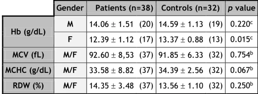
To further characterize the study population, we analyzed the hemogram parameters (Table 19).
Table 19 – Hemogram parameters of PD and HC groups. For the variables that depend on gender values for males and females are presented. Due to errors in blood analysis requisition, verified at the time of the sample collection, some parameters were not analyzed in all the patients. Normal ranges are presented on Annex 7. Male (M). Female (F).
b Mann-Whitney U test; c T-test.
Gender Patients (n=38) Controls (n=32) p value Hb (g/dL) M 14.06 1.51 (20) 14.59 1.13 (19) 0.220 c F 12.39 1.12 (17) 13.37 0.88 (13) 0.015c MCV (fL) M/F 92.60 8,53 (37) 91.85 6.33 (32) 0.754b MCHC (g/dL) M/F 33.58 8.82 (37) 34.39 2.56 (32) 0.067b RDW (%) M/F 14.35 3.48 (37) 13.56 1.10 (32) 0.250b
All values are within the reference values for both groups (Annex 7). The results obtained show that hemoglobin (Hb) levels from PD are lower than the levels in HC, mainly in females (Graph 6).
Graph 6 - Hb from PD and HC groups: females (a) and males (b) – scatter plot graphs. The remain hemogram parameters scatter plot graphs for both groups are on Annex 8.
Comparison with a control group
4. Discussion
PD is a neurodegenerative disease, affecting more the male population (27) and people over 60 years old, with mean age at diagnosis approximately 70.5 years (28).
The PD population of our study was also composed mostly by men, with mean age at disease onset of 73.8 years. It can be considered representative of the PD population, because it has demographic characteristics similar to those of epidemiologic studies, with an earlier onset of the disease of approximately 4 years. HC group has an equal gender distribution and it is younger than the PD group (Tables 3 and 4). The age difference is probably explained by the fact that HC male population was mostly recruited from Urology sector, in which men population is usually old, and HC female population was mostly recruited from Gynecology sectors, in which women population is usually young.
Comparing medical history, PD group has more comorbidities associated (Table 5), probably because it is an older population and/or because their health status is better characterized due to its high medical surveillance (they have regular Neurology appointments).
The diagnosis of PD, accordingly with NICE current guidelines (7), is established through UKPDSBBC, with the presence of bradykinesia and at least one of the other cardinal symptoms (rigidity, rest tremor and postural instability) (8). In what concern the symptoms, tremor is the most frequent, followed by rigidity (22). Postural instability is the last one to appear, resulting from an impairment of centrally mediated postural reflexes (3).
PD patients of our study had an onset symptomatology similar to those described in literature, with tremor being the most common symptom (84.21%; Graph 1). Rigidity was the second most common, present in 10.53% of cases (Graph 1). However, at evaluation rigidity became the most frequent symptom (81.58%; Graph 2). This could be explained by the fact that tremor is probably the symptom more visible to the observer and the one with the highest improvement with treatment, so it is highlighted at onset and then patients experience a real decrease in tremor.
As expected, none of the patients presented postural instability as onset symptom, but at evaluation, it emerges on 4 patients (10.53%), with disease duration 10 years (Graph 2). We noticed that patients within Late Stage (mH&Yss 2) or Long Duration disease (disease duration 10 years), who took a low dose of L-DOPA (<300 mg/day) presented more tremor at evaluation. We can assume that, for those patients, tremor could be controlled by increasing L-DOPA dose. Tremor was also the symptom with the highest improvement, which could explain the decreased number of patients with tremor on evaluation.
Physiopathology underlying the DN degeneration are still not completely understood, however several studies proved that neuroinflammation plays a central role on PD physiopathology, due to microglia chronic activation (10, 17). Activated microglia synthesize inflammatory markers, including cytokines, which through autocrine signalling create a self-propagating cycle of microglia activation. (9, 12) Several studies shown significantly higher levels of cytokines in serum and CSF of PD patients when compared with HC,(9) including pro-inflammatory cytokines, like IL-6 (12, 17), IL-12 (19), TNF-, (12, 18) and interferon- (12), and anti-inflammatory cytokines, namely IL-2, IL-4 (12) and IL-10 (12, 19). There are also evidences of slightly higher levels of pro-inflammatory (IL-1β, IL-6, IL-8 and IL-12) (18, 19) and anti-inflammatory (IL-10) (18) cytokines in PD patients’ serum, without statistical significance. The studies with a similar method of sample collection, storage and cytokine measurement [Cytometric Bead Array (CBA)] (12, 18) presented results not consistent. Koziorowski et al.(18) obtained cytokine values with a much lower magnitude order and
TNF- and IL-6 were undetectable for HC. Two of those studies (12, 18) shown detectable values of the others cytokines in HC.
Extending our literature search to other areas, researches with CBA method have inconsistent results too. Several studies detected IL-10, IL-12p70 and IL-6 values bellow our LLOD. (29, 30) Although, it is described that cytokine levels in plasma are higher than in serum (30).
In order to approach this neuroinflammatory side of PD, we proposed to evaluate serum concentration of IL-1β, IL-8, IL-6, IL-10, TNF, and IL-12p70 in PD patients and HC and to compare the cytokine pattern between those two groups. Contrary to what we expected, all cytokines were below the LLOD for both PD and HC groups. The serum samples were kept refrigerated within 5 hours at maximum, before being stored at -80ºC, thus strongly limiting sample degradation. CBA assay method has advantages over conventional ELISA methodology. It measures the concentration of an unknown analyte in less time and using fewer sample dilutions, because of the broad dynamic range of fluorescent detection via flow cytometry and the efficient capturing of analytes via suspended particles. Our methodology was accurate, because we obtained linear calibration curves, as we can see on Annex 6. However, the assay has limitations. The theoretical limit of detection of BDCBAHICK is comparable to conventional ELISA, but the actual limit of detection in a given experiment may vary slightly due to the complexity and kinetics of this multi-analyte assay. It could be interesting to repeat the cytokines measurement on plasma instead of serum and simultaneously increase the study population.
In order to complement our research about inflammation in PD, we also analyze unspecific systemic inflammatory markers routinely used in clinic, namely CRP and ESR (Table 9). CRP was higher in PD patients, but still much low than the reference value. We did not find differences between ESR values, however male from PD population presented lower values, which stands against the findings on CRP. We have to emphasize that we collected a lower number of sample for ESR measurement, so to further conclusions we should could increase the number of samples.
Comparison with a control group
Still, there is doubt about the truly origin of increased cytokines in the PD patients serum. It might involve the efflux of cytokines from the CNS through the blood brain barrier (12), which presents a higher permeability in PD patients (31). However, immune activation in PD is not restricted to CNS (17) and peripheral activated lymphocytes could produce the cytokines, which is explained by a relationship between lymphocyte number and the elevation of serum cytokines (12). There are also changes in subpopulations of blood lymphocytes, with reduced levels of T helper and B lymphocytes (32). In addition, there are evidences of higher levels of leukocytes in PD patients than in HC (12).
To explore those facts, we analyse leukogram of study population. At first sight, we detect a higher level of neutrophils in PD population (Table 11). Regarding lymphocytes, in PD patients their value decreases with progression of disease duration and elevation of L-DOPA dose. This leads us to a question: the decrease of lymphocytes values results from temporal evolution of disease or it is an adverse effect from L-DOPA. Considering the disease physiopathology is possible to assume that changes in immune system will become severe with temporal disease progression. It is not described any L-DOPA adverse effect related with lymphocytes levels. In order to explore this question, we divided PD patients into subgroups according with disease stage, disease duration and L-DOPA dose and we compare lymphocyte levels between those subgroups. We found that patients with Long Duration disease and High Doses of L-DOPA presented significantly lower values of lymphocytes (Table 12 and 13). As patients with a more prolonged disease usually use higher doses of L-DOPA, we analyzed the relationship between lymphocyte number and disease duration for different L-DOPA dose subgroups. There is a tendency to observe a lower number of lymphocytes for higher doses of L-DOPA in all the groups, but it is not statistically significant (Table 14). Then, we ca not take conclusions about the truly cause of lymphocytes variations in PD patients. It would be interesting to do a differential analysis of PD patients’ lymphocytes, in order to assess which phenotype has the greater variance. The duration of L-DOPA treatment could also give us interesting results.
Exploring another aspect, oxidative stress can also play a key role on PD physiopathology and antioxidants may be an endogenous defence against the disease (13). Urate, the end product of purine metabolism, it is a potent antioxidant, peroxynitrite scavenger, iron chelator, and ascorbate stabilizer (31, 33). Evidences suggest that high blood and CSF urate concentrations are related with a reduced risk of PD (31, 33) and with a decreases disease progression rate, including cognitive decline (33). This inverse association was independent from age, smoking, caffeine consumption, and other aspects of lifestyle that have been related to both PD and uraemia (31). Those data could establish urate as the first molecular predictor of clinical progression in PD (33). However, there is still not sufficient data to support a recommendation for urate increase as therapeutic measure (31, 33).
As expected, urate presented lower values on PD group, especially on male population (Table 15). Our urate values for PD patients are consistent with other studies (31, 33). Late Stage
and Long Duration PD patients presented lower values, again specially on male population (Table 16 and 17). In order to understand if urate could be a marker of PD initial phase, we compare Early Stage and Short Duration PD patients with HC group. We detect that male population had slightly lower values of urate, as expected. However, female population present the inverse. (Table 18).We must emphasize the fact that our study presents limitations at this level, because we only proceeded to a single measurement of urate and we did not control confounding factors related to lifestyle. Medical records review and repeated urate measurements for PD patients will probably give us interesting results. However, we can assume that urate could be a putative marker of disease severity, at least on male population. Our data are promising and increasing the number of the study population, we could probably corroborate our findings.
Additional information about oxidative stress could be obtain through the measurement of an oxidative stress marker on peripheral blood, such as 8-hydroxyguanosine, which results from nucleoside oxidation, since recent studies shown high levels of these substances in PD peripheral blood (34).
Besides PD etiology remains largely unknown (1, 2, 9), some risk factors have been already described. Anemia is one of them, with slightly but consistently lower levels of Hb during life, when compared with controls (35). On the other hand, smoking and vitamin E13 are associated
with a lower risk of developing PD (1).
In our study, PD patients presented lower Hb values, especially female participants, but still with no anemia criteria (Table 19 and Annex 7). However we can not interpret these results, because PD females are older and confounding factors were not eliminated [such as cigarette smoking, exposure to pesticides, or hysterectomy (35)].
Looking towards our work, we can point some limitations. First, the small sample size and single-point measurements of serum cytokines and biochemical data limited our statistical power in the analyses. Second, PD group sample was older than HC, making difficult to compare and to take conclusions about some of the variables. Third, mH&Yss and disease duration are rough methods to describe disease progression. For last, we included a large number of variables, making impossible to control the confounding factors for each one of them.
Nevertheless, we used medical record of study participants, making possible to analyze with certainty the disease variables for PD patients; confounding factors for cytokines analysis were eliminated (see Material and Methods – Exclusion criteria); and exclusion criteria were much more restrictive for HC group, whereby our sample is representative of a healthy population with the age and gender distribution described.
Comparison with a control group
In summary, we can highlight the following points. In concern of characterization of PD population of CHCB, it is an elderly population, with mean age of 73.8 years, mainly composed by men. Regarding inflammatory aspects of the disease, our study showed that cytokine pattern is not sensitive or specific to assess disease progression, because results are very inconsistent. However, our results are in favor of the involvement of immune system and probably a deeper investigation could bring us more information about temporal disease progression and a way to better characterize the disease. On the other hand, antioxidant urate presents us as a putative biomarker of disease severity.
This study is only a starting point. It allowed us characterize PD population of the region and start a research that, in the future, should be divided in order to better describe each of the issues addressed. Furthermore, more subjects should be included in both groups, with the aim of collecting the necessary information to extrapolate our results with confidence.